Article

Whole Genome Sequence Analysis of Mycobacterium bovis Cattle Isolates, Algeria

1
Laboratoire d’Agro Biotechnologie et de Nutrition des Zones Semi Arides, Université Ibn Khaldoun, Tiaret 14000, Algeria
2
Institut des Sciences Vétérinaires, Université de Blida 1, Blida 09000, Algeria
3
Institut Hospitalo-Universitaire Méditerranée Infection, 13005 Marseille, France
4
Faculté de Médecine, Aix-Marseille-Université, IHU Méditerranée Infection, 13005 Marseille, France
5
Laboratoire National de Référence pour la Tuberculose et Mycobactéries, Institut Pasteur d’Algérie, Alger 16015, Algeria
6
Laboratoire de Reproduction des Animaux de la Ferme, Université Ibn Khaldoun, Tiaret 14000, Algeria
*
Author to whom correspondence should be addressed.
Pathogens 2021, 10(7), 802; https://doi.org/10.3390/pathogens10070802
Submission received: 20 April 2021 / Revised: 11 June 2021 / Accepted: 18 June 2021 / Published: 24 June 2021

Abstract

:
Mycobacterium bovis (M. bovis), a Mycobacterium tuberculosis complex species responsible for tuberculosis in cattle and zoonotic tuberculosis in humans, is present in Algeria. In Algeria however, the M. bovis population structure is unknown, limiting understanding of the sources and transmission of bovine tuberculosis. In this study, we identified the whole genome sequence (WGS) of 13 M. bovis strains isolated from animals exhibiting lesions compatible with tuberculosis, which were slaughtered and inspected in five slaughterhouses in Algeria. We found that six isolates were grouped together with reference clinical strains of M. bovis genotype-Unknown2. One isolate was related to M. bovis genotype-Unknown7, one isolate was related to M. bovis genotype-Unknown4, three isolates belonged to M. bovis genotype-Europe 2 and there was one new clone for two M. bovis isolates. Two isolates from Blida exhibited no pairwise differences in single nucleotide polymorphisms. None of these 13 isolates were closely related to four zoonotic M. bovis isolates previously characterized in Algeria. In Algeria, the epidemiology of bovine tuberculosis in cattle is partly driven by cross border movements of animals and animal products.

1. Introduction

Bovine tuberculosis is a chronic, deadly infection most often caused by Mycobacterium bovis (M. bovis) and Mycobacterium caprae (M. caprae), two species belonging to the Mycobacterium tuberculosis complex [1,2]. M. bovis most often infects cattle but also causes tuberculosis in other animal species and is a re-emerging cause of zoonotic tuberculosis in humans [3,4]. This disease poses a major threat to public health and creates socio-economic problems, including loss of meat due to seizures in slaughterhouses and lower milk yields [5,6]. Accordingly, bovine tuberculosis has an impact upon the international trade of animals and animal products [6,7], and bovine tuberculosis must be notified to the World Organization for Animal Health, as mentioned in its Terrestrial Animal Health Code [8].
In Algeria, some of the 2,049,652 heads of cattle [9] in the country are suspected to be infected by M. bovis [10]. Cases of bovine tuberculosis are often reported in slaughterhouse registers, and slaughterhouse workers exhibit a higher prevalence of M. bovis tuberculosis than the general population [11] (F. Tazerart, unpublished data). In Algeria, ante-mortem tuberculin skin test screening and post-mortem examinations for suspicious lesions at the slaughterhouse are the official diagnostic procedures for the disease [12]. Nevertheless, in the absence of systematic laboratory diagnosis, definitive confirmation of the disease is lacking. Consequently, bovine tuberculosis remains a neglected infectious disease in Algeria, and few microbiology studies have been reported [10]. The identification of the circulation of M. bovis strains and their hosts and geographical distribution are poorly understood.
This study aimed to start bridging this gap, using whole genome sequencing (WGS) to investigate a few M. bovis strains currently circulating in Algeria.

2. Results

2.1. Data Analysis

Of the 928 cattle routinely slaughtered and inspected, 94 (10.13%, CI 8.24–12.34%) animals had visible lesions compatible with tuberculosis. The prevalence of suspected lesions significantly varied across the five studied slaughterhouses (p = 0.001). Prevalence was 20.9% at the Médéa slaughterhouse, which was the most affected site, and 5.8% at the Bgayet slaughterhouse, which was the least affected site (Figure 1). Males accounted for 617/928 (66.5%) of slaughtered animals and females for 311/928 (33.5%) (p < 10−4), although the prevalence of tuberculous lesions was higher in females than in males (n = 41/311 (13.18%) compared to n = 53/617 (8.59%), p = 0.038). Most of the slaughtered animals were aged <2 years (n = 427/928; 46%), followed by animals aged 2–5 years (n = 399/928; 43%) and animals older than 5 years (n = 102/928; 11%). Older animals >5 years were most affected (25.5%), followed by young animals (<2 years) at 9.83%, then adult animals (2–5 years) at 6.51%. The differences among prevalence values were significant (p < 10−4). Most slaughtered animals were of average weight (score 2.5–3) (536/928; 57.75%), followed by lean animals (286/928; 30.81%). The prevalence of the suspected lesions was 60/536 (11.2%) in average weight animals, 31/286 (10.8%) in lean animals and 03/106 (2.8%) in fatty animals (p = 0.03). Tuberculous lesions were mainly located on the lymph nodes (95.7%) and 4.3% in the viscera (Table 1).

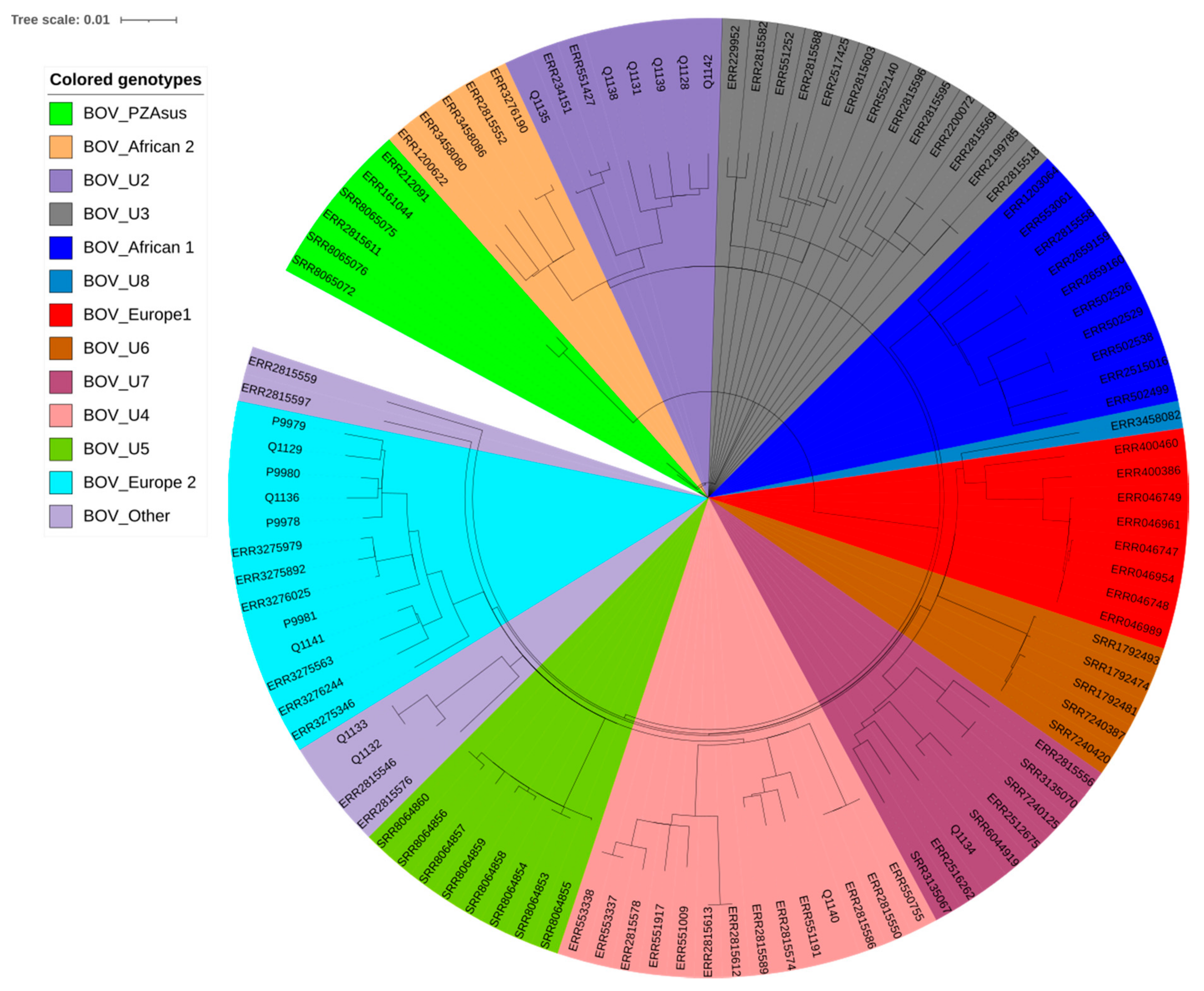
2.2. Genomic Typing Analysis

A total of 94 samples, including 90 lymph node samples, three lung samples and one liver sample, yielding 13 cultures obtained on Löwenstein–Jensen culture medium, Middlebrook 7H10 solid medium and Coletsos medium were identified as M. tuberculosis complex by partial rpoB PCR-sequencing. No other mycobacterial species was cultured from these samples. Further WGS refined the identification of the 13 isolates as M. bovis. Previously, the work of Loiseau et al. indicated that the diversity of M. bovis in Africa has been underestimated, after their discovery of new clonal complexes in the continent [13]. Genomic comparison of the Algerian M. bovis isolates to the ones reviewed in the work of Loiseau et al. indicated four different clonal complexes of M. bovis in Algeria—three of which were the newly discovered ones, and one of which was known—the Eu2 clonal complex commonly found in western Europe. Specifically, we found six isolates, namely Q1128, Q1131, Q1135, Q1138, Q1139 and Q1142, which were grouped together with reference clinical strains of M. bovis clonal complex-Unknown2. One isolate, Q1134, was related to M. bovis clonal complex-Unknown7; one isolate, Q1140, was related to M. bovis clonal complex-Unknown4; three isolates, Q1141, Q1129 and Q1136, belonged to M. bovis clonal complex-Europe 2; and there was one new clone for two isolates, Q1132 and Q1133, which did not belong to any of the clonal complexes identified by Loiseau et al. (Figure 2, Table S1) [13]. Based on available data for 3364 clinical isolates of M. bovis (Tables S1 and S3) [13], African countries other than Algeria included Ethiopia, Malawi, Eritrea, Morocco, Tunisia and South Africa. European countries included the UK, Switzerland, Sweden, Spain, France, Italy, Germany, Netherlands and Belgium. American countries included the USA, Canada, Brazil and Mexico. Asian countries include Lebanon. The proportions of M. bovis genotypes observed in Algeria were significantly different than those observed in other countries (p < 0.001), other African countries (p = 0.045) and American countries (p < 0.001) but did not significantly differ from European countries (p = 0.245). Using a 12 SNPs cut-off to detect potential cross-transmission [14], we detected one group of transmission involving isolates Q1132 and Q1133 exhibiting 215-SNPs, and 0-SNPs after removing repetitive regions (Figure 2, Table S2).
Further comparison of bovine isolates with four available Algerian patients’ M. bovis isolates previously determined as belonging to the European 2 group (P9978, P9979, P9980 and P9981) [15] indicated no evidence of zoonotic infection of these isolates alone using the same SNP cut-off as above (12-SNPs). Statistical information on the genome sequences are reported in Supplementary Data Table S1. Reads were deposed in GenBank with accession number PRJNA715078.

3. Discussion

In Algerian slaughterhouses, the inspection of slaughtered animals to look for lesions compatible with tuberculosis differs from one slaughterhouse to another.
Here, more males than females were slaughtered, in accordance with regulations prohibiting the slaughter of female animals other than in cases of necessity. However, we observed a significantly higher prevalence of tuberculosis in females than in males, an observation that may also reflect an increased susceptibility of females that go through gestation, parturition and lactation, and live a long, productive life [16]. Most slaughtered animals were under two years old, reflecting the high organoleptic value of meat in young animals, which is in great demand. However, older animals over the age of five years had a significantly higher prevalence of lesions suspected of being tuberculosis. Indeed, the chances of exposure to tuberculosis increases with age [17]. Most slaughtered animals were of average weight (score 2.5–3) reflecting consumer preference for meat containing a reasonable amount of fat. In addition, we observed a significant difference between the three body weight scores of animals suspected of tuberculosis; those of average weight were most affected, followed by those that were lean, then fatty animals, potentially explained by the wasting nature of this disease [18].
In this context, this report on the WGS-based identification and typing of a collection of M. bovis isolates updates the 2007 snapshot of bovine tuberculosis in Algeria [10]. The prevalence of bovine tuberculosis, as estimated from the veterinary inspection of slaughtered cattle, was 10.13%, higher than previously reported in Algeria in 2007 (3.6%) and in neighboring Morocco in 2014–2015 (3.7%) [10,19].
In this study, WGS revealed the presence of four different genotypes of M. bovis and one new genotype in Algeria compared to 12 genotypes reported by Loiseau et al. (Figure 2, Table S1) [13]. We detected two isolates exhibiting a closely related WGS pattern, suggesting these two isolates, Q1132 and Q1133, were issued from the same M. bovis clone. These two isolates were isolated from the same slaughterhouse (Mouzaia in Blida).
Unsurprisingly, these data illustrate the cross-border spread of M. bovis and the fact that Algeria regularly imports live cattle from Europe, notably from France and Spain, each supplying 40,000 heads of cattle per year [20,21]. These data indicate that part of the epidemiology of bovine tuberculosis in cattle in Algeria is driven by the cross-border movement of animals.
However, limited information about slaughtered animals, such as their origin and movements, limits the possibility of carrying out a spatial epidemiological study on the distribution of bovine tuberculosis. Illegal slaughter further complicates the establishment of such a bovine tuberculosis map. Additionally, only 13 M. bovis isolates were available for this first ever WGS analysis of animal M. bovis in Algeria. Such a small sample is likely to underestimate pathogen diversity in Algeria and is therefore unlikely to ascertain epidemiological linkages between cases.
Finally, despite the above-reported limitations, this founding WGS-based study in Algeria indicates the contribution of WGS to tracing animal M. bovis isolates in that country. This study suggests that it would be of interest to establish an Algerian national M. bovis WGS database for tracing sources of infection, including the potential role of cross-border importation and of wild fauna, as reported in other countries such as the UK [22,23], and the cross-transmission in farms. Further comparisons between human [15] and animal isolates would also help to trace sources of zoonotic M. bovis tuberculosis in patients from a medical prevention perspective.
Given such a small number of isolates, this analysis could be very skewed, and a larger number of studied isolates may provide similarities with other locations. This indicates that continued research in this area is required.
Despite this limitation, we did find similarities with European nations, which is consistent with cattle being imported into Algeria from Europe and the historical links between these regions

4. Materials and Methods

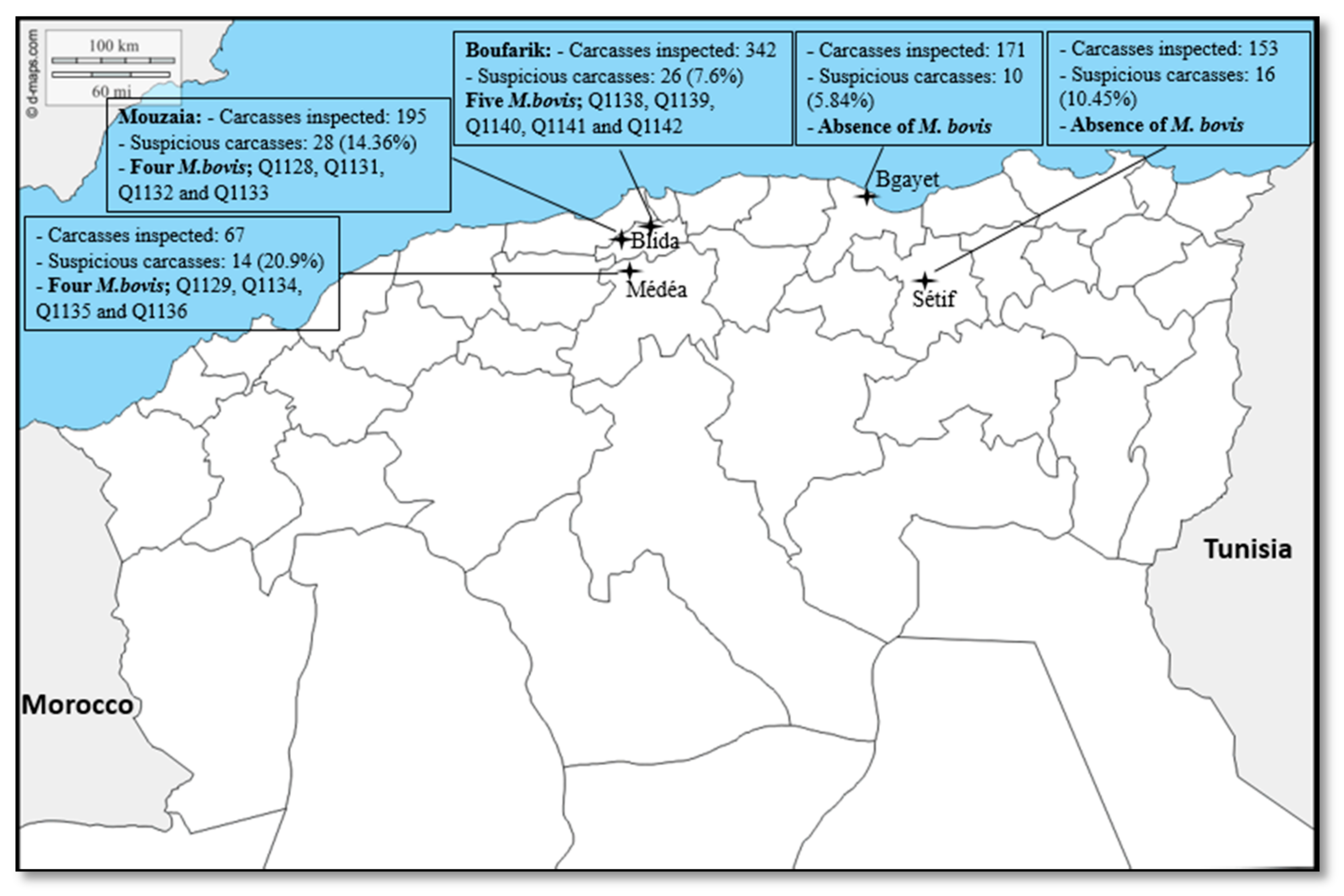
4.1. Study Area and Sample Collection

The study was conducted during different periods of 2017 and 2018 in five slaughterhouses in Algeria, namely the slaughterhouses in Bgayet (January–May 2018), Sétif (January–May 2018), Médéa (December 2016–April 2017), Mouzaia (March–May 2017) and the slaughterhouses in Boufarik, which are in the department of Blida (Figure 1) (March–May 2018). The distance between each slaughterhouse was estimated to be between 27 km (Mouzaia–Boufarik) and 315 km (Sétif–Mouzaia). In detail, these 13 isolates were obtained from three slaughterhouses, namely the Mouzaia slaughterhouse in Blida (Q1128, Q1129, Q1131, Q1132 and Q1133), the Médéa slaughterhouse (Q1134, Q1135 and Q1136) and the Boufarik slaughterhouse in Blida (Q1138, Q1139, Q1140, Q1141 and Q1142). The choice of these slaughterhouses was justified by their accessibility and their relative size in the region in terms of cattle slaughter. Cattle slaughtered in these slaughterhouses came from the neighboring regions or sometimes from remote regions. An information sheet was systematically established for each animal slaughtered, including sex, age, breed, body score and origin. During the study period, lesions of suspected tuberculosis were collected from slaughtered cattle and conserved for 11–15 months at −20 °C until further processing.

4.2. Tissue Preparation and Culture

Some of these samples were processed at the Institut Pasteur d’Algérie in Algiers, and the rest were processed in the biosafety level 3 laboratory (BSL3) at the Institut Hospitalier Universitaire, Méditerranée Infection in Marseille. At the Institut Pasteur d’Algérie, gross visible lesions were thawed, dissected and crushed using a sterile pestle and mortar. Crushed tissues were decontaminated by adding 4 mL of 4% NaOH [24] and washed with sterile distilled water. Pellets were inoculated in a Löwenstein–Jensen culture medium containing glycerol (three tubes) and were incubated at 37 °C for 12 weeks. At the Institut Hospitalier Universitaire, gross visible lesions were thawed, and aliquots were decontaminated into a triple volume of 1% chlorhexidine (chlorhexidine digluconate, Sigma-Aldrich, St. Louis, MO, USA) [25] incubated for 30 min at room temperature with manual agitation every five minutes. After eliminating chlorhexidine, 10 mL of a neutralizing solution (1000 mL sterile phosphate buffered saline (PBS), 3 g egg lecithin and 10 mL Tween 80) were added, and the mixture was left at room temperature for 10 min with agitation to inactivate the remaining chlorhexidine. After three sterile PBS washes, the decontaminated tissue was finely crushed using Potter’s crusher and inoculated in four types of culture media including Middlebrook 7H10 solid medium containing 10% (v/v) oleic acid, albumin, dextrose and catalase (OADC) (Becton Dickinson, Sparks, MD, USA) and 0.5% (v/v) glycerol (Euromedex, Souffelweyersheim, France); 10% OADC-Middlebrook 7H10 solid medium containing sodium pyruvate (4.16 mg/mL) (Sigma-Aldrich) [26,27]; 10% OADC-Middlebrook 7H9 liquid medium (Becton Dickinson), containing 0.05% Tween 80 (Sigma-Aldrich) [28]; and Coletsos medium (Bio-Rad, Marnes-la-Coquette, France). Cultures were incubated at 37 °C for 12 weeks with a 10-day follow-up of colony growth.

4.3. Isolate Identification

Any culture growing on any of the solid media was dissolved in 200 μL PBS and inactivated at 100 °C for one hour before further handling in the biosafety level 2 (BSL2) laboratory. Total DNA was extracted by vortexing the suspension with glass powder (Sigma-Aldrich) using the FastPrep apparatus (MP Biomedicals, Santa Ana, CA, USA) and extracting DNA using a Qiagen kit (Qiagen, Courtaboeuf, France), as previously described [29]. Partial rpoB gene PCR-sequencing was then performed to confirm the M. tuberculosis complex, as previously described [30].

4.4. Genome Sequence Analyses

As for whole genome sequence (WGS) experiments, DNA was extracted using the InstaGene™ Matrix (Bio-Rad, Marnes-la-Coquette, France) and quantified using a Qubit assay with the high sensitivity kit (Life technologies, Carlsbad, CA, USA). A total of 0.2 µg/µL of DNA was then sequenced by Illumina MiSeq runs (Illumina Inc., San Diego, USA). DNA was fragmented and amplified by limited PCR (12 cycles), introducing dual-index barcodes and sequencing adapters. After purification on AMPure XP beads (Beckman Coulter Inc., Fullerton, USA), the libraries were normalized and pooled for sequencing on the MiSeq. Paired-end sequencing and automated cluster generation with dual indexed 2 × 250-bp reads were performed. Output sequencing reads were analyzed using MTBseq [31] to identify species, lineages and sub-lineages and to calculate the SNP distance between study strains. MTBseq was used also to extract and align the SNPs between the genome studies. Identification results were supported by other tools such as TB Profiler and Mykrobe Predictor-TB [32,33]. A phylogenetic tree based on extracted SNPs between 105 M. bovis genomes was generated using PhyML 3.0 online tools (http://phylogeny.lirmm.fr/phylo_cgi/one_task.cgi?task_type=phyml; accessed on 1 October 2020).

4.5. Statistical Analysis

The data collected were analyzed using SPSS (Statistical Package for Social Sciences, version 23.0) and Microsoft Office Excel software programs. As a criterion, reliability difference indicators used the profile p < 0.05. Statistical analyses were performed using Chi-square tests to compare the presence of risk factors between suspected cases of tuberculosis (Table 1) and to compare the proportions of M. bovis genotypes, among other genotypes in different geographic regions/countries. We delineated four groups of data: Group 1, all countries in the 3364-isolates database; Group 2, all African countries from the database; Group 3, all American countries; and Group 4, all European countries.

Supplementary Materials

The following are available online at https://www.mdpi.com/article/10.3390/pathogens10070802/s1, Table S1: Statistical genomes data of study strains extracted using MTBseq. Table S2: SNPs distance between study strains using MTBseq. Table S3: Published data of several M. bovis genotypes detected in Algeria.

Author Contributions

F.T., J.S. and M.D. conceived and designed the experiments, conceived the methodology and wrote the manuscript. F.T. carried out the investigations, performed the experiments and the laboratory manipulations. J.S. carried out genome characterization and bio-informatic analysis. F.T. and J.S. carried out the formal analysis and processed the data on software. D.Y. and M.D. set up the material resources. N.S., A.N. and M.D. supervised the project. All authors have read and agreed to the published version of the manuscript.

Funding

This study was supported by the Algerian Ministry of Higher Education and Scientific Research under the Exceptional National Programme (P.N.E.) and by the University of Blida 1. This study was also supported by the French Government under the Investissements d’Avenir (Investments for the Future) programme managed by the Agence Nationale de la Recherche (ANR, fr: National Agency for Research) (reference: Méditerranée Infection 10-IAHU-03).

Institutional Review Board Statement

According to Algerian law, no Ethics Committee approval is required when analyzing tissues collected post-mortem after an animal has been slaughtered in an official slaughterhouse.

Informed Consent Statement

Not applicable.

Data Availability Statement

Not applicable.

Acknowledgments

The authors thank staff and veterinarians of the five slaughterhouses in Algeria for their support and help with our work. The authors acknowledge the help of Zolikha Ali Mahine, Wahiba Akkouche, Yanis Dissi, Amir Affoune, Fatima Aissaoui, Kenza Medini and Abderrahmane Benkouar for their participation and collection of samples. We would also like to offer our thanks to Sofiane Tahrikt for his help in preparing and sending samples from Blida to IHU in Marseille.

Conflicts of Interest

The authors declare no conflict of interest.

References

  1. Boukary, A.R.; Thys, E.; Rigouts, L.; Matthys, F.; Berkvens, D.; Mahamadou, I.; Yenikoye, A.; Saegerman, C. Ris factors associated with bovine tuberculosis and molecular characterization of Mycobacterium bovis strains in urban settings in Niger. Transbound. Emerg. Dis. 2012, 59, 490–502. [Google Scholar] [CrossRef]
  2. Lorente-Leal, V.; Liandris, E.; Castellanos, E.; Bezos, J.; Domínguez, L.; de Juan, L.; Romero, B. Validation of a Real-Time PCR for the Detection of Mycobacterium tuberculosis Complex Members in Bovine Tissue Samples. Front. Vet. Sci. 2019, 6, 61. [Google Scholar] [CrossRef] [PubMed]
  3. Eroksuz, Y.; Baydar, E.; Otlu, B.; Dabak, M.; Eroksuz, H.; Karabulut, B.; Incili, C.A.; Timurkan, M.O. Case report: Systemic tuberculosis caused by Mycobacterium bovis in a cat. BMC Vet. Res. 2019, 15, 9. [Google Scholar] [CrossRef] [PubMed]
  4. Nebreda-Mayoral, T.; Brezmes-Valdivieso, M.F.; Gutiérrez-Zufiaurre, N.; García-de Cruz, S.; Labayru-Echeverría, C.; López-Medrano, R.; López-Urrutia-Lorente, L.; Tinajas-Puertas, A.; Rivero-Lezcano, O. Human Mycobacterium bovis infection in Castile and León (Spain), 2006–2015. Enferm. Infecc. Microbiol. Clin. (Engl. Ed.) 2019, 37, 19–24. [Google Scholar] [CrossRef] [PubMed]
  5. Belkhiri, M.; Tlidjane, M.; Benhathat, Y.; Meziane, T. Histopathological study and pulmonary classification of bovine lesions. Afr. J. Agric. Res. 2009, 4, 584–591. [Google Scholar]
  6. Boland, F.; Kelly, G.E.; Good, M.; More, S.J. Bovine tuberculosis and milk production in infected dairy herds in Ireland. Prev. Vet. Med. 2010, 93, 153–161. [Google Scholar] [CrossRef]
  7. Cousins, D.V. Mycobacterium bovis infection and control in domestic livestock. Rev. Sci. Tech. Off. Int. Epiz. 2001, 20, 71–85. [Google Scholar] [CrossRef] [PubMed]
  8. World Organisation for Animal Health. Bovine Tuberculosis. 2020. Available online: https://www.oie.int/en/animal-health-in-the-world/animal-diseases/bovine-tuberculosis/#A (accessed on 19 February 2020).
  9. Ministry of Agriculture and Rural Development and Fishing of Algeria. Department of Veterinary Services. Establishment of an Animal Identification, Registration and Monitoring System in Algeria. K. Boughalem CVO Algeria Algiers, Algeria—November 24 and 25, 2015. CPC/REMESA. Available online: https://docplayer.fr/43882308-Dr-k-boughalem-cvo-algerie-alger-algerie-24-et-25-novembre-2015-cpc-remesa.html?fbclid=IwAR37DQnECLe-WWs85AseZi5iXI31I8cFkFxFUNkMbA5o19_FJdprpa-uPog (accessed on 19 February 2020).
  10. Sahraoui, N.; Müller, B.; Guetarni, D.; Boulahbal, F.; Yala, D.; Ouzrout, R.; Berg, S.; Smith, N.H.; Zinsstag, J. Molecular characterization of Mycobacterium bovis strains isolated from cattle slaughtered at two abattoirs in Algeria. BMC Vet. Res. 2009, 5, 4. [Google Scholar] [CrossRef] [Green Version]
  11. Michel, A.L.; Müller, B.; van Helden, P.D. Mycobacterium bovis at the animal–human interface: A problem, or not? Vet. Microbiol. 2010, 140, 371–381. [Google Scholar] [CrossRef] [Green Version]
  12. Sahraoui, N.; Muller, B.; Mamache, B.; Yala, D.; Boulahbal, F.; Zinsstag, J.; Guetarni, D. Tuberculosis in cattle and Goat of North Algeria. Vet. Res. 2011, 4, 100–103. [Google Scholar]
  13. Loiseau, C.; Menardo, F.; Aseffa, A.; Hailu, E.; Gumi, B.; Ameni, G.; Berg, S.; Rigouts, L.; Robbe-Austerman, S.; Zinsstag, J.; et al. An African origin for Mycobacterium bovis. Evol. Med. Public Health 2020, 2020, 49–59. [Google Scholar] [CrossRef] [PubMed] [Green Version]
  14. Yang, C.; Luo, T.; Shen, X.; Wu, J.; Gan, M.; Xu, P.; Wu, Z.; Lin, S.; Tian, J.; Liu, Q.; et al. Transmission of multidrug-resistant Mycobacterium tuberculosis in Shanghai, China: A retrospective observational study using whole-genome sequencing and epidemiological investigation. Lancet Infect. Dis. 2017, 17, 275–284. [Google Scholar] [CrossRef] [Green Version]
  15. Tazerart, F.; Saad, J.; Niar, A.; Sahraoui, N.; Drancourt, M. Mycobacterium bovis Pulmonary Tuberculosis, Algeria. Emerg. Infect. Dis. 2021, 27, 972. [Google Scholar] [CrossRef]
  16. Teklu, A.; Asseged, B.; Yimer, E.; Gebeyehu, M.; Woldesenbet, Z. Tuberculous lesions not detected by routine abattoir inspection: The experience of the Hossana municipal abattoir, southern Ethiopia. Rev. Sci. Tech. 2004, 23, 957–964. [Google Scholar] [CrossRef] [PubMed] [Green Version]
  17. Gebrezgabiher, G.; Romha, G.; Ameni, G. Prevalence study of bovine tuberculosis and genus typing of its causative agents in cattle slaughtered at Dilla Municipal abattoir, Southern Ethiopia. Afr. J. Basic Appl. Sci. 2014, 6, 103–109. [Google Scholar]
  18. Terefe, D. Gross pathological lesions of bovine tuberculosis and efficiency of meat inspection procedure to detect-infected cattle in Adama municipal abattoir. J. Vet. Med. Anim. Health 2014, 6, 48–53. [Google Scholar]
  19. Yahyaoui-Azami, H.; Aboukhassib, H.; Bouslikhane, M.; Berrada, J.; Rami, S.; Reinhard, M.; Gagneux, S.; Feldmann, J.; Borrell, S.; Zinsstag, J. Molecular characterization of bovine tuberculosis strains in two slaughterhouses in Morocco. BMC Vet. Res. 2017, 13, 272. [Google Scholar] [CrossRef] [Green Version]
  20. Chougar, L.; Amor, N.; Farjallah, S.; Harhoura, K.; Aissi, M.; Alagaili, A.N.; Merella, P. New insight into genetic variation and haplotype diversity of Fasciola hepatica from Algeria. Parasitol. Res. 2019, 118, 1179–1192. [Google Scholar] [CrossRef]
  21. Ministry of Agriculture, Food and Environment of Spain. Livestock Foreign Trade: MAPAMA’s Role. Conference on the Export of Live Cattle to Third Countries. 2017. Available online: https://www.mapa.gob.es/es/ganaderia/temas/comercio-exterior-ganadero/01comercioexteriorganaderopapeldelmapama_tcm30-426534.pdf (accessed on 25 March 2020).
  22. Pavlik, I.; Machackova, M.; Yayo Ayele, W.; Lamka, J.; Parmova, I.; Melicharek, I.; Hanzlikova, M.; Körmendy, B.; Nagy, G.; Cvetnic, Z.; et al. Incidence of bovine tuberculosis in wild and domestic animals other than cattle in six Central European countries during 1990–1999. Vet. Med. Praha 2002, 47, 122–131. [Google Scholar] [CrossRef] [Green Version]
  23. Krajewska-Wędzina, M.; Didkowska, A.; Sridhara, A.A.; Elahi, R.; Johnathan-Lee, A.; Radulski, Ł.; Lipiec, M.; Anusz, K.; Lyashchenko, K.P.; Miller, M.A.; et al. Transboundary tuberculosis: Importation of alpacas infected with Mycobacterium bovis from the United Kingdom to Poland and potential for serodiagnostic assays in detecting tuberculin skin test false-negative animals. Transbound. Emerg. Dis. 2020, 67, 1306–1314. [Google Scholar] [CrossRef]
  24. Petroff, S.A. Some cultural studies on the tubercle bacillus. Bull. Johns Hopkins Hosp. 1915, 26, 276–279. [Google Scholar]
  25. Asmar, S.; Drancourt, M. Chlorhexidine decontamination of sputum for culturing Mycobacterium tuberculosis. BMC Microbiol. 2015, 15, 155. [Google Scholar] [CrossRef] [Green Version]
  26. Chambers, M.A.; Williams, A.; Gavier-Widén, D.; Whelan, A.; Hall, G.; Marsh, P.D.; Bloom, B.R.; Jacobs, W.R.; Hewinson, G. Identification of a Mycobacterium bovis BCG Auxotrophic Mutant That Protects Guinea Pigs against M. bovis and Hematogenous Spread of Mycobacterium tuberculosis without Sensitization to Tuberculin. Infect. Immun. 2000, 68, 7094–7099. [Google Scholar] [CrossRef] [PubMed] [Green Version]
  27. Dean, G.S.; Clifford, D.; Whelan, A.O.; Tchilian, E.Z.; Beverley, P.C.; Salguero, F.J.; Xing, Z.; Vordermeier, H.M.; Villarreal-Ramos, B. Protection induced by simultaneous subcutaneous and endobronchial vaccination with BCG/BCG and BCG/adenovirus expressing antigen 85A against Mycobacterium bovis in cattle. PLoS ONE 2015, 10, e0142270. [Google Scholar]
  28. García-Barragán, Á.; Gutiérrez-Pabello, J.A.; Alfonseca-Silva, E. Calcitriol increases nitric oxide production and modulates microbicidal capacity against Mycobacterium bovis in bovine macrophages. Comp. Immunol. Microbiol. Infect. Dis. 2018, 59, 17–23. [Google Scholar] [CrossRef]
  29. Angelakis, E.; Roux, V.; Raoult, D.; Rolain, J.-M. Real-time PCR strategy and detection of bacterial agents of lymphadenitis. Eur. J. Clin. Microbiol. Infect. Dis. 2009, 28, 1363–1368. [Google Scholar] [CrossRef]
  30. Adékambi, T.; Colson, P.; Drancourt, M. rpoB-based identification of nonpigmented and late-pigmenting rapidly growing mycobacteria. J. Clin. Microbiol. 2003, 41, 5699–5708. [Google Scholar] [CrossRef] [Green Version]
  31. Kohl, T.A.; Utpatel, C.; Schleusener, V.; Filippo, M.R.D.; Beckert, P.; Cirillo, D.M.; Niemann, S. MTBseq: A comprehensive pipeline for whole genome sequence analysis of Mycobacterium tuberculosis complex isolates. PeerJ 2018, 6, e5895. [Google Scholar] [CrossRef] [Green Version]
  32. Coll, F.; McNerney, R.; Preston, M.D.; Guerra-Assunção, J.A.; Warry, A.; Hill-Cawthorne, G.; Mallard, K.; Nair, M.; Miranda, A.; Alves, A.; et al. Rapid determination of anti-tuberculosis drug resistance from whole-genome sequences. Genome Med. 2015, 7, 51. [Google Scholar] [CrossRef] [Green Version]
  33. Hunt, M.; Bradley, P.; Lapierre, S.G.; Heys, S.; Thomsit, M.; Hall, M.B.; Malone, K.M.; Wintringer, P.; Walker, T.M.; Cirillo, D.M.; et al. Antibiotic resistance prediction for Mycobacterium tuberculosis from genome sequence data with Mykrobe. Wellcome Open Res. 2019, 4, 191. [Google Scholar] [CrossRef] [Green Version]
Figure 1. Location of the five slaughterhouses in four departments in northern Algeria.
Pathogens 10 00802 g001
Figure 2. Phylogeny tree of 105 genome sequence of Mycobacterium bovis based on 2881 variable positions extracted using MTBseq. The scale bar indicates the number of substitutions per polymorphic site. The tree was generated using PhyML 3.0 online tools.
Pathogens 10 00802 g002
Table 1. Distribution of suspected cases of tuberculosis according to the variation factors.
Variation FactorsNumber of Suspect CarcassesRate (%)Totalp
GenderMale538.596170.038
Female4113.18311
AgeYoung (<2 years)429.83427<10−4
Adults (2–5 years)266.51399
Elderly (>5 years)2625.5102
Weight (score)Lean (1–2)3110.832860.03
Middle (2.5–3)6011.19536
Fat (3.5–5)32.83106
LocationLymph nodes9095.74--
Viscera4 (3 lungs, 1 liver)4.26-
Total9410.13928
Publisher’s Note: MDPI stays neutral with regard to jurisdictional claims in published maps and institutional affiliations.

