
Protozoan and Microbial Pathogens of House Cats in the Province of Tekirdag in Western Turkey


Abstract
:1. Introduction
2. Method
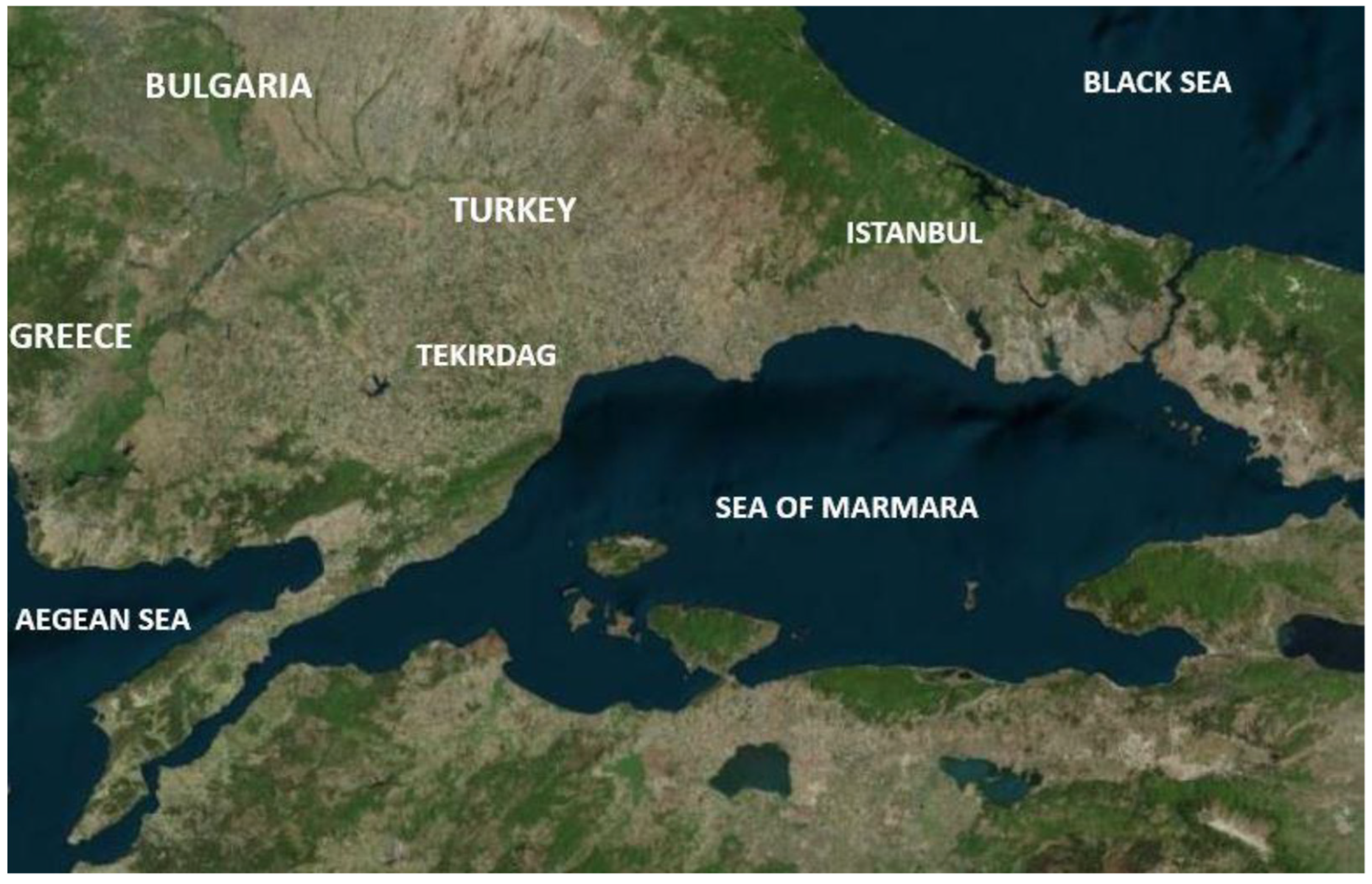
2.1. Sampling Area
2.2. Animals and Sample Collection
2.3. DNA Extraction
2.4. Agarose Gel Electrophoresis
2.5. Statistical Analysis
3. Results
4. Discussion
5. Conclusions
Author Contributions
Funding
Data Availability Statement
Conflicts of Interest
References
- Anonymous. World Cat Day. BBC News (in Turkish). 2021. Available online: https://bbc.com/turkce/haberler-dunya-56099552 (accessed on 14 July 2021).
- Baneth, G.; Thamsborg, S.M.; Otranto, D.; Guillot, J.; Blaga, R.; Deplazes, P.; Solano-Gallego, L. Major parasitic zoonoses associated with dogs and cats in Europe. J. Comp. Pathol. 2016, 155, 54–74. [Google Scholar] [CrossRef] [Green Version]
- Inci, A.; Doganay, M.; Ozdarendeli, A.; Duzlu, O.; Yildirim, A. Overview of zoonotic diseases in Turkey: The One health concept and future threats. Turk. J. Parasitol. 2018, 42, 39–80. [Google Scholar] [CrossRef] [Green Version]
- Moritz, E.D.; Winton, C.S.; Tonnetti, L.; Townsend, R.L.; Berardi, V.P.; Hewins, M.E.; Weeks, K.E.; Dodd, R.Y.; Stramer, S.L. Screening for Babesia microti in the U.S. blood supply. N. Engl. J. Med. 2016, 8, 2236–2245. [Google Scholar] [CrossRef] [Green Version]
- Pennisi, M.G.; Alongi, A.; Agnone, A.; Vitale, F.; Reale, S.; Torina, A. Cats as reservoir of Babesia microti. Parassitologia 2007, 49, 100. [Google Scholar]
- Criado-Fornelio, A.; Martinez-Marcos, A.; Buling-Saraña, A.; Barba-Carretero, J.C. Presence of Mycoplasma haemofelis, Mycoplasma haemominutum and piroplasmids in cats from southern Europe: A molecular study. Vet. Microbiol. 2003, 10, 307–317. [Google Scholar] [CrossRef]
- Malagon, F.; Tapia, J.L. Experimental transmission of Babesia microti infection by the oral route. Parasitol. Res. 1994, 80, 645–648. [Google Scholar] [CrossRef] [PubMed]
- Dogan, M. Investigation of zoonotic pathogens in patients with tick attachment in Tekirdag, Turkey. Klimik. J. 2019, 32, 324–328. [Google Scholar] [CrossRef]
- Baneth, G.; Sheiner, A.; Eyal, O.; Hahn, S.; Beaufils, J.P.; Anug, Y.; Talmi-Frank, D. Redescription of Hepatozoon felis (Apicomplexa: Hepatozoidae) based on phylogenetic analysis, tissue and blood form morphology, and possible transplacental transmission. Parasit. Vectors. 2013, 15, 102. [Google Scholar] [CrossRef] [Green Version]
- Wang, J.L.; Li, T.T.; Liu, G.H.; Zhu, X.Q.; Yao, C. Two tales of Cytauxzoon felis infections in domestic cats. Clin. Microbiol. Rev. 2017, 30, 861–885. [Google Scholar] [CrossRef] [PubMed] [Green Version]
- Reif, K.E.; Macaluso, K.R. Ecology of Rickettsia felis: A review. J. Med. Entomol. 2009, 46, 723–736. [Google Scholar] [CrossRef] [Green Version]
- Rolain, J.M.; Franc, M.; Davoust, B.; Raoult, D. Molecular detection of Bartonella quintana, B. koehlerae, B. henselae, B. clarridgeiae, Rickettsia felis, and Wolbachia pipientis in cat fleas, France. Emerg. Infect. Dis. 2003, 9, 338–342. [Google Scholar] [CrossRef] [PubMed]
- Wechtaisong, W.; Bonnet, S.I.; Lien, Y.Y.; Chuang, S.T.; Tsai, Y.L. Transmission of Bartonella henselae within Rhipicephalus sanguineus: Data on the potential vector role of the tick. PLoS Negl. Trop. Dis. 2020, 1, e0008664. [Google Scholar]
- Król, N.; Militzer, N.; Stobe, E.; Nijhof, A.M.; Pfeffe, M.; Kempf, V.A.J.; Obiegala, A. Evaluating transmission paths for three different Bartonella spp. in Ixodes ricinus ticks using artificial deeding. Microorganisms 2021, 22, 901. [Google Scholar] [CrossRef]
- Snellgrove, A.N.; Krapiunaya, I.; Ford, S.L.; Stanley, H.M.; Wickson, A.G.; Hartzer, K.L.; Levin, M.L. Vector competence of Rhipicephalus sanguineus sensu stricto for Anaplasma platys. Ticks Tick Borne Dis. 2020, 11, 101517. [Google Scholar] [CrossRef]
- Dahlgren, F.S.; Heitman, K.N.; Drexler, N.A.; Massung, R.F.; Behravesh, C.B. Human granulocytic anaplasmosis in the United States from 2008 to 2012: A summary of national surveillance data. Am. J. Trop. Med. Hyg. 2015, 93, 66–72. [Google Scholar] [CrossRef] [PubMed] [Green Version]
- Pennisi, M.G.; Hofmann-Lehmann, R.; Radford, A.D.; Tasker, S.; Belák, S.; Addie, D.D.; Boucraut-Baralon, C.; Egberink, H.; Frymus, T.; Gruffydd-Jones, T.; et al. Anaplasma, Ehrlichia and Rickettsia species infections in cats: European guidelines from the ABCD on prevention and management. J. Feline Med. Surg. 2017, 19, 542–548. [Google Scholar] [CrossRef] [PubMed] [Green Version]
- Ng-Nguyen, D.; Hii, S.F.; Hoang, M.T.; Nguyen, V.T.; Rees, R.; Stenos, J.; Traub, R.J. Domestic dogs are mammalian reservoirs for the emerging zoonosis flea-borne spotted fever, caused by Rickettsia felis. Sci. Rep. 2020, 5, 4151. [Google Scholar] [CrossRef]
- Stanek, G.; Strle, F. Lyme borreliosis-from tick bite to diagnosis and treatment. FEMS Microbiol. Rev. 2018, 42, 233–258. [Google Scholar] [CrossRef] [Green Version]
- Pal, U.; Kitsou, C.; Drecktrah, D.; Yas, O.B.; Fikrig, E. Interactions between ticks and lyme disease spirochetes. Curr. Issues Mol. Biol. 2021, 42, 113–144. [Google Scholar] [PubMed]
- Maggi, R.G.; Compton, S.M.; Trull, C.L.; Mascarelli, P.E.; Mozayeni, B.R.; Breitschwerdt, E.B. Infection with hemotropic Mycoplasma species in patients with or without extensive arthropod or animal contact. J. Clin. Microbiol. 2013, 51, 3237–3241. [Google Scholar] [CrossRef] [PubMed] [Green Version]
- Torres-Guerrero, E.; Quintanilla-Cedillo, M.R.; Ruiz-Esmenjaud, J.; Arenas, R. Leishmaniasis: A review. F1000Res 2017, 26, 750. [Google Scholar] [CrossRef] [PubMed]
- Pennisi, M.G.; Cardoso, L.; Baneth, G.; Bourdeau, P.; Koutinas, A.; Miró, G.; Oliva, G.; Solano-Gallego, L. LeishVet update and recommendations on feline leishmaniosis. Parasite Vectors 2015, 4, 302. [Google Scholar] [CrossRef] [Green Version]
- Morganti, G.; Veronesi, F.; Stefanetti, V.; Di Muccio, T.; Fiorentino, E.; Diaferia, M.; Passamonti, F.; Gramiccia, M. Emerging feline vector-borne pathogens in Italy. Parasite Vectors 2019, 2, 193. [Google Scholar] [CrossRef]
- Morelli, S.; Colombo, M.; Dimzas, D.; Barlaam, A.; Traversa, D.; Di Cesare, A.; Russi, I.; Spoletini, R.; Paoletti, B.; Diakou, A. Leishmania infantum Seroprevalence in Cats From Touristic Areas of Italy and Greece. Front. Vet. Sci. 2020, 11, 616566. [Google Scholar] [CrossRef]
- Spada, E.; Perego, R.; Vitale, F.; Bruno, F.; Castelli, G.; Tarantola, G.; Baggiani, L.; Magistrelli, S.; Proverbio, D. Feline Leishmania spp. infection in a non- endemic area of Northern Italy. Animals 2020, 10, 817. [Google Scholar] [CrossRef]
- Can, H.; Doskaya, M.; Ozdemir, H.G.; Sahar, E.A.; Karakavuk, M.; Pektas, B.; Karakus, M.; Toz, S.; Caner, A.; Doskaya, A.D.; et al. Seroprevalence of Leishmania infection and molecular detection of L. tropica and L. infantum in stray cats of Izmir, Turkey. Exp. Parasitol. 2016, 167, 109–114. [Google Scholar] [CrossRef] [PubMed]
- Kalmár, Z.; Dumitrache, M.O.; D’Amico, G.; Matei, I.A.; Ionică, A.M.; Gherman, C.M.; Lupșe, M.; Mihalca, A.D. Multiple tick-borne pathogens in Ixodes ricinus ticks collected from humans in Romania. Pathogens 2020, 19, 390. [Google Scholar] [CrossRef] [PubMed]
- Persing, D.H.; Mathiesen, D.; Marshall, W.F.; Telford, S.R.; Spielman, A. Detection of B.microti by polymerase chain reaction. J. Clin. Microbiol. 1992, 30, 2097–2103. [Google Scholar] [CrossRef] [PubMed] [Green Version]
- Duarte, S.C.; Linhares, G.F.C.; Romanowsky, T.N.; Neto, O.J.S.; Borges, L.M.F. Assessment of primers designed for the subspecies-specific discrimination among Babesia c. canis, B. c. vogeli and B. C. rossi by PCR assay. Vet. Parasitol. 2008, 152, 16–20. [Google Scholar] [CrossRef]
- Brown, H.M.; Latimer, K.S.; Erikson, L.E.; Cashwell, M.E.; Britt, J.O. Detection of persistent Cytauxzoon felis infection by polymerase chain reaction in three asymptomatic domestic cats. J. Vet. Diag. Investig. 2008, 20, 485–488. [Google Scholar] [CrossRef] [Green Version]
- Cabello, J.; Altet, L.; Napolitano, C.; Sastre, N.; Hidalgo, E. Survey of infectious agents in the endangered Darwin’s fox (Lycalopex fulvipes): High prevalence and diversity of hemotrophic mycoplasmas. Vet. Microbiol. 2013, 27, 448–542. [Google Scholar] [CrossRef]
- Lima, M.L.F.I.; Soares, P.T.I.; Ramos, C.A.N.; Araujo, F.R.; Ramos, R.A.N. Molecular detection of A. platys in a naturally-infected cat in Brazil. Brazil. J. Microbiol. 2010, 41, 381–385. [Google Scholar] [CrossRef] [Green Version]
- de la Fuente, J.; Massung, R.F.; Wong, S.J.; Chu, F.K.; Lutz, H. Analysis of the msp4 gene of A. phagocytophilum strains. J. Clin. Microbiol. 2005, 43, 1309–1317. [Google Scholar] [CrossRef] [PubMed] [Green Version]
- Prusinski, M.A.; Kokas, J.E.; Hukey, K.T.; Kogut, S.J.; Lee, J. Prevalence of Borrelia burgdorferi, Anaplasma phagocytophilum, and Babesia microti in Ixodes scapularis collected from recreational lands in the Hudson valley region, New York State. J. Med. Entomol. 2014, 51, 226–236. [Google Scholar] [CrossRef] [PubMed]
- Leulmi, H.; Aouadi, A.; Bitam, I.; Bessas, A.; Benakhla, A. Detection of B. tamiae, C. burnetii and rickettsiae in arthropods and tissues from wild and domestic animals in northeastern Algeria. Parasite Vectors 2016, 20, 27. [Google Scholar] [CrossRef]
- Isozumi, R.; Fukui, M.; Kaneko, A.; Chan, C.W.; Kawamoto, F. Improved detection of malaria cases in island settings of Vanuatu and Kenya by PCR that targets the Plasmodium mitochondrial cytochrome c oxidase III gene. Parasitol. Internat. 2015, 64, 304–308. [Google Scholar] [CrossRef] [PubMed]
- Sharief, A.H.; Khalil El-Tahir, A.G.; Barker, D.C.; Omer, S.A.; Abdalla, H. Simple and direct characterization of L. donovani isolates based on cytochrome oxidase II gene sequences. Open Trop. Med. J. 2011, 4, 1–5. [Google Scholar] [CrossRef]
- Wagner, E.R.; Bremer, W.G.; Rikihisa, Y.; Ewing, S.A.; Needham, G.R. Development of a p28-based PCR assay for E. chaffeensis. Mol. Cell Probes. 2004, 18, 111–116. [Google Scholar] [CrossRef] [Green Version]
- Diniz, P.P.; Schulz, B.S.; Hartmann, K.; Breitschwerdt, E.B. “C Neoehrlichia mikurensis” infection in a dog from Germany. J. Clin. Microbiol. 2011, 49, 2059–2062. [Google Scholar]
- Usluca, S.; Celebi, B.; Karasartova, D.; Gureser, A.S.; Matur, F.; Oktem, M.A.; Sozen, M.; Karatas, A.; Babur, C.; Mumcuoglu, K.Y.; et al. Molecular survey of Babesia microti (Aconoidasida: Piroplasmida) in wild rodents in Turkey. J. Med. Entomol. 2019, 28, 1605–1609. [Google Scholar] [CrossRef]
- Penzhorn, B.L.; Oosthuizen, M.C. Babesia species of domestic cats: Molecular characterization has opened pandora’s box. Front. Vet. Sci. 2020, 27, 134. [Google Scholar] [CrossRef]
- Duzlu, O.; Inci, A.; Yildirim, A.; Onder, Z.; Ciloglu, A. The investigation of some tick-borne protozoon and rickettsial infections in dogs by Real Time PCR and the molecular characterizations of the detected isolates. Ankara Univ. Vet. Fac. J. 2014, 61, 275–282. [Google Scholar]
- Karasartova, D.; Gureser, A.S.; Gokce, T.; Celebi, B.; Yapar, D.; Keskin, A.; Celik, S.; Ece, Y.; Erenler, A.K.; Usluca, S.; et al. Bacterial and protozoal pathogens found in ticks collected from humans in Corum province of Turkey. PLoS Negl. Trop. Dis. 2018, 12, e0006395. [Google Scholar] [CrossRef] [PubMed]
- Kaya, M. Investigation of Frequency of Babesiosis among Human Who Have Tick Bite History Living in Tatvan Region. Master’s Thesis, Erciyes University, Kayseri, Turkey, 2011. [Google Scholar]
- Attipa, C.; Papasouliotis, K.; Solano-Gallego, L. Prevalence study and risk factor analysis of selected bacterial, protozoal and viral, including vector-borne, pathogens in cats from Cyprus. Parasite Vectors 2017, 10, 130. [Google Scholar] [CrossRef] [Green Version]
- Giannelli, A.; Latrofa, M.S.; Nachum-Biala, Y. Three different Hepatozoon species in domestic cats from southern Italy. Ticks Tick Borne Dis. 2017, 8, 721–724. [Google Scholar] [CrossRef]
- Ortuño, A.; Castellà, J.; Criado-Fornelio, A. Molecular detection of a Hepatozoon species in stray cats from a feline colony in North-Eastern Spain. Vet. J. 2018, 177, 134–136. [Google Scholar] [CrossRef]
- Vilhena, H.; Martinez-Díaz, V.L.; Cardoso, L. Feline vector-borne pathogens in the north and centre of Portugal. Parasite Vectors. 2013, 6, 99. [Google Scholar] [CrossRef] [PubMed] [Green Version]
- Tuna, G.E.; Bakirci, S.; Dinler, C.; Battal, G.; Ulutas, B. Molecular identification and clinicopathological findings of Hepatozoon sp. infection in a cat: First report from Turkey. Turk. Parasitol. J. 2018, 42, 286–289. [Google Scholar] [CrossRef]
- Ceylan, O.; Xuan, X.; Sevinc, F. Primary tick-borne protozoan and rickettsial infections of animals in Turkey. Pathogens. 2021, 19, 231. [Google Scholar] [CrossRef]
- Rizzi, T.E.; Reichard, M.V.; Cohn, L.A.; Birkenheuer, A.J.; Taylor, J.D. Prevalence of Cytauxzoon felis infection in healthy cats from enzootic areas in Arkansas, Missouri, and Oklahoma. Parasite Vectors 2015, 8, 13–16. [Google Scholar] [CrossRef] [PubMed] [Green Version]
- Zou, F.C.; Li, Z.; Yang, J.F.; Chang, J.Y.; Liu, G.H.; Lv, Y.; Zhu, X.Q. Cytauxzoon felis infection in domestic cats, Yunnan province, China. Emerg. Infect. Dis. 2019, 25, 353–354. [Google Scholar] [CrossRef] [Green Version]
- Rahmati, M.M.; Zaeemi, M.; Razmi, G.R. Preliminary study of Cytauxzoon felis infection in outdoor cats in Mashhad, Iran. Parasitol. Res. 2020, 119, 4177–4183. [Google Scholar] [CrossRef]
- Karaca, M.; Akkan, H.A.; Tutuncu, M.; Ozdal, N.; Deger, S. Cytauxzoonosis in Van cats. Yuz. Y. Univ. Vet. Med. J. 2007, 18, 37–39. [Google Scholar]
- Celebi, B.; Kilic, S.; Aydin, N. Investigation of Bartonella henselae in cats in Ankara, Turkey. Zoonoses Public Health 2009, 56, 169–175. [Google Scholar] [CrossRef] [PubMed]
- Sigirci, B.D.; Ilgaz, A. Detection of the Presence of Bartonella henselae in cats in Istanbul. J. Fac. Vet. Med. Istanbul Univ. 2019, 39, 209–217. [Google Scholar]
- Kirkan, S.; Parin, U.; Yuksel, H.T. Investigation of Bartonella henselae and Bartonella clarridgeiae prevalance in cats. Ann. Rev. Res. 2019, 5, 64–69. [Google Scholar]
- Maden, M.; Dogan, M.; Altıntas, G.; Yıldız, E.E.; Ekik, M.; Ince, E.M.; Kose, S.I. Prevalence of Bartonella henselae in pet and stray cats from the aspect of public health: A research sample in the concept of one medicine-one health: A research sample in the concept of one medicine—one health. Kafkas Univ. Vet. Fac. J. 2015, 21, 313–317. [Google Scholar]
- Guzel, M.; Celebi, B.; Yalcin, E.; Koenhemsi, L.; Mamak, N.; Pasa, S.; Aslan, O. A serological investigation of Bartonella henselae infection in cats in Turkey. J. Vet. Med. Sci. 2011, 73, 1513–1516. [Google Scholar] [CrossRef] [Green Version]
- Aydin, N.; Korkmazgil, B.; Kirkan, S.; Telli, M.; Eyigor, M.; Aksoy, A.M.; Parin, U.; Tekbiyik, S. Seropositivity of Bartonella henselae in risky human population, cats and dogs. Meandros Med. Dent. J. 2019, 20, 51–56. [Google Scholar] [CrossRef]
- Sayin-Kutlu, S.; Ergin, C.; Kutlu, M. Bartonella henselae seroprevalence in cattle breeders and veterinarians in the rural areas of Aydin and Denizli, Turkey. Zoon. Publ. Health. 2012, 59, 445–449. [Google Scholar] [CrossRef]
- Yilmaz, C. Borrelia Henselae Seroprevalence in Blood Donors from Denizli Area (in Turkish); Tip Uzmanlik Tezi. P.Ü: Denizli, Türkiye, 2008. [Google Scholar]
- Souza, A.M.; Almosny, N.R.P.; Favacho, A.R.M.; Almeida, D.N.P.; Ferreira, R.F.; Ferreira, E.O.; Moreira, N.S.; Lemos, E.R.S. Bartonella spp. and hematological changes in privately owned domestic cats from Rio de Janeiro. Brazil. J. Infect. Dev. Ctries 2017, 5, 591–596. [Google Scholar] [CrossRef] [PubMed] [Green Version]
- Pons, I.; Sanfeliu, I.; Quesada, M.; Anton, E.; Sampere, M.; Font, B.; Pla, J.; Segura, F. Prevalence of Bartonella henselae in cats in Catalonia, Spain. Am. J. Trop. Med. Hyg. 2005, 72, 453–457. [Google Scholar] [CrossRef] [Green Version]
- Gutiérrez, R.; Morick, D.; Gross, I.; Winkler, R.; Abdeen, Z.; Harrus, S. Bartonellae in domestic and stray cats from Israel: Comparison of bacterial cultures and high-resolution melt real-time PCR as diagnostic methods. Vector Borne Zoonotic Dis. 2013, 13, 857–864. [Google Scholar] [CrossRef]
- Bergmans, A.M.; de Jong, C.M.; van Amerongen, G. Prevalence of Bartonella species in domestic cats in the Netherlands. J. Clin. Microbiol. 1997, 35, 2256–2261. [Google Scholar] [CrossRef] [Green Version]
- Morelli, S.; Crisi, P.E.; Di Cesare, A.; De Santis, F.; Barlaam, A.; Santoprete, G.; Parrinello, C.; Palermo, S.; Mancini, P.; Traversa, D. Exposure of client-owned cats to zoonotic vector-borne pathogens: Clinic-pathological alterations and infection risk analysis. Comp. Immunol. Microbiol. Infect. Dis. 2019, 66, 101344. [Google Scholar] [CrossRef]
- Diakou, A.; Di Cesare, A.; Accettura, P.M. Intestinal parasites and vector-borne pathogens in stray and free-roaming cats living in continental and insular Greece. PLoS Negl. Trop. Dis. 2017, 11, e0005335. [Google Scholar] [CrossRef] [Green Version]
- Chmielewski, T.; Podsiadły, E.; Tylewska-Wierzbanowska, S. Presence of Bartonella spp. in various human populations. Pol. J. Microbiol. 2007, 56, 33–38. [Google Scholar]
- Giladi, M.; Shapira, L.; Trever, A.; Velan, A.; Rasis, M.; Ephros, M. How do humans acquire cat scratch disease? A study among stray cats and human patients. In Proceedings of the 9th International Conference. Bartonnella as Emerging Pathogens, Paris, France, 18–20 September 2019; pp. 40–42. [Google Scholar]
- Guptill, L.; Wu, C.C.; Hogenesch, H. Prevalence, risk factors, and genetic diversity of Bartonella henselae infections in pet cats in four regions of the United States. J. Clin. Microbiol. 2004, 42, 652–659. [Google Scholar] [CrossRef] [Green Version]
- Nelson, C.A.; Saha, S.; Mead, P.S. Cat-Scratch Disease in the United States, 2005−2013. Emerg. Infect. Dis. 2016, 22, 1741–1746. [Google Scholar] [CrossRef]
- Massei, F.; Messina, F.; Gori, L. High prevalence of antibodies to Bartonella henselae among Italian children without evidence of cat-scratch disease. Clin. Infect. Dis. 2004, 38, 145–148. [Google Scholar] [CrossRef] [Green Version]
- Celebi, B. Bartonella henselae and its infections. Mikrobiyol. Bul. 2008, 42, 163–175. [Google Scholar]
- Sen, E.; Uchishima, Y.; Okamoto, Y. Molecular detection of A. phagocytophilum and Borrelia burgdorferi in Ixodes ricinus ticks from Istanbul metropolitan area and rural Trakya (Thrace) region of northwestern Turkey. Ticks Tick-Borne Dis. 2011, 2, 94–98. [Google Scholar] [CrossRef] [PubMed]
- Kar, S.; Yilmazer, N.; Midilli, K. Borrelia burgdorferi s.l. and Rickettsia spp. in ticks collected from European part of Turkey. Kafkas Univ. Vet. Fak. Derg. 2013, 19, 19–24. [Google Scholar] [CrossRef] [Green Version]
- Gulanber, E.G.; Gulanber, A.; Albayrak, R. Lyme disease (Borreliosis) in a Saint Bernard dog: First clinical case in Turkey. J. Vet. Anim. Sci. 2007, 31, 367–369. [Google Scholar]
- Orkun, O.; Karaer, Z.; Cakmak, A.; Nalbantoglu, S. Identification of tick-borne pathogens in ticks feeding on humans in Turkey. PLoS Negl. Trop. Dis. 2014, 7, e3067. [Google Scholar] [CrossRef] [PubMed]
- Calisir, B.; Polat, E.; Guney, G.; Gonenc, L. Investigation on the species composition of the ixodid ticks from Belgrade forest in Istanbul and their role as vectors of Borrelia burgdorferi. Acta Zool. Bulg. 2000, 52, 23–28. [Google Scholar]
- Geurden, T.; Becskei, C.; Six, R.H. Detection of tick-borne pathogens in ticks from dogs and cats in different European countries. Ticks Tick Borne Dis. 2018, 9, 1431–1436. [Google Scholar] [CrossRef]
- Little, S.E.; Barrett, A.W.; Nagamori, Y. Ticks from cats in the United States: Patterns of infestation and infection with pathogens. Vet. Parasitol. 2018, 15, 15–20. [Google Scholar] [CrossRef] [PubMed]
- Cora, M.; Kaklikkaya, N.; Topbas, M.; Can, G.; Yavuzyilmaz, A.; Tosun, I.; Aydin, F. Determination of seroprevalence of Borrelia burgdorferi IgG in adult population living in Trabzon. Balkan Med. J. 2017, 34, 47–52. [Google Scholar] [CrossRef]
- Rudenko, N.; Golovchenko, M. Sexual transmission of Lyme borreliosis? The question that calls for an answer. Trop. Med. Infect. Dis. 2021, 24, 87. [Google Scholar] [CrossRef]
- Littman, M.P.; Gerber, B.; Goldstein, R.E. ACVIM consensus update on Lyme borreliosis in dogs and cats. J. Vet. Intern. Med. 2018, 32, 887–903. [Google Scholar] [CrossRef] [PubMed]
- Žákovská, A.; Schánilec, P.; Treml, F. Seroprevalence of antibodies against Borrelia burgdorferi and L. interrogans in cats in district of Brno and its environs, the Czech Republic. Ann. Agric. Environ. Med. 2020, 11, 356–360. [Google Scholar] [CrossRef]
- Yucesan, B.C.; Babur, C.; Sezen, F.; Nalbantoglu, S. Ticks species biting humans in Ankara: Species diversity, hosts and geographical distribution. Turk. Hyg. Exp. Biol. J. 2019, 76, 3–14. [Google Scholar] [CrossRef]
- Schäfer, I.; Kohn, B. Anaplasma phagocytophilum infection in cats: A literature review to raise clinical awareness. J. Feline Med. Surg. 2020, 22, 428–441. [Google Scholar] [CrossRef] [PubMed] [Green Version]
- Gunes, T.; Poyraz, O.; Ataş, M.; Turgut, N.H. The seroprevalence of Anaplasma phagocytophilum in humans from two different climatic regions of Turkey and its co-seroprevalence rate with Borrelia burgdorferi. Turk. J. Med. Sci. 2011, 41, 903–908. [Google Scholar]
- Ongut, G.; Ogunc, D.; Mutlu, G.; Colak, D.; Gultekin, M.; Gunseren, F.; Donmez, L.; Tuncer, D. Seroprevalence of antibodies to Anaplasma phagocytophilum in Antalya, Turkey. Infection 2006, 34, 107–109. [Google Scholar] [CrossRef]
- Kilic, H.; Gurcan, S.; Kunduracilar, H.; Eskiocak, M. Anaplasmosis seropositivity in people exposured to tick bite. Thrace Univ. Med. Fac. J. 2010, 27, 79–82. [Google Scholar]
- Eisenstein, T. Human Granulocytic Anaplasmosis in Connecticut, 2014–2019: An Analysis of Surveillance Datas and Future Recommendations. 2020, Public Health Theses. Available online: https://elischolar.library.yale.edu/cgi/viewcontent.cgi?article=1929&context=ysphtdl (accessed on 14 July 2021).
- Moniuszko-Malinowska, A.; Dunaj, J.; Andersson, M.O.; Chmielewski, T.; Czupryna, P.; Groth, M.; Grygorczuk, S.; Zajkowska, J.; Kondrusik, M.; Kruszewska, E.; et al. Anaplasmosis in Poland—analysis of 120 patients. Ticks Tick Borne Dis. 2021, 2, 101763. [Google Scholar] [CrossRef]
- Azagi, T.; Hoornstra, D.; Kremer, K.; Hovius, J.W.R.; Sprong, H. Evaluation of disease causality of rare Ixodes ricinus-borne infections in Europe. Pathogens 2020, 24, 150. [Google Scholar] [CrossRef] [Green Version]
- Oter, K.; Cetinkaya, H.; Vurusener, C. Molecular detection and typing of Anaplasma species in small ruminants in Thrace Region of Turkey. Kafkas Univ. J. Vet. Med. 2016, 22, 133–138. [Google Scholar]
- Cetinkaya, H.; Matur, E.; Akyazi, I.; Ekiz, E.E.; Aydin, L.; Toparlak, M. Serological and molecular investigation of Ehrlichia spp. and Anaplasma spp. in ticks and blood of dogs, in the Thrace Region of Turkey. Ticks Tick Borne Dis. 2016, 7, 706–714. [Google Scholar] [CrossRef] [PubMed]
- Aktas, M.; Vatansever, Z.; Altay, K. Molecular evidence for Anaplasma phagocytophilum in Ixodes ricinus from Turkey. Trans. R. Soc. Trop. Med. Hyg. 2010, 1, 10–15. [Google Scholar] [CrossRef]
- Gargili, A.; Palomar, A.M.; Midilli, K.; Portillo, A.; Kar, S.; Oteo, J.A. Rickettsia species in ticks removed from humans in Istanbul, Turkey. Vector Borne Zoonotic Dis. 2012, 12, 938–941. [Google Scholar] [CrossRef]
- Hoque, M.M.; Barua, S.; Kelly, P.J.; Chenoweth, K.; Kaltenboeck, B.; Wang, C. Identification of Rickettsia felis DNA in the blood of domestic cats and dogs in the USA. Parasite Vectors 2020, 18, 581. [Google Scholar] [CrossRef] [PubMed]
- Lappin, M.R. Update on flea and tick associated diseases of cats. Vet. Parasitol. 2018, 30, 26–29. [Google Scholar] [CrossRef] [PubMed]
- Borsan, S.D.; Ionică, A.M.; Galon, C. High diversity, prevalence, and co-infection rates of tick-borne pathogens in ticks and wildlife hosts in an urban area in Romania. Front. Microbiol. 2021, 12, 645–650. [Google Scholar]
- Stanley, H.; Rhodes, D.V.L. Presence of Rickettsia species in ticks collected from companion animals in Northeastern Georgia, United States. Vet. Sci. 2021, 26, 37. [Google Scholar] [CrossRef]
- Dougas, G.; Tsakris, A.; Billinis, C. Molecular detection of R. felis in common fleas in Greece and comparative evaluation of genotypic methods. J. Microbiol. Methods. 2021, 180, 106104. [Google Scholar] [CrossRef]
- Capelli, G.; Montarsi, F.; Porcellato, E. Occurrence of Rickettsia felis in dog and cat fleas (Ctenocephalides felis ) from Italy. Parasite Vectors 2009, 2, S8. [Google Scholar] [CrossRef] [Green Version]
- Pascucci, I.; Di Domenico, M.; Curini, V.; Cocco, A.; Averaimo, D.; D’Alterio, N.; Cammà, C. Diversity of Rickettsia in ticks collected in Abruzzi and Molise Regions (Central Italy). Microorganisms 2019, 13, 696. [Google Scholar] [CrossRef] [Green Version]
- Zurita, A.; Benkacimi, L.; El Karkouri, K.; Cutillas, C.; Parola, P.; Laroche, M. New records of bacteria in different species of fleas from France and Spain. Comp. Immunol. Microbiol. Infect. Dis. 2021, 76, 101648. [Google Scholar] [CrossRef] [PubMed]
- Teoh, Y.T.; Hii, S.F.; Stevenson, M.A. Serological evidence of exposure to Rickettsia felis and Rickettsia typhi in Australian veterinarians. Parasite Vectors 2017, 13, 129. [Google Scholar] [CrossRef] [PubMed] [Green Version]
- Hun, L.; Troyo, A. An update on the detection and treatment of Rickettsia felis. Res. Rep. Trop. Med. 2012, 21, 47–55. [Google Scholar] [CrossRef] [PubMed] [Green Version]
- Tuzer, E.; Goksu, K.; Bilal, T.; Yesildere, T. A Case of haemobartonellosis in a cat in Istanbul. J. Protozool. Res. 1993, 3, 69–70. [Google Scholar]
- Aslan, O. Hemotropic mycoplasmas: From Haemobartonella to Mycoplasma. J. Adv. Vet. Biol Sci. Tec. 2016, 1, 31–40. [Google Scholar]
- Cetinkaya, H.; Haktanir, D.; Arun, S.; Vurusaner, C. Molecular detection and prevalence of feline hemotropic mycoplasmas in Istanbul, Turkey. Acta Parasitol. 2016, 61, 165–171. [Google Scholar] [CrossRef] [PubMed]
- Sarvani, E.; Tasker, S.; Kovacević, M.F. Prevalence and risk factor analysis for feline haemoplasmas in cats from Northern Serbia, with molecular subtyping of feline immunodeficiency virus. JFMS Open Rep. 2018, 22, 2055116918770037. [Google Scholar] [CrossRef]
- Imre, M.; Văduva, C.; Dărăbuș, G. Molecular detection of hemotropic mycoplasmas (hemoplasmas) in domestic cats in Romania. BMC Vet. Res. 2020, 22, 399–401. [Google Scholar] [CrossRef]
- Jenkins, K.S.; Dittmer, K.E.; Marshall, J.C. Prevalence and risk factor analysis of feline haemoplasma infection in New Zealand domestic cats using a real-time PCR assay. J. Feline Med. Surg. 2013, 15, 1063–1069. [Google Scholar] [CrossRef]
- Ravagnan, S.; Carli, E.; Piseddu, E. Prevalence and molecular characterization of canine and feline hemotropic mycoplasmas (hemoplasmas) in northern Italy. Parasite Vectors 2017, 13, 132–136. [Google Scholar] [CrossRef] [Green Version]


| No | Identified Pathogens | Primer Sequences (5′–3′) | Annealing/°C | Product Size bp | Reference |
|---|---|---|---|---|---|
| 1 | Babesia microti | F: ATAGGTCAGAAACTTGAATGATACA R: CTTAGTATAAGCTTTTATACAGC | 55 | 238 | [29] |
| 2 | Babesia canis canis | F: GTGAACCTTATCACTTAAAGG R: CTACACAGAGCACACAGCC | 56 | 746 | [30] |
| 3 | Cytauxzoon felis | F: CCAGCTCCAATAGCGTATATT R: AGGATGAACTCGATGAATGCA | 61 | 431 | [31] |
| 4 | Hepatozoon felis | F: CTTACCGTGGCAGTGACGGT R: TGTTATTTCTTGTCACTACCTCTCTTATGC | 58 | 146 | [32] |
| 5 | Anaplasma platys | F: GATTTTTGTCGTAGCTTGCTATG R: TAGCACTCATCGTTTACAGC | 55 | 678 | [33] |
| 6 | A. phagocytophilum | F: ATGAATTACAGAGAATTGCTTGTAGG R: TTAATTGAAAGCAAATCTTGCTCCTATG | 54 | 849 | [34] |
| 7 | Borrelia burgdorferi | F: AATAGGTCTAATATTAGCCTTAATAGC R: TCAAGTCTGGTTCCGTCTGCTC | 60 | 417 | [35] |
| 8 | Bartonella henselae | F: TTCCGYCTTATGGGTTTTGG R: CATTTCTGTTGGAAATCCTAG | 52 | 246 | [12] |
| 9 | Rickettsia felis | F: CCGATTCAGCAGGTTCTTCAA R: ATGTTCGGGCTTCCGGTATG | 57 | 120 | [36] |
| 10 | Hemotropic Mycoplasma spp | F: GCCCATATTCCTACGGGAAGCAGCAGT R: CTCCACCACTTGTTCAGGTCCCCGTC | 68 | 620 | [21] |
| 11 | Plasmodium spp | F: CCTGTTATCCCCGGCGAACCTTC R: CTCGCCATTTGATAGCGGTTAACC | 63 | 937 | [37] |
| 12 | Leishmaniadonovani | F: GGCATAAATCCATGTAAGA R: TGGCTTTTATATTATCATTTT | 54 | 540 | [38] |
| 13 | Ehrlichia chaffeensis | F: AGATACTTCAAGCTCTATTC R: AGGTAGTGGTATTAACGG | 49 | 277 | [39] |
| 14 | Neoehrlichia mikurensis | F: AACAGGTGAAACACTAGATAAGTCCAT R: TTCTACTTTGAACATTTGAAGAATTACTAT | 58 | 950 | [40] |
| Pathogens | Frequency | ||||
|---|---|---|---|---|---|
| − | + | + (Row %) | χ2 | p Value | |
| Babesia microti | 163 | 4 | 2.4 | 152.26 | <0.001 |
| Adjusted residual (z-score) | 5.5 | −5.5 | |||
| p value * | <0.001 | <0.001 | |||
| Babesia canis canis | 127 | 40 | 24.0 | ||
| Adjusted residual (z-score) | −2.1 | 2.1 | |||
| p value * | 0.036 | 0.036 | |||
| Cytauxzoon felis | 156 | 11 | 6.6 | ||
| Adjusted residual (z-score) | 4.1 | −4.1 | |||
| p value * | <0.001 | <0.001 | |||
| Hepatozoon felis | 149 | 18 | 10.8 | ||
| Adjusted residual (z-score) | 2.6 | −2.6 | |||
| p value * | 0.009 | 0.009 | |||
| Anaplasma phagocytophilum | 155 | 12 | 7.2 | ||
| Adjusted residual (z-score) | 3.8 | −3.8 | |||
| p value * | <0.001 | <0.001 | |||
| Anaplasma platys | 116 | 51 | 30.5 | ||
| Adjusted residual (z-score) | −4.4 | 4.4 | |||
| p value * | <0.001 | <0.001 | |||
| Rickettsia felis | 123 | 44 | 26.3 | ||
| Adjusted residual (z-score) | −2.9 | 2.9 | |||
| p value * | 0.004 | 0.004 | |||
| Bartonella henselae | 100 | 67 | 40.1 | ||
| Adjusted residual (z-score) | −7.8 | 7.8 | |||
| p value * | <0.001 | <0.001 | |||
| Borrelia burgdorferi | 132 | 35 | 21.0 | ||
| Adjusted residual (z-score) | −1.0 | 1.0 | |||
| p value * | 0.3 | 0.3 | |||
| Hemotropic Mycoplasma | 148 | 19 | 11.4 | ||
| Adjusted residual (z-score) | 2.4 | −2.4 | |||
| p value * | 0.02 | 0.02 | |||
| Frequency | χ2 | p Value | Fisher′s Exact Test p Value | |||
|---|---|---|---|---|---|---|
| Group | – | + | + (Row %) | |||
| Babesia microti | 0.362 | |||||
| Male | 84 | 1 | 1.2 | |||
| Female | 79 | 3 | 3.7 | |||
| Babesia canis canis | 3.78 | 0.052 | ||||
| Male | 70 | 15 | 17.6 | |||
| Female | 57 | 25 | 30.5 | |||
| Cytauxzoon felis | 0.14 | 0.709 | ||||
| Male | 80 | 5 | 5.9 | |||
| Female | 76 | 6 | 7.3 | |||
| Hepatozoon felis | 1.16 | 0.281 | ||||
| Male | 78 | 7 | 8.2 | |||
| Female | 71 | 11 | 13.4 | |||
| Anaplasma phagocytophilum | 3.47 | 0.062 | ||||
| Male | 82 | 3 | 3.5 | |||
| Female | 73 | 9 | 11.0 | |||
| Anaplasma platys | 1.05 | 0.307 | ||||
| Male | 56 | 29 | 34.1 | |||
| Female | 60 | 22 | 26.8 | |||
| Rickettsia felis | 0.318 | 0.573 | ||||
| Male | 61 | 24 | 28.2 | |||
| Female | 62 | 20 | 24.4 | |||
| Bartonella henselae | 1.68 | 0.195 | ||||
| Male | 55 | 30 | 35.3 | |||
| Female | 45 | 37 | 45.1 | |||
| Borrelia burgdorferi | 3.35 | 0.067 | ||||
| Male | 72 | 13 | 15.3 | |||
| Female | 60 | 22 | 26.8 | |||
| Hemotropic Mycoplasma | 10.6 | 0.11 | 0.744 | |||
| Male | 76 | 9 | 12.2 | |||
| Female | 72 | 10 | ||||
| Frequency | χ2 | p Value | Fisher′s Exact Test p Value | |||
|---|---|---|---|---|---|---|
| Group | − | + | + (Row %) | |||
| Babesia microti | 1.000 | |||||
| <1 year | 77 | 2 | 2.5 | |||
| >1 year | 86 | 2 | 2.3 | |||
| Babesia canis canis | 3.20 | 0.074 | ||||
| <1 year | 65 | 14 | 17.7 | |||
| >1 year | 62 | 26 | 29.5 | |||
| Cytauxzoon felis | 1.90 | 0.169 | ||||
| <1 year | 76 | 3 | 3.8 | |||
| >1 year | 80 | 8 | 9.1 | |||
| Hepatozoon felis | 0.55 | 0.458 | ||||
| <1 year | 69 | 10 | 12.7 | |||
| >1 year | 80 | 8 | 9.1 | |||
| Anaplasma phagocytophilum | 7.88 | 0.005 | ||||
| <1 year | 78 | 1 | 1.3 | |||
| >1 year | 77 | 11 | 12.5 | |||
| Anaplasma platys | 0.94 | 0.333 | ||||
| <1 year | 52 | 27 | 34.2 | |||
| >1 year | 64 | 24 | 27.3 | |||
| Rickettsia felis | 0.08 | 0.774 | ||||
| <1 year | 59 | 20 | 25.3 | |||
| >1 year | 64 | 24 | 27.3 | |||
| Bartonella henselae | 0.28 | 0.592 | ||||
| <1 year | 49 | 30 | 38.0 | |||
| >1 year | 51 | 37 | 42.0 | |||
| Borrelia burgdorferi | 0.95 | 0.330 | ||||
| <1 year | 65 | 14 | 17.7 | |||
| >1 year | 67 | 21 | 23.9 | |||
| Hemotropic Mycoplasma | 0.23 | 0.630 | ||||
| <1 year | 71 | 8 | 10.1 | |||
| >1 year | 77 | 11 | 12.5 | |||
| Frequency | χ2 | p Value | |||
|---|---|---|---|---|---|
| Group | − | + | + (Row %) | ||
| Male | 714 | 136 | 16.0 | 4.80 | 0.028 |
| Female | 655 | 165 | 20.1 | ||
| <1 year | 661 | 129 | 16.3 | 2.92 | 0.088 |
| >1 year | 708 | 172 | 19.5 | ||
| Bm | Bcc | Cf | Hf | Ap | Apl | Rf | Bh | Bb | HM | |
|---|---|---|---|---|---|---|---|---|---|---|
| Bm | 1.000 | 0.279 | 0.116 | −0.054 | 0.108 | 0.151 | −0.094 | 0.111 | 0.112 | 0.067 |
| p value | <0.001 | 0.135 | 0.485 | 0.165 | 0.051 | 0.228 | 0.151 | 0.150 | 0.388 | |
| Bcc | 1.000 | 0.134 | 0.031 | 0.061 | 0.115 | 0.047 | 0.170* | 0.228 | −0.024 | |
| p value | 0.085 | 0.689 | 0.432 | 0.138 | 0.550 | 0.028 | 0.003 | 0.755 | ||
| Cf | 1.000 | −0.092 | 0.207 | 0.034 | 0.060 | 0.029 | 0.041 | 0.057 | ||
| p value | 0.236 | 0.007 | 0.667 | 0.438 | 0.711 | 0.597 | 0.465 | |||
| Hf | 1.000 | −0.022 | 0.021 | 0.055 | 0.109 | 0.011 | −0.003 | |||
| p value | 0.778 | 0.787 | 0.479 | 0.159 | 0.890 | 0.970 | ||||
| Ap | 1.000 | 0.017 | −0.061 | 0.103 | 0.028 | −0.027 | ||||
| p value | 0.829 | 0.432 | 0.184 | 0.723 | 0.732 | |||||
| Apl | 1.000 | 0.046 | 0.253 | 0.010 | 0.049 | |||||
| p value | 0.554 | 0.001 | 0.898 | 0.529 | ||||||
| Rf | 1.000 | 0.037 | 0.160* | 0.171 | ||||||
| p value | 0.632 | 0.039 | 0.027 | |||||||
| Bh | 1.000 | 0.239 | –0.024 | |||||||
| p value | 0.002 | 0.759 | ||||||||
| Bb | 1.000 | –0.046 | ||||||||
| p value | 0.559 | |||||||||
| HM | 1.000 |
Publisher’s Note: MDPI stays neutral with regard to jurisdictional claims in published maps and institutional affiliations. |
© 2021 by the authors. Licensee MDPI, Basel, Switzerland. This article is an open access article distributed under the terms and conditions of the Creative Commons Attribution (CC BY) license (https://creativecommons.org/licenses/by/4.0/).
Share and Cite
Muz, M.N.; Erat, S.; Mumcuoglu, K.Y. Protozoan and Microbial Pathogens of House Cats in the Province of Tekirdag in Western Turkey. Pathogens 2021, 10, 1114. https://doi.org/10.3390/pathogens10091114
Muz MN, Erat S, Mumcuoglu KY. Protozoan and Microbial Pathogens of House Cats in the Province of Tekirdag in Western Turkey. Pathogens. 2021; 10(9):1114. https://doi.org/10.3390/pathogens10091114
Chicago/Turabian StyleMuz, Mustafa Necati, Serkan Erat, and Kosta Y. Mumcuoglu. 2021. "Protozoan and Microbial Pathogens of House Cats in the Province of Tekirdag in Western Turkey" Pathogens 10, no. 9: 1114. https://doi.org/10.3390/pathogens10091114