Article

SsNEP2 Contributes to the Virulence of Sclerotinia sclerotiorum

1
Hunan Provincial Key Laboratory of Phytohormones and Growth Development, Hunan Agricultural University, Changsha 410128, China
2
College of Bioscience and Biotechnology, Hunan Agricultural University, Changsha 410128, China
*
Author to whom correspondence should be addressed.
Pathogens 2022, 11(4), 446; https://doi.org/10.3390/pathogens11040446
Submission received: 27 February 2022 / Revised: 1 April 2022 / Accepted: 5 April 2022 / Published: 7 April 2022

Abstract

:
Sclerotinia sclerotiorum is a notorious soilborne fungal pathogen that causes serious economic losses globally. The necrosis and ethylene-inducible peptide 1 (NEP1)-like proteins (NLPs) were previously shown to play an important role in pathogenicity in fungal and oomycete pathogens. Here, we generated S. sclerotiorum necrosis and ethylene-inducible peptide 2 (SsNEP2) deletion mutant through homologous recombination and found that SsNEP2 contributes to the virulence of S. sclerotiorum without affecting the development of mycelia, the formation of appressoria, or the secretion of oxalic acid. Although knocking out SsNEP2 did not affect fungal sensitivity to oxidative stress, it did lead to decreased accumulation of reactive oxygen species (ROS) in S. sclerotiorum. Furthermore, Ssnlp24SsNEP2 peptide derived from SsNEP2 triggered host mitogen-activated protein kinase (MAPK) activation, increased defense marker gene expression, and enhanced resistance to Hyaloperonospora arabidopsidis Noco2. Taken together, our data suggest that SsNEP2 is involved in fungal virulence by affecting ROS levels in S. sclerotiorum. It can serve as a pathogen-associated molecular pattern (PAMP) and trigger host pattern triggered immunity to promote the necrotrophic lifestyle of S. sclerotiorum.

1. Introduction

Sclerotinia sclerotiorum (Lib.) De Bary is a notorious soilborne ascomycete pathogen that causes stem rot disease [1,2] and can colonize more than 600 plant species, mainly in the Brassicaceae, Solanaceae, Asteraceae, and Fabaceae families [3,4]. S. sclerotiorum infects plants by secreting plant cell wall-degrading enzymes (PCWDEs) which soften, hydrolyze, and degrade plant cell tissues [5,6,7,8]. After the plant cell wall is destroyed, oxalic acid (OA) secreted by S. sclerotiorum further affects the normal metabolism of host cells. Therefore, the production of oxalic acid is crucial for the virulence of S. sclerotiorum [9], while the level of reactive oxygen species (ROS) in plant cells promotes S. sclerotiorum infection [10,11]. However, recent studies have shown that OA promotes S. sclerotiorum invasion by regulating the pH of the infection environment, rather than the OA itself [12,13].
In a constant struggle with plant immune systems, pathogens have evolved to produce a multitude of effectors, proteins, and small molecules to manipulate the host’s cellular processes and establish a parasitic relationship [14,15,16]. One important family of secreted proteins in pathogenic microorganisms is termed necrosis and ethylene-inducible peptide 1 (NEP1)-like proteins (NLPs) [17]. The first identified member of the NLP family, NEP1 from Fusarium oxysporum, causes cell necrosis and induces ethylene production in coca leaves [18,19]. NLPs are highly conserved in bacteria, oomycetes, and fungi [20]. Almost all NLP family members carry an N-terminal secretory signal peptide and a necrosis-inducing Phytophthora protein 1 (NPP1) domain (PF05630) [21]. The predicted signal peptide directs the soluble protein to be secreted into the extracellular matrix. NLPs contain two types of conserved amino acid structures. One is a heptapeptide motif with the sequence GHRHDWE in the central region of the protein, of which -HRH-W- is the conserved core. The other is two highly conserved cysteine residues at the N-terminal half of NLPs [22]. Cytotoxic NLPs bind to glycosyl-inositol phosphoceramide (GIPC) sphingolipids, which are abundant in the outer part of the plant plasma membrane, making NLPs cytolytic in eudicots [22]. The sphingolipids are not just abundant in the outer plasma membrane, they have long hydrophobic lipid chains, they aggregate and form lipid rafts. Most importantly, specific ceramides (GIPC) have been recently defined as NLP protein receptors [23]. For instance, NLP1 and NLP2 of Verticillium dahlia are cytotoxic, and knockout of NLP1 and NLP2 reduced their virulence on tomato (Solanum lycopersicum) and Arabidopsis thaliana plants [24]. In addition, VdNLP1 affects vegetative growth and conidiospore production [24]. Similarly, silencing of single cytotoxic NLP genes in Pectobacterium atroseptica and P. carotovora showed reduced virulence on potato tubers, but only the mutant of P. atroseptica showed decreased virulence on potato stems [25,26].
NLPs also carry a pattern of 20/24 amino acid residues (nlp20/24), which can be recognized by receptor-like protein 23 (RLP23) in plants [27]. Upon nlp20 induction, pattern-triggered immunity (PTI) responses can be induced, including MAP kinase activation, defense marker gene expression, and enhanced host immunity against virulent pathogens [27,28]. Synthetic nlp24 peptides derived from the oomycete Hyaloperonospora arabidopsidis NLP3, nlp24 from Botrytis cinerea NEP2, and NPP1 from Bacillus subtilis can also induce ethylene production and trigger plant immunity [29,30].
NLPs also have functions beyond plant–pathogen interactions; they affect conidia formation and play an important role in maintaining cell membrane integrity [24,31]. Although NLPs have been studied in different fungal pathogens, their role and molecular mechanism in S. sclerotiorum are still unclear. In a previous study, SsNEP1 and SsNEP2 from S. sclerotiorum were both classified as NLPs, and overexpression of SsNEPs in Nicotiana benthamiana was reported to cause necrotic lesions on leaves [32]. Here, we discuss the function of SsNEP2 through genetic knockout and complementation analysis. Our results suggest that SsNEP2 contributes to the virulence of S. sclerotiorum and nlp24 derived from SsNEP2 could trigger host plant immunity.

2. Results

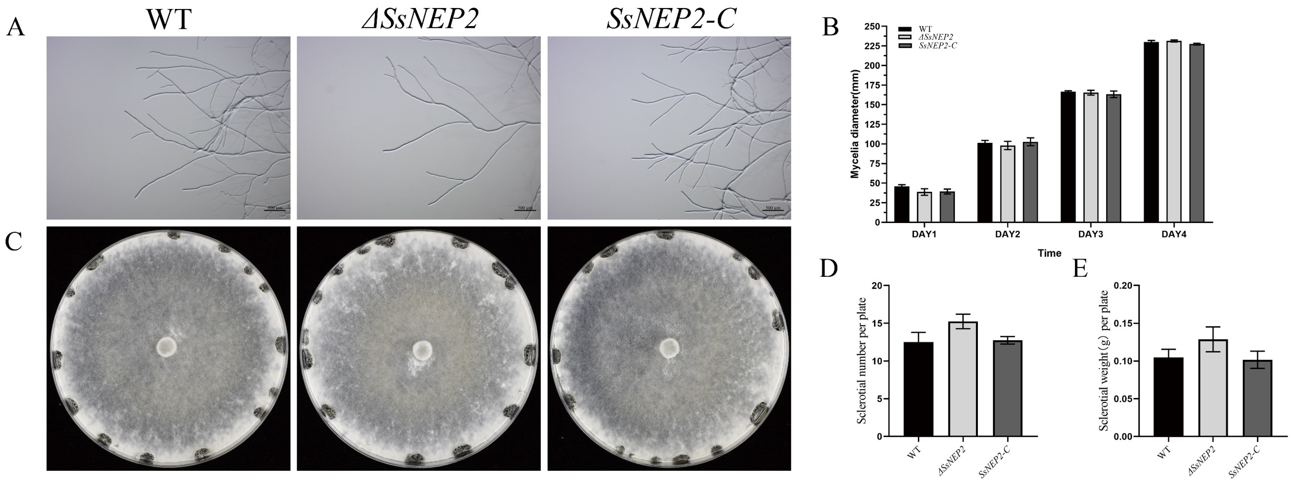
2.1. Knockout and Complementation of SsNEP2 in S. sclerotiorum

Although both SsNEP1 and SsNEP2 belong to NLPs [32], the alignment results show that they only share 35% identity in their amino acid sequences (Supplementary Figure S1A,B). As predicted, both SsNEP1 and SsNEP2 contain N-terminal secretory signal peptide and NPP1 domain (Supplementary Figure S1C). However, only SsNEP2 is abundantly induced upon infection [32], indicating that it may play a more important role in pathogenicity. We replaced SsNEP2 with the hygromycin-resistance gene in situ by the split marker method to obtain a deletion mutant (Figure 1A). Afterwards, complementation-transformed strains were obtained by means of agrobacterium-induced transformation in S. sclerotiorum. PCR results showed that SsNEP2 was completely deleted and not expressed at all, indicating that ΔSsNEP2 is a real knockout allele (Figure 1B,C), whereas the complementary strain (SsNEP2-C) was restored at the transcription level (Figure 1C).
To test whether SsNEP2 affects S. sclerotiorum growth, we examined both the hyphal and sclerotial phenotypes of the mutant. ΔSsNEP2 did not affect the mycelium morphology (Figure 2A). The results show that knockout of SsNEP2 had little effect on sclerotial formation, as there was no significant difference in the number or weight of sclerotia per plate among ΔSsNEP2, wild-type, and SsNEP2-C strain (Figure 2C–E). Similarly, no significant difference was detected in the mycelium growth rate (Figure 2B), indicating that SsNEP2 does not contribute to the growth of mycelium and the formation of sclerotia.

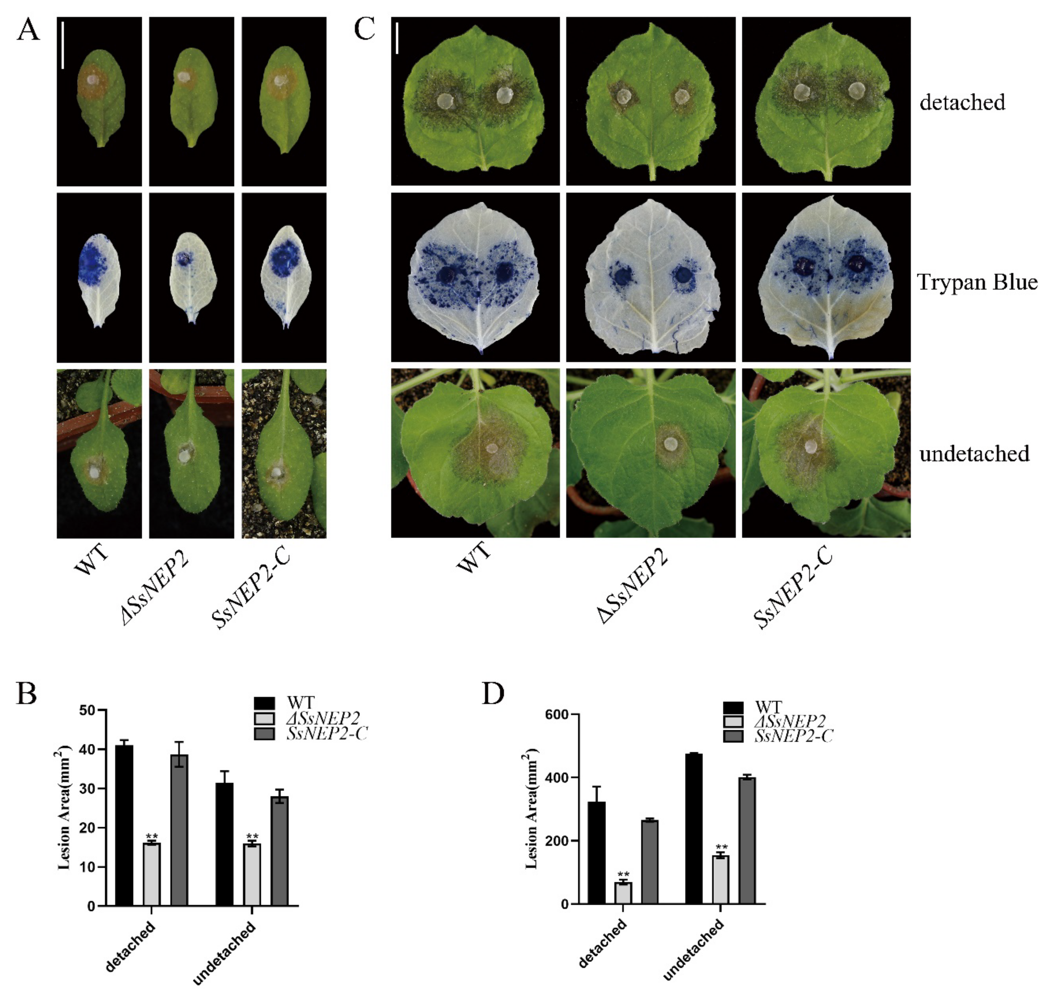
2.2. Loss of SsNEP2 Compromises Virulence of S. sclerotiorum

Pathogenicity is vital for successful infection by pathogens. To determine whether pathogenicity is affected by SsNEP2 knockout in S. sclerotiorum, mutant and wild-type ΔSsNEP2 and complementary strain SsNEP2-C were inoculated on detached leaves of A. thaliana (Figure 3A) and Nicotiana benthamiana (Figure 3C). After 24 h, the lesions caused by ΔSsNEP2 were significantly smaller (<50%) than those caused by the wild-type strain, while the lesions caused by the complementary strain SsNEP2-C were similar to those caused by the wild-type strain (Figure 3B,D). After detached leaves were stained with trypan blue, the areas of stained dead cells were consistent with the areas of corresponding lesions of infected leaves. Similar results were obtained for infection on undetached leaves of living plants in A. thaliana (Figure 3A) and N. benthamiana (Figure 3A,C), indicating that the loss of SsNEP2 compromised the virulence of S. sclerotiorum, whereas transformation of the WT copy of SsNEP2 restored the virulence in ΔSsNEP2 mutant.

2.3. SsNEP2 Does Not Affect Oxalate Secretion and Appressorium Formation

As both oxalic acid content and changes in pH value are very important factors affected the infection process of S. sclerotiorum [33], we inoculated ΔSsNEP2 mutant on bromophenol blue containing PDA for 48 h to check whether the reduced virulence was caused by oxalic acid production. As shown in Figure 4C, ΔSsNEP2 mutant had the same phenotype as the wild-type and SsNEP2-C strains, demonstrating that the mutant secretes oxalate normally and causes a pH change around the hyphae. These experiments suggest that the compromised virulence of ΔSsNEP2 is not due to a loss of the ability to secrete oxalic acid or to regulate ambient pH.
The appressorium plays an important role in the disease cycle, and reduced appressorium formation leads to decreased virulence of S. sclerotiorum [34,35]. To test whether the reduced virulence of ΔSsNEP2 is due to reduced appressorium formation, we carefully observed the formation and number of appressoria in the mutant. On the slide surface, the ΔSsNEP2, wild-type, and SsNEP2-C strains formed appressoria normally, and the quantities did not show statistical differences (Figure 4A). After 16 h of infection on onion epidermis, the appressoria were stained with trypan blue, and we found that the mutant could still form appressoria normally during the infection process, which was not significantly different from wild-type and SsNEP2-C (Figure 4B). Thus, the reduced virulence of ΔSsNEP2 is not related to the formation of appressorium.

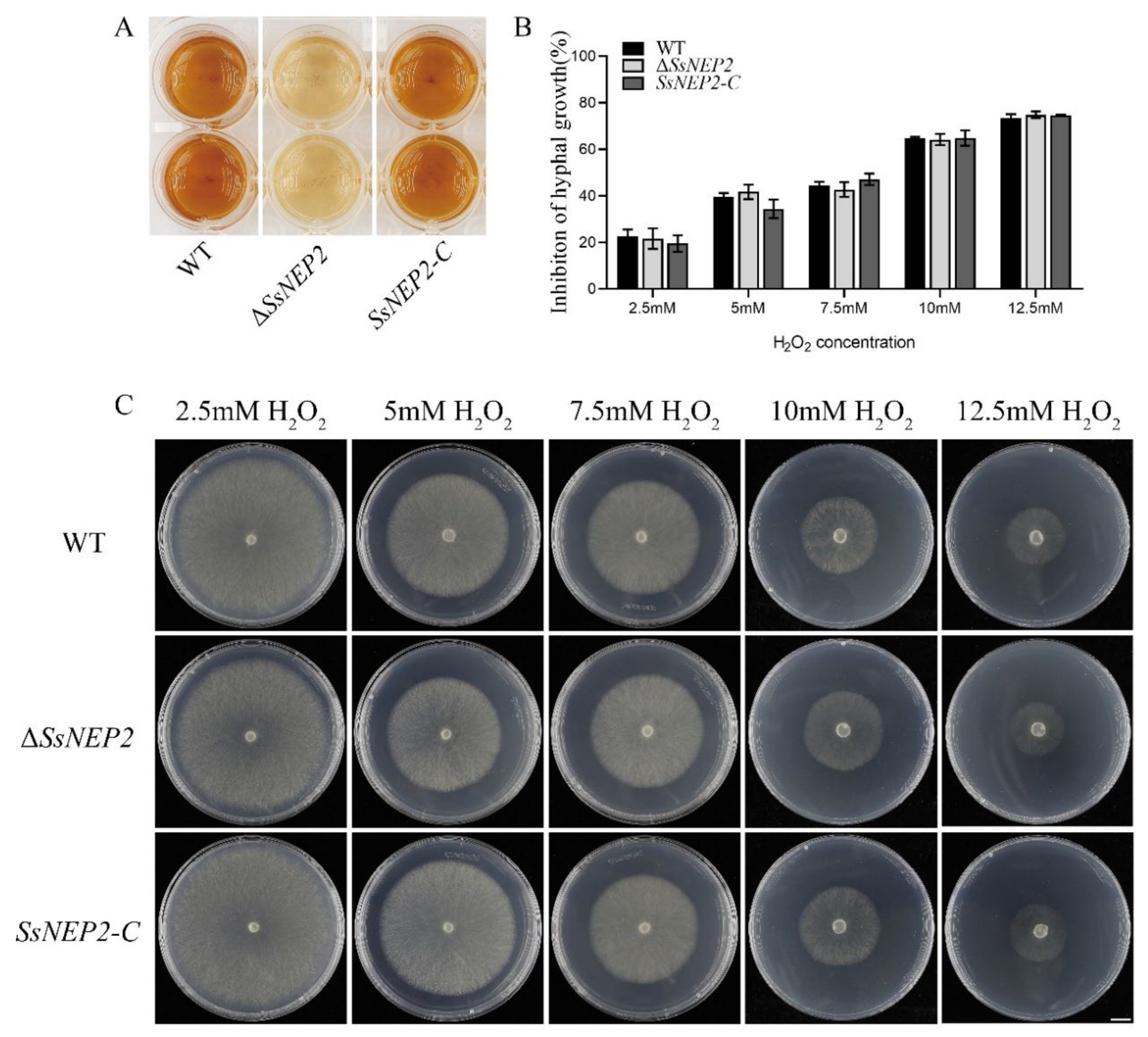
2.4. Knockout of SsNEP2 Affects ROS Accumulation

Cytotoxic NLPs can cause H2O2 accumulation in plants, leading to elevated ROS levels, which triggers programmed cell death (PCD) [31,36]. As a member of the NLPs, SsNEP2 may also have such a function. A. thaliana was infected with WT, ΔSsNEP2, and SsNEP2-C for 8 h, and then stained with 3,3′-diaminobenzidine (DAB). It was found that significantly fewer brown complexes formed in the leaves of plants infected with ΔSsNEP2 than the wild type, indicating that SsNEP2 may induce increased H2O2 content in plants (Supplementary Figure S2). Pathogens can also secrete ROS, and the developmental differentiation and virulence of fungi are affected by intracellular ROS levels [37,38]. As shown in Figure 5A, the color of ΔSsNEP2 was lighter than that of the wild type, whereas the color of the complemented strain SsNEP2-C was not significantly different, indicating that H2O2 content in the mutant strains was lower than that in the wild-type strain, and SsNEP2 knockout did affect ROS accumulation (Figure 5A).
In addition to acting as effectors, NLPs also affect the growth and development of pathogens and their responses to abiotic stresses [24,31]. To test the function of SsNEP2 in response to peroxide stress, we inoculated WT, ΔSsNEP2, and SsNEP2-C on PDA medium containing different gradient concentrations of H2O2. Inoculation of ΔSsNEP2 and wild type on PDA medium containing H2O2 cultured for 48 h also showed no difference in the inhibition of mycelial growth at different molar concentrations of H2O2 among ΔSsNEP2 mutant, wild-type, and SsNEP2-C strains (Figure 5B,C). This suggests that deletion of SsNEP2 does not affect tolerance to H2O2 of S. sclerotiorum.

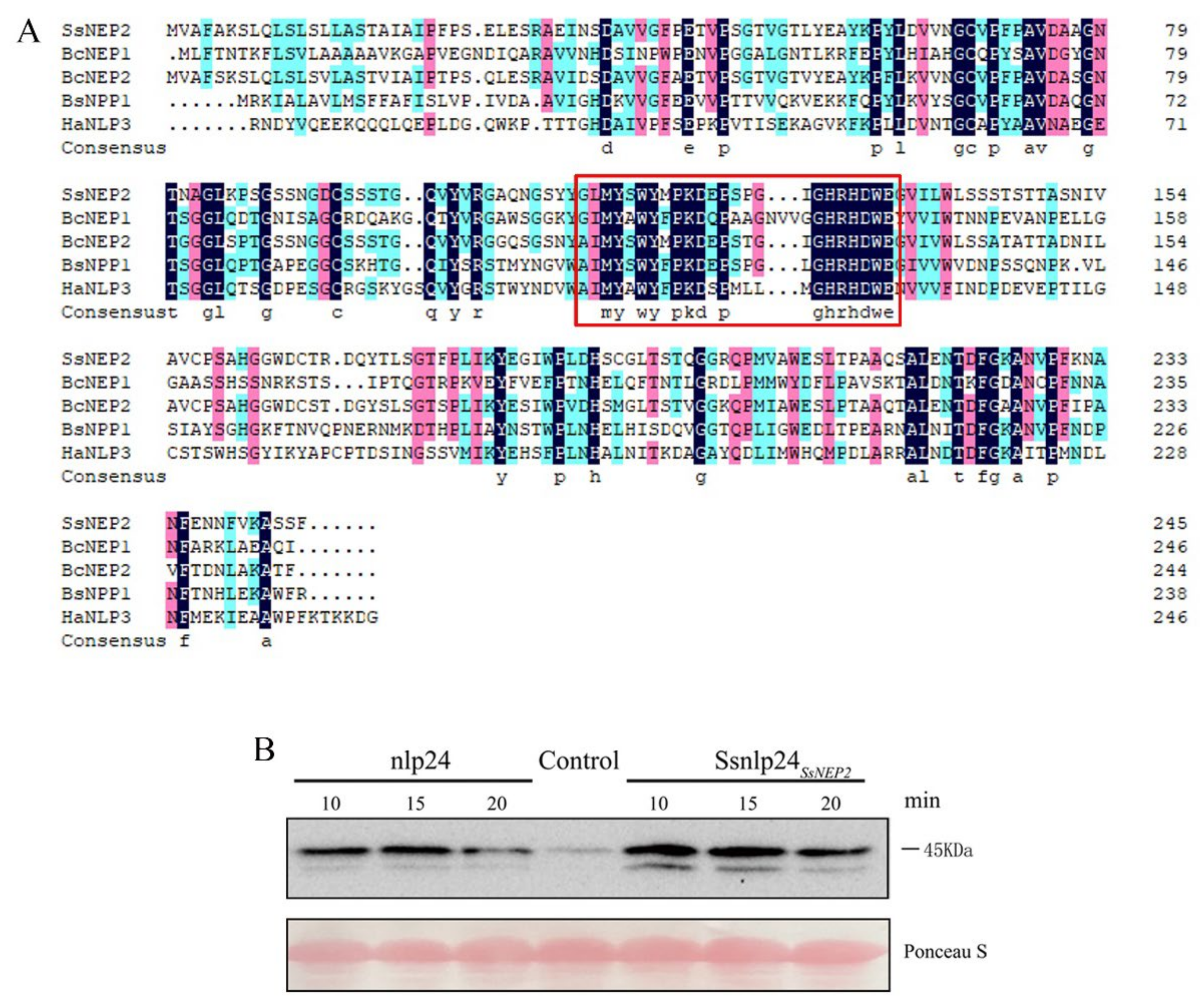
2.5. Ssnlp24 Derived from SsNEP2 Triggers MAPK Activation in Arabidopsis

Just as the conserved 24 amino acids of NLP were reported to activate Arabidopsis immunity [27], SsNEP2 contains the homologous sequence of nlp24 (named Ssnlp24SsNEP2); therefore, we performed multiple alignment of the amino acid sequences of SsNEP2, BcNEP1, BcNEP2, BsNPP1, and HaNLP3. As shown in Figure 6A, these proteins had a high similarity in their amino acid sequences.
To test whether Ssnlp24SsNEP2 can trigger plant immunity, we collected Arabidopsis leaves after treatment with each synthetic nlp peptide for 10–20 min. From the Western blot results, we found that nlp24 induced MAPK activation, as reported previously (Figure 6B). Notably, Ssnlp24SsNEP2 also induced MAPK activation, to the same extent as nlp24 (Figure 6B). These results demonstrate that Ssnlp24SsNEP2 induces a PAMP-triggered immune response similarly to nlp24, and the recognition of Ssnlp24SsNEP2 in the early infection phase contributes to host immunity against S. sclerotiorum.

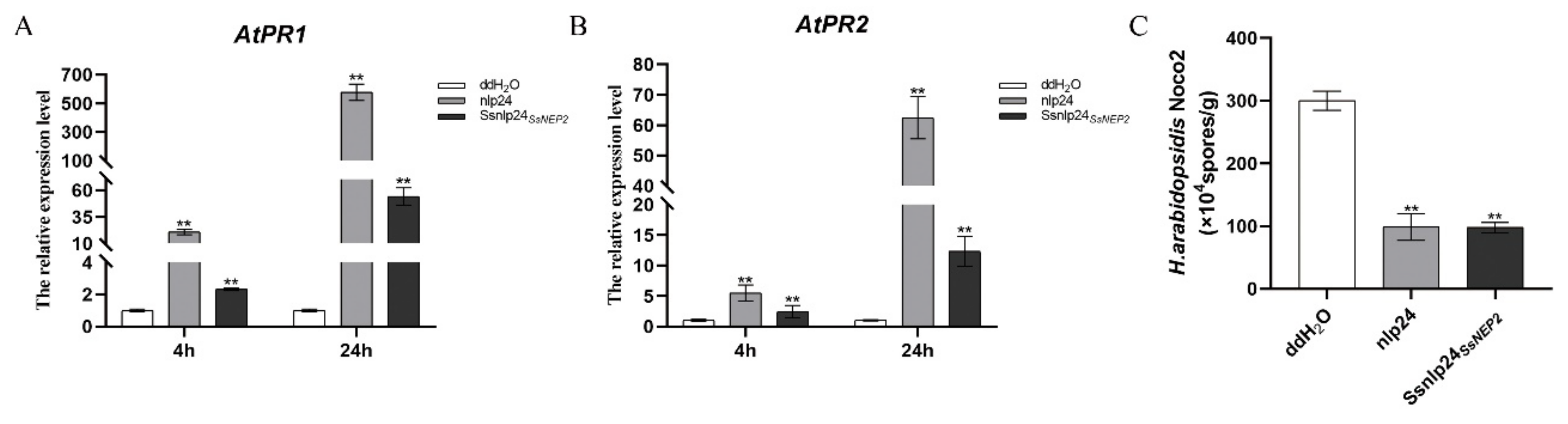
2.6. Ssnlp24SsNEP2 Acts as PAMP Signal to Induce Plant Immunity

Treatment with the synthetic nlp20/nlp24 peptide induces PR gene expression and ethylene production in A. thaliana through the pattern-triggered immunity (PTI) pathway [28,30]. To test whether Ssnlp24SsNEP2 also upregulates PR gene expression as in conserved nlp24, we took samples pretreated with 1 μM nlp24 or Ssnlp24SsNEP2 after 4 or 24 h, respectively. As expected, Ssnlp24SsNEP2 enhanced the expression levels of AtPR1 and AtPR2 (Figure 7A,B). Over time, the expression levels of both AtPR1 and AtPR2 further increased (Figure 7A,B).
To further check whether enhanced PR gene expression leads to increased resistance to pathogens, we sprayed spores of H. arabidopsidis (H. a.) Noco2 on seedlings pretreated with Ssnlp24SsNEP2 and found that resistance against H. a. Noco2 was enhanced, similar to the nlp24-induced immune response (Figure 7C). These results suggest that Ssnlp24SsNEP2 can be recognized as a PAMP and induces plant immunity though PTI.

3. Discussion

The virulence of S. sclerotiorum is regulated by multiple factors [39]. A key factor is the secretion of proteins out of cells, and the NLP family makes up most of the secreted protein [40]. The results of our assays show that the virulence of the deletion mutant was significantly reduced. The complemented strain, however, showed recovered infectivity at the same level as the wild type. Therefore, SsNEP2, as part of a class of cytotoxic NLPs, is related to the virulence of S. sclerotiorum. Deletion of this gene results in decreased virulence. PyaNLP can bind eudicot GIPCs to exert its cytolytic activity [23]. Whether SsNEP2 has the function of binding to the GIPCs of eudicot requires more in-depth study. However, ΔSsNEP2 may also secrete OA normally and change the pH value around the hyphae. At the same time, appressoria of the mutant strain can be formed normally and they are no different from that of WT in quantity and quality. Therefore, we demonstrated that the reduced virulence of ΔSsNEP2 may be due to other reasons that are not related to oxalate secretion or appressorium formation.
In the process of interacting with pathogens, ROS accumulation in plants may occur for two reasons. On the one hand, plants will generate ROS as a molecular signal to improve their own defense status after recognizing pathogens [41]; on the other hand, pathogens can secrete ROS by themselves, which leads to excessive ROS in the plants and triggers programmed cell death [42,43]. After inoculation with the deletion mutant ΔSsNEP2, the hydrogen peroxide content in Arabidopsis plants was significantly lower compared to those infected with the wild-type strain. S. sclerotiorum can manipulate host redox environment to facilitate its infection. For example, by secreting oxalic acid which regulates the production of ROS in plants and leading to PCD [10], or reducing the accumulation of ROS in plants by reducing the production of ROS in S. sclerotiorum [44]. Since the mutant does not affect the production of oxalic acid, we speculate that SsNEP2, as a toxic protein, can increase the ROS level in plants and cause cell death, which is beneficial to subsequent S. sclerotiorum infection. In addition, we found that SsNEP2 also affected H2O2 accumulation in S. sclerotiorum. Deletion of SsNEP2 may decrease the content of H2O2 released by S. sclerotiorum into plants, thus reducing the amount of death cell and the damaged area in plants. It is suggested that SsNEP2 may affect virulence by regulating the ROS concentration in the plant and/or S. sclerotiorum. The oxygen burst produced by the plant’s immune system also affects the growth of pathogens, which have evolved scavenging systems to maintain the ROS balance in their own bodies [43,45]. We then simulated oxidative stress on S. sclerotiorum as it encountered an oxygen burst in plants. However, the performance of ΔSsNEP2 was not different from the wild type, indicating that deleting SsNEP2 does not affect the clearance of H2O2 in S. sclerotiorum.
As one of the earliest signals generated after plants sense pathogens’ invasion, MAPK activation plays an important role in regulating plant immunity [46]. Ssnlp24SsNEP2, which is derived from SsNEP2 contains the homologous sequence of nlp24 and can induce plant immunity such as nlp24. After spraying synthesized small peptides on the surface of plants for 10 min, MAPK cascade was activated. The expression of AtPR1 and AtPR2 was upregulated 4 h after induction. Even 24 h later, the expression levels were further increased. This series of reactions is beneficial to improve plants’ resistance to some pathogens. Thus, Ssnlp24SsNEP2 treatment increases the resistance to H.a. Noco2. Considering that S. sclerotiorum is a necrotic pathogen, the programmed cell death in the plant caused by MAPK activation will benefit its growth. Therefore, we speculate that Ssnlp24SsNEP2, as a pathogen-associated molecular pattern, can be recognized by plant receptors, thus triggering plant immunity and rapidly increasing ROS levels, resulting in cell damage and PCD, which is more conducive to infection caused by S. sclerotiorum.
Previous research has shown that SsNEP2 was abundantly induced upon infection. After infecting Arabidopsis with wild-type strains for 24 h, we measured the expression of SsNEP2 and found that the expression of SsNEP2 was significantly up-regulated upon infection (Supplementary Figure S3). The results were consistent with previous studies, suggesting that SsNEP2 is involved in the S. sclerotiorum infection process. Combined with the protein domain prediction of SsNEP2, we speculate that SsNEP2 may be involved in the infection of S. sclerotiorum as a secreted protein. Furthermore, SsNEP1 expression was increased in ΔSsNEP2, either grown on PDA or infected on Arabidopsis (Supplementary Figure S3B). We hypothesize that SsNEP1 and SsNEP2 may function redundantly, but this requires more experiments to prove it. Based on the above results, we speculate that SsNEP2 has an effect on the infection process of S. sclerotiorum in two ways. On the one hand, SsNEP2 is involved in S. sclerotiorum virulence as a secreted protein. On the other hand, the small peptide Ssnlp24SsNEP2 derived from SsNEP2 triggers plant immunity, induces ROS production, and causes host cell death to promote the necrotrophic life of S. sclerotiorum.

4. Materials and Methods

4.1. Fungal Strains and Culture Conditions

Wild-type strain 1980 was subcultured on potato dextrose agar (PDA), and deletion mutants and complemented strains were cultured on PDA containing 150 μg/mL hygromycin B (Roche) in an incubator at 20 °C for daily storage and 4 °C for long-term storage.

4.2. Plant Materials and Growth Conditions

The A. thaliana and N. benthamiana used for S. sclerotiorum virulence assays were cultured in a culture room at 22 °C with 16 h of light and 8 h of darkness; 4-week-old plants were used for the assays.

4.3. Identification and Sequence Analysis of SsNEP2

The NLP sequences of Fusarium oxysporum Fo47, Verticillium dahliae, Magnaporthe oryzae, Botrytis cinerea B05.10, Aspergillus fumigatus, Stagonospora sp. SRC1lsM3a, and S. sclerotiorum were downloaded from the Ensembl Fungi database. Multiple sequence alignment of the NLP protein sequences was performed by Clustal software and opened with MEGA7.0 software, then the neighbor-joining (NJ) method was used to construct an evolutionary tree. Bootstrap was set to 1000. Protein sequence numbers are shown in the evolutionary tree. The domains contained in the sequence were analyzed by PredictProtein (https://predictprotein.org/ accessed on 16 February 2022).

4.4. Gene Replacement and Complementation

The SsNEP2 gene was knocked out from the genome of S. sclerotiorum using the split-marking approach. Primers 1F: ss12g090490UF (ATCTTCAAAGTTTCCGCCACA)/2R: ss12g090490 UR (GTGCTCCTTCAA-TATCATCTTCTGCAAGAATGGACATCAAGCTC) were designed to amplify approximately 1000 bp of the 5′ untranslated region of SsNEP2. Primers 3F: ss12g090490 DF (GTTTAGAGGTAATCCTTCTTTTGGGGTCGAGGTGGAGTGA)/4R: ss12g090490 DR (CAGGAAACCTTGAATGGCTTGA) were used to amplify the 3′ UTR of SsNEP2 with approximately 1300 bp, using the genomic DNA of Sclerotinia as a template. Fragment 1 was composed of the 5’ UTR of SsNEP2 and 5′ part of hygromycin phosphotransferase cassette, and fragment 2 was composed of the 3’ UTR region of SsNEP2 and 3′ part of hygromycin phosphotransferase cassette. These two fragments were then inserted into the T-vector (pEASY®-Blunt Cloning Kit, TransGen, Beijing, China). The resulting vector, T-NEP2 was used as a template to amplify two split-marker fragments using primers ss12g090490 UF (ATCTTCAAAGTTTCCGCCACA)/HY-R (AAATTGCCGTCAACCAAGCTC) and YG-F (TTTCAGCTTCGATGTAGGAGG)/ss12g090490 DR (CAGGAAACCTTGAATGGCTTGA). These two fragments overlapped in the hygromycin resistance gene fragment and were used to co-transform into wild-type Sclerotinia protoplasts according to the method of Rollins [47]. ss12g090490 DDF (CGCTCGTCCGTGCATCAAGACT)/HY-R and YG-F/ss12g090490 DDR (GCTGGATTGAAATGCGATGCT) two pairs of primers are used to test whether the hygromycin-resistance gene is inserted at the correct site. Transformants were purified by hyphal tip transfer at least 3 times. Amplified full-length SsNEP2 sequence was used to verify gene deletion strains.
For ΔSsNEP2 complementation, the binary vector pCH-EF-1-NEP2 was constructed using the backbone of pCH-EF-1 (shared by D. Jiang from Huazhong Agricultural University). The full-length SsNEP2 gene, including promoter and coding sequence (CDS), was amplified from WT genomic DNA. Full-length SsNEP2 gene fragment and pCH-EF-1 vector were digested by using restriction enzyme XhoI and SacI, then linked by homologous recombinase (ClonExpress® II One Step Cloning Kit, Vazyme, Nanjing, China) to generate the pCH-EF-1-NEP2 construct. Then the plasmid was used for SsNEP2 transformation using the polyethylene glycol (PEG)-mediated transformation method [47,48].

4.5. RT-qPCR Analysis

Gene expression levels in Arabidopsis leaves were detected at 4 and 24 h after spraying nlp20 and Ssnlp24SsNEP2. The RNA of test samples was extracted using an EastepTM Super Total RNA Extraction Kit and GoScript™ Reverse Transcription System Kit for cDNA synthesis (Promega, Madison, WI, USA). Quantitative expression assays were performed using SYBR® Green Premix Pro Taq HS qPCR Kit II (AG11702, Accurate Biotechnology (Hunan)Co.,Ltd, Changsha, China) with StepOneTM Real-time PCR Instrument Thermal Cycling Block. Primers of AtPR1, AtPR2, and Actin1 used for qPCR were as follows. PR1-F: (GCTAACTACAACTACGCTGC)/PR1-R: (CTCGTTCACATAATTCCCAC); PR2-F: (CGTTGGAAATGAGGTGAAA)/PR2-R: (CAGTGGTGGTGTCAGTGGC); and ACTIN1-RT-F: (CGATGAAGCTCAATCCAAACGA)/ACTIN1-RT-R: (CAGAGTCGAGCACAATACCG). Actin1 was used as internal reference.
Mycelia of S. sclerotiorum were cultured on cellophane and collect Arabidopsis leaves which inoculated S. sclerotiorum after 24 h for SsNEP1 and SsNEP2 gene expression assay. Primers of SsNEP1, SsNEP2 and β-tubulin used for qPCR were: SsNEP1 qF: (GAGGGCAACGACATCCAAG)/SsNEP1 qR: (ACGGCAGCCAGCGGAGATA), SsNEP2 qF: (CCGCCATAGCCATTCCCTTC)/SsNEP2 qR: (ATCACCATTGCTACTGCCACTT) and SsTubqF: (ACCTCCATCCAAGAACTC)/SsTubqR: (GAACTCCATCTCGTCCAT). β-tubulin was used as internal reference. The program setting included holding stage (95 °C, 2 min), cycling stage (95 °C, 20 s; 55 °C, 20 s; 72 °C, 20 s; 40 cycles), and melt curve stage (95 °C, 15 s; 60 °C, 1 min). The transcript level of the gene of interest was calculated from the threshold cycle using the 2ΔΔCT method [49] with three replicates, and the data were analyzed using SPSS Statistics v.24.0 (IBM, Armonk, NY, USA).

4.6. Analysis of Virulence

To assess virulence, mycelial plugs (2 or 5 mm in diameter) from wild-type, ΔSsNEP2, and SsNEP2-C strains were harvested from the edges of colonies maintained in PDA cultures for 48 h and then inoculated on leaves, with 2-mm plugs for A. thaliana and 5-mm plugs for N. benthamiana. Lesion areas were measured 48 h later and counted with ImageJ. Two leaves were used per experiment and each experiment was repeated 3 times. The data were analyzed using SPSS Statistics v.24.0 (IBM, Armonk, NY, USA).
The infected plant leaves were placed in 1% trypan blue solution and soaked at room temperature for at least 2 h. After that, they were decolorized with 90–100% alcohol and photographed and stored in 60% glycerol.

4.7. Compound Appressoria Observation and OA Analysis

A 5-mm mycelia plug of S. sclerotiorum was placed on a glass slide and cultured for 16 h to observe the formation and number of appressora. After 16 h of inoculation with S. sclerotiorum, onion epidermis was soaked in 0.5% trypan blue solution for 30 min and then decolorized using bleaching solution (ethanol:acetic acid:glycerol = 3:1:1). Samples were examined and photographed under the light microscope (Axio Imager 2, ZEISS, Oberkochen, Germany).
S. sclerotiorum was inoculated on PDA medium containing 100 μg/mL bromophenol blue to detect whether it secreted oxalic acid.

4.8. DAB Staining

Using a sterile punch (5 mm), the mycelia-colonized plugs were punched out from the wild-type, ΔSsNEP2, and SsNEP2-C strains of S. sclerotiorum (9 pieces each) and placed separately in a 24-well plate. A total of 2 mL of 1 mg/mL DAB solution was added into each well, and then the samples was incubated for 30 min at 25 °C in the dark and observed immediately.
After 8 h of inoculation with S. sclerotiorum, Arabidopsis leaves were soaked in 1 mg/mL DAB solution overnight and then decolorized with bleaching solution and photographed (Stemi 508, ZEISS, Oberkochen, Germany).

4.9. Pathogen Infection Assay

To analyze nlp20- and Ssnlp24SsNEP2-induced pattern-triggered immunity, leaves of 3-week-old Arabidopsis were sprayed with 1 µM nlp20 or Ssnlp24SsNEP2. After 24 h, the seedlings were sprayed with H.a. Noco2 spore suspension at 30,000 spores/mL H2O. Plants were then covered with a transparent lid and grown in a short-day incubator at 18 °C for 7 days. Using a cell counting plate, the number of conidia spores per leaf was counted. Experiments were conducted three independent times, and two biological replicates were used each time.

4.10. MAPK Activity Assay

Two-week-old Arabidopsis seedlings grown on 1/2 MS medium plates were sprayed with 1 µM of nlp20 or Ssnlp24SsNEP2, and samples were collected 0, 10, 15, and 20 min after each treatment. After grinding in liquid nitrogen, protein extract buffer (200 mM pH = 6.8 Tris-Cl, 8% SDS, 0.4% bromophenol blue, 40% glycerol, 20% β-mercaptoethanol) was added to extract total protein. Phosphorylation of MAPK was detected by Western blot with anti-pERK (no. 4370S, 1:2500 dilution; Cell Signaling Technology, Boston, MA, USA). All experiments were repeated at least 3 times.

4.11. Oxidative Stress Response

To test the response of ΔSsNEP2 to oxidative stress, wild-type, ΔSsNEP2, and SsNEP2-C strains were grown on PDA medium containing H2O2 (2.5, 5, 7.5, 10, and 12.5 mM). After 48 h, the diameter and growth inhibition rate of mycelia were measured: Inhibition rate (%) = 100 × (colony diameter of strain without H2O2—colony diameter of strain with H2O2)/(colony diameter of strain without H2O2).

Supplementary Materials

The following supporting information can be downloaded at: https://www.mdpi.com/article/10.3390/pathogens11040446/s1, Figure S1: Bioinformatics analysis of SsNEP1 and SsNEP2 protein; Figure S2: ΔSsNEP2 reduces H2O2 accumulation in Arabidopsis leaves; Figure S3: SsNEP2 is involved in the infection process of S. sclerotiorum.

Author Contributions

C.Y. and S.X. designed the experiments, and C.Y. performed most of the described experiments. C.Y., W.L., X.H., X.T. and Y.X. performed the screening. X.T., L.Q. and Z.P. conducted the infection experiments. W.L. and Y.L. analyzed the ROS data. C.Y. and S.X. wrote the manuscript. All authors have read and agreed to the published version of the manuscript.

Funding

National Natural Science Foundation of China (grant 31971836) and Natural Science Foundation of Hunan Province (grant 2019JJ40126).

Institutional Review Board Statement

Not applicable.

Informed Consent Statement

Not applicable.

Data Availability Statement

Not applicable.

Acknowledgments

We cordially thank Xin Li (University of British Columbia) for critical reading of the manuscript, Daohong Jiang (Huazhong Agricultural University) for sharing pCH-EF-1 plasmid, and Jeffrey Rollins (University of Florida) for sharing WT S. sclerotiorum strain 1980. We would like to thank National Natural Science Foundation of China (grant 31971836) and the Natural Science Foundation of Hunan Province (grant 2019JJ40126) for funds awarded to S.X. and Z.P.

Conflicts of Interest

The authors declare no conflict of interest.

References

  1. Zhao, J.; Meng, J. Genetic analysis of loci associated with partial resistance to Sclerotinia sclerotiorum in rapeseed (Brassica napus L.). Theor. Appl. Genet. 2003, 106, 759–764. [Google Scholar] [CrossRef] [PubMed]
  2. Xia, S.; Xu, Y.; Hoy, R.; Zhang, J.; Qin, L.; Li, X. The Notorious Soilborne Pathogenic Fungus Sclerotinia sclerotiorum: An Update on Genes Studied with Mutant Analysis. Pathogens 2019, 9, 27. [Google Scholar] [CrossRef] [PubMed] [Green Version]
  3. Liang, X.; Rollins, J.A. Mechanisms of Broad Host Range Necrotrophic Pathogenesis in Sclerotinia sclerotiorum. Phytopathology 2018, 108, 1128–1140. [Google Scholar] [CrossRef] [PubMed] [Green Version]
  4. Boland, G.J.; Hall, R. Index of plant hosts of Sclerotinia sclerotiorum. Can. J. Plant Pathol. 1994, 16, 93–108. [Google Scholar] [CrossRef]
  5. Zhang, H.; Wu, Q.; Cao, S.; Zhao, T.; Chen, L.; Zhuang, P.; Zhou, X.; Gao, Z. A novel protein elicitor (SsCut) from Sclerotinia sclerotiorum induces multiple defense responses in plants. Plant Mol. Biol. 2014, 86, 495–511. [Google Scholar] [CrossRef] [PubMed]
  6. Yu, Y.; Xiao, J.; Du, J.; Yang, Y.; Bi, C.; Qing, L. Disruption of the Gene Encoding Endo-beta-1, 4-Xylanase Affects the Growth and Virulence of Sclerotinia sclerotiorum. Front. Microbiol. 2016, 7, 1787. [Google Scholar] [CrossRef] [PubMed] [Green Version]
  7. Lang, C.; Dornenburg, H. Perspectives in the biological function and the technological application of polygalacturonases. Appl. Microbiol. Biotechnol. 2000, 53, 366–375. [Google Scholar] [CrossRef]
  8. Dallal Bashi, Z.; Rimmer, S.R.; Khachatourians, G.G.; Hegedus, D.D. Factors governing the regulation of Sclerotinia sclerotiorum cutinase A and polygalacturonase 1 during different stages of infection. Can. J. Microbiol. 2012, 58, 605–616. [Google Scholar] [CrossRef]
  9. Godoy, G.; Steadman, J.R.; Dickman, M.B.; Dam, R. Use of mutants to demonstrate the role of oxalic acid in pathogenicity of Sclerotinia sclerotiorum on Phaseolus vulgaris. Physiol. Mol. Plant Pathol. 1990, 37, 179–191. [Google Scholar] [CrossRef]
  10. Williams, B.; Kabbage, M.; Kim, H.J.; Britt, R.; Dickman, M.B. Tipping the balance: Sclerotinia sclerotiorum secreted oxalic acid suppresses host defenses by manipulating the host redox environment. PLoS Pathog. 2011, 7, e1002107. [Google Scholar] [CrossRef] [Green Version]
  11. Kim, K.S.; Min, J.Y.; Dickman, M.B. Oxalic acid is an elicitor of plant programmed cell death during Sclerotinia sclerotiorum disease development. Mol. Plant Microbe. Interact. 2008, 21, 605–612. [Google Scholar] [CrossRef] [Green Version]
  12. Xu, L.S.; Li, G.Q.; Jiang, D.H.; Chen, W.D. Sclerotinia sclerotiorum: An Evaluation of Virulence Theories. Annu. Rev. Phytopathol. 2018, 56, 311–338. [Google Scholar] [CrossRef] [PubMed]
  13. Liang, X.; Liberti, D.; Li, M.; Kim, Y.T.; Hutchens, A.; Wilson, R.; Rollins, J.A. Oxaloacetate acetylhydrolase gene mutants of Sclerotinia sclerotiorum do not accumulate oxalic acid, but do produce limited lesions on host plants. Mol. Plant Pathol. 2015, 16, 559–571. [Google Scholar] [CrossRef] [PubMed]
  14. Varden, F.A.; De la Concepcion, J.C.; Maidment, J.H.; Banfield, M.J. Taking the stage: Effectors in the spotlight. Curr. Opin Plant Biol. 2017, 38, 25–33. [Google Scholar] [CrossRef] [PubMed]
  15. Tang, L.; Yang, G.; Ma, M.; Liu, X.; Li, B.; Xie, J.; Fu, Y.; Chen, T.; Yu, Y.; Chen, W.; et al. An effector of a necrotrophic fungal pathogen targets the calcium-sensing receptor in chloroplasts to inhibit host resistance. Mol. Plant Pathol. 2020, 21, 686–701. [Google Scholar] [CrossRef] [Green Version]
  16. Yang, G.; Tang, L.; Gong, Y.; Xie, J.; Fu, Y.; Jiang, D.; Li, G.; Collinge, D.B.; Chen, W.; Cheng, J. A cerato-platanin protein SsCP1 targets plant PR1 and contributes to virulence of Sclerotinia sclerotiorum. New Phytol. 2018, 217, 739–755. [Google Scholar] [CrossRef] [Green Version]
  17. Pemberton, C.L.; Salmond, G.P. The Nep1-like proteins-a growing family of microbial elicitors of plant necrosis. Mol. Plant Pathol. 2004, 5, 353–359. [Google Scholar] [CrossRef]
  18. Bailey, B.A. Purification of a protein from culture filtrates of Fusarium oxysporum that induces ethylene and necrosis in leaves of Erythroxylum coca. Phytopathology 1995, 85, 1250–1255. [Google Scholar] [CrossRef]
  19. Bailey, B.A.; Apel-Birkhold, P.C.; Luster, D.G. Expression of NEP1 by Fusarium oxysporum f. sp. erythroxyli After Gene Replacement and Overexpression Using Polyethylene Glycol-Mediated Transformation. Phytopathology 2002, 92, 833–841. [Google Scholar] [CrossRef] [Green Version]
  20. Ottmann, C.; Luberacki, B.; Kufner, I.; Koch, W.; Brunner, F.; Weyand, M.; Mattinen, L.; Pirhonen, M.; Anderluh, G.; Seitz, H.U.; et al. A common toxin fold mediates microbial attack and plant defense. Proc. Natl. Acad. Sci. USA 2009, 106, 10359–10364. [Google Scholar] [CrossRef] [Green Version]
  21. Fellbrich, G.; Romanski, A.; Varet, A.; Blume, B.; Brunner, F.; Engelhardt, S.; Felix, G.; Kemmerling, B.; Krzymowska, M.; Nurnberger, T. NPP1, a Phytophthora-associated trigger of plant defense in parsley and Arabidopsis. Plant J. 2002, 32, 375–390. [Google Scholar] [CrossRef]
  22. Seidl, M.F.; Van den Ackerveken, G. Activity and Phylogenetics of the Broadly Occurring Family of Microbial Nep1-Like Proteins. Annu. Rev. Phytopathol. 2019, 57, 367–386. [Google Scholar] [CrossRef] [PubMed]
  23. Lenarcic, T.; Albert, I.; Bohm, H.; Hodnik, V.; Pirc, K.; Zavec, A.B.; Podobnik, M.; Pahovnik, D.; Zagar, E.; Pruitt, R.; et al. Eudicot plant-specific sphingolipids determine host selectivity of microbial NLP cytolysins. Science 2017, 358, 1431–1434. [Google Scholar] [CrossRef] [PubMed] [Green Version]
  24. Santhanam, P.; van Esse, H.P.; Albert, I.; Faino, L.; Nurnberger, T.; Thomma, B.P. Evidence for functional diversification within a fungal NEP1-like protein family. Mol. Plant Microbe Interact. 2013, 26, 278–286. [Google Scholar] [CrossRef] [PubMed] [Green Version]
  25. Mattinen, L.; Tshuikina, M.; Mae, A.; Pirhonen, M. Identification and characterization of Nip, necrosis-inducing virulence protein of Erwinia carotovora subsp. carotovora. Mol. Plant Microbe Interact. 2004, 17, 1366–1375. [Google Scholar] [CrossRef] [Green Version]
  26. Pemberton, C.L.; Whitehead, N.A.; Sebaihia, M.; Bell, K.S.; Hyman, L.J.; Harris, S.J.; Matlin, A.J.; Robson, N.D.; Birch, P.R.; Carr, J.P.; et al. Novel quorum-sensing-controlled genes in Erwinia carotovora subsp. carotovora: Identification of a fungal elicitor homologue in a soft-rotting bacterium. Mol. Plant Microbe Interact. 2005, 18, 343–353. [Google Scholar] [CrossRef] [Green Version]
  27. Albert, I.; Bohm, H.; Albert, M.; Feiler, C.E.; Imkampe, J.; Wallmeroth, N.; Brancato, C.; Raaymakers, T.M.; Oome, S.; Zhang, H.; et al. An RLP23-SOBIR1-BAK1 complex mediates NLP-triggered immunity. Nat. Plants 2015, 1, 15140. [Google Scholar] [CrossRef]
  28. Bohm, H.; Albert, I.; Oome, S.; Raaymakers, T.M.; Van den Ackerveken, G.; Nurnberger, T. A conserved peptide pattern from a widespread microbial virulence factor triggers pattern-induced immunity in Arabidopsis. PLoS Pathog. 2014, 10, e1004491. [Google Scholar] [CrossRef]
  29. Ono, E.; Mise, K.; Takano, Y. RLP23 is required for Arabidopsis immunity against the grey mould pathogen Botrytis cinerea. Sci. Rep. 2020, 10, 13798. [Google Scholar] [CrossRef]
  30. Oome, S.; Raaymakers, T.M.; Cabral, A.; Samwel, S.; Bohm, H.; Albert, I.; Nurnberger, T.; Van den Ackerveken, G. Nep1-like proteins from three kingdoms of life act as a microbe-associated molecular pattern in Arabidopsis. Proc. Natl. Acad. Sci. USA 2014, 111, 16955–16960. [Google Scholar] [CrossRef] [Green Version]
  31. Liu, J.; Nie, J.; Chang, Y.; Huang, L. Nep1-like Proteins from Valsa mali Differentially Regulate Pathogen Virulence and Response to Abiotic Stresses. J. Fungi 2021, 7, 830. [Google Scholar] [CrossRef]
  32. Dallal Bashi, Z.; Hegedus, D.D.; Buchwaldt, L.; Rimmer, S.R.; Borhan, M.H. Expression and regulation of Sclerotinia sclerotiorum necrosis and ethylene-inducing peptides (NEPs). Mol. Plant Pathol. 2010, 11, 43–53. [Google Scholar] [CrossRef]
  33. Xu, L.; Xiang, M.; White, D.; Chen, W. pH dependency of sclerotial development and pathogenicity revealed by using genetically defined oxalate-minus mutants of Sclerotinia sclerotiorum. Environ. Microbiol 2015, 17, 2896–2909. [Google Scholar] [CrossRef]
  34. Kabbage, M.; Yarden, O.; Dickman, M.B. Pathogenic attributes of Sclerotinia sclerotiorum: Switching from a biotrophic to necrotrophic lifestyle. Plant Sci. 2015, 233, 53–60. [Google Scholar] [CrossRef]
  35. Jiao, W.; Yu, H.; Cong, J.; Xiao, K.; Zhang, X.; Liu, J.; Zhang, Y.; Pan, H. Transcription factor SsFoxE3 activating SsAtg8 is critical for sclerotia, compound appressoria formation, and pathogenicity in Sclerotinia sclerotiorum. Mol. Plant Pathol. 2022, 23, 204–217. [Google Scholar] [CrossRef]
  36. Schouten, A.; Van Baarlen, P.; Van Kan, J.A.L. Phytotoxic Nep1-like proteins from the necrotrophic fungus Botrytis cinerea associate with membranes and the nucleus of plant cells. New Phytol. 2008, 177, 493–505. [Google Scholar] [CrossRef]
  37. Qin, G.; Tian, S.; Chan, Z.; Li, B. Crucial role of antioxidant proteins and hydrolytic enzymes in pathogenicity of Penicillium expansum: Analysis based on proteomics approach. Mol. Cell Proteom. 2007, 6, 425–438. [Google Scholar] [CrossRef] [Green Version]
  38. Kim, K.H.; Willger, S.D.; Park, S.W.; Puttikamonkul, S.; Grahl, N.; Cho, Y.; Mukhopadhyay, B.; Cramer, R.A., Jr.; Lawrence, C.B. TmpL, a transmembrane protein required for intracellular redox homeostasis and virulence in a plant and an animal fungal pathogen. PLoS Pathog. 2009, 5, e1000653. [Google Scholar] [CrossRef] [Green Version]
  39. Ding, L.N.; Li, T.; Guo, X.J.; Li, M.; Liu, X.Y.; Cao, J.; Tan, X.L. Sclerotinia Stem Rot Resistance in Rapeseed: Recent Progress and Future Prospects. J. Agric. Food Chem. 2021, 69, 2965–2978. [Google Scholar] [CrossRef]
  40. Pirc, K.; Hodnik, V.; Snoj, T.; Lenarcic, T.; Caserman, S.; Podobnik, M.; Bohm, H.; Albert, I.; Kotar, A.; Plavec, J.; et al. Nep1-like proteins as a target for plant pathogen control. PLoS Pathog. 2021, 17, e1009477. [Google Scholar] [CrossRef]
  41. Lehmann, S.; Serrano, M.; L’Haridon, F.; Tjamos, S.E.; Metraux, J.P. Reactive oxygen species and plant resistance to fungal pathogens. Phytochemistry 2015, 112, 54–62. [Google Scholar] [CrossRef] [PubMed] [Green Version]
  42. Liu, L.; Wang, Q.; Sun, Y.; Zhang, Y.; Zhang, X.; Liu, J.; Yu, G.; Pan, H. Sssfh1, a Gene Encoding a Putative Component of the RSC Chromatin Remodeling Complex, Is Involved in Hyphal Growth, Reactive Oxygen Species Accumulation, and Pathogenicity in Sclerotinia sclerotiorum. Front. Microbiol. 2018, 9, 1828. [Google Scholar] [CrossRef] [PubMed]
  43. Wang, Y.; Ji, D.; Chen, T.; Li, B.; Zhang, Z.; Qin, G.; Tian, S. Production, Signaling, and Scavenging Mechanisms of Reactive Oxygen Species in Fruit-Pathogen Interactions. Int. J. Mol. Sci. 2019, 20, 2994. [Google Scholar] [CrossRef] [PubMed] [Green Version]
  44. Kim, H.J.; Chen, C.; Kabbage, M.; Dickman, M.B. Identification and characterization of Sclerotinia sclerotiorum NADPH oxidases. Appl. Environ. Microbiol. 2011, 77, 7721–7729. [Google Scholar] [CrossRef] [PubMed] [Green Version]
  45. Heller, J.; Tudzynski, P. Reactive oxygen species in phytopathogenic fungi: Signaling, development, and disease. Annu. Rev. Phytopathol. 2011, 49, 369–390. [Google Scholar] [CrossRef]
  46. Meng, X.; Zhang, S. MAPK cascades in plant disease resistance signaling. Annu. Rev. Phytopathol. 2013, 51, 245–266. [Google Scholar] [CrossRef]
  47. Rollins, J.A. The Sclerotinia sclerotiorum pac1 gene is required for sclerotial development and virulence. Mol. Plant Microbe Interact. 2003, 16, 785–795. [Google Scholar] [CrossRef] [Green Version]
  48. Xu, Y.; Ao, K.; Tian, L.; Qiu, Y.; Huang, X.; Liu, X.; Hoy, R.; Zhang, Y.; Rashid, K.Y.; Xia, S.; et al. A Forward Genetic Screen in Sclerotinia sclerotiorum Revealed the Transcriptional Regulation of Its Sclerotial Melanization Pathway. Mol. Plant Microbe Interact. 2022, 35, MPMI10210254R. [Google Scholar] [CrossRef]
  49. Livak, K.J.; Schmittgen, T.D. Analysis of relative gene expression data using real-time quantitative PCR and the 2(-Delta Delta C(T)) Method. Methods 2001, 25, 402–408. [Google Scholar] [CrossRef]
Figure 1. Generation of knockout and complementation strain of SsNEP2 in S. sclerotiorum. (A) Split marker method technical process. Fragment 1 is composed of upstream of target gene and the first half of hygromycin-resistance gene (HY), and fragment 2 is composed of the second half of hygromycin (YG) and downstream of the target gene. Both fragments replace the target gene with hygromycin-resistance gene by homologous recombination. (B) Full-length SsNEP2 gene amplification in genomes of WT (1), ΔSsNEP2 (2), and SsNEP2-C (3). (C) Relative expression level of SsNEP2. β-tubulin was used as internal reference. Relative expression of SsNEP2 in WT strain was set as control. WT, wild type; ΔSsNEP2, knockout strain; SsNEP2-C, complementation strain.
Pathogens 11 00446 g001
Figure 2. SsNEP2 deletion does not affect growth phenotype. (A) Mycelial morphology of SsNEP2 knockout and complementation strain. Bar = 0.5 mm. (B) Mycelial growth of WT, ΔSsNEP2, and SsNEP2-C. Strains were grown on PDA medium and colony diameters were measured every 24 h. (C) Colony morphology of WT, ΔSsNEP2, and SsNEP2-C strains after 14 days. (D) Number of sclerotia per plate. (E) Sclerotial weight per plate. WT, wild-type strain; ΔSsNEP2, knockout strain; SsNEP2-C, complementation strain. Experiments were conducted three times. Error bars indicate standard deviation (SD). Statistical significance was analyzed using Student’s t-test between wild-type and knockout mutant or complementation strain.
Pathogens 11 00446 g002
Figure 3. SsNEP2 is associated with virulence. (A,C) Inoculated lesions and trypan blue staining of WT, SsNEP2 deletion, and complementation strains of detached and undetached leaves of A. thaliana and N. benthamiana. Data were recorded at 24 hpi. Bar = 10 mm. (B,D) Lesion areas of WT, ΔSsNEP2, and SsNEP2-C on leaves of A. thaliana and N. benthamiana. WT, wild-type strain; ΔSsNEP2, knockout strain; SsNEP2-C, complementation strain. ImageJ was used to analyze lesion areas. Experiments were conducted three times. Error bars represent SD. Statistical significance was analyzed using Student’s t-test between wild-type and knockout mutant or complementation strain (** p < 0.01).
Pathogens 11 00446 g003
Figure 4. SsNEP2 does not affect oxalate secretion and appressorium formation. (A) WT, ΔSsNEP2, and SsNEP2-C placed on glass slides and cultured for 16 h to observe formation and number of appressoria. (B) Penetration assay of WT, ΔSsNEP2, and SsNEP2-C on onion epidermis cells. Invasion hyphae were stained with trypan blue. Bar = 0.2 mm. (C) Mycelium of WT, ΔSsNEP2, and SsNEP2-C grown on PDA medium containing bromophenol blue. Experiments were conducted three times. WT, wild-type strain; ΔSsNEP2, knockout strain; SsNEP2-C, complementation strain.
Pathogens 11 00446 g004
Figure 5. SsNEP2 affects ROS accumulation. (A) DAB staining of WT, ΔSsNEP2, and SsNEP2-C. (B) Growth inhibition rate of WT, ΔSsNEP2, and SsNEP2-C. (C) Colonial morphology and mycelial growth of WT, ΔSsNEP2, and SsNEP2-C under different concentrations of H2O2. Bar = 10 mm. Experiments were conducted three times. WT, wild type; ΔSsNEP2, knockout strain; SsNEP2-C, complementation strain.
Pathogens 11 00446 g005
Figure 6. Ssnlp24 triggers MAPK activation in Arabidopsis. (A) Multiple sequence alignment of SsNEP2 (SS1G_11912), BcNEP1(XP_001555180), BcNEP2(XP_001551049), BsNPP1(WP_019714591.1). and HaNLP3(6QBE_A). Red box: conserved nlp24 region. (B) Ssnlp24SsNEP2-induced MAPK activation. Seedlings were treated with 1 μM nlp24 or Ssnlp24SsNEP2. “Control” refers to the untreated “0” time point. Western blot with anti-pERK antibody is used for analysis of MAPK activation. Equal loading confirmed by Ponceau staining of Rubisco. Experiments were conducted three times.
Pathogens 11 00446 g006
Figure 7. Ssnlp24SsNEP2 enhances plant immunity. (A,B) Relative expression levels of PR1 and PR2 in Col-0 after treatment with 1 μM nlp24 or Ssnlp24SsNEP2 for 4 or 24 h. Error bars represent SD. Statistical significance was analyzed using Student’s t-test between wild-type and knockout mutant or complementary strain (** p < 0.01). (C) Three-week-old soil-grown plants of indicated genotypes were pretreated with H2O and 1 μM nlp20 or Ssnlp24SsNEP2 and sprayed with H. a. Noco2 spores (30,000 spores/mL) 24 h later. Experiments were conducted three times. Error bars represent SD. Statistical significance was analyzed using Student’s t-test between ddH2O, nlp24 and Ssnlp24SsNEP2 (** p < 0.01).
Pathogens 11 00446 g007
Publisher’s Note: MDPI stays neutral with regard to jurisdictional claims in published maps and institutional affiliations.

