Article

Antimicrobial Resistance and Genetic Diversity of Pseudomonas aeruginosa Strains Isolated from Equine and Other Veterinary Samples

1
Research Department, LABÉO, 14053 Caen, France
2
Inserm UMR 1311, Dynamicure, Normandie University, UNICAEN, UNIROUEN, 14000 Caen, France
3
CHU de Caen, Service de Microbiologie, Avenue de la Côte de Nacre, 14033 Caen, France
4
Anses, Normandy Laboratory for Animal Health, Physiopathology and Epidemiology of Equine Diseases Unit, 14430 Goustranville, France
5
CHU de Caen, Service d’Hygiène Hospitalière, Avenue de la Côte de Nacre, 14033 Caen, France
*
Author to whom correspondence should be addressed.
These authors contributed equally to this work.
Pathogens 2023, 12(1), 64; https://doi.org/10.3390/pathogens12010064
Submission received: 26 November 2022 / Revised: 20 December 2022 / Accepted: 26 December 2022 / Published: 30 December 2022
(This article belongs to the Section Bacterial Pathogens)

Abstract

:
Pseudomonas aeruginosa is one of the leading causes of healthcare-associated infections in humans. This bacterium is less represented in veterinary medicine, despite causing difficult-to-treat infections due to its capacity to acquire antimicrobial resistance, produce biofilms, and persist in the environment, along with its limited number of veterinary antibiotic therapies. Here, we explored susceptibility profiles to antibiotics and to didecyldimethylammonium chloride (DDAC), a quaternary ammonium widely used as a disinfectant, in 168 P. aeruginosa strains isolated from animals, mainly Equidae. A genomic study was performed on 41 of these strains to determine their serotype, sequence type (ST), relatedness, and resistome. Overall, 7.7% of animal strains were resistant to carbapenems, 10.1% presented a multidrug-resistant (MDR) profile, and 11.3% showed decreased susceptibility (DS) to DDAC. Genomic analyses revealed that the study population was diverse, and 4.9% were ST235, which is considered the most relevant human high-risk clone worldwide. This study found P. aeruginosa populations with carbapenem resistance, multidrug resistance, and DS to DDAC in equine and canine isolates. These strains, which are not susceptible to antibiotics used in veterinary and human medicine, warrant close the setting up of a clone monitoring, based on that already in place in human medicine, in a one-health approach.

1. Introduction

Pseudomonas aeruginosa (P. aeruginosa) is a Gram-negative bacterium commonly found in water and soil and is considered an opportunistic pathogen for humans, animals, and plants [1,2]. It can simply be carried in humans or can lead to various types of acute or chronic infection, typically nosocomial infections such as hospital-acquired pneumonia, bloodstream infections, and urinary tract infections, especially in urinary catheterization in humans and in immunocompromised individuals [3].
In veterinary medicine, even though P. aeruginosa naturally colonizes animal surface tissues [4], infection is relatively uncommon. However, it does cover a diverse spectrum, from otitis, ulcerative keratitis, urinary tract infections, and pyoderma in dogs and cats [2,5,6,7], to mastitis in dairy cows, sheep and goats [2,8], hemorrhagic pneumonia in mink, otitis in chinchillas, and necrotic upper and lower respiratory tract lesions in snakes [9]. P. aeruginosa infections in animals, and particularly dog otitis cases, occur following improperly administered antibiotics [4]. In 2021 in France, according to the Résapath data, Pseudomonas spp. represented 10% and 8% of antibiograms performed on dog and horse samples, respectively [10].
P. aeruginosa is reported as a rare pathogen in horses, but it can cause opportunistic infections of the reproductive tract, lower respiratory tract, eyes, skin, and guttural pouch, especially after antimicrobial therapy [2,11,12,13].
Worldwide, in the horse breeding industry, P. aeruginosa and Klebsiella pneumoniae (K. pneumoniae) infections are screened via surveillance for Taylorella equigenitalis [14], which is the causative agent of contagious equine metritis [15]. For these three bacteria, the severity of the infection is variable in the mare. P. aeruginosa can cause endometritis [16,17,18] and infertility [19,20]. In 13 studies performed between 1996 and 2021 in different countries, P. aeruginosa represented between 0 and 11.7% of bacterial species found from either a uterine swab or uterine lavage [20]. The mare can be either a simple carrier in the absence of clinical signs, or infected in acute or chronic forms [21], characterized by vulvar discharge ranging from slight to abundant. Stallion do not usually show clinical signs of infection. Carriers (mare and stallion) remain vectors of transmission, as the bacterial cells can be found on the upper surface of the clitoris, in the clitoral fossa and sinuses, on the penis and sheath or, less frequently, in the urethra and bladder. Semen may also be contaminated by colonization of the stallion’s sex glands [14]. Biofilm production, which can lead to persistent infections, is also involved in this type of infection in mares [21].
P. aeruginosa can be found in various environments and be carried in the intestinal tract of healthy animals, considered as reservoirs for the bacteria [2,22], due to its ubiquity and versatile metabolism [23,24]. The scale of P. aeruginosa dissemination and infection cases can be reduced by judicious use of antimicrobial therapy and by control of the human and animal hospital environment through prophylactic measures and cleaning, drying, disinfection, and sterilization processes.
One of the challenges posed by P. aeruginosa infections is that they are difficult to treat effectively. Indeed, in addition to a natural resistance to many usual classes of antibiotics, the bacterium can also acquire resistances that can lead to multi-resistant phenotypes and ultimately extensive resistance [25]. The natural resistance of P. aeruginosa is mostly explained by the low permeability of its outer membrane, expression of the chromosomic cephalosporinase AmpC, and constitutive expression of the efflux pump MexAB-OprM [26]. Intrinsic resistance, which is associated with other resistance mechanisms, such as gene acquisition or gene mutational processes that modify the expression and function of chromosomally encoded mechanisms, can lead to high-level resistance and even therapeutic deadlock [27,28,29]. Other factors that increase the difficulty of treating P. aeruginosa infections include its ability to adhere to tissues, intracellular accumulation, and biofilm formation [30]. Antimicrobial resistance in P. aeruginosa has reached such a point that the World Health Organization has listed carbapenem-resistant P. aeruginosa among the critical priority pathogens for new antibiotic research and development [31]. In veterinary medicine, despite a lower prevalence of multidrug resistance in bacteria than found in human medicine, the low number of antibiotic treatments authorized impacts the issue of antimicrobial resistance [2,32].
Due to cross-resistance and/or co-resistance mechanisms, the overuse and misuse of disinfectants has also led to decreased susceptibility to important antimicrobials, such as chlorhexidine, hypochlorite, and didecyldimethylammonium chloride (DDAC) [33,34,35,36,37]. Decreased susceptibility can occur via natural selection or by reinforcement of an acquired resistance mechanism that allows for adaptation to the new environment [38]. Both for veterinary medicine in general and livestock in particular, antiseptics and disinfectants are critical elements in infectious agent management, including zoonosis and antimicrobial-resistant micro-organisms [39]. In the European Union, there are almost 250 different chemical compounds that are used, either alone or in combination, in disinfectant products, including quaternary-ammonium-based disinfectants [40]. DDAC is a quaternary ammonium compound employed as a biocide in various applications for private and professional use, from food and agriculture to leisure and medical equipment [41]. In veterinary settings, DDAC is considered one of the most commonly used biocides in Europe [42], where it is used for its detergent and disinfectant actions on floors, walls, accessories, examination tables, medicated equipment, and noninvasive medical devices [43].
Here, we studied animal P. aeruginosa strains isolated from 1996 to 2020 to: (1) determine their antimicrobial resistance profile and susceptibility to DDAC detergent–disinfectant; and (2) describe the genetic diversity of circulating populations and their resistome.

2. Materials and Methods

2.1. P. aeruginosa Bacterial Strains

We used 4 reference strains with the available genomes: ATCC15442 and ATCC27853 were obtained from the American Type Culture Collection (ATCC), and PAO1 and PA14 from the Institut Pasteur collection (Paris, France). ATCC15442 is recommended for disinfectant susceptibility testing [44], ATCC27853 is the reference for Pseudomonas spp. antibiotic susceptibility testing [45], PAO1 is the reference genome for the P. aeruginosa species [23], and PA14 is a highly virulent strain that represents the most common P. aeruginosa clonal group worldwide [46].
A further 168 P. aeruginosa strains isolated from animals (135 from equid origin, 30 from canine, 2 from feline, and 1 from bovine) were selected retrospectively (Figure 1 and Supplementary Table S1 Part 1): 24 were collected at the Anses, Normandy Laboratory for Animal Health (Goustranville, France), and the other 144 were isolated at the LABÉO diagnostic laboratory (Saint-Contest, France), from samples received for diagnostic (ante or postmortem) or screening analysis. Selected strains covered a diverse set in terms of the year of isolation and origin (animal species and type of sample). The Anses strains represented all isolated P. aeruginosa strains during the 1996–2017 period and included 22 from necropsies and two from other/unspecified sampling types. The LABÉO strains represented all veterinary antibiotic-resistant strains isolated and conserved during the 2017–2018 period (n = 27), and then all isolated P. aeruginosa strains during the 2019–2020 period (n = 117). The LABÉO’s strains were isolated from genital samples (n = 89), auricular samples (n = 27), respiratory samples (n = 12), cutaneous/wound samples (n = 8), ocular samples (n = 5), other/unspecified samples (n = 2), and digestive samples (n = 1). All strains were stored at −65 to −80 °C. Species identification was confirmed using matrix-assisted laser desorption/ionization-time-of-flight mass spectrometry (MALDI-TOF) (Microflex; Bruker Daltonik, Bremen, Germany).
From these 168 strains, we selected 41 strains to constitute the panel for genomic characterization, including 31 equine strains, 7 canine strains, 2 feline strains, and 1 bovine-associated strain (Figure 1 and Table S1 Part 2). Selection was based on antibiotic susceptibility tests (AST), disinfectant minimum inhibitory concentration (MIC), and diversity in year of isolation and origin (animal species and type of sample) of the strains. Appendix A gives a summary of the temporal distribution and origin of the strains.

2.2. Antimicrobial Susceptibility Testing

2.2.1. Antibiotic Susceptibility Testing

For the 168-strain study panel, ASTs were performed for 16 antipseudomonal antibiotics (Bio-Rad, Hercules, CA, USA), in association or not with a B-lactamase inhibitor: penicillin (piperacillin: PIL, piperacillin-tazobactam: PTZ, ticarcillin: TIC, ticarcillin-clavulanic acid: TCC), carbapenems (imipenem: IPM, meropenem: MEM), monobactams (aztreonam: ATM), cephalosporins (cefepime: FEP, ceftazidime: CZD, ceftolozane/tazobactam: CLT), phosphonic acid (fosfomycin: FOS), aminoglycosides (amikacin: AKN, gentamicin: GMN, tobramycin: TMN) and fluoroquinolones (ciprofloxacin: CIP, levofloxacin: LVX). The ASTs were performed on Mueller–Hinton agar (Becton Dickinson, Franklin Lakes, NJ) following the EUCAST guidelines for the Pseudomonas spp. disk diffusion method [45]. Other ASTs were performed for four veterinary antibiotics according to the NF U47-107 standard [47]: cephalosporins (cefquinome: CEQ, ceftiofur: XNL) and fluoroquinolones (enrofloxacin: ENR, marbofloxacin: MAR). For the 41 whole-genome-sequenced strains, ASTs were performed for the same panel and for one additional antibiotic: aminoglyco-side (netilmicin: NTM).
Figure 1. Selection diagram for the strains of the study. 1: six classes tested by disc diffusion method, i.e., 16 antibiotics for human and/or veterinary use according to the CASFM/EUCAST 2021 V1.0 list for Pseudomonas spp. and 4 antibiotics for veterinary use according to the CASFM 2013; 2: didecyldimethylammonium chloride tested by the broth dilution method; 3: in total 21 antibiotics for hospital or veterinary use. Anses: Agence nationale de sécurité sanitaire de l’alimentation, de l’environnement et du travail, B: bovine, C: canine, DDAC: didecyldimethylammonium chloride, E: equine, F: feline, MALDI-TOF: matrix-assisted laser desorption/ionization–time-of-flight, MIC: minimum inhibitory concentration, P. aeruginosa: Pseudomonas aeruginosa, WGS: whole-genome sequencing.
Pathogens 12 00064 g001
Antibiotic resistance breakpoints were obtained following the recommendations of EUCAST, CASFM, or veterinary CASFM, as summarized in Appendix B. For antibiotics with human medical uses (seven classes, all antibiotics tested except the veterinary-use antibiotics CEQ, XNL, ENR, and MAR), strains showing resistance in at least three antibiotic classes were considered multidrug-resistant (MDR). Strains with remaining susceptibility in one or two antibiotic categories were considered extensively drug-resistant (XDR) [48].

2.2.2. Quaternary Ammonium Compound Susceptibility Testing

For the 168-strain study panel, the MIC of the detergent–disinfectant DDAC was assessed in triplicate per strain by the reference broth microdilution method [49] using cation-adjusted Mueller–Hinton Broth w/TES (Thermo Fisher Scientific, Waltham, MA, USA) at concentrations ranging from 0.5 to 1024 mg/L. Here, the threshold of decreased susceptibility (DS) was set at MIC > 62.9 mg/L, corresponding to the concentration of DDAC when combined with an alkylamine in a routine disinfectant diluted according to the manufacturer’s instructions for use.

2.3. Whole Genome Sequencing and Bioinformatics Analysis

Genomes of the reference strains ATCC27853 and PA14 were obtained from the European Nucleotide Archive (ENA) database with accession numbers CP015117 and ASWV01000001, respectively. The ATCC15442 and PAO1 sequences were obtained from GenBank with accession numbers GCF_000504485.1 and GCA_000006765.1, respectively.
The 41 strains were sequenced by the “Plateforme de Microbiologie Mutualisée P2M” (Institut Pasteur, Paris, France). A MagNA Pure 96 instrument (Roche Diagnostics, Meylan, France) was used for DNA extraction, a Nextera XT library kit (Illumina Inc., San Diego, CA, USA) was used for NGS library construction, and a NextSeq500 (Illumina Inc., San Diego, CA, USA) was used for sequencing. FastQC V0.11.0 [50] and MultiQC V1.9 [51] software were used for quality control checks on raw sequence data. The paired-end reads were preprocessed (filtered and trimmed) using fqCleaner [52,53,54,55,56,57,58,59], with a minimal read size of 70 bp and a Phred quality score of 28. De novo assembly was performed using SPAdes 3.12 [60] with a 50× minimum average sequencing depth, and Quast software V5.0 [61] was used for final assembly quality checks.
Species identifications based on the sequences were validated with Ribosomal Multilocus Sequence Typing (rMLST), available at https://pubmlst.org/ (accessed on 25 November 2022) [62]. In silico, P. aeruginosa strain serotyping was performed using PAst1.0 software, available at https://cge.cbs.dtu.dk/services/PAst-1.0/ (accessed on 25 November 2022) [63,64]. Then, multilocus sequence typing (MLST) was performed on the sequence variation of 7 housekeeping genes using MLST2.0 software (version 2.0.4, 2019/05/08; database version 2021/10/04). This software uses the MLST allele sequence and profile obtained from PubMLST.org [63,65,66,67,68,69,70]. Finally, the core genome MLST was determined based on the P. aeruginosa MLST scheme, targeting 3867 loci (available on cgmlst.org at https://www.cgmlst.org/ncs/schema/16115339/locus/, accessed on 25 November 2022, database version 2021/05/26) [71]. Chewbacca software version 2.8.5 [72] was used for the cgMLST scheme conversion and allele calling. Finally, a neighbor-joining tree based on cgMLST was visualized with iTOL v6.3.3 [73]. The nucleotide sequences were submitted to AMRFinderPlus analysis (version 3.10.30, database version 2022-05-26.1) [74], with a minimal identity of 80% and minimal coverage of 50%, to identify antimicrobial resistance genes and known resistance-associated point mutations. The workflow steps from library construction and sequencing to sequence analysis were performed using the bioinformatics pipeline summarized in Appendix C (Figure A1). All 41 assembled genomes were deposited as BioProject PRJNA887012.

2.4. Statistical Analysis

All statistical tests were performed with GraphPad Prism version 9.0.0 for macOS (GraphPad Software, San Diego, CA, USA).
Populations were tested for independence using Fisher’s exact test to determine whether DS to DDAC was linked to the nonsusceptibility of strains to antibiotics (MDR or XDR profiles). Then, to assess whether the distribution of the DS-to-DDAC phenotype was significant, Fisher’s tests were performed by comparing the numbers of each animal species to the numbers of the remaining population. The same method was applied to compare the distribution of the DS-to-DDAC phenotype by type of sample. For a given category (animal species and then type of sample), if the total number was less than five, it was not given a p-value as it was considered non-representative.

3. Results

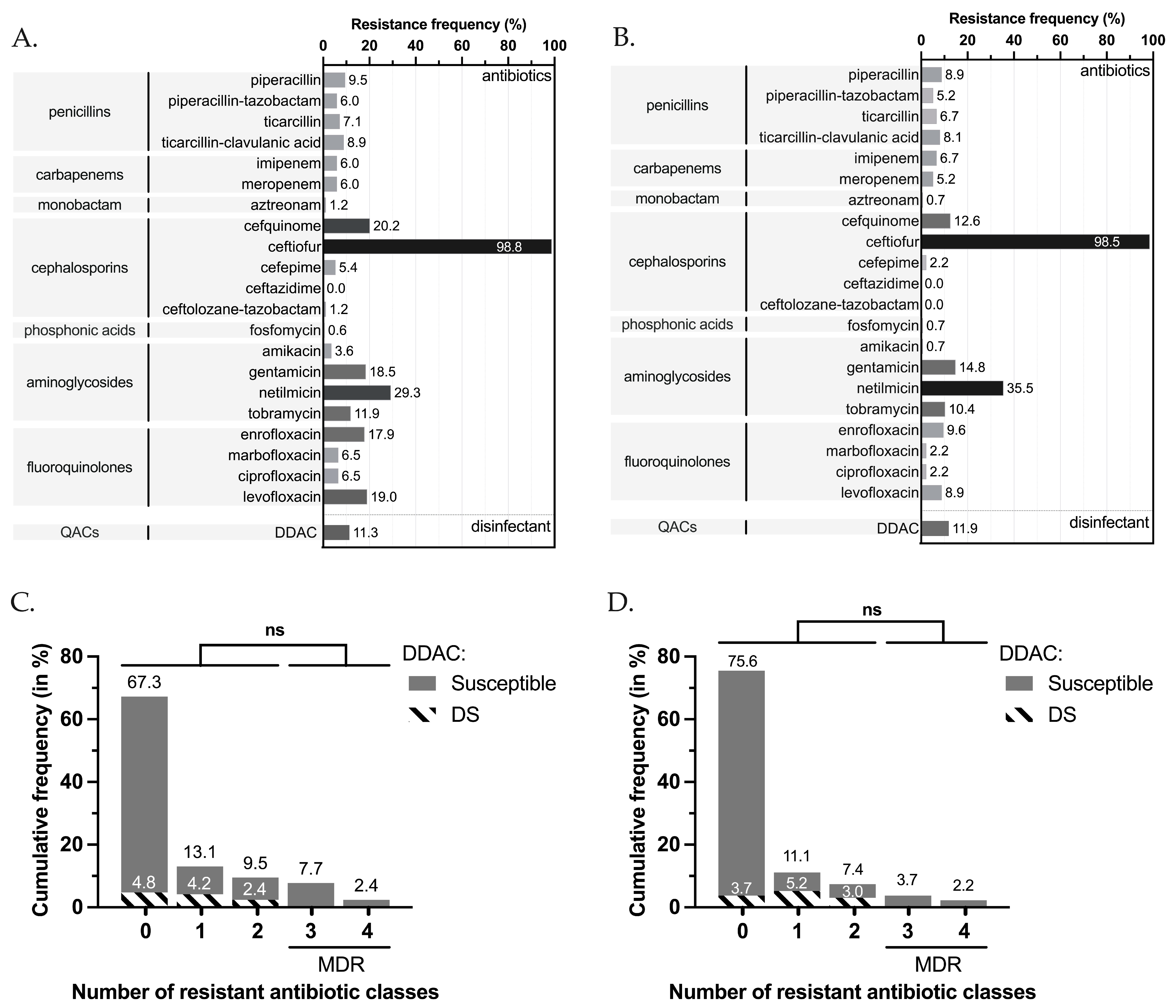
3.1. Antimicrobial and DDAC Resistance Phenotypes

The highest rates of resistance were observed for the veterinary antibiotic XNL (98.8%), followed by NTM (29.3%), CEQ (20.2%), LVX (19.0%), GMN (18.5%), ENR (17.9%) and TMN (11.9%). Frequency of resistance was less than 10.0% for other antibiotics tested (Figure 2A). For equine strains, which represented a major part of our population, the rates were 98.5% for XNL, 35.5% for NTM, 14.8% for GMN, 12.6% for CEQ, and 10.4% for TMN (Figure 2B).
Among the sequenced strains (n = 41), we found numerous genes conferring antibiotic resistance: some were constitutive for P. aeruginosa, and others were acquired. At least one resistance gene was associated with each main antibiotic class (Figure 3). A total of 11.9% (n = 20) of all veterinary strains were resistant to anti-Pseudomonas penicillins (at least one resistance among PIL, PTZ, TIC, TCC), 7.7% (n = 13) were resistant to carbapenems (IPM and/or MEM), 1.2% (n = 2) were resistant to monobactams (ATM), and 20.2% (n = 34) were resistant to cephalosporins (FEP, CZD, CLT, CEQ) (Figure 2A). For equine strains, 11.1% (n=15) were resistant to penicillins, 7.4% (n = 10) to carbapenems, and only one strain was resistant to monobactams (Figure 2B). For both classes of antibiotics, canine strains were the most resistant, with 16.7% (n = 5),10.0% (n = 3) and 3.3% (n = 1) of resistance, respectively (Figure 2B). These resistances were associated with the presence of resistance genes for β-lactams, including those coding CARB-2, the PSE family carbenicillin-hydrolyzing class A beta-lactamase (blaCARB-2), PDC, the cephalosporin-hydrolyzing class-C β-lactamase (blaPDC variants), and the OXA-family oxacillin-hydrolyzing class-D β-lactamases (blaOXA). The genes indicated as blaOXA-like and blaPDC-like were variants of blaOXA and blaPDC, respectively, that had less than 100% identity but were not known from the database. Note that no carbapenemase-coding genes were identified in the sequenced strains (Figure 3). For fosfomycin, only 0.6% (n = 1) of the veterinary population showed resistance, whereas the fosfomycin resistance glutathione transferase (fosA) coding gene was found in all sequenced strains. It was observed that 18.5% (n = 31) of the veterinary strains tested showed at least one resistance to aminoglycosides (AKN, GMN, NTM, TMN) (Figure 2A). Overall, 14.8% (n = 20) of equine strains were resistant to at least one aminoglycoside and 36.7% (n = 11) of canine strains (Figure 2B). Various antimicrobial resistance genes targeting this class of antibiotics were identified in the tested strains, including genes encoding aminoglycoside O-phosphotransferases (aph variants), N-acetyltransferases (aac variants), and aminoglycoside nucleotidyltransferases (aad variants) (Figure 3). For fluoroquinolones (CIP, LVX), at least one resistance was observed in 19.0% (n = 32) of veterinary strains. This rate was close (22.0%) when also considering veterinary antibiotics (ENR, MAR) (Figure 2A). The equine strains were resistant in 8.9% of cases (n = 12), although canine strains were resistant in 66.8% of cases (n = 20) (Figure 2B). For fluoroquinolone resistance, only gene-encoding protein CrpP was retrieved. For quinolone resistance, we identified genes encoding for the pentapeptide repeat protein QnrVC1 and modification of the amino acid sequence in the quinolone-resistance-determining region (QRDR) due to sequence alterations in position 473 of the parE gene. For sulfonamide, we found only the resistance gene encoding dihydropteroate synthase (sul1). For phenicols, we found type B chloramphenicol O-acetyltransferase (catB variants) and chloramphenicol efflux major facilitator superfamily (MFS) transporters (cmlA/floR variants and cmx). For tetracycline, genes coding the efflux MFS transporter Tet(G) and resistance-nodulation-division (RND) transporter efflux pump MexCD-OprJ were found. Interestingly, the qacEdelta1, qacG2, and qacL genes encoding small multidrug resistance (SMR) protein transporters were also found (Figure 3).
The study panel population showed various resistance profiles, with 67.3% (n = 113) of the strains displaying susceptibility to all 17 human antibiotics tested (seven classes), 13.1% (n = 22), and 9.5% (n = 16) showing nonsusceptibility to one and two antibiotic classes, respectively; 10.1% (n = 17) of strains were considered MDR (Figure 2B) but no strains were found to be XDR. The MDR profile concerned 30.0% (n = 9/30) of the canine-associated strains and 5.9% (n = 8/135) of the equine-associated strains, but was not associated with any other animal. Strain origins were auricular (n = 7/27; 25.9%), cutaneous/wound (n = 2/8; 25.0%), other/unspecified (n = 1/4; 25.0%), ocular (n = 1/5; 20.0%), respiratory (n = 1/12; 8.3%) and genital (n = 5/89; 5.6%). The feline- and bovine-associated strains did not present any resistance to antibiotics except the veterinary antibiotic ceftiofur. Note that the Anses strains were mostly susceptible to all the antibiotics tested, except for two strains, one of which was MDR.
Regarding DDAC, the MICs ranged from 8 to 128 mg/L, and decreased susceptibility (DS, MIC > 62.9 mg/L) was observed in 11.3% of tested strains (n = 19/168) (Figure 2C) and 11.9% of equine strains (n = 16/135) (Figure 2D). The DS to DDAC phenotypes was associated with patterns of antimicrobial drug resistance to up to two antibiotic classes: 4.8% showed no antibiotic resistance (3.7% for equine strains), 4.2% were resistant to one antibiotic class (5.2% for equine strains), and 2.4% were resistant to two antibiotic classes (3.0% for equine strains) (Figure 2C,D). The DS to DDAC phenotypes were observed since at least 2017 (from two in 2017 to seven in 2020; Table 1), largely in equine strains (n = 16) but also in feline (n = 2) and canine strains (n = 1) (Table 1). Before 2017, its presence cannot be evaluated, due to a lack of representativeness of the population. The DS to DDAC phenotype was significantly found in respiratory samples (n = 8, Fisher’s exact test, p < 0.0001), and it was also found in genital samples (n = 7); both were specifically associated with equines. The DS to DDAC phenotypes was also observed from ocular, cutaneous/wound, auricular, and digestive strains, but each only once (Table 1).

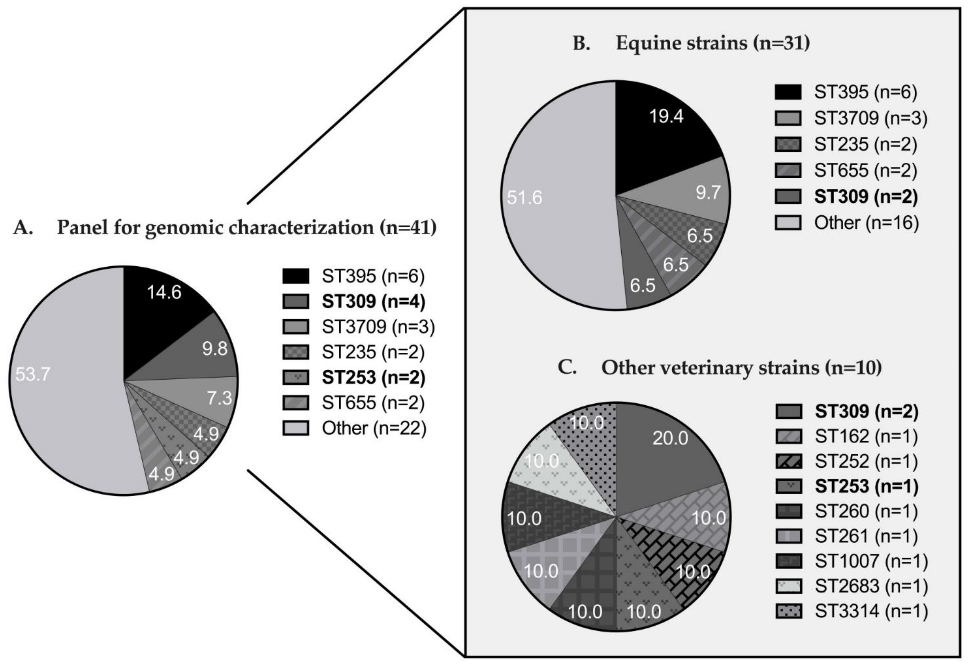
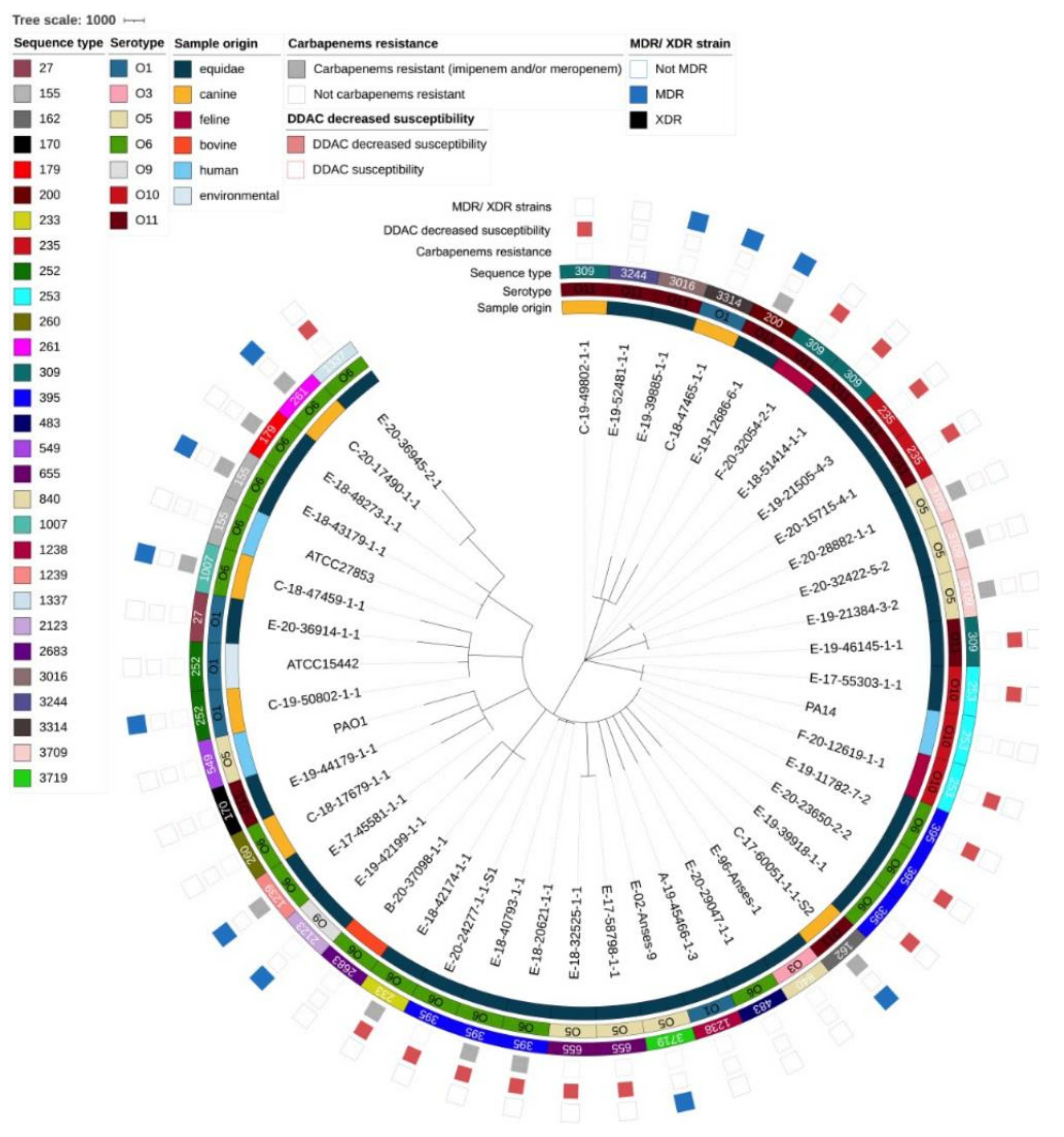
3.2. Genomic Diversity and Resistome Analysis

The 41 strains of the panel for genomic characterization were distributed into seven serotypes (Table 2). The three main serotypes were the same for all veterinary strains, and for the Equidae population in particular: serotypes O6 (39.0%), O11 (26.8%), and O5 (14.6%). Only O6 and O11, like serotypes O1 and O10, were found in more than one animal species.
The 41 sequenced strains were distributed among 28 sequence types (ST) (Figure 4A): primarily ST395 (14.6%), ST309 (9.8%), ST3709 (7.3%), ST235 (4.9%), ST253 (4.9%), and ST655 (4.9%). The other 22 STs were represented once. A closer focus on the equine strains (Figure 4B) showed that the main STs were ST395 (19.4%), ST3709 (9.7%), and ST235-ST309-ST655 (6.5% for each), with the other 16 ST represented by one strain. In the other animal species, only ST309 was represented twice, and ST162, ST252, ST253, ST260, ST261, ST1007, ST2683, and ST3314 were represented once (Figure 4C). Only ST309 and ST253 were found in more than one animal species.
This apparent diversity in serotypes and sequence types was confirmed by the cgMLST results charted in Figure 4. Of a total of 3867 loci searched, 3314 loci were present in the genome of all strains. Overall, strains diverged in distance from 14 to 3256 loci (average distance was 2893) and were well distributed according to origin (equine, canine, feline, bovine, human or environmental), year of collection, serotype, sequence type, carbapenem-resistance phenotype (major antibiotics reserved for human medicine), DS to DDAC phenotype, and whether or not the strain was multidrug-resistant (Figure 5).
None of the 41 sequenced strains showed a high level of antibiotic resistance (carbapenem resistance and MDR status) associated with DS to DDAC. However, three equine strains (E-18-20621-1-1, E-18-40793-1-1, and E-18-42174-1-1) showed a DS to DDAC associated with resistance to carbapenems. These strains were serotype O6: two were ST395, and one was ST233. The other equine strains presented either a DS-to-DDAC profile (n = 13), an MDR profile associated or not with resistance to carbapenems (n = 3 and n = 5, respectively), carbapenem resistance only (n = 4), or none of these phenotypes (n = 107). MDR status was assigned to 5 out of 7 of tested canine strains: three had resistance to carbapenems, and only one had DS to DDAC (C-19-49802-1-1). These strains were not associated with a single serotype or ST. The two feline strains, F-20-32054-2-1 (O11 and ST309) and F-20-12619-1-1 (O10 and ST253) did not show resistance to carbapenems or an MDR profile but had DS to DDAC. The bovine strain B-20-37098 (O6 and ST2683) was susceptible to antibiotics and DDAC.

4. Discussion

In France, since the 1990s, the surveillance of antimicrobial resistance in animals has been performed by the RESAPATH network [https://resapath.anses.fr/ accessed on 25 November 2022]. In 2020, the RESAPATH collected results from 71 member laboratories of 51,736 antibiograms performed on strains from dogs (27.3%), cattle (19.7%), poultry (19.7%), cats (10.8%), horses (7.4%) and pigs (7%) [75]. The Anses–Normandy Laboratory for Animal Health and LABÉO are both members of RESAPATH, with LABÉO providing the French network with the majority of horse antibiograms. In the 2006–2021 period, LABÉO performed 35,686 antibiograms on equine samples, and P. aeruginosa was the fourth most important pathogen isolated after group C streptococci, Escherichia coli, and Staphylococcus aureus. P. aeruginosa represented 3.6% (1367/37,686) of total isolated bacteria [76,77]. The majority of P. aeruginosa strains were isolated from respiratory tract samples (36.2%; 495/1367), followed by genital swabs (35.7%; 488/1367) and cutaneous samples (17.1%; 234/1367) [76,77].
This study set out to review the level of resistance of veterinary strains to the main human and veterinary antibiotics and to a common disinfectant, and to highlight the lack of anti-Pseudomonas therapies available in the veterinary field. It also enabled us to investigate and report the genomic diversity of these populations and the different antimicrobial resistance genes represented in them, and argue for the need to jointly study the Human–Animal–Environmental reservoirs. We selected 111/263 (42.0%) of the strains isolated from 2017 to 2020 at LABÉO to ensure a diversity of sample years and origins. This panel was completed by 24 equine strains provided by Anses and isolated during necropsies performed in the 1996–2017 period. Several animal species were included in this study; however, the small number of feline and bovine strains ruled out including these strains in a cross-species comparison. However, these feline and bovine strains did make it possible to determine whether some phenotypes are specific to one of the species.
For ASTs, the only antibiotics cited for testing by the veterinary CASFM 2021 [78] are gentamicin, amikacin, and ciprofloxacin. The observed resistance rates in our population were 18.5% for gentamicin and less than 10% for amikacin and ciprofloxacin. However, among the list of anti-pseudomonas antibiotics tested in this study, only ceftiofur, cefquinome, gentamicin, marbofloxacin, and enrofloxacin are currently marketed for veterinary use according to the Index of Veterinary Medicines authorized in France by the Anses [79]. Polymyxins can also be used but were not tested here. In this population, resistance rates for these antibiotics are above 20% for cephalosporins (up to 98.8% for ceftiofur) but less than 20% for fluoroquinolones.
We expected to find this high rate (up to 98.8%) of resistance to ceftiofur [80,81,82], but as this antibiotic is taken into consideration for the choice of therapy by some diagnostic laboratories, we wanted to provide additional evidence of the low activity of ceftiofur on P. aeruginosa. For gentamicin and fluoroquinolones, the rates obtained were lower than those found by van Spijk et al. [83] between 2012 and 2015 on an equine hospital population. Note that for some of these antibiotics, there is no marketed medicine suitable for use in every animal species. Consequently, there is a significant lack of antibiotic-based solutions against P. aeruginosa in veterinary medicine. For all classes of antibiotics tested, canine strains had higher resistance rates than equine strains. This was particularly marked for fluoroquinolones (57.9% difference) and aminoglycosides (21.9% difference). The resistance values obtained here can be compared with a previous study performed at the nearby Caen University Hospital on strains isolated from patients between 2011 and 2020 [84]. The resistance rates found in P. aeruginosa strains were much lower than those obtained in human medicine for phosphonic acid (−32.7% for the veterinary population) and penicillins (−15.2%), and measurably lower for monobactams (−5.2%), cephalosporins (−4.8%) and carbapenems (−1.8%), but higher for fluoroquinolones (+3.4%) and aminoglycosides (+7.2%). It is surprising to note such a small difference in resistance rates in respect of carbapenems between veterinary and human populations, especially as carbapenems are not authorized and used in veterinary medicine in Europe, except in exceptional cases in university clinics. As found in other studies, the presence of a carbapenem resistance phenotype in animals is rarely associated with the presence of a carbapenemase. The presumed mechanisms would be the decreased permeability by deficiency of the outer membrane protein OprD2 [85], hyperproduction of the chromosomal cephalosporinase AmpC [86,87,88], or preferential overexpression of efflux pumps [89]. However, some rare cases of carbapenemase expression, especially VIM-2, have been reported in different countries in P. aeruginosa strains isolated from dogs, cattle, and fowl [90,91], but to our knowledge not yet in horses. Some studies even suggest the existence of zoonotic transmission from animals to humans [92] and from humans to animals [93,94], but in an anecdotal manner, including the case of a transmission of a VIM-2 strain from humans to animals in Brazil [95].
No pandrug-resistant or extensively drug-resistant strains were found, but 10.1% of the strains studied were categorized as MDR. Note that MDR strains were found in the more recent strains isolated at LABÉO, suggesting that equine P. aeruginosa strains have adapted to 3–4 classes of antibiotics. This finding corroborates similar previous studies [89] showing the existence of MDR strains in the veterinary population and extends this problem to Equidae. In comparison, in human medicine, 12.6% of P. aeruginosa strains analyzed have either MDR (11.9%) or XDR (0.7%) profiles [84].
From a genomic point of view, the 41 veterinary strains were quite diverse, which is consistent with the strain sampling system. These strains came from different types of environments depending on animal species and geographical location. In total, 28 different STs were identified. Note that among these STs, ST395 (n = 6), ST235 (n = 2), ST253 (n = 2), ST233 (n = 1), and ST27 (n = 1) were also found in the hospital isolates in a previous human study (patient only) [84]. Thus, 29.3% of the animal strains sequenced in this study shared an ST identified in patients at the University Hospital of Caen. ST235, representing 4.9% of the sequenced strains, was even considered in 2020 as among the top 10 P. aeruginosa high-risk clones worldwide based on prevalence, global spread, and association with MDR/XDR profiles and the extended-spectrum β-lactamases and carbapenemases [96]. However, in our study, ST235 strains were neither XDR, MDR, nor carbapenem-resistant, but only associated with a DS-to-DDAC phenotype. ST235 had already been found in Japan in dogs and cats at a rate of up to 21.1% [94]. Four ST309 strains were also identified in this study and were found in the human hospital environment [84]. It would be informative to study the genetic distance between veterinary and hospital strains in more detail, in particular using a cgMLST approach.
In terms of cgMLST, note that strains isolated from different animal species could have greater genomic proximity than strains isolated from the same animal species, which would be quite diversified. However, the data collected on our samples do not allow us to distinguish between cases of infection or colonization in the animal. This factor would determine whether or not strains from colonization and/or infection would be grouped on the phylogenetic tree. It would be also interesting to determine how close the human and animal strains were in order to get a picture of their potential capacity for human–animal transmission.
Concerning the resistome of the strains, some genes were only found to be associated with one animal species or within one ST. Except for aph(3′)-IIb, which is systematically present in P. aeruginosa, all the aminoglycoside resistance genes were specific to equine strains, as was also the case for some oxacillinases and the blaCARB-2 gene and for the genes for resistance to sulfonamides, phenicols (except catB7), tetracycline, the qnrVC1 gene targeting quinolones, and resistance genes to quaternary ammonium. The resistome associated with the other species was thus less diverse. On the whole, for the same ST, the strains had similar, but not always identical, resistomes. In ST252, ST655, and ST3709, only one resistome was found by ST, whereas for ST155, ST235, ST253, ST309, and ST395, various antimicrobial resistance gene profiles were found in each, although similar. Moreover, the same resistome could lead to various resistance profiles. For example, the canine-associated MDR strain C-19-50802-1-1 presented the same resistome as the non-MDR reference strain ATCC15442, implying potentially mutational mechanisms conferring higher resistance to the canine-associated strain.
This study also determined the MIC of P. aeruginosa strains to a quaternary ammonium compound, DDAC, that is widely used in veterinary hospital disinfectants and as a biocide in various applications. Considering a MIC > 62.9 mg/L (corresponding to the concentration of DDAC in the commercial disinfectant solution), there was DS to DDAC in 11.0% of our strains and this DS was not associated with MDR P. aeruginosa profiles. MICs of DDAC greater than or equal to our DS threshold have also been shown for veterinary strains isolated between 1994 and 2003 in the USA, but on a smaller scale (2.9% of 175 P. aeruginosa strains) [81]. In contrast, at Caen University Hospital, this profile was found for 38.9% of strains, spanning both human strains and hospital-environment strains, that were significantly more associated with MDR and XDR P. aeruginosa profiles and more prevalent in the hospital environment (62.5% of them) than in human strains (28.2%) [84]. In our population, the fact that we included strains potentially from individuals and non-hospital veterinary environments likely leads to a lower calculated rate of resistance. These strains must have been less frequently exposed to DDAC than strains from the hospital environment, which is obviously regularly disinfected. The underlying molecular mechanisms of this phenotype are not yet fully elucidated, but the initial evidence points to MexAB-OprM pump efflux overexpression [84].

5. Conclusions

To the best of our knowledge, this is the biggest and most representative phenotypic and genomic study on P. aeruginosa strains isolated from Equidae. With the implementation of whole-genome sequencing and genomic approaches, we were able to assess the diversity and the resistome of the different strains. Such a strategy is destined to become an indispensable tool for monitoring infections and the dissemination of resistance. In contrast to hospital-acquired human infections, our results point to a high diversity of P. aeruginosa populations as causative agents in equine infections. Because more than 10% of animal strains showed an MDR phenotype, horses may be considered reservoirs of antimicrobial resistance in P. aeruginosa. Moreover, a significant number of isolates were resistant to carbapenems (7.7%), which are antimicrobials non-authorized in veterinary medicine. This highlights the need for a global approach in epidemiological studies. Our data also pointed out that attention should be paid to the use of disinfectants such as DDAC, constituting a selective pressure for the persistence of less susceptible strains. Our robust data are the foundation for further monitoring P. aeruginosa resistant strains and optimizing antimicrobial therapies in veterinary medicine via a one-health approach.

Supplementary Materials

The following supporting information can be downloaded at: https://www.mdpi.com/article/10.3390/pathogens12010064/s1, Table S1: Characteristics of the strains used in the study (Part 1) and for genomic characterization (Part 2). Table S2: Excel file of antimicrobial resistance-associated genes of strains from the panel for genomic characterization and for reference strains (n = 41 + 4).

Author Contributions

Conceptualization, M.P., S.L.H. and A.L.; methodology, M.P., S.C., G.L., C.S. and F.G.; software, M.P. and F.G.; validation, F.G., S.L.H. and A.L.; formal analysis, M.P. and F.G.; investigation, M.P. and A.L.; data curation, M.P., A.L., F.G. and S.L.H.; writing—original draft preparation, M.P. and A.L.; writing—review and editing, M.P., S.C., F.G., S.P., J.-C.G., S.L.H. and A.L.; visualization, M.P., S.C., F.G., S.P., J.-C.G., S.L.H. and A.L.; supervision, A.L. and S.L.H.; project administration, A.L. and S.L.H.; funding acquisition, A.L. and S.L.H. All authors have read and agreed to the published version of the manuscript.

Funding

This research was funded by the Normandy Regional Council grant number AMI2019RIN50_MPOTTIER and the French Ministry of Agriculture and Food (ÉcoAntibio2 Plan) grant number 2020-206-29-062.

Institutional Review Board Statement

Not applicable.

Informed Consent Statement

Not applicable.

Data Availability Statement

The datasets generated and analyzed during the current study are available via https://doi.org/10.6084/m9.figshare.c.6350123 accessed on 25 November 2022. All the DNA sequences of the present study can be downloaded in the BioProject PRJNA887012.

Conflicts of Interest

The authors declare no conflict of interest. The funders had no role in the design of the study; in the collection, analyses, or interpretation of data; in the writing of the manuscript; or in the decision to publish the results.

Appendix A

Table A1. Distribution of the decreased susceptibility-to-DDAC phenotype in the panel for genomic characterization (n = 41), according to the year of strain isolation, animal species and sampling.
Year of SamplingDDAC StatusTotal
n
Susceptible
n
DS
n
19961-1
20021-1
2017224
20185510
20198513
20206612
Total231841
Sample originDDAC statusTotal
n
Susceptible
n
DS
n
Equine161531
Canine617
Feline-22
Bovine1-1
Total231841
Type of sampleDDAC statusTotal
n
Susceptible
n
DS
n
Genital10616
Respiratory189
Auricular415
Cutaneous/wound415
Other/not specified2-2
Ocular112
Digestive-11
Necropsy1-1
Total231841
DDAC: didecyldimethylammonium chloride; DS: decreased susceptibility.

Appendix B

Table A2. Zone diameter breakpoints used for clinical interpretation of inhibition zone diameters for all P. aeruginosa strains.
AntibioticsUsed for MDR/XDR CategorizationSourceZone Diameter Breakpoints (mm)
S≥R<ATU
PenicillinsPiperacillinyesEUCAST 2021, Pseudomonas spp. [97]501818–19
Piperacillin-tazobactamyesEUCAST 2021, Pseudomonas spp. [97]501818–19
TicarcillinyesEUCAST 2021, Pseudomonas spp. [97]5018
Ticarcillin-clavulanic acidyesEUCAST 2021, Pseudomonas spp. [97]5018
CarbapenemsImipenemyesEUCAST 2021, Pseudomonas spp. [97]5020
MeropenemyesEUCAST 2021, Pseudomonas spp. [97]2418
MonobactamAztreonamyesEUCAST 2021, Pseudomonas spp. [97]5018
CephalosporinsCefquinomenoCASFM veterinary 2013, Enterobacteriaceae [98]2219
CeftiofurnoCASFM veterinary 2013, Enterobacteriaceae [98]2118
CefepimeyesEUCAST 2021, Pseudomonas spp. [97]5021
CeftazidimeyesEUCAST 2021, Pseudomonas spp. [97]5017
Ceftolozane-tazobactamyesEUCAST 2021, Pseudomonas spp. [97]2323
Phosphonic acidsFosfomycinyesEUCAST 2021, Pseudomonas spp.
Based on clinical observations [97]
127
AminoglycosidesAmikacinyesEUCAST 2021, Pseudomonas spp. [97]1515
GentamicinyesEUCAST 2019, Pseudomonas spp. [99]1515
NetilmicinyesEUCAST 2019, Pseudomonas spp. [99]1212
TobramycinyesEUCAST 2021, Pseudomonas spp. [97]1818
FluoroquinolonesEnrofloxacinnoCASFM veterinary 2013, Enterobacteriaceae [98]2217
MarbofloxacinnoCASFM veterinary 2013, Enterobacteriaceae [98]1815
Ciprofloxacin *yesEUCAST 2021, Pseudomonas spp. [97]5026
LevofloxacinyesEUCAST 2021, Pseudomonas spp. [97]5022
ATU: area of technical uncertainty; CASFM: Comité de l’Antibiogramme de la Société Française de Microbiologie; DDAC: didecyldimethylammonium chloride; DS: decreased susceptibility; EUCAST: European Committee on Antimicrobial Susceptibility Testing; MDR: multidrug-resistant; R: resistance; S: susceptibility; XDR: extensively drug-resistant. *: EUCAST 2021 recommendations.

Appendix C

Figure A1. Bioinformatics pipeline for whole-genome sequencing and sequence analysis. [50,51,52,53,54,55,56,57,58,59,60,61,62,63,64,65,66,67,68,69,70,71,72,73,74]. cg: core genome; MLST: multilocus sequence typing; WGS: whole-genome sequencing.
Pathogens 12 00064 g0a1

References

  1. Walker, T.S.; Bais, H.P.; Déziel, E.; Schweizer, H.P.; Rahme, L.G.; Fall, R.; Vivanco, J.M. Pseudomonas aeruginosa-plant root interactions, pathogenicity, biofilm formation, and root exudation. Plant Physiol. 2004, 134, 320–331. [Google Scholar] [CrossRef] [PubMed] [Green Version]
  2. Lupo, A.; Haenni, M.; Madec, J.-Y. Antimicrobial resistance in Acinetobacter spp. and Pseudomonas spp. Microbiol. Spectr. 2018, 6. [Google Scholar] [CrossRef] [PubMed]
  3. WHO Regional Office for Europe/European Centre for Disease Prevention and Control. Antimicrobial Resistance Surveillance in Europe: 2022–2020 Data; WHO Regional Office for Europe: Copenhagen, Denmark, 2022; p. 164. [Google Scholar]
  4. Résapath. Réseau d’Epidémiosurveillance de l’Antibiorésistance des Bactéries Pathogènes Animales, Bilan 2014; Agence Nationale de Sécurité Sanitaire de l’Alimentation, de l’Environnement et du Travail (Anses): Lyon, France; Ploufragan-Plouzané-Niort Laboratory: Ploufragan, France, 2015; p. 168. [Google Scholar]
  5. Hariharan, H.; Coles, M.; Poole, D.; Lund, L.; Page, R. Update on antimicrobial susceptibilities of bacterial isolates from canine and feline otitis externa. Can. Vet. J. 2006, 47, 3. [Google Scholar]
  6. Edwards, S.G.; Maggs, D.J.; Byrne, B.A.; Kass, P.H.; Lassaline, M.E. Effect of topical application of 0.5% proparacaine on corneal culture results from 33 dogs, 12 cats, and 19 horses with spontaneously arising ulcerative keratitis. Vet. Ophthalmol. 2019, 22, 415–422. [Google Scholar] [CrossRef] [PubMed] [Green Version]
  7. Poonsuk, K.; Chuanchuen, R. Contribution of the MexXY multidrug efflux pump and other chromosomal mechanisms on aminoglycoside resistance in Pseudomonas aeruginosa isolates from canine and feline infections. J. Vet. Med. Sci. 2012, 74, 1575–1582. [Google Scholar] [CrossRef] [Green Version]
  8. Sela, S.; Hammer-Muntz, O.; Krifucks, O.; Pinto, R.; Weisblit, L.; Leitner, G. Phenotypic and genotypic characterization of Pseudomonas aeruginosa strains isolated from mastitis outbreaks in dairy herds. J. Dairy Res. 2007, 74, 425–429. [Google Scholar] [CrossRef]
  9. Aleksandrov, M.; Petkov, A. Case of Pseudomonas aeruginosa infection in tropical snakes. Vet.-Meditsinski Nauk. 1985, 22, 53–61. [Google Scholar]
  10. Résapath. Réseau d’Epidémiosurveillance de l’Antibiorésistance des Bactéries Pathogènes Animales, Bilan 2021; Agence Nationale de Sécurité Sanitaire de l’Alimentation, de l’Environnement et du Travail (Anses): Lyon, France; Ploufragan-Plouzané-Niort Laboratory: Ploufragan, France, 2022; p. 48. [Google Scholar]
  11. Govan, J.R.; Sarasola, P.; Taylor, D.J.; Tatnell, P.J.; Russell, N.J.; Gacesa, P. Isolation of a mucoid alginate-producing Pseudomonas aeruginosa strain from the equine guttural pouch. J. Clin. Microbiol. 1992, 30, 595–599. [Google Scholar] [CrossRef] [Green Version]
  12. Moore, J.E.; Buckley, T.C.; Millar, B.C.; Gibson, P.; Cannon, G.; Egan, C.; Cosgrove, H.; Stanbridge, S.; Anzai, T.; Matsuda, M.; et al. Molecular surveillance of the incidence of Taylorella equigenitalis and Pseudomonas aeruginosa from horses in Ireland by sequence-specific PCR. Equine Vet. J. 2001, 33, 319–322. [Google Scholar] [CrossRef]
  13. Coman, D. Horserace Betting Levy Board: Codes of practice update. Vet Equine 2022, 6, 159–162. [Google Scholar] [CrossRef]
  14. Horserace Betting Levy Board Codes of Practice; HBLB: London, UK, 2004; p. 24.
  15. Whitlock, F.M.; Newton, J.R. A practitioner’s guide to understanding equine infectious disease diagnostics in the United Kingdom. Part 1: How to optimise sampling approaches and a guide to agent detection testing methods. Equine Vet. Educ. 2022, 34, 330–336. [Google Scholar] [CrossRef]
  16. RESPE. Saison de Monte 2021, Bilan Annuel; Réseau d’Epidémio-Surveillance en Pathologie Équine: Saint-Contest, France, 2021; p. 6. [Google Scholar]
  17. Allen, J.L.; Begg, A.P.; Browning, G.F. Outbreak of equine endometritis caused by a genotypically identical strain of Pseudomonas aeruginosa. J. Vet. Diagn. Investig. 2011, 23, 1236–1239. [Google Scholar] [CrossRef]
  18. Bruyas, J.-F.; Puyt, J.-D.; Hermange, T.; Betsch, J.-M.; Maillard, K.; Destrumelle, S. Thérapeutique anti-infectieuse raisonnée des métrites et endométrites de la jument. Prat. Vét. Equine 2013, 45, 7–16. [Google Scholar]
  19. Tiago, G.; Júlio, C.; António, R. Conception rate, uterine infection and embryo quality after artificial insemination and natural breeding with a stallion carrier of Pseudomonas aeruginosa: A Case Report. Acta Vet. Scand. 2012, 54, 20. [Google Scholar] [CrossRef] [Green Version]
  20. Omar, H.; Hambidge, M.; Firmanes, B.; Shabandri, A.M.; Wilsher, S. Bacteria isolated from equine uteri in the United Arab Emirates: A retrospective study. J. Equine Vet. Sci. 2022, 115, 104029. [Google Scholar] [CrossRef]
  21. Díaz-Bertrana, M.L.; Deleuze, S.; Pitti Rios, L.; Yeste, M.; Morales Fariña, I.; Rivera del Alamo, M.M. Microbial prevalence and antimicrobial sensitivity in equine endometritis in field conditions. Animals 2021, 11, 1476. [Google Scholar] [CrossRef]
  22. Mena, K.D.; Gerba, C.P. Risk assessment of Pseudomonas aeruginosa in water. In Reviews of Environmental Contamination and Toxicology; Whitacre, D.M., Ed.; Springer: Boston, MA, USA, 2009; Volume 201, pp. 71–115. ISBN 978-1-4419-0031-9. [Google Scholar]
  23. Stover, C.K.; Pham, X.Q.; Erwin, A.L.; Mizoguchi, S.D.; Warrener, P.; Hickey, M.J.; Brinkman, F.S.; Hufnagle, W.O.; Kowalik, D.J.; Lagrou, M.; et al. Complete genome sequence of Pseudomonas aeruginosa PAO1, an opportunistic pathogen. Nature 2000, 406, 959–964. [Google Scholar] [CrossRef] [Green Version]
  24. Gellatly, S.L.; Hancock, R.E.W. Pseudomonas aeruginosa: New insights into pathogenesis and host defenses. Pathog. Dis. 2013, 67, 159–173. [Google Scholar] [CrossRef] [Green Version]
  25. Azam, M.W.; Khan, A.U. Updates on the pathogenicity status of Pseudomonas aeruginosa. Drug Discov. Today 2019, 24, 350–359. [Google Scholar] [CrossRef]
  26. Botelho, J.; Grosso, F.; Peixe, L. Antibiotic resistance in Pseudomonas aeruginosa-mechanisms, epidemiology and evolution. Drug Resist. Updates 2019, 44, 100640. [Google Scholar] [CrossRef]
  27. Reig, S.; Le Gouellec, A.; Bleves, S. What is new in the anti–Pseudomonas aeruginosa clinical development pipeline since the 2017 WHO alert? Front. Cell. Infect. Microbiol. 2022, 12, 909731. [Google Scholar] [CrossRef] [PubMed]
  28. Adhimi, R.; Tayh, G.; Ghariani, S.; Chairat, S.; Chaouachi, A.; Boudabous, A.; Slama, K.B. Distribution, diversity and antibiotic resistance of Pseudomonas spp. isolated from the water dams in the north of Tunisia. Curr. Microbiol. 2022, 79, 188. [Google Scholar] [CrossRef] [PubMed]
  29. Fangous, M.-S.; Gosset, P.; Galakhoff, N.; Gouriou, S.; Guilloux, C.-A.; Payan, C.; Vallet, S.; Héry-Arnaud, G.; Le Berre, R. Priming with intranasal Lactobacilli prevents Pseudomonas aeruginosa acute pneumonia in mice. BMC Microbiol. 2021, 21, 195. [Google Scholar] [CrossRef]
  30. Oyardi, O.; Savage, P.B.; Guzel, C.B. Effects of ceragenins and antimicrobial peptides on the A549 cell line and an in vitro co-culture model of A549 cells and Pseudomonas aeruginosa. Pathogens 2022, 11, 1044. [Google Scholar] [CrossRef] [PubMed]
  31. Tacconelli, E.; Carrara, E.; Savoldi, A.; Harbarth, S.; Mendelson, M.; Monnet, D.L.; Pulcini, C.; Kahlmeter, G.; Kluytmans, J.; Carmeli, Y.; et al. Discovery, research, and development of new antibiotics: The WHO priority list of antibiotic-resistant bacteria and tuberculosis. Lancet Infect. Dis. 2018, 18, 318–327. [Google Scholar] [CrossRef]
  32. Commitee for Medicinal Products for Veterinary. Categorisation of Antibiotics in the European Union; European Medicines Agency: Amsterdam, The Netherlands, 2019. [Google Scholar]
  33. Russell, A. Biocide use and antibiotic resistance: The relevance of laboratory findings to clinical and environmental situations. Lancet Infect. Dis. 2003, 3, 794–803. [Google Scholar] [CrossRef]
  34. Yazdankhah, S.P.; Scheie, A.A.; Høiby, E.A.; Lunestad, B.-T.; Heir, E.; Fotland, T.Ø.; Naterstad, K.; Kruse, H. Triclosan and antimicrobial resistance in bacteria: An overview. Microb. Drug Resist. 2006, 12, 83–90. [Google Scholar] [CrossRef]
  35. Forbes, S.; Latimer, J.; Bazaid, A.; McBain, A.J. Altered competitive fitness, antimicrobial susceptibility, and cellular morphology in a triclosan-induced small-colony variant of Staphylococcus aureus. Antimicrob. Agents Chemother. 2015, 59, 4809–4816. [Google Scholar] [CrossRef] [Green Version]
  36. Wieland, N.; Boss, J.; Lettmann, S.; Fritz, B.; Schwaiger, K.; Bauer, J.; Hölzel, C.S. Susceptibility to disinfectants in antimicrobial-resistant and -susceptible isolates of Escherichia coli, Enterococcus faecalis and Enterococcus faecium from poultry-ESBL/AmpC-phenotype of E. coli is not associated with resistance to a quaternary ammonium compound, DDAC. J. Appl. Microbiol. 2017, 122, 1508–1517. [Google Scholar] [CrossRef]
  37. Rozman, U.; Pušnik, M.; Kmetec, S.; Duh, D.; Šostar Turk, S. Reduced susceptibility and increased resistance of bacteria against disinfectants: A systematic review. Microorganisms 2021, 9, 2550. [Google Scholar] [CrossRef]
  38. Nasr, A.M.; Mostafa, M.S.; Arnaout, H.H.; Elshimy, A.A.A. The effect of exposure to sub-inhibitory concentrations of hypochlorite and quaternary ammonium compounds on antimicrobial susceptibility of Pseudomonas aeruginosa. Am. J. Infect. Control 2018, 46, e57–e63. [Google Scholar] [CrossRef] [Green Version]
  39. Lloyd, D.H.; Page, S.W. Antimicrobial stewardship in veterinary medicine. Microbiol. Spectr. 2018, 6. [Google Scholar] [CrossRef] [Green Version]
  40. Yemiş, F.; Harmancı, N.Y. Classification, uses and environmental implications of disinfectants. Pak. J. Anal. Environ. Chem. 2020, 21, 179–192. [Google Scholar] [CrossRef]
  41. Kampf, G. Didecyldimethylammonium chloride. In Antiseptic Stewardship; Springer International Publishing: Cham, Switzerland, 2018; pp. 371–394. ISBN 978-3-319-98784-2. [Google Scholar]
  42. Samreen; Ahmad, I.; Malak, H.A.; Abulreesh, H.H. Environmental antimicrobial resistance and its drivers: A potential threat to public health. J. Glob. Antimicrob. Resist. 2021, 27, 101–111. [Google Scholar] [CrossRef]
  43. National Center for Biotechnology Information. PubChem Compound Summary for CID 23558, Didecyldimethylammonium Chloride. Available online: https://pubchem-ncbi-nlm-nih-gov.ezproxy.normandie-univ.fr/compound/Didecyldimethylammonium-chloride (accessed on 24 September 2022).
  44. Association Française de Normalisation. NF EN 13727+A2-Antiseptiques et Désinfectants Chimiques-Essai Quantitatif de Suspension pour l’Evaluation de l’Activité Bactéricide en Médecine; Association Française de Normalisation: Paris, France, 2015; p. 54. [Google Scholar]
  45. European Committee on Antimicrobial Susceptibility Testing. Antimicrobial Susceptibility Testing EUCAST Disk Diffusion Method; European Committee on Antimicrobial Susceptibility Testing: Växjö, Sweden, 2021; p. 22. [Google Scholar]
  46. Mikkelsen, H.; McMullan, R.; Filloux, A. The Pseudomonas aeruginosa reference strain PA14 displays increased virulence due to a mutation in ladS. PLoS ONE 2011, 6, e29113. [Google Scholar] [CrossRef] [Green Version]
  47. Association Française de Normalisation. NF U47-107-Méthodes D’analyse en Santé Animale-Guide de Réalisation des Antibiogrammes par la Méthode de Diffusion en Milieu Gélosé; Association Française de Normalisation: Paris, France, 2012; p. 13. [Google Scholar] [CrossRef] [Green Version]
  48. Magiorakos, A.-P.; Srinivasan, A.; Carey, R.B.; Carmeli, Y.; Falagas, M.E.; Giske, C.G. Multidrug-resistant, extensively drug-resistant and pandrug-resistant bacteria: An international expert proposal for interim standard definitions for acquired resistance. Clin. Microbiol. Infect. 2012, 18, 268–281. [Google Scholar] [CrossRef] [Green Version]
  49. ISO 20776-1:2019; Susceptibility Testing of Infectious Agents and Evaluation of Performance of Antimicrobial Susceptibility Test Devices—Part 1: Broth Micro-Dilution Reference Method for Testing the In Vitro Activity of Antimicrobial Agents against Rapidly Growing Aerobic Bacteria Involved in Infectious Diseases. International Organization for Standardization: Geneva, Switzerland, 2019; p. 19.
  50. Simon Andrews Babraham Bioinformatics-FastQC a Quality Control Tool for High Throughput Sequence Data. Available online: https://www.bioinformatics.babraham.ac.uk/projects/fastqc/ (accessed on 14 January 2022).
  51. Ewels, P.; Magnusson, M.; Lundin, S.; Käller, M. MultiQC: Summarize analysis results for multiple tools and samples in a single report. Bioinformatics 2016, 32, 3047–3048. [Google Scholar] [CrossRef] [Green Version]
  52. Brown, C.T.; Howe, A.; Zhang, Q.; Pyrkosz, A.B.; Brom, T.H. A reference-free algorithm for computational normalization of shotgun sequencing data. arXiv 2012, arXiv:1203.4802. [Google Scholar]
  53. Criscuolo, A.; Brisse, S. AlienTrimmer: A tool to quickly and accurately trim off multiple short contaminant sequences from high-throughput sequencing reads. Genomics 2013, 102, 500–506. [Google Scholar] [CrossRef] [Green Version]
  54. Durai, D.A.; Schulz, M.H. Improving in silico normalization using read weights. Sci. Rep. 2019, 9, 5133. [Google Scholar] [CrossRef] [Green Version]
  55. Legrand, V.N.; Kergrohen, T.N.; Joly, N.N.; Criscuolo, A.N. ROCK: Digital normalization of whole genome sequencing data. J. Open Source Softw. 2022, 7, 3790. [Google Scholar] [CrossRef]
  56. Liu, Y.; Schröder, J.; Schmidt, B. Musket: A multistage k-mer spectrum-based error corrector for Illumina sequence data. Bioinformatics 2013, 29, 308–315. [Google Scholar] [CrossRef] [PubMed] [Green Version]
  57. Magoč, T.; Salzberg, S.L. FLASH: Fast length adjustment of short reads to improve genome assemblies. Bioinformatics 2011, 27, 2957–2963. [Google Scholar] [CrossRef] [PubMed]
  58. Roguski, Ł.; Deorowicz, S. DSRC 2—Industry-oriented compression of fastq files. Bioinformatics 2014, 30, 2213–2215. [Google Scholar] [CrossRef] [PubMed] [Green Version]
  59. Wedemeyer, A.; Kliemann, L.; Srivastav, A.; Schielke, C.; Reusch, T.B.; Rosenstiel, P. An improved filtering algorithm for big read datasets and its application to single-cell assembly. BMC Bioinform. 2017, 18, 324. [Google Scholar] [CrossRef] [Green Version]
  60. Bankevich, A.; Nurk, S.; Antipov, D.; Gurevich, A.A.; Dvorkin, M.; Kulikov, A.S.; Lesin, V.M.; Nikolenko, S.I.; Pham, S.; Prjibelski, A.D.; et al. SPAdes: A new genome assembly algorithm and its applications to single-cell sequencing. J. Comput. Biol. J. Comput. Mol. Cell Biol. 2012, 19, 455–477. [Google Scholar] [CrossRef] [Green Version]
  61. Gurevich, A.; Saveliev, V.; Vyahhi, N.; Tesler, G. QUAST: Quality assessment tool for genome assemblies. Bioinformatics 2013, 29, 1072–1075. [Google Scholar] [CrossRef] [Green Version]
  62. Jolley, K.A.; Bliss, C.M.; Bennett, J.S.; Bratcher, H.B.; Brehony, C.; Colles, F.M.; Wimalarathna, H.; Harrison, O.B.; Sheppard, S.K.; Cody, A.J.; et al. Ribosomal multilocus sequence typing: Universal characterization of bacteria from domain to strain. Microbiol. Read. Engl. 2012, 158, 1005–1015. [Google Scholar] [CrossRef]
  63. Camacho, C.; Coulouris, G.; Avagyan, V.; Ma, N.; Papadopoulos, J.; Bealer, K.; Madden, T.L. BLAST+: Architecture and applications. BMC Bioinform. 2009, 10, 421. [Google Scholar] [CrossRef] [Green Version]
  64. Thrane, S.W.; Taylor, V.L.; Lund, O.; Lam, J.S.; Jelsbak, L. Application of whole-genome sequencing data for O-specific antigen analysis and in silico serotyping of Pseudomonas aeruginosa isolates. J. Clin. Microbiol. 2016, 54, 1782–1788. [Google Scholar] [CrossRef] [Green Version]
  65. Lemee, L.; Dhalluin, A.; Pestel-Caron, M.; Lemeland, J.-F.; Pons, J.-L. Multilocus sequence typing analysis of Human and animal Clostridium difficile isolates of various toxigenic types. J. Clin. Microbiol. 2004, 42, 2609–2617. [Google Scholar] [CrossRef] [Green Version]
  66. Bartual, S.G.; Seifert, H.; Hippler, C.; Luzon, M.A.D.; Wisplinghoff, H.; Rodríguez-Valera, F. Development of a multilocus sequence typing scheme for characterization of clinical isolates of Acinetobacter baumannii. J. Clin. Microbiol. 2005, 43, 4382–4390. [Google Scholar] [CrossRef] [Green Version]
  67. Wirth, T.; Falush, D.; Lan, R.; Colles, F.; Mensa, P.; Wieler, L.H.; Karch, H.; Reeves, P.R.; Maiden, M.C.J.; Ochman, H.; et al. Sex and virulence in Escherichia coli: An evolutionary perspective. Mol. Microbiol. 2006, 60, 1136–1151. [Google Scholar] [CrossRef]
  68. Jaureguy, F.; Landraud, L.; Passet, V.; Diancourt, L.; Frapy, E.; Guigon, G.; Carbonnelle, E.; Lortholary, O.; Clermont, O.; Denamur, E.; et al. Phylogenetic and genomic diversity of human bacteremic Escherichia coli strains. BMC Genom. 2008, 9, 560. [Google Scholar] [CrossRef] [Green Version]
  69. Griffiths, D.; Fawley, W.; Kachrimanidou, M.; Bowden, R.; Crook, D.W.; Fung, R.; Golubchik, T.; Harding, R.M.; Jeffery, K.J.M.; Jolley, K.A.; et al. Multilocus sequence typing of Clostridium difficile. J. Clin. Microbiol. 2010, 48, 770–778. [Google Scholar] [CrossRef] [Green Version]
  70. Larsen, M.V.; Cosentino, S.; Rasmussen, S.; Friis, C.; Hasman, H.; Marvig, R.L.; Jelsbak, L.; Sicheritz-Pontén, T.; Ussery, D.W.; Aarestrup, F.M.; et al. Multilocus sequence typing of total-genome-sequenced bacteria. J. Clin. Microbiol. 2012, 50, 1355–1361. [Google Scholar] [CrossRef] [Green Version]
  71. Tönnies, H.; Prior, K.; Harmsen, D.; Mellmann, A. Establishment and evaluation of a core genome multilocus sequence typing scheme for whole-genome sequence-based typing of Pseudomonas aeruginosa. J. Clin. Microbiol. 2021, 59, e01987-20. [Google Scholar] [CrossRef]
  72. Silva, M.; Machado, M.P.; Silva, D.N.; Rossi, M.; Moran-Gilad, J.; Santos, S.; Ramirez, M.; Carriço, J.A. ChewBBACA: A complete suite for gene-by-gene schema creation and strain identification. Microb. Genom. 2018, 4, e000166. [Google Scholar] [CrossRef]
  73. Letunic, I.; Bork, P. Interactive Tree Of Life (ITOL) v5: An online tool for phylogenetic tree display and annotation. Nucleic Acids Res. 2021, 49, W293–W296. [Google Scholar] [CrossRef]
  74. Feldgarden, M.; Brover, V.; Gonzalez-Escalona, N.; Frye, J.G.; Haendiges, J.; Haft, D.H.; Hoffmann, M.; Pettengill, J.B.; Prasad, A.B.; Tillman, G.E.; et al. AMRFinderPlus and the Reference Gene Catalog facilitate examination of the genomic links among antimicrobial resistance, stress response, and virulence. Sci. Rep. 2021, 11, 12728. [Google Scholar] [CrossRef]
  75. Résapath. Réseau d’Epidémiosurveillance de l’Antibiorésistance des Bactéries Pathogènes Animales, Bilan 2020; Agence Nationale de Sécurité Sanitaire de l’Alimentation, de l’Environnement et du Travail (Anses): Lyon, France; Ploufragan-Plouzané-Niort Laboratory: Ploufragan, France, 2021; p. 42. [Google Scholar]
  76. Duchesne, R.; Castagnet, S.; Maillard, K.; Petry, S.; Cattoir, V.; Giard, J.-C.; Leon, A. In vitro antimicrobial susceptibility of equine clinical isolates from France, 2006–2016. J. Glob. Antimicrob. Resist. 2019, 19, 144–153. [Google Scholar] [CrossRef] [PubMed]
  77. Léon, A.; Castagnet, S.; Maillard, K.; Paillot, R.; Giard, J.-C. Evolution of in vitro antimicrobial susceptibility of equine clinical isolates in France between 2016 and 2019. Anim. Open Access J. 2020, 10, 812. [Google Scholar] [CrossRef] [PubMed]
  78. Comité de l’Antibiogramme de la Société Française de Microbiologie (CA-SFM). Recommandations Vétérinaires 2021; Société Française de Microbiologie: Paris, France, 2021; p. 15. [Google Scholar]
  79. European Committee on Antimicrobial Susceptibility Testing. Breakpoint Tables for Interpretation of MICs and Zone Diameters; European Committee on Antimicrobial Susceptibility Testing: Växjö, Sweden, 2021; p. 116. [Google Scholar]
  80. Anses. Index des RCP (Résumé des Caractéristiques du Produit). Available online: http://www.ircp.anmv.anses.fr/ (accessed on 22 October 2022).
  81. Scott, A.; Pottenger, S.; Timofte, D.; Moore, M.; Wright, L.; Kukavica-Ibrulj, I.; Jeukens, J.; Levesque, R.C.; Freschi, L.; Pinchbeck, G.L.; et al. Reservoirs of resistance: Polymyxin resistance in veterinary-associated companion animal isolates of Pseudomonas aeruginosa. Vet. Rec. 2019, 185, 206. [Google Scholar] [CrossRef] [PubMed] [Green Version]
  82. Beier, R.C.; Foley, S.L.; Davidson, M.K.; White, D.G.; McDermott, P.F.; Bodeis-Jones, S.; Zhao, S.; Andrews, K.; Crippen, T.L.; Sheffield, C.L.; et al. Characterization of antibiotic and disinfectant susceptibility profiles among Pseudomonas aeruginosa veterinary isolates recovered during 1994–2003. J. Appl. Microbiol. 2015, 118, 326–342. [Google Scholar] [CrossRef] [PubMed]
  83. Rubin, J.; Walker, R.D.; Blickenstaff, K.; Bodeis-Jones, S.; Zhao, S. Antimicrobial resistance and genetic characterization of fluoroquinolone resistance of Pseudomonas aeruginosa isolated from canine infections. Vet. Microbiol. 2008, 131, 164–172. [Google Scholar] [CrossRef] [PubMed]
  84. Van Spijk, J.N.; Schmitt, S.; Fürst, A.; Schoster, A. A retrospective analysis of antimicrobial resistance in bacterial pathogens in an equine hospital (2012–2015). Schweiz Arch. Tierheilkd 2016, 158, 433–442. [Google Scholar] [CrossRef] [Green Version]
  85. Pottier, M.; Gravey, F.; Castagnet, S.; Auzou, M.; Bénédicte, L.; Guérin, F.; Giard, J.-C.; Léon, A.; Hello, S.L. A ten-year microbiological study of Pseudomonas aeruginosa strains revealed diffusion of carbapenems and quaternary ammonium compounds resistant populations. medRxiv 2022. [Google Scholar] [CrossRef]
  86. Shen, J.; Pan, Y.; Fang, Y. Role of the outer membrane protein OprD2 in carbapenem-resistance mechanisms of Pseudomonas aeruginosa. PLoS ONE 2015, 10, e0139995. [Google Scholar] [CrossRef]
  87. Yoneyama, H.; Nakae, T. Mechanism of efficient elimination of protein D2 in outer membrane of imipenem-resistant Pseudomonas aeruginosa. Antimicrob. Agents Chemother. 1993, 37, 2385–2390. [Google Scholar] [CrossRef] [Green Version]
  88. Yoon, E.-J.; Jeong, S.H. Mobile carbapenemase genes in Pseudomonas aeruginosa. Front. Microbiol. 2021, 12, 614058. [Google Scholar] [CrossRef]
  89. Aurilio, C.; Sansone, P.; Barbarisi, M.; Pota, V.; Giaccari, L.G.; Coppolino, F.; Barbarisi, A.; Passavanti, M.B.; Pace, M.C. Mechanisms of action of carbapenem resistance. Antibiotics 2022, 11, 421. [Google Scholar] [CrossRef]
  90. Haenni, M.; Bour, M.; Châtre, P.; Madec, J.-Y.; Plésiat, P.; Jeannot, K. Resistance of animal strains of Pseudomonas aeruginosa to carbapenems. Front. Microbiol. 2017, 8, 1847. [Google Scholar] [CrossRef]
  91. Sellera, F.P.; Da Silva, L.C.B.A.; Lincopan, N. Rapid Spread of critical priority carbapenemase-producing pathogens in companion animals: A One Health challenge for a post-pandemic world. J. Antimicrob. Chemother. 2021, 76, 2225–2229. [Google Scholar] [CrossRef]
  92. Al Bayssari, C.; Dabboussi, F.; Hamze, M.; Rolain, J.-M. Emergence of carbapenemase-producing Pseudomonas aeruginosa and Acinetobacter baumannii in livestock animals in Lebanon. J. Antimicrob. Chemother. 2015, 70, 950–951. [Google Scholar] [CrossRef] [Green Version]
  93. Abd El-Ghany, W.A. Pseudomonas aeruginosa infection of avian origin: Zoonosis and One Health implications. Vet. World 2021, 14, 2155–2159. [Google Scholar] [CrossRef]
  94. Wang, Y.; Wang, X.; Schwarz, S.; Zhang, R.; Lei, L.; Liu, X.; Lin, D.; Shen, J. IMP-45-producing multidrug-resistant Pseudomonas aeruginosa of canine origin. J. Antimicrob. Chemother. 2014, 69, 2579–2581. [Google Scholar] [CrossRef] [Green Version]
  95. Hayashi, W.; Izumi, K.; Yoshida, S.; Takizawa, S.; Sakaguchi, K.; Iyori, K.; Minoshima, K.; Takano, S.; Kitagawa, M.; Nagano, Y.; et al. Antimicrobial resistance and Type III secretion system virulotypes of Pseudomonas aeruginosa isolates from dogs and cats in primary veterinary hospitals in Japan: Identification of the international high-risk clone Sequence Type 235. Microbiol. Spectr. 2021, 9, e00408-21. [Google Scholar] [CrossRef]
  96. Fernandes, M.R.; Sellera, F.P.; Moura, Q.; Carvalho, M.P.N.; Rosato, P.N.; Cerdeira, L.; Lincopan, N. Zooanthroponotic transmission of drug-resistant Pseudomonas aeruginosa, Brazil. Emerg. Infect. Dis. 2018, 24, 1160–1162. [Google Scholar] [CrossRef] [Green Version]
  97. Del Barrio-Tofiño, E.; López-Causapé, C.; Oliver, A. Pseudomonas aeruginosa epidemic high-risk clones and their association with horizontally-acquired β-lactamases: 2020 update. Int. J. Antimicrob. Agents 2020, 56, 106196. [Google Scholar] [CrossRef]
  98. Comité de l’Antibiogramme de la Société Française de Microbiologie (CA-SFM). Recommandations Vétérinaires 2013; Société Française de Microbiologie: Paris, France, 2013; p. 13. [Google Scholar]
  99. European Committee on Antimicrobial Susceptibility Testing. Breakpoint Tables for Interpretation of MICs and Zone Diameters; European Committee on Antimicrobial Susceptibility Testing: Växjö, Sweden, 2019; p. 100. [Google Scholar]
Figure 2. Antimicrobial resistance for veterinary strains. Frequency of resistance in P. aeruginosa strains tested for antibiotic susceptibility and antimicrobial resistance for the study panel (n = 168) (A) and for equine strains (n = 135) (B). Frequency histogram of the number of strains (in percent) showing at least one resistance for each different antibiotic class for the study panel (n = 168) (C) and for equine strains (n = 135) (D). Except for netilmicin, ceftiofur, cefquinome, marbofloxacin, and enrofloxacin, tested only for the panel for genomic characterization (n = 41), all other antimicrobials were tested for the study panel (n = 168). The four veterinary antibiotics (ceftiofur, cefquinome, marbofloxacin, and enrofloxacin) were excluded from the class-based resistance analysis for figure (B) Populations were tested for independence for DS to DDAC and loss of susceptibility to more than three categories of antibiotics using Fisher’s exact test. Ns: p-value > 0.05. DDAC: didecyldimethylammonium chloride; DS: decreased susceptibility; MDR: multidrug-resistant.
Pathogens 12 00064 g002
Figure 3. Antimicrobial-resistance-associated genes of strains from the panel for genomic characterization and for reference strains (n = 41 + 4). Strains were organized according to the cgMLST minimum distance tree. The figure lists serotype, sequence type, presence of a carbapenem resistance (imipenem and/or meropenem) phenotype, whether the strain was multidrug-resistant or not, and presence of decreased susceptibility to DDAC phenotype. The sub-variants of the aph(3′) gene were grouped, as well as the sub-variants of aac(6′). Co-occurrences of these sub-variants are indicated and separated by a backslash. The “-like genes” were variants not referenced on AMRFinder that had less than 100% shared identity. C: canine, B: bovine, DDAC: didecyldimethylammonium chloride, E: equine, F: feline, MDR: multidrug-resistant, O: serotype, ST: sequence type.
Pathogens 12 00064 g003
Figure 4. Multilocus sequence typing (MLST) of 41 sequenced strains. Representation of (A) the whole panel for genomic characterization, (B) equine strains, and (C) strains from other animal species (i.e., canine, feline, and bovine). The sequence types written in bold were found to be associated with several animal species. In figures (A,B), sequence types represented only once were grouped together. Figure (A) contained ST27, ST155, ST162, ST170, ST179, ST200, ST233, ST252, ST260, ST261, ST483, ST840, ST1007, ST1238, ST1239, ST1337, ST2123, ST2683, ST3016, ST3244, ST3314, and ST3719; figure (B) contained ST27, ST155, ST170, ST179, ST200, ST233, ST253, ST483, ST840, ST1238, ST1239, ST1337, ST2123, ST3016, ST3244, and ST3719. ST: Sequence type.
Pathogens 12 00064 g004
Figure 5. Minimum spanning tree of the strains from the panel for genomic characterization and reference strains (n = 41 + 4). Core genome MLST clustering according to the cgMLST Pseudomonas aeruginosa scheme previously published [71] and based on 3076 genes. The reference strains are ATCC15442, ATCC27853, PA14, and PAO1. The tree charts: origin of the strains (equine, canine, feline, bovine, human or environmental), serotype, sequence type, carbapenem resistance phenotype, DS to DDAC phenotype, and whether or not the strain was multidrug-resistant. C: canine, B: bovine, DDAC: didecyldimethylammonium chloride, E: equine, F: feline, MDR: multidrug-resistant, O: serotype, ST: sequence type, XDR: extensively drug-resistant.
Pathogens 12 00064 g005
Table 1. Distribution of the decreased-susceptibility-to-DDAC phenotype in the study panel (n = 168) according to the year of strain isolation, animal species, and sample type. p-values of Fisher’s test of independence comparing the category against the rest of the population.
Year of
Sampling
DDAC Status
SusceptibleDS
n (%)
19961 (100.0)-
19971 (100.0)-
19982 (100.0)-
19992 (100.0)-
20022 (100.0)-
20032 (100.0)-
20043 (100.0)-
20053 (100.0)-
20071 (100.0)-
20081 (100.0)-
20103 (100.0)-
20111 (100.0)-
20151 (100.0)-
20177 (77.8)2 (22.2)
201814 (73.7)5 (26.3)
201961 (92.4)5 (7.6)
202044 (86.3)7 (13.7)
Total149 (88.7)19 (11.3)
Sample OriginDDAC Statusp-values
SusceptibleDS
n (%)
Equine119 (88.1)16 (11.9)>0.9999
Canine29 (96.7)1 (3.3)0.2023
Feline-2 (100)-
Bovine1 (100)--
Total149 (88.7)19 (11.3)-
Type of SampleDDAC statusp-values
SusceptibleDS
n (%)
Genital82 (92.1)7 (7.9)0.1504
Other animal species 1 (100)--
Equine81 (92)7 (8)-
Auricular26 (96.3)1 (3.7)0.3164
Other animal species 26 (96.3)1 (3.7)-
Necropsy22 (100)-0.139
Equine22 (100)--
Respiratory4 (33.3)8 (66.7)<0.0001
Equine4 (33.3)8 (66.7)-
Cutaneous/wound7 (87.5)1 (12.5)>0.9999
Other animal species 1 (100)--
Equine6 (85.7)1 (14.3)-
Ocular4 (80)1 (20)0.4555
Other animal species 1 (50)1 (50)-
Equine3 (100)--
Other/not specified4 (100)--
Other animal species 1 (100)--
Equine3 (100)--
Digestive-1 (100)-
Other animal species -1 (100)-
Total149 (88.7)19 (11.3)-
DDAC: didecyldimethylammonium chloride; DS: decreased susceptibility.
Table 2. P. aeruginosa serotype distribution (n = 41, panel for genomic characterization).
Strain OriginSerotype
O1O3O5O6O9O10O11Total
Equine2161211831
Canine2 3 27
Feline 112
Bovine 1 1
Total n (%)4 (9.8)1 (2.4)6 (14.6)16 (39.0)1 (2.4)2 (4.9)11 (26.8)41 (100.0)
O: serotype.
Disclaimer/Publisher’s Note: The statements, opinions and data contained in all publications are solely those of the individual author(s) and contributor(s) and not of MDPI and/or the editor(s). MDPI and/or the editor(s) disclaim responsibility for any injury to people or property resulting from any ideas, methods, instructions or products referred to in the content.

