
Natural History and Hepatitis B Virus Surface Antigen (HBsAg) Spontaneous Seroclearance in Hepatitis B Virus e-Antigen (HBeAg)-Negative Patients with Inactive Chronic Infection: A Multicenter Regional Study from South Italy
 , ,
, ,
Abstract
:1. Introduction
2. Materials and Methods
2.1. Study Design
2.2. Laboratory Data and Transient Elastography
2.3. Statistical Analysis
3. Results
4. Discussion
5. Conclusions
Author Contributions
Funding
Institutional Review Board Statement
Informed Consent Statement
Data Availability Statement
Conflicts of Interest
References
- European Association for the Study of the Liver. EASL 2017 Clinical Practice Guidelines on the management of hepatitis B virus infection. J. Hepatol. 2017, 67, 370–398. [Google Scholar] [CrossRef] [PubMed]
- Chu, C.M.; Liaw, Y.F. Hepatitis B surface antigen seroclearance during chronic HBV infection. Antivir. Ther. 2010, 15, 133–143. [Google Scholar] [CrossRef] [PubMed]
- Yeo, Y.H.; Tseng, T.C.; Hosaka, T.; Cunningham, C.; Fung, J.Y.Y.; Ho, H.J.; Kwak, M.S.; Trinh, H.N.; Ungtrakul, T.; Yu, M.L.; et al. Incidence, Factors, and Patient-Level Data for Spontaneous HBsAg Seroclearance: A Cohort Study of 11,264 Patients. Clin. Transl. Gastroenterol. 2020, 11, e00196. [Google Scholar] [CrossRef] [PubMed]
- Brunetto, M.R.; Oliveri, F.; Colombatto, P.; Moriconi, F.; Ciccorossi, P.; Coco, B.; Romagnoli, V.; Cherubini, B.; Moscato, G.; Maina, A.M.; et al. Hepatitis B surface antigen serum levels help to distinguish active from inactive hepatitis B virus genotype D carriers. Gastroenterology 2010, 139, 483–490. [Google Scholar] [CrossRef] [PubMed]
- Brouwer, W.P.; Chan, H.L.; Brunetto, M.R.; Martinot-Peignoux, M.; Arends, P.; Cornberg, M.; Cherubini, B.; Thompson, A.J.; Liaw, Y.F.; Marcellin, P.; et al. Good Practice in using HBsAg in Chronic Hepatitis B Study Group (GPs-CHB Study Group). Repeated Measurements of Hepatitis B Surface Antigen Identify Carriers of Inactive HBV During Long-term Follow-up. Clin. Gastroenterol. Hepatol. 2016, 14, 1481–1489.e5. [Google Scholar] [CrossRef] [PubMed]
- Chu, C.M.; Hung, S.J.; Lin, J.; Tai, D.I.; Liaw, Y.F. Natural history of hepatitis B e antigen to antibody seroconversion in patients with normal serum aminotransferase levels. Am. J. Med. 2004, 116, 829–834. [Google Scholar] [CrossRef] [PubMed]
- Yoshida, K.; Enomoto, M.; Tamori, A.; Nishiguchi, S.; Kawada, N. Combination of Entecavir or Tenofovir with Pegylated Interferon-α for Long-Term Reduction in Hepatitis B Surface Antigen Levels: Simultaneous, Sequential, or Add-on Combination Therapy. Int. J. Mol. Sci. 2021, 22, 1456. [Google Scholar] [CrossRef] [PubMed]
- European Association for the Study of the Liver. EASL clinical practice guidelines: Management of chronic hepatitis B virus infection. J. Hepatol. 2012, 57, 167–185. [Google Scholar] [CrossRef] [PubMed]
- Khetsuriani, N.; Mosina, L.; Van Damme, P.; Mozalevskis, A.; Datta, S.; Tohme, R.A. Progress Toward Hepatitis B Control—World Health Organization European Region, 2016–2019. MMWR Morb. Mortal. Wkly. Rep. 2021, 70, 1029–1035. [Google Scholar] [CrossRef] [PubMed]
- Mazzitelli, M.; Greco, G.; Serapide, F.; Scaglione, V.; Morrone, H.; Marascio, N.; Giancotti, A.; Liberto, M.; Matera, G.; Trecarichi, E. Outcome of HBV screening and vaccination in a migrant population in southern Italy. Infez. Med. 2021, 29, 236–241. [Google Scholar] [PubMed]
- Hadziyannis, S.; Papatheodoridis, G.V. Hepatitis B e antigen-negative chronic hepatitis: Natural history and treatment. Semin. Liver Dis. 2006, 26, 130–141. [Google Scholar] [CrossRef] [PubMed]
- Zarski, J.P.; Marcellin, P.; Leroy, V.; Trepo, C.; Samuel, D.; Ganne-Carrie, N.; Barange, K.; Canva, V.; Doffoel, M.; Cales, P.; et al. Characteristics of patients with chronic hepatitis B in France: Predominant frequency of HBe antigen negative cases. J. Hepatol. 2006, 45, 355–360. [Google Scholar] [CrossRef] [PubMed]
- Tseng, T.C.; Liu, C.J.; Chen, C.L.; Yang, H.C.; Su, T.H.; Wang, C.C.; Yang, W.T.; Kuo, S.F.; Liu, C.H.; Chen, P.J.; et al. Risk stratification of hepatocellular carcinoma in hepatitis B virus e antigen-negative carriers by combining viral biomarkers. J. Infect. Dis. 2013, 208, 584–593. [Google Scholar] [CrossRef] [PubMed]
- Han, Z.G.; Qie, Z.H.; Qiao, W.Z. HBsAg spontaneous seroclearance in a cohort of HBeAg-seronegative patients with chronic hepatitis B virus infection. J Med Virol. 2016, 88, 79–85. [Google Scholar] [CrossRef] [PubMed]
- Chien, T.L.; Wang, J.H.; Kee, K.M.; Chen, C.H.; Hung, C.H.; Lu, S.N. Factors Predicting HBsAg Seroclearance and Alanine Transaminase Elevation in HBeAg-Negative Hepatitis B Virus-Infected Patients with Persistently Normal Liver Function. PLoS ONE 2016, 11, e0166543. [Google Scholar] [CrossRef] [PubMed]
- Norder, H.; Couroucé, A.M.; Coursaget, P.; Echevarria, J.M.; Lee, S.D.; Mushahwar, I.K.; Robertson, B.H.; Locarnini, S.; Magnius, L.O. Genetic diversity of Hepatitis B virus strains derived worldwide: Genotypes, subgenotypes and HBsAg subtypes. Intervirology 2004, 47, 289–309. [Google Scholar] [CrossRef] [PubMed]
- De Franchis, R.; Meucci, G.; Vecchi, M.; Tatarella, M.; Colombo, M.; Del Ninno, E.; Rumi, M.G.; Donato, M.F.; Ronchi, G. The natural history of asymptomatic hepatitis B surface antigen carriers. Ann. Intern. Med. 1993, 118, 191–194. [Google Scholar] [CrossRef] [PubMed]
- Manno, M.; Camma, C.; Schepis, F.; Bassi, F.; Gelmini, R.; Giannini, F.; Miselli, F.; Grottola, A.; Ferretti, I.; Vecchi, C.; et al. Natural history of chronic HBV carriers in Northern Italy: Morbidity and mortality after 30 years. Gastroenterology 2004, 127, 756–763. [Google Scholar] [CrossRef] [PubMed]

| Baseline HBsAg Levels | |||||
|---|---|---|---|---|---|
| Total (n = 146) | <100 IU/mL (n = 46) | 100–1000 IU/mL (n = 47) | >1000 IU/mL (n = 53) | p Value | |
| Age, mean ± SD, years | 57.7 ± 11.0 | 62.3 ± 8.7 | 58.9 ± 9.5 | 52.7 ± 12.1 | <0.0001 |
| Male/Female, n | 69/77 | 27/19 | 22/25 | 20/33 | 0.11 |
| BMI, mean ± SD, Kg/m2 * | 26.4 ± 4.6 | 27.8 ± 4.6 | 26.3 ± 4.4 | 25.1 ± 4.4 | 0.04 |
| Follow-up, mean ± SD, months | 87.1 ± 32.1 | 89.8 ± 33.3 | 88.5 ± 34.3 | 83.5 ± 29.0 | 0.58 |
| HBsAg, median (IQR), IU/mL | 425 (84–1682) | 21 (10–80) | 400 (201–527) | 2243 (1500–5422) | n.a. |
| ALT, median (IQR), U/L | 21 (18–29) | 23 (18–29) | 20 (18–28) | 21 (17–28) | 0.63 |
| AST, median (IQR), U/L | 20 (17–25) | 21 (17–24) | 20 (17–23) | 22 (19–27) | 0.21 |
| GGT, median (IQR), U/L | 19 (14–28) | 21 (16–31) | 17 (14–22) | 22 (14–29) | 0.33 |
| HBV-DNA, median (IQR), IU/mL * | 567 (143–1639) | 211 (72–775) | 706 (201–1785) | 739 (270–4031) | 0.002 |
| FibroScan, median (IQR), kPa | 4.8 (4.3–5.5) | 4.7 (4.0–5.5) | 4.9 (4.3–5.7) | 4.7 (4.3–5.4) | 0.76 |
| Liver steatosis, n/N (%) * | 47/113 (41.6) | 14/32 (43.8) | 16/34 (47.1) | 17/47 (36.2) | 0.59 |
| Diabetes or hyperglycemia, n/N (%) * | 22/113 (19.5) | 9/32 (28.1) | 6/34 (17.7) | 7/47 (14.9) | 0.33 |
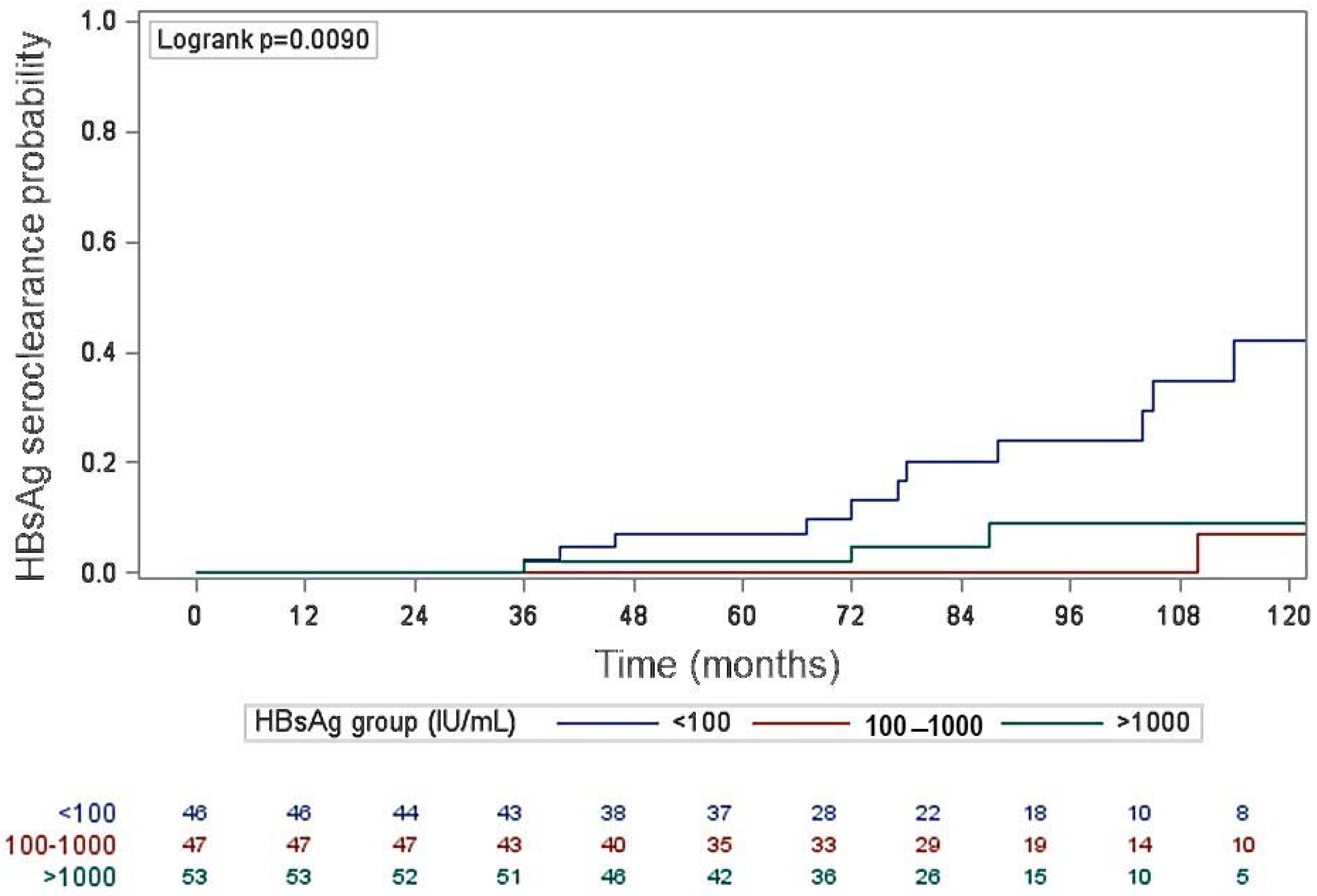
| HBsAg Seroclearance | Crude HR (95%CI) | Cumulative Incidence (95%CI) | p Value | |||
|---|---|---|---|---|---|---|
| Yes | No | 5 Years | 10 Years | |||
| N. of subjects (%) | 17 (11.6) | 129 (88.4) | - | 3.0% (1.2–7.9%) | 17.4% (10.8–27.6%) | - |
| HBsAg levels, n (%) | 0.009 | |||||
| <100 IU/mL | 11 (23.9) | 35 (76.1) | 3.89 (1.08–14.00) | 7.0% (2.3–20.1%) | 35.1% (20.9–54.8%) | |
| 100–1000 IU/mL | 3 (6.4) | 44 (93.6) | 0.87 (0.17–4.35) | 0% | 4.4% (0.6–27.1%) | |
| >1000 IU/ml | 3 (5.7) | 50 (94.3) | 1.00 * | 2.1% (0.3–23.9%) | 10.1% (3.2–29.2%) | |
| HBsAg Seroclearance | Crude HR (95%CI) | p Value | Adjusted HR + (95%CI) | p Value | ||
|---|---|---|---|---|---|---|
| Yes (n = 17) | No (n = 129) | |||||
| Baseline HBsAg levels, n (%) | 0.002 | 0.01 | ||||
| <100 IU/mL | 11 (23.9) | 35 (76.1) | 4.18 (1.55–11.32) | 3.53 (1.29–9.69) | ||
| ≥100 IU/mL | 6 (6.0) | 94 (94.0) | 1.00 * | 1.00 * | ||
| Sex, n (%) | 0.02 | 0.06 | ||||
| Male | 13 (18.8) | 56 (81.2) | 3.51 (1.14–10.81) | 2.84 (0.90–8.90) | ||
| Female | 4 (5.2) | 73 (94.8) | 1.00 * | 1.00 * | ||
| Age, mean ± SD, years | 61.4 ± 7.7 | 57.2 ± 11.4 | 1.04 (0.99–1.09) | 0.12 | 1.03 (0.98–1.09) | 0.30 |
| Baseline BMI, mean ± SD, Kg/m2 § | 27.7 ± 4.0 | 26.2 ± 4.6 | 1.11 (0.98–1.25) | 0.20 | ||
| Follow-up, mean ± SD, months | 100.6 ± 31.6 | 85.3 ± 31.8 | 0.97 (0.94–1.00) | 0.23 | ||
| Baseline HBV-DNA, median (IQR), IU/mL | 310 (104–1043) | 572 (164–1930) | 1.00 (0.99–1.01) | 0.78 | ||
| Baseline FibroScan, median (IQR), Kpa | 4.3 (3.8–5.3) | 4.9 (4.3–5.6) | 0.86 (0.49–1.50) | 0.78 | ||
| Liver steatosis, n/N (%) § | 7/10 (70.0) | 40/103 (38.8) | 3.13 (0.81–12.13) | 0.08 | 2.12 (0.54–8.37) | 0.26 |
Disclaimer/Publisher’s Note: The statements, opinions and data contained in all publications are solely those of the individual author(s) and contributor(s) and not of MDPI and/or the editor(s). MDPI and/or the editor(s) disclaim responsibility for any injury to people or property resulting from any ideas, methods, instructions or products referred to in the content. |
© 2023 by the authors. Licensee MDPI, Basel, Switzerland. This article is an open access article distributed under the terms and conditions of the Creative Commons Attribution (CC BY) license (https://creativecommons.org/licenses/by/4.0/).
Share and Cite
Barone, M.; Iannone, A.; Mezzapesa, M.; Milella, M.; Di Gennaro, F.; Niro, G.; Cotugno, R.; Cozzolongo, R.; Mennea, G.; Rendina, M.; et al. Natural History and Hepatitis B Virus Surface Antigen (HBsAg) Spontaneous Seroclearance in Hepatitis B Virus e-Antigen (HBeAg)-Negative Patients with Inactive Chronic Infection: A Multicenter Regional Study from South Italy. Pathogens 2023, 12, 1198. https://doi.org/10.3390/pathogens12101198
Barone M, Iannone A, Mezzapesa M, Milella M, Di Gennaro F, Niro G, Cotugno R, Cozzolongo R, Mennea G, Rendina M, et al. Natural History and Hepatitis B Virus Surface Antigen (HBsAg) Spontaneous Seroclearance in Hepatitis B Virus e-Antigen (HBeAg)-Negative Patients with Inactive Chronic Infection: A Multicenter Regional Study from South Italy. Pathogens. 2023; 12(10):1198. https://doi.org/10.3390/pathogens12101198
Chicago/Turabian StyleBarone, Michele, Andrea Iannone, Martino Mezzapesa, Michele Milella, Francesco Di Gennaro, Grazia Niro, Rosa Cotugno, Raffaele Cozzolongo, Giuseppe Mennea, Maria Rendina, and et al. 2023. "Natural History and Hepatitis B Virus Surface Antigen (HBsAg) Spontaneous Seroclearance in Hepatitis B Virus e-Antigen (HBeAg)-Negative Patients with Inactive Chronic Infection: A Multicenter Regional Study from South Italy" Pathogens 12, no. 10: 1198. https://doi.org/10.3390/pathogens12101198