Article

Lysine-Derived Maillard Reaction Products Inhibit the Growth of Salmonella enterica Serotype Typhimurium

Food, Nutrition and Health, University of British Columbia, 241-2205 East Mall, Vancouver, BC V6T 1Z4, Canada
*
Author to whom correspondence should be addressed.
These authors contributed equally to this work.
Pathogens 2023, 12(2), 215; https://doi.org/10.3390/pathogens12020215
Submission received: 6 January 2023 / Revised: 20 January 2023 / Accepted: 27 January 2023 / Published: 30 January 2023
(This article belongs to the Section Bacterial Pathogens)

Abstract

:
An emerging consumer trend to purchase minimally heated and ready-to-eat food products may result in processing methods that do not effectively reduce pathogenic populations. Crude Maillard reaction products (MRPs) are naturally generated compounds that have been shown to display antimicrobial effects against pathogens. Crude MRPs were generated from reducing sugars (fructose (Fru), glucose (Glc), ribose (Rib) or xylose (Xyl)) with lysine and the melanoidin equivalence was measured using an absorbance of 420 nm (Ab420). The relative antimicrobial activity of each MRP was measured by examining both the length of lag phase and maximum growth rate. MRPs were found to significantly shorten the lag phase and decrease the maximum growth rate of S. Typhimurium (p < 0.05). Glucose-lysine MRP (GL MRP) was determined to have the highest relative melanoidin (1.690 ± 0.048 at Ab420) and its efficacy against S. Typhimurium populations was measured at 37 °C and at pH 7.0 and estimated on xylose lysine deoxycholate (XLD) agar. GL MRP significantly reduced S. Typhimurium populations by >1 log CFU/mL at 8 and 24 h after inoculation (p < 0.05). GL MRPs also further decreased S. Typhimurium populations significantly under thermal stress condition (55 °C) compared to optimal (37 °C) by ~1 log CFU/mL (p < 0.05). Overall, GL MRP demonstrated effective antimicrobial activity against S. Typhimurium at 37 °C and 55 °C.

1. Introduction

Salmonella enterica (S. enterica) is a facultative, Gram-negative bacteria that has been reported as one of the top pathogens causing foodborne illnesses in North America [1]. From 2017–2022, S. enterica has been implicated in numerous foodborne outbreaks in North America with foods such as chicken, ground beef, ground turkey, fish, frozen cooked shrimp, seafood, etc. [2]. S. enterica is resilient at a wide range of temperatures from −2 °C to 54 °C, with an optimum growth temperature at 37 °C and is able to survive in high acidic environments such as a pH of 2 when entering the stomach of a host [3]. A previous study indicated that S. enterica serovar Typhimurium displayed high acid resistance, with a survival level > 10% after being exposed for 2 h at pH 2.5 [4]. With the consideration that S. enterica is able to survive under severe conditions, controlling and minimizing S. enterica populations in the food system has been a major challenge in the past few decades.
An emerging consumer trend is to purchase minimally processed and ready-to-eat food products [5]. Minimally processed and ready-to-eat food products require less heat treatment. However, heat treatment is one of the most effective techniques to kill foodborne pathogens [6]. Consequently, issues have risen over the safety of minimally processed food products. A cooking method that has gained popularity in recent years is the sous vide cooking method. Sous vide cooking refers to having the food vacuum-sealed in heat-stable plastic pouches and then submerged in a water bath or a hot steam oven at temperatures lower than the minimum required cooking temperatures to kill pathogens presented by the U.S. Department of Agriculture Food Safety and Inspection Service and the Government of Canada [7,8,9]. Examples include heating red meat at 56 °C for less than 4 h and an average of 55 °C for fish and shellfish for sous vide cooking, while the recommended minimum cooking temperature is 62.8 °C for meat, fish and shellfish [7,8]. In 2014, there were three S. enterica Enteritidis illnesses in British Columbia, Canada, potentially related to consuming sous vide cooked foods [10]. From the three illnesses, two were potentially from sous vide style cooked eggs from one restaurant and the last was from sous vide cooked duck breast from another restaurant [10]. The investigation found the sous vide cooking method to be improperly carried out which amplified the Salmonella Enteritidis risk for illness in consumers [10].
There is currently no additional method to be used in concurrent with sous vide cooking temperatures to help reduce S. enterica populations. An important concern is if the initial S. enterica populations on the raw food ingredients are sufficiently high then mild sous vide cooking temperatures would be insufficient to decrease the populations to a safe level [11]. A proposed method of reducing S. enterica populations is the use of Maillard reaction products (MRPs) derived from foods thermally processed with time and temperature being factors that generate a complex network of compounds perceived important for improving the taste and appearance of foods. An additional important aspect of MRPs is the antimicrobial activity displayed by some; however, knowledge on specific conditions to elicit activity related to S. enterica is limited [12,13,14].
Previous studies have reported that the high molecular weight (HMW) compounds of MRP have relatively greater antimicrobial activity compared to lower or intermediate weight products [15,16]. One explanation for this finding is the potentially greater affinity of HMW products to exhibit metal-chelating activity which has been shown to result in cell membrane damage in Gram-negative bacteria [15,16,17]. Moreover, the efficacy of MRP to inhibit S. enterica growth under acidic and thermal stress conditions that are relevant to the food industry is currently unclear. Therefore, the objective of this study was to evaluate the effectiveness of crude, water-soluble MRP extracts generated from different reducing sugar-lysine model combinations for antimicrobial activity towards S. enterica Typhimurium under acidic and thermal stress conditions.

2. Materials and Methods

2.1. Preparation of Crude MRPs

MRPs were conducted using reducing sugars (fructose (Fru), glucose (Glc), ribose (Rib) and xylose (Xyl)) (Sigma-Aldrich Co., St Louis, MO, USA) heated in the presence of lysine (Sigma-Aldrich Co., St Louis, MO, USA) at a ratio of 1:1 at 180 °C for 60 min in a Blue M convection oven (Blue Island, IL, USA) to generate the following crude MRP stock solutions: fructose-lysine (FL), glucose-lysine (GL), ribose-lysine (RL) and xylose-lysine (XL). The concentration of the crude MRP stock solutions were 500 mg/mL and the samples were subsequently collected and prepared by dissolving 0.4 g of crude MRP powder in 8 mL sterile deionized water. Samples were then homogenized and sonicated for 10 min in a FS-110 ultrasonic cleanser (Fisher Scientific, Hampton, NH, USA). Samples were then filtered through a 0.45 µm membrane filter (VWR International, Radnor, PA, USA) and stored at −20 °C for further chemical and microbiological experiments.

2.2. Determination of Crude MRP Stock Absorbance at 420 nm

A commonly used indicator to characterize the stages of Maillard reaction is the use of spectrophotometric measurements. At an absorbance of 420 nm (Ab420), brown pigments which are indicative of melanoidin equivalents are detected [18,19]. HMW MRPs, termed melanoidins for the purpose of this study, were measured in a 96-well plate at Ab420 at 23 °C using a microplate reader (SpectraMax M2; Molecular Devices, Sunnyvale, CA, USA).

2.3. Bacterial Strain and Growth Conditions

The bacterial strain used in this experiment was a Salmonella enterica subsp. enterica serotype Typhimurium FSL S5-536 (S. Typhimurium) from the ILSI Salmonella collection. The strain was maintained for long-term storage at −80 °C in tryptic soy broth (TSB, Becton, Dickinson and Company, Sparks, MD, USA) supplemented with 20% glycerol (VWR International, Radnor, PA, USA). Working stock of the strain was stored at 4 °C on brain heart infusion (BHI) agar (Becton, Dickinson and Company, Sparks, MD, USA) for a maximum of one month. An isolated colony of S. Typhimurium was inoculated into 5 mL of BHI broth (Becton, Dickinson and Company, Sparks, MD, USA) and the culture was incubated for 18 h at 37 °C with orbital shaking at 175 rpm. Approximately 109 colony-forming units (CFU)/mL were obtained by diluting the S. Typhimurium culture with BHI broth until OD600 reached 0.8 ± 0.02. Subsequently, the S. Typhimurium culture was centrifuged at 1800 × g for 5 min at 20 °C using Microcentrifuge 5424R (Eppendorf, Harmburg, Germany) and washed twice with phosphate-buffered saline ((PBS); VWR International, Radnor, PA, USA) for the following steps in Section 2.4 and Section 2.5.

2.4. Growth of S. Typhimurium with MRP under Acidic and Thermal Stress Conditions

To assess the growth parameters (length of lag phase and maximum growth rate) of S. Typhimurium under acidic stress, pH 5.5 and control, pH 7.2 conditions were used. To reach pH 5.5, 1 M hydrochloric acid (VWR International, Radnor, PA, USA) was added to the BHI broth using an AccumetTM AB 15+ Basic pH meter (Fisher Scientific, Hampton, NH, USA). One hundred and seventy µL of BHI broth with pH treatment (pH 5.5) and without pH treatment (pH 7.2) were dispensed into individual wells of a 96-well microtiter plate (VWR International, Radnor, PA, USA) with 20 µL of MRP stock solutions (FL, GL, RL and XL) from 2.1 and 20 µL of sterilized water (blank), respectively. The washed S. Typhimurium culture from Section 2.3 was then diluted to achieve a concentration of 104 CFU/mL using buffered peptone water ((BPW); Becton, Dickinson and Company, Sparks, MD, USA) and 10 µL of the S. Typhimurium culture was added to each of the wells containing: (1) acidified BHI broth (pH 5.5) with MRP stock solutions, (2) acidified BHI broth (pH 5.5) without MRP stock solutions, (3) BHI broth (pH 7.2) with MRP stock solutions and (4) BHI broth (pH 7.2) without MRP stock solutions. The final concentrations of MRPs with S. Typhimurium were 0.5 mg/mL. The microtiter plate was then placed in a SpectraMax M2 microplate reader (Molecular Devices, Sunnyvale, CA, USA) for OD600 nm readings for 24 h with 10 min intervals at 37 °C.
Thermal resistance assay was performed as mentioned above, but 10 µL of the S. Typhimurium culture was added to each of the wells containing: (1) BHI broth (pH 7.2) with MRP stock solutions and (2) BHI broth (pH 7.2) without MRP stock solutions at 42 °C. Growth curves generated from the current study were fitted with the Gompertz model using the grofit package in R, version 4.1.1 (R, Inc., Boston, MA, USA) to estimate the length of lag phase (λ) and maximum growth rate (µ). Lag phase is the period of time in which the cells are adjusting to the environment before exponential growth [20]. Maximum growth rate is the highest rate of increase under favorable conditions for the cell [21].

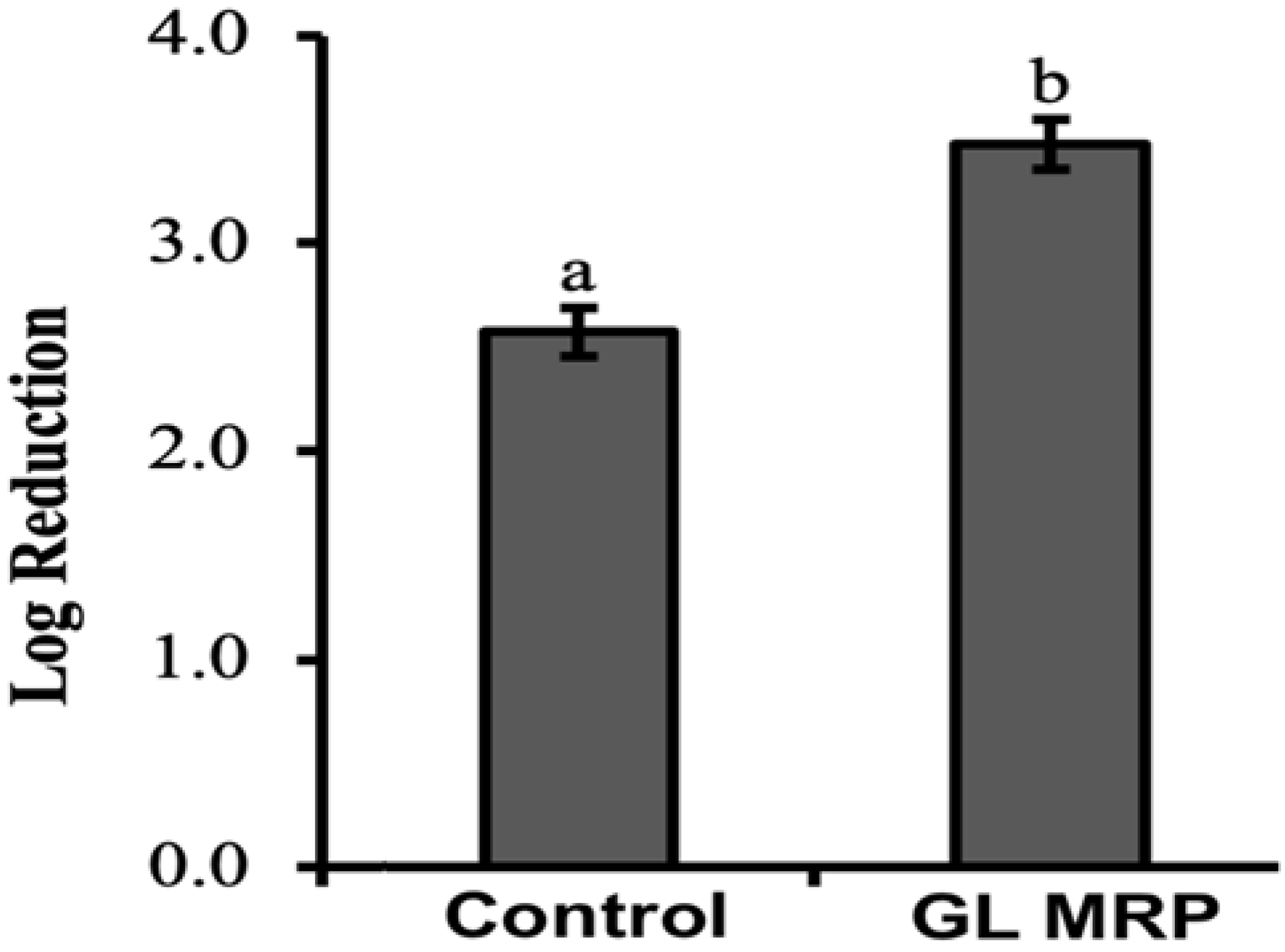
2.5. Efficacy of GL MRP against S. Typhimurium

A total of 5 mL of the S. Typhimurium culture from 2.3 was inoculated into 5 mL of BHI broth and 100 µL of GL MRP. A blank was used with 10 mL of BHI broth and 100 µL of sterile deionized water to ensure no cross-contamination occurred. All samples were incubated at 37 °C and the S. Typhimurium population was estimated at 0, 4, 8, 12 and 24 h after inoculation on xylose lysine deoxycholate ((XLD); Becton, Dickinson and Company, Sparks, MD, USA) agar. The agar was incubated at 37 °C for 24 h before retrieval for bacterial population estimation.

2.6. Growth of S. Typhimurium with GL MRP at 37 °C and 55 °C

Thermal resistance was performed 8 h after the inoculation of S. Typhimurium into 5 mL of BHI broth and 100 µL of GL MRP from 2.5. Eight h was chosen because the data from 2.5 indicated it was the end of the exponential phase. Ten µL of the S. Typhimurium inoculum from 2.5 was added to 990 mL PBS to reach 104 CFU/mL. The diluted S. Typhimurium suspension was plated on the XLD agar prior to incubation as the negative control. The remaining diluted S. Typhimurium suspension was washed twice with PBS and the precipitated pellet was used for further experiments. To study the thermal resistance of S. Typhimurium, the pellet was dissolved in BHI broth and half of the samples were heated at 37 °C and the other half at 55 °C for 10 min. Afterwards, S. Typhimurium populations from the samples were estimated on XLD agar after incubation at 37 °C for 24 h.

2.7. Statistical Analysis

The melanoidin equivalence based on Ab420 from different sugar-lysine model systems from 2.2 was conducted with seven replicates and the absorbance was analyzed by one way analysis of variance (ANOVA), followed by Tukey’s Honest Significant Difference (Tukey’s HSD) for means separation. For 2.4, the experiment was similarly conducted with seven biological replicates and the length of lag phase and maximum growth rate of S. Typhimurium was analyzed by one-way ANOVA, followed by Tukey’s HSD for means separation. Three biological replicates were conducted for 2.5 and a one-way ANOVA was used to determine the effect of GL MRP on S. Typhimurium with Tukey’s HSD for means separation. Similarly, three biological replicates were conducted for 2.6 but the difference in S. Typhimurium populations with GL MRP at 37 °C compared to 55 °C was analyzed with a Student’s t-test. All samples were considered statistically significant with p values < 0.05. All statistical analyses were conducted with JMP, version 11 (JMP, Cary, NC, USA).

3. Results and Discussion

3.1. Sugar-Amino Acid Combinations to Generate HMW MRP Products

The melanoidin equivalent based on Ab420 from different sugar-lysine model systems is presented in Table 1. Different reducing sugars, such as Fru, Glu, Rib and Xyl, were reacted with lysine to generate crude MRPs (FL, GL, RL and XL) at 180 °C for 60 min. Of these four sugar–lysine combinations, it was the GL MRP that had the significantly highest melanoidin content (p < 0.05; Table 1). This is similar to a previous study that found model GL to contain a 45% and 15% higher melanoidin content compared to XL at pH 5.5 and 8.0, respectively [22].

3.2. Effect of MRPs on the Survival of S. Typhimurium under Sublethal Acidic and Thermal Stress Conditions

The two conditions tested in this study to evaluate the sensitivity of S. Typhimurium to different reaction models used to generate HMW MRPs included altering pH and temperature; both of which are commonly used to control foodborne pathogens in the food industry. Turbidity measurements (OD600) were used to assess cell density without differentiating between live or dead cells.
The lag phase of a bacterial growth cycle reflected the time it took for the bacteria to adjust to a new environment before it could undergo exponential growth [23]. All four MRP supplementations had no significant differences compared to control at pH 7.2 and at 37 °C (p > 0.05; Table 2). In contrast, in the acidic environment of pH 5.5, S. Typhimurium cells exposed to GL MRP had a shorter lag phase compared to other treatments (p < 0.05; Table 2). At 42 °C and at pH 7.2, samples with MRP treatment except for FL had a significantly decreased lag phase compared to controls (p < 0.05; Table 2). Thus, S. Typhimurium exhibited the fastest adaption to the acidic environment with GL MRP supplementation. When under thermal stress, S. Typhimurium exhibited the fastest adaption with GL, RL and XL MRP supplementations.
It is important to note that the composition of melanoidins from different sugar–lysine combinations may differ in respect to the physical, chemical or biological properties of pre-melanoidin components that form a complex mixture of HMW melanoidins [24]. These complex set of reactions define both intermediate (pre-melanoidin) and final (melanoidin) stages, that are sugar-specific in terms of final composition [19,24]. In addition to temperature, the pH is also a factor, as under slightly basic conditions, Schiff base and Amadori rearrangement, products will tend to lead to a 2,3-enolization reaction [24]. This is different if the conditions have an acidic pH, where the Amadori rearrangement is facilitated to a 1,2 enolization pathway [24]. Further dehydration and fragmentation results in numerous additional pre-melanoidin products formed during these intermediate stages, which collectively influence the final composition of MRPs and thus could contribute for the significant differences in antimicrobial activity and the length of the lag phase against S. Typhimurium.
Previous studies indicated that rpoS, a virulence factor, was known to support S. enterica survival in harsh environmental conditions and the expression of rpoS could be upregulated under acidic stress conditions [25,26,27]. Chalova et al. (2012) observed a 3.43-fold upregulation of rpoS expression when S. Typhimurium was exposed to MRP, which indicated the potential of MRP to modulate the stress response [28]. Recent studies indicated that deletion of rpoS in S. Typhimurium resulted in a longer lag phase when exposed to acidic stress conditions [25]. Overall, four MRPs in this study shortened the length of lag phase to different extents, compared to a control, which supports a previous study that demonstrated it was the variation on melanoidin content that contributed to different levels of antimicrobial activity [29].
All four sources of MRPs significantly decreased the maximum growth rate compared to the control at pH 5.5 and pH 7.2, at 37 °C, respectively, and also at pH 7.2 at 42 °C (p < 0.05; Table 3). However, the significant differences observed for MRP treatments derived from specific sugar–lysine combinations could not be distinguished despite the apparent difference in melanoidin content (Table 1 and Table 3). This result was surprising because different MRP sources can vary in component compositions, which could lead to unique differences in relative efficacies to inhibit the maximum growth rate of S. Typhimurium. The results showed that this was not the case with the MRP crude fractions recovered in this study. An important factor to note is the formation of hydrogen peroxide (H2O2) as a product of MRP synthesis [24,30]. H2O2 is a common disinfectant against pathogens such as S. Typhimurium and previous research has found H2O2 produced from MRP synthesis causing cell damage [30,31,32,33]. H2O2 was produced from all four sources of MRPs [24,30] and this would result in a common, non-specific-MRP effect on the maximum growth rate of S. Typhimurium. This could explain the insignificant effects noted between different MRP treatments, albeit significantly different from the control (p < 0.05; Table 3).
Hauser et al. (2014) showed that the concentration of MRP was an important factor towards an antimicrobial activity. The MRP used was recovered from 85% ethanol and at a reconstituted concentration of 16.1 g/L, was shown to completely inhibit Escherichia coli (E. coli) after 24 h [34]. An MRP concentration of 6.5 g/L had no effect on E. coli growth compared to control, after a 21-h treatment [34]. Rufian-Henares and De La Cueva (2009) reported similar findings where the highest concentration of melanoidin (8.0 mg/mL) inhibited the highest growth of E. coli ATCC 35150, followed by 5.0 mg/mL, 4.0 mg/mL, 2.5 mg/mL and 1.0 mg/mL [35]. From Table 1, although it was shown that GL MRP had the highest melanoidin content, based on an Ab420 index, the capacity to mitigate the maximum growth rate of S. Typhimurium was not significantly different from other sugar–lysine sources of MRPs (Table 3). A possible explanation for this finding is that the MRPs were relatively crude mixtures of HMW components [21] that, when combined, exceeded the threshold concentration to show differences in the maximum growth rate of S. Typhimurium between the different MRP treatments [19].

3.3. GL MRP Inhibits the Growth of S. Typhimurium at Optimal Conditions

In the present study, the GL model system was selected to generate MRPs, indexed by using Ab420 as an estimate of the highest melanoidin content (Table 1) in order to evaluate growth patterns of S. Typhimurium in a 24-h time frame at 37 °C. Figure 1 showed the growth of S. Typhimurium with GL MRP and without GL MRP supplementation (Control) on XLD agar. The GL MRP-treated S. Typhimurium populations were significantly reduced by ~1.5 log CFU/mL by 8 h and ~1 log CFU/mL by 12 h in comparison to the control after inoculation on XLD agar (p < 0.05; Figure 1).
The antimicrobial activity of HMW MRPs generated from reducing sugar–amino acid reactions have been studied previously with selected pathogens, and melanoidin content was identified as the antimicrobial component of MRPs [29,35,36,37,38]. Kukuminato et al. (2021) used melanoidin xylose-Phe and xylose-Pro against Listeria monocytogenes ATCC 1911 at 25 °C and found reductions of ~1 log CFU/mL with xylose-Pro and ~3 log CFU/mL with xylose-Phe after 12 h [29]. Another study used MRPs with glucose and fructose and found different concentrations of MRPs to have varying effects on E. coli and methicillin-resistant Staphylococcus aureus (MRSA) [38]. Glucose MRP reduced E. coli and MRSA by ~12% and 5% when using 3% glucose MRP, respectively [38]. The antimicrobial activity was increased when using 5% glucose MRP as E. coli and MRSA populations were reduced by ~45% and ~43%, respectively [38]. Fructose MRP was more effective against E. coli as 3% fructose MRP reduced E. coli and MRSA populations by ~17% and ~15%, respectively [38]. When fructose MRP concentrations were increased to 5%, E. coli populations were reduced by 60% but 5% fructose MRP was less effective against MRSA compared to 5% glucose MRP as populations were reduced by only 20% [37]. Bhattacharjee et al. (2021) found low concentrations of MRP (0.06X and 0.12X) could extend lag time by around 12 h and high concentrations of MRP (0.4X and 0.5X) were effective in decreasing populations of Aggregatibacter actinomycetemcomitans by up to 8 logs [37]. Ruffian-Henares and Morales (2008) found HMW MRPs exhibited a higher antimicrobial activity against E. coli compared to low molecular weight (LMW) MRPs [36].
Melanoidins are known to be negatively charged end-products of the Maillard reaction with metal-chelating activity for Mg2+, Fe3+ and Cu2+- ions [35]. Reducing cation availability, especially Fe+ on the outer membrane of Gram-negative bacteria, can initiate a sub-lethal injury, where irreversible changes in membrane function adversely affects nutrient transport to support growth [17,35,36]. One important example of an antimicrobial activity attributed to MRP -Fe3+ chelation concerns the potential interaction with siderophore-Fe+3 complexes [35]. The fact that S. Typhimurium is a Gram-negative bacterium is particularly important in this example, since Gram-negative bacteria secrete siderophores to bind Fe3+ [24]. Receptors and transporters located on the outer membrane of Gram-negative bacteria are required to utilize free Fe3+ [24]. The capacity of MRPs to chelate free Fe+ will interrupt Fe3+ binding to siderophores in Gram-negative bacteria, thus affecting cell membrane function for S. Typhimurium that results in reduced replication efficiency [24,35,39,40].
The Baranyi model for bacterial growth was selected over the Gompertz model due to better predictive capabilities that involve varying environmental conditions over time. There were four parameters that were assessed in the analyses: (1) maximum growth rate in CFU/mL/h (μmax); (2) initial cell density in CFU/mL (No); (3) maximum cell density in CFU/mL (Nmax) and (4) the difference of the maximum and minimum cell density in CFU/mL (ΔN). Based on the parameters obtained from the Baranyi model, all four parameters between control and GL MRP-treated S. Typhimurium cells were significantly lower compared to the control (p < 0.05; Table 4). These are compelling results to show a GL MRP-related antimicrobial effect against S. Typhimurium.
Figure 2 shows the log reduction in S. Typhimurium without GL MRP (Control) and with GL MRP treatment at optimal (37 °C) and thermal stress conditions (55 °C). Log reduction was obtained by calculating the difference in S. Typhimurium populations between 37 °C and 55 °C. Exposure of S. Typhimurium to GL MRP caused a significantly higher log reduction from 37 °C to 55 °C compared to the control, indicating that GL MRP accelerated the population decrease in S. Typhimurium at 55 °C (p < 0.05; Figure 2). Since high temperatures may accelerate molecular motion, the binding activity between GL MRP and Mg2+, Fe3+ and Cu2+ may be accelerated, which consequently aids in decreasing the population of S. Typhimurium [35]. The use of GL MRP against S. Typhimurium at 55 °C could be considered as a novel processing aid for foods heat-processed at lower temperatures. Recent food trends have been geared towards minimally processed foods to retain a high nutritional value and natural sensory qualities and a popular cooking method for this trend is sous vide cooking [8]. The temperature of sous vide cooking for certain food products such as fish, seafood and meat averages at ~55 °C and the food is maintained at this temperature for many hours or days [8]. However, the minimum recommended temperatures for cooking meat are to reach 62.8–71.1 °C and 62.8 °C for fish and shellfish internally [7,9]. If there is a sous vide cooking error and the fish, seafood or meat product is not heated for the entire duration of the time required, then foodborne pathogens may still be present and cause foodborne illness for the consumer. The surface application of GL MRP to foods that are cooked at temperatures lower than recommended temperatures could be considered as a potential aid to reduce foodborne illness risks in these food products.

4. Conclusions

The results of the current study have shown that the crude water-soluble MRPs have antimicrobial activity on S. Typhimurium. Due to the use of Ab420 as an estimation of melanoidin content, the compounds that form HMW MRPs could not be individually characterized. Future work should be completed to chemically characterize the HMW MRP components that contributed to the observed antimicrobial activity in order to determine the structure and functional activity of these compounds. From the MRPs used in this study, GL MRP was found to have the relatively highest melanoidin content and was able to reduce S. Typhimurium populations by >1 log CFU/mL after 8 and 24 h after inoculation. Therefore, the data support previous findings of an MRP antimicrobial activity. When S. Typhimurium was exposed to GL MRP, the difference in log populations on XLD of populations heated at 55 °C and 37 °C decreased by ~1 log CFU/mL compared to the non-GL MRP control. This supports the idea that GL MRP could be used as an effective aid when applied to the surface of a food product to reduce S. Typhimurium populations, when the temperatures used to process these foods are lower than the minimum-required cooking temperatures to kill foodborne pathogens. Future research on the efficacy of GL MRP against pathogens on foods heated at lower than required cooking temperatures is recommended for food products such as fish, seafood and meat at 55 °C; a common practice used for sous vide cooking. In addition, organoleptic testing will also need to be conducted on such products to test if distinct color and taste features considered important for seafood sensory quality are compromised in the presence of GL MRPs.

Author Contributions

Conceptualization, S.W.; methodology, S.W.; software, S.W. and K.M.; validation, K.M. and C.W.Y.W.; formal analysis, C.W.Y.W. and K.M.; investigation, S.W., K.M. and C.W.Y.W.; resources, S.W. and D.D.K.; data curation, C.W.Y.W.; writing—original draft preparation, C.W.Y.W.; writing—review and editing, C.W.Y.W., K.M., D.D.K. and S.W.; visualization, C.W.Y.W.; supervision, S.W.; project administration, S.W.; funding acquisition, S.W. and D.D.K. All authors have read and agreed to the published version of the manuscript.

Funding

This research was funded by the NSERC-Discovery Grants (RGPIN 38349 and RGPIN 04871). No competing financial interests exit. The funders had no role in the design of the study; in the collection, analyses, or interpretation of data in the writing of the manuscript, or in the decision to publish the results.

Institutional Review Board Statement

Not applicable.

Informed Consent Statement

Not applicable.

Data Availability Statement

Not applicable.

Acknowledgments

We thank Hyun Soo Seo for technical assistance with the experiments. The Salmonella strain used in this study was provided by Martin Wiedmann at Cornell University.

Conflicts of Interest

The research was conducted in the absence of any commercial or financial associations that can be misconstrued as potential conflicts of interest.

References

  1. Scallan, E.; Hoekstra, R.M.; Angulo, F.J.; Tauxe, R.v.; Widdowson, M.-A.; Roy, S.L.; Jones, J.L.; Griffin, P.M. Foodborne Illness Acquired in the United States—Major Pathogens. Emerg. Infect. Dis. 2011, 17, 7–15. [Google Scholar] [CrossRef] [PubMed]
  2. Centers for Disease Control and Prevention Reports of Selected Salmonella Outbreak Investigations. Available online: https://www.cdc.gov/salmonella/outbreaks.html (accessed on 3 February 2019).
  3. Pradhan, D.; Devi Negi, V. Stress-Induced Adaptations in Salmonella: A Ground for Shaping Its Pathogenesis. Microbiol. Res. 2019, 229, 126311. [Google Scholar] [CrossRef] [PubMed]
  4. Berk, P.A.; de Jonge, R.; Zwietering, M.H.; Abee, T.; Kieboom, J. Acid Resistance Variability Among Isolates of Salmonella Enterica Serovar Typhimurium DT104. J. Appl. Microbiol. 2005, 99, 859–866. [Google Scholar] [CrossRef] [PubMed]
  5. Madruga, M.; Steele, E.M.; Reynolds, C.; Levy, R.B.; Rauber, F. Trends in Food Consumption According to the Degree of Food Processing Among the UK Population Over 11 Years. Br. J. Nutr. 2022, 1–8. [Google Scholar] [CrossRef]
  6. Błaszczyk, I.; Iciek, J. Selection of Heat Treatment Conditions and Prevention of Secondary Microbial Contamination of Liquid Sugar: Practical Remarks. J. Food Sci. Technol. 2021, 58, 2842–2846. [Google Scholar] [CrossRef]
  7. U.S. Department of Agriculture. Food Safety and Inspection Service Safe Minimum Internal Temperature Chart. Available online: https://www.fsis.usda.gov/food-safety/safe-food-handling-and-preparation/food-safety-basics/safe-temperature-chart (accessed on 25 December 2022).
  8. Kilibarda, N.; Brdar, I.; Baltic, B.; Markovic, V.; Mahmutovic, H.; Karabasil, N.; Stanisic, S. The Safety and Quality of Sous Vide Food. Meat Technol. 2018, 59, 38–45. [Google Scholar] [CrossRef] [Green Version]
  9. Government of Canada Safe Cooking Temperatures. Available online: https://www.canada.ca/en/health-canada/services/general-food-safety-tips/safe-internal-cooking-temperatures.html (accessed on 3 January 2023).
  10. McIntyre, L.; Jorgenson, V.; Ritson, M. Sous Vide Style Cooking Practices Linked to Salmonella Enteritidis Illnesses. Environ. Health Rev. 2017, 60, 42–49. [Google Scholar] [CrossRef] [Green Version]
  11. Public Health Ontario Case Study: Sous Vide. Available online: https://www.publichealthontario.ca/-/media/Documents/C/2016/case-study-sous-vide.pdf?rev=bf5d650a459d431aabf5493d4cdd2e03&sc_lang=en (accessed on 26 December 2022).
  12. Einarsson, H.; Snygg, B.G.; Eriksson, C. Inhibition of Bacterial Growth by Maillard Reaction Products. J. Agric. Foodchem. 1983, 31, 1043–1047. [Google Scholar] [CrossRef]
  13. Kundinger, M.M.; Zabala-Díaz, I.B.; Chalova, V.I.; Ricke, S.C. Effects of Maillard Reaction Products on HilA Expression in Salmonella Typhimurium. J. Food Sci. 2008, 73, M32–M35. [Google Scholar] [CrossRef]
  14. Sheikh-Zeinoddin, M.; Perehinec, T.M.; Hill, S.E.; Rees, C.E.D. Maillard Reaction Causes Suppression of Virulence Gene Expression in Listeria Monocytogenes. Int. J. Food Microbiol. 2000, 61, 41–49. [Google Scholar] [CrossRef]
  15. Gu, F.L.; Kim, J.M.; Abbas, S.; Zhang, X.M.; Xia, S.Q.; Chen, Z.X. Structure and Antioxidant Activity of High Molecular Weight Maillard Reaction Products from Casein-Glucose. Food Chem. 2010, 120, 505–511. [Google Scholar] [CrossRef]
  16. Yoshimura, Y.; Iijima, T.; Watanabe, T.; Nakazawa, H. Antioxidative Effect of Maillard Reaction Products Using Glucose-Glycine Model System. J. Agric. Food Chem. 1997, 45, 4106–4109. [Google Scholar] [CrossRef]
  17. Ramonaityte, D.T.; Keršiene, M.; Adams, A.; Tehrani, K.A.; Kimpe, N. de The Interaction of Metal Ions with Maillard Reaction Products in a Lactose-Glycine Model System. Food Res. Int. 2009, 42, 331–336. [Google Scholar] [CrossRef]
  18. Hirano, M.; Miura, M.; Gomyo, T. A Tentative Measurement of Brown Pigments in Various Processed Foods. Biosci. Biotechnol. Biochem. 1996, 60, 877–879. [Google Scholar] [CrossRef] [PubMed]
  19. Kitts, D.D. Antioxidant and Functional Activities of MRPs Derived from Different Sugar–Amino Acid Combinations and Reaction Conditions. Antioxidants 2021, 10, 1840. [Google Scholar] [CrossRef]
  20. Yates, G.T.; Smotzer, T. On the Lag Phase and Initial Decline of Microbial Growth Curves. J. Theor. Biol. 2007, 244, 511–517. [Google Scholar] [CrossRef]
  21. Westoby, M.; Nielsen, D.A.; Gillings, M.R.; Litchman, E.; Madin, J.S.; Paulsen, I.T.; Tetu, S.G. Cell Size, Genome Size, and Maximum Growth Rate Are Near-Independent Dimensions of Ecological Variation Across Bacteria and Archaea. Ecol. Evol. 2021, 11, 3956–3976. [Google Scholar] [CrossRef]
  22. Mikami, Y.; Murata, M. Effects of Sugar and Buffer Types, and PH on Formation of Maillard Pigments in the Lysine Model System. Food Sci. Technol. Res. 2015, 21, 813–819. [Google Scholar] [CrossRef] [Green Version]
  23. Rolfe, M.D.; Rice, C.J.; Lucchini, S.; Pin, C.; Thompson, A.; Cameron, A.D.S.; Alston, M.; Stringer, M.F.; Betts, R.P.; Baranyi, J.; et al. Lag Phase Is a Distinct Growth Phase That Prepares Bacteria for Exponential Growth and Involves Transient Metal Accumulation. J. Bacteriol. 2012, 194, 686–701. [Google Scholar] [CrossRef] [PubMed] [Green Version]
  24. Mu, K.; Wang, S.; D Kitts, D. Evidence to Indicate That Maillard Reaction Products Can Provide Selective Antimicrobial Activity. Integr. Food Nutr. Metab. 2016, 3, 330–335. [Google Scholar] [CrossRef]
  25. Shiroda, M.; Pratt, Z.L.; Döpfer, D.; Wong, A.C.L.; Kaspar, C.W. RpoS Impacts the Lag Phase of Salmonella Enterica During Osmotic Stress. Fems Microbiol. Lett. 2014, 357, 195–200. [Google Scholar] [CrossRef] [PubMed] [Green Version]
  26. Soo Lee, I.; Lin, J.; Hall, H.K.; Bearson, B.; Foster, J.W. The Stationary-phase Sigma Factor ΣS (RpoS) Is Required for a Sustained Acid Tolerance Response in Virulent Salmonella Typhimurium. Mol. Microbiol. 1995, 17, 155–167. [Google Scholar] [CrossRef]
  27. Fang, F.C.; Libby, S.J.; Buchmeiert, N.A.; Loewen, P.C.; Switala, J.; Harwood, J.; Guiney, D.G. The Alternative (a Factor KatF (RpoS) Regulates Salmonella Virulence. Proc. Natl. Acad. Sci. USA 1992, 89, 11978–11982. [Google Scholar] [CrossRef] [Green Version]
  28. Chalova, V.I.; Hernández-Hernández, O.; Muthaiyan, A.; Sirsat, S.A.; Natesan, S.; Sanz, M.L.; Moreno, F.J.; O’Bryan, C.A.; Crandall, P.G.; Ricke, S.C. Growth and Transcriptional Response of Salmonella Typhimurium LT2 to Glucose-Lysine-Based Maillard Reaction Products Generated Under Low Water Activity Conditions. Food Res. Int. 2012, 45, 1044–1053. [Google Scholar] [CrossRef] [Green Version]
  29. Kukuminato, S.; Koyama, K.; Koseki, S. Antibacterial Properties of Melanoidins Produced from Various Combinations of Maillard Reaction against Pathogenic Bacteria. Am. Soc. Microbiol. 2021, 9, e01142-21. [Google Scholar] [CrossRef] [PubMed]
  30. Hegele, J.; Münch, G.; Pischetsrieder, M. Identification of Hydrogen Peroxide as a Major Cytotoxic Component in Maillard Reaction Mixtures and Coffee. Mol. Nutr. Food Res. 2009, 53, 760–769. [Google Scholar] [CrossRef] [PubMed]
  31. Ruiz-Roca, B.; Delgado-Andrade, C.; Pilar Navarro, M.; Seiquer, I. Effects of Maillard Reaction Products from Glucose-Lysine Model Systems on Oxidative Stress Markers and Against Oxidative Induction by Hydrogen Peroxide in Caco-2 Cells. J. Food Nutr. Res. 2011, 50, 237–248. [Google Scholar]
  32. Ünlütürk, A.; Turantasl, F. Bactericidal Effect of Hydrogen Peroxide on Salmonella Typhimurium in Liquid Whole Egg. J. Appl. Bacteriol. 1987, 62, 25–28. [Google Scholar] [CrossRef]
  33. Ukuku, D.O.; Pilizota, V.; Sapers, G.M. Effect of Hot Water and Hydrogen Peroxide Treatments on Survival of Salmonella and Microbial Quality of Whole and Fresh-Cut Cantaloupe †. J. Food Prot. 2004, 67, 432–437. [Google Scholar] [CrossRef]
  34. Hauser, C.; Müller, U.; Sauer, T.; Augner, K.; Pischetsrieder, M. Maillard Reaction Products as Antimicrobial Components for Packaging Films. Food Chem. 2014, 145, 608–613. [Google Scholar] [CrossRef]
  35. Rufian-Henares, J.A.; de La Cueva, S.P. Antimicrobial Activity of Coffee Melanoidins—A Study of Their Metal-Chelating Properties. J. Agric. Food Chem. 2009, 57, 432–438. [Google Scholar] [CrossRef] [PubMed]
  36. Rurián-Henares, J.A.; Morales, F.J. Antimicrobial Activity of Melanoidins against Escherichia Coli Is Mediated by a Membrane-Damage Mechanism. J. Agric. Food Chem. 2008, 56, 2357–2362. [Google Scholar] [CrossRef] [PubMed] [Green Version]
  37. Bhattacharjee, M.K.; Mehta, B.S.; Akukwe, B. Maillard Reaction Products Inhibit the Periodontal Pathogen Aggregatibacter Actinomycetemcomitans by Chelating Oron. Arch. Oral. Biol. 2021, 122, 104989. [Google Scholar] [CrossRef] [PubMed]
  38. Ji, L.; Zhang, Y.; Song, W.; Cai, L.; Wang, Y.; Guo, J. Analysis on Antibacterial Activities and Volatile Compounds of Maillard Reaction Products Derived From Squid Skin. E3s Web Conf. 2020, 145, 1–6. [Google Scholar] [CrossRef] [Green Version]
  39. Amarante-Mendes, G.P.; Adjemian, S.; Branco, L.M.; Zanetti, L.C.; Weinlich, R.; Bortoluci, K.R. Pattern Recognition Receptors and the Host Cell Death Molecular Machinery. Front. Immunol. 2018, 9, 2379. [Google Scholar] [CrossRef] [Green Version]
  40. Morales, F.J.; Fernández-Fraguas, C.; Jiménez-Pérez, S. Iron-Binding Ability of Melanoidins from Food and Model Systems. Food Chem. 2005, 90, 821–827. [Google Scholar] [CrossRef]
Figure 1. Growth of S. Typhimurium under no GL MRP supplementation (Control) and GL MRP supplementation at 37 °C on XLD Agar. Error bars represent standard deviation between three biological replicates.
Pathogens 12 00215 g001
Figure 2. Log reduction in S. Typhimurium populations eight hours after inoculation without GL MRP supplementation (Control) and GL MRP supplementation heated at optimal condition (37 °C) and thermal condition (55 °C) for 10 min. Log reduction was obtained by calculating the difference in S. Typhimurium populations (log CFU/mL) on XLD agar between 55 °C and 37 °C. Means and standard deviations were calculated using data from three biological replicates. Different superscripts (a–b) indicate significant differences (p < 0.05) between treatments.
Pathogens 12 00215 g002
Table 1. Melanoidin equivalence (absorbance at 420 nm) of crude MRP (FL, GL, RL and XL) stock solutions heated at 180 °C for 60 min.
Crude MRPAbsorbance at 420 nm 1,2
FL1.030 ± 0.006 b
GL1.690 ± 0.048 a
RL0.578 ± 0.011 d
XL0.813 ± 0.037 c
1 All values are presented as mean ± standard deviation from seven replicates; 2 Values followed by different lower-case letters (a–d) indicate significant differences (p < 0.05).
Table 2. Length of lag phase λ (hours) of S. Typhimurium growth at pH 5.5 and pH 7.2 at 37 °C and pH 7.2 at 42 °C when incubated with MRP (FL, GL, RL and XL) treatments 1 for 24 h.
Crude MRPλ at 37 °Cλ at 42 °C
pH 5.5 2pH 7.2pH 7.2 2
Control (No MRP)5.54 ± 0.11 a5.06 ± 0.05 a5.54 ± 0.29 a
FL5.23 ± 0.30 a4.70 ± 0.26 a4.95 ± 0.40 a,b
GL4.51 ± 0.31 b4.78 ± 0.29 a4.79 ± 0.26 b
RL5.18 ± 0.57 a4.59 ± 0.31 a4.83 ± 0.19 b
XL5.09 ± 0.33 a,b4.60 ± 0.28 a4.93 ± 0.45 b
1 All values are presented as mean ± standard deviation from seven biological replicates. 2 Values within each column followed by different lower-case letters (a–b) indicate significant differences (p < 0.05).
Table 3. Maximum growth rate µ (/hour) of S. Typhimurium growth at pH 5.5 and pH 7.2 at 37 °C and pH 7.2 at 42 °C when incubated with MRP (FL, GL, RL and XL) treatments 1 for 24 h.
Crude MRPµ at 37 °Cµ at 42 °C
pH 5.5 2pH 7.2 2pH 7.2 2
Control (No MRP)0.128 ± 0.004 a0.230 ± 0.011 a0.163 ± 0.010 a
FL0.105 ± 0.005 b0.160 ± 0.042 b0.116 ± 0.046 b
GL0.087 ± 0.013 b0.160 ± 0.060 b0.097 ± 0.008 b
RL0.094 ± 0.017 b0.144 ± 0.032 b0.092 ± 0.004 b
XL0.095 ± 0.009 b0.127 ± 0.007 b0.101 ± 0.015 b
1 All values are presented as mean ± standard deviation from seven biological replicates; 2 Values within each column followed by different superscripts (a–b) indicate significant differences (p < 0.05).
Table 4. The four parameters obtained from the Baranyi model for both control (No MRP) and GL MRP-treated S. Typhimurium on XLD Agar 1.
µmax 2No 2Nmax 2ΔN 2
Control 1.653 ± 0.059 a9.569 ± 0.261 a14.108 ± 0.084 a4.538 ± 0.205 a
GL MRP1.323 ± 0.251 b9.268 ± 0.068 b13.263 ± 0.125 b3.996 ± 0.132 b
1 All values are presented as mean ± standard deviation with three biological replicates. 2 Values within each column followed by different lower-case letters (a–b) indicate significant differences (p < 0.05).
Disclaimer/Publisher’s Note: The statements, opinions and data contained in all publications are solely those of the individual author(s) and contributor(s) and not of MDPI and/or the editor(s). MDPI and/or the editor(s) disclaim responsibility for any injury to people or property resulting from any ideas, methods, instructions or products referred to in the content.

