Article

Resistance Profiles and Virulence Determinants in Biofilm-Forming Enterococcus faecium Isolated from Raw Seafood in Bangladesh

Department of Microbiology and Hygiene, Faculty of Veterinary Science, Bangladesh Agricultural University, Mymensingh 2202, Bangladesh
*
Author to whom correspondence should be addressed.
Pathogens 2023, 12(9), 1101; https://doi.org/10.3390/pathogens12091101
Submission received: 9 August 2023 / Revised: 25 August 2023 / Accepted: 27 August 2023 / Published: 28 August 2023
(This article belongs to the Special Issue Surveillance and Control of Foodborne Pathogens)

Abstract

:
Pathogenic, antibiotic-resistant, and biofilm-forming bacteria can be transferred to humans through the consumption of contaminated seafood. The present study was carried out to determine antibiotic resistance profiles and virulence determinants in biofilm-forming Enterococcus faecium isolated from seafood in Bangladesh. A total of 150 seafood samples, including shrimp (n = 50), crabs (n = 25), and marine fish (n = 75), were screened using cultural, staining, biochemical, polymerase chain reaction (PCR), Congo red (CR), and disk diffusion (DD) assays. In PCR, E. faecium was detected in 27.3% (41/150; CI95% 20.8; 34.9) of samples, where marine fish (34.7%, CI95% 24.9; 45.9) had the highest prevalence (p < 0.05) compared to crabs (32%, CI95% 17.2; 51.6) and shrimp (14%, CI95% 7.0; 26.1). Thirty-two (78.1%, CI95% 63.3; 88.0) of the E. faecium isolates were determined to be biofilm formers in the CR test, where 43.9% (18/41, CI95% 29.9; 59.0) and 34.2% (14/41, CI95% 21.6; 49.5) of the isolates were strong and intermediate biofilm formers, respectively. In PCR, virulence genes, i.e., pil (100%), ace (92.7%), agg (68.3%), fsrA (65.9%), gelE (63.4%), sprE (53.7%), fsrB (51.2%), and fsrC (43.9%), were detected in E. faecium isolates. All the E. faecium isolates were phenotypically resistant to ≥3 antimicrobial categories and ≥3 antibiotics, including WHO-classified reserve antibiotics linezolid (70.7%) and fosfomycin (19.5%). Moreover, the multiple antibiotic resistance index ranged up to 0.8, showing resistance to ten antibiotics and eight antibiotic classes. In this study, the prevalence of virulence genes and antibiotic resistance was significantly greater (p < 0.05) in strong biofilm-forming E. faecium strains as compared to strains with intermediate and non-biofilm-forming abilities. As far as we know, this study, for the first time in Bangladesh, determined antibiotic resistance and detected virulence genes in biofilm-forming E. faecium isolated from seafood samples. The data from this study could play a significant role in evaluating potential health hazards linked to the ingestion of uncooked or minimally processed seafood.

1. Introduction

Bangladesh possesses significant potential for both catch fisheries and aquaculture because of its extensive network of inland lakes and rivers. With the world’s largest flooded wetland, and ranking as Asia’s third-largest hub of aquatic biodiversity after China and India, Bangladesh is highly regarded as a prime global location for fishing [1]. Consumption of seafood has been on the rise in Bangladesh. Furthermore, the development of marine fish production in the country has made a substantial contribution to its overall fisheries output, constituting around 15% of the total 4.8 million metric tons [2]. However, the significant nutritional content of sea products makes them an important carrier of pathogenic bacteria [3].
Seafood possesses the ability to transport enterococci from various origins such as rivers, lakes, and ocean water, which are regularly exposed to urban wastewater and water from aquaculture that may be contaminated. The presence of enterococci in foods, including raw food, is primarily due to their ability to thrive in challenging environmental conditions associated with food production and storage [4]. Furthermore, these bacteria are widely distributed and can be commonly found in both saltwater and seafood [5]. Enterococci, including Enterococcus faecium and E. faecalis, are increasingly emerging as important pathogens for humans [6]. They are recognized as a major contributor to hospital-acquired infections. About 90% of clinical infections and over 10% of hospital-acquired infections in humans are attributed to E. faecium and E. faecalis [7]. Interestingly, E. faecium ranks as the fourth most prevalent human pathogen worldwide [8].
Bacteria frequently utilize biofilms as a tactic to endure difficult environmental situations. These biofilms develop when groups of microbial cells cluster together and are surrounded by extracellular polymeric substances. In contrast to solitary planktonic cells, bacteria that create biofilms possess numerous benefits, including enhanced resistance to hostile environmental circumstances, disinfectants, and antimicrobial agents [9,10]. Enterococci are recognized for their capacity to develop biofilms, a phenomenon wherein groups of cells tightly attach to a variety of surfaces, encompassing both animate and inanimate substrates [11]. Moreover, studies have indicated that certain genes associated with virulence, such as fsrA, fsrB, and fsrC (quorum-sensing genes), pil (pili gene), gelE (gelatinase gene), sprE (serine protease gene), ace (gene connected to enterococcal adhesion to collagen), agg (aggregation substance gene), and cyl (cytolysin gene), play an important role in the pathogenicity of enterococci by allowing the microbes to bind to target cells and facilitating genetic material transfer between cells [12].
The escalating issue of antimicrobial resistance (AMR) is a notable and increasing worry in the realm of public health, posing a significant barrier to both human and veterinary medicine [13]. The rising occurrence of AMR is progressively causing heightened apprehension within public health circles due to its ability to undermine the effectiveness of antibiotics and complicate the management of bacterial infections [14]. Enterococci inherently display resistance to multiple antimicrobial agents typically employed for addressing infections caused by Gram-positive microorganisms. Furthermore, the capacity of enterococci to gain antibiotic resistance by means of plasmid and transposon transfer, exchange of chromosomal material, or mutation is also noteworthy [15]. The notable rise of bacteria resistant to multiple antibiotics detected in diverse aquatic environments could hold substantial implications for public health, given that treating human infections caused by these organisms might become challenging using pharmaceutical interventions. In recent years, there has been a growing focus on E. faecium, mainly because of its ability to acquire multiple determinants for antibiotic resistance, a trait that sets it apart from other fecal bacteria released into the environment [16].
In contrast to various food origins, limited data are accessible concerning AMR and virulence in biofilm-forming E. faecium detected from seafood in Bangladesh. The present study was carried out to address the aforementioned gaps since, as far as we know, there are no data in Bangladesh concerning the antibiotic resistance and virulence characteristics of biofilm-forming E. faecium in seafood.

2. Materials and Methods
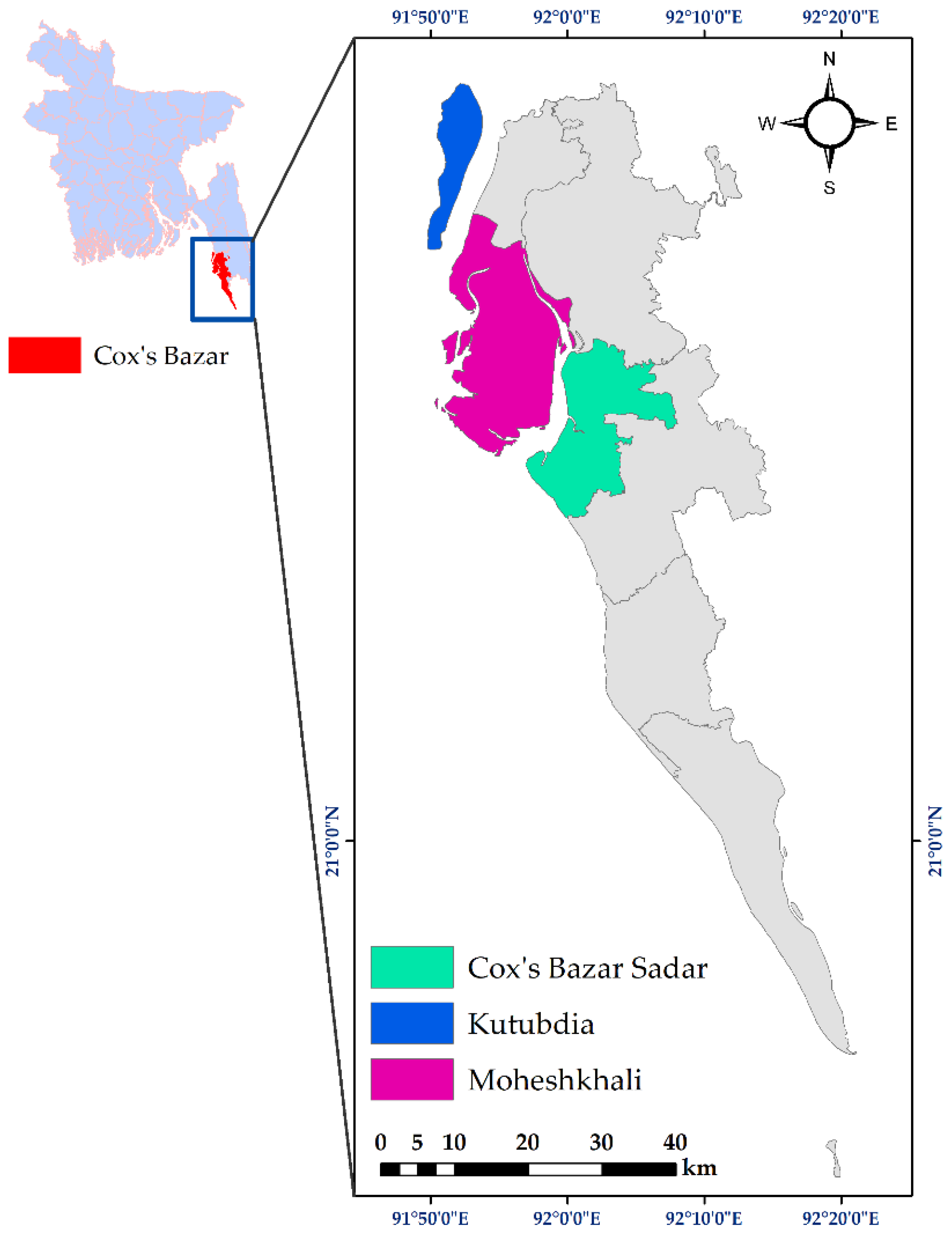
2.1. Sampling Sites

The samples were collected during the period of January 2022 to March 2023 from three coastal areas, i.e., Cox’s Bazar Sadar (21.4272° N, 92.0061° E), Moheshkhali (21.5374° N, 91.9418° E), and Kutubdia (21.8167° N, 91.8583° E) Upazilas (Figure 1) in the Chattogram region of Bangladesh. These areas were selected based on different factors, such as the availability of seafood in those areas, easy access to seafood, and their high demand in those areas.

2.2. Sample Collection and Processing

In this study, a total of 150 raw seafood samples, including shrimp (n = 50), crab (Scylla serrata, n = 25), and sea fish (n = 75), were collected. Shrimp included white shrimp (Penaeus indicus, n = 25) and tiger shrimp (Penaeus monodon, n = 25), while the fish species constituted rupchanda (Pampus chinensis, n = 25), tuna (Euthynnus affinis, n = 25), and loitta (Harpadon nehereus, n = 25). During the study duration, sampling visits were carried out every two weeks in various retailed fish markets, and each sample was promptly placed into a sterile zipper bag, followed by keeping them on ice for transportation to the Microbiology laboratory, Department of Microbiology and Hygiene, BAU, Mymensingh (24.7245° N, 90.4372° E). In the laboratory, the samples’ outer surface was treated with 70% (v/v) alcohol for cleaning purposes. For sea fish, each sample weighing 25 g was homogenized with 225 mL of buffered peptone broth (BPB) (HiMedia, Mumbai, Maharashtra, India), and the mixture was left to incubate overnight at 37 °C [15]. For shrimp and crabs, the brain, leg, muscle, and intestine from each sample were blended, and 25 g of the resulting blend was homogenized and incubated using similar procedures [17].

2.3. Isolation and Molecular Detection of Enterococcus faecium

The initial isolation of E. faecium was performed by culturing the bacterial sample on an enterococcus agar base (EAB) (HiMedia, Mumbai, Maharashtra, India) medium. A single loopful of the bacterial cultured broth was streaked on EAB medium and then incubated aerobically overnight at 37 °C. Colonies appearing as white or pale pink on the EAB medium were initially considered to be E. faecium. Subsequently, the presumed colonies underwent Gram staining and various biochemical tests, including sugar fermentation, Voges–Proskauer, indole, and catalase tests [18]. Finally, E. faecium isolates were analyzed using a simplex polymerase chain reaction (PCR) to detect the presence of E. faecium by targeting the ddlE. faecium gene (Table 1).
The PCR procedure involved obtaining genomic DNA from isolated E. faecium using the boiling method, following the methodology previously described [21]. In brief, 1 mL of the enriched culture underwent centrifugation at 5000 rpm for 5 min. After discarding the supernatant, a suspension was formed by adding 200 μL of phosphate buffer solution (PBS). The subsequent steps encompassed boiling the suspension and allowing it to cool for 10 min at each step. This was followed by another round of centrifugation at 10,000 rpm for 10 min. The resultant supernatant, which held the genomic DNA, was collected and preserved at a temperature of −20 °C to facilitate future analysis.
Each PCR reaction was conducted within a final volume of 20 µL (nuclease-free water: 3 µL; 2X master mixture (Promega, Madison, WI, USA): 10 µL; forward and reverse primers: 1 µL each; and DNA template: 5 µL). The PCR-positive controls encompassed E. faecium genomic DNA that had previously tested positive for the relevant gene. For PCR-negative controls, non-template controls were implemented, employing PBS instead of genomic DNA.
Once the amplification process was completed, the PCR outputs underwent electrophoresis on a 1.5% agarose gel. This gel was treated with ethidium bromide for staining purposes and then visualized using an ultraviolet transilluminator (Biometra, Göttingen, Germany). As a point of reference to confirm the anticipated sizes of the amplified PCR products, a 100 bp DNA ladder (Promega, Madison, WI, USA) was employed.

2.4. Determination of Biofilm-Forming Abilities of Enterococcus faecium

Biofilm formation in E. faecium was characterized phenotypically using the Congo Red (CR) test, following the methodology previously outlined [22]. The CR test involved culturing the E. faecium isolates on Congo Red Agar (CRA) plates. Initially, E. faecium cultures were streaked on CRA plates and then incubated at 37 °C for 24 h. Based on the colony appearance on the CRA plates, isolates exhibiting dry filamentous crusty black colonies, pink colonies with a dark center, and smooth pink colonies were classified as strong, intermediate, and non-biofilm producers, respectively [10].

2.5. Molecular Detection of Virulence Genes in Enterococcus faecium

The presence of virulence-related genes commonly observed in E. faecium, such as fsrA, fsrB, fsrC, pil, gelE, sprE, ace, agg, and cyl, was identified using a simplex PCR assay (Table 1). The PCR protocols used to detect these genes were the same as those described earlier in Section 2.3. Moreover, the PCR-positive controls encompassed E. faecium genomic DNA that had previously tested positive for the relevant virulence genes. For PCR-negative controls, non-template controls were implemented, employing PBS instead of genomic DNA.

2.6. Antibiotic Susceptibility Test

As per the Clinical and Laboratory Standards Institute (CLSI) guidelines [23], the disk diffusion method [24] was performed to determine the antibiotic sensitivity patterns of E. faecium isolates. To conduct the antibiotic susceptibility test (AST), bacterial colonies were grown on EAB agar plates at 37 °C for 18–24 h. A suspension of 2–3 bacterial colonies was then prepared in 0.85% sterile normal saline solution and adjusted to a final concentration of 0.5 McFarland standard units. After an additional 24 h incubation at 37 °C, the bacterial inoculum was spread onto Mueller–Hinton agar plates using sterile cotton swabs, and finally, previously selected antibiotics were dispensed onto the surface of the inoculated agar plate. In this study, we selected thirteen commercially available antibiotics (HiMedia, Mumbai, Maharashtra, India) belonging to ten different antibiotic classes. The three World Health Organization (WHO)-classified antibiotic groups, i.e., access, watch, and reserve, were also focused on during the antibiotic selection. Access antibiotics included penicillins (penicillin—10 μg and ampicillin—10 μg), tetracyclines (tetracycline—30 μg), nitrofurans (nitrofurantoin—300 μg), and amphenicols (chloramphenicol—30 μg); watch antibiotics were glycopeptides (vancomycin—30 μg), macrolides (erythromycin—15 μg), fluoroquinolones (ciprofloxacin—5 μg, levofloxacin—5 μg, and norfloxacin—10 μg), and ansamycins (rifampin—5 μg); and reserve antibiotics included phosphonic acids (fosfomycin—200 μg) and oxazolidinones (linezolid—30 μg). The bacterial strain Escherichia coli ATCC25922 was employed as the designated control strain. Isolates that displayed resistance to at least three antimicrobial categories were classified as multidrug-resistant (MDR) [25]. The following formula [26] was utilized to calculate the multiple antibiotic resistance (MAR) indices:
MAR   index = T h e   c o u n t   o f   a n t i b i o t i c s   t o   w h i c h   a n   i s o l a t e s   s h o w e d   r e s i s t a n c e T h e   t o t a l   n u m b e r   o f   a n t i b i o t i c s   e m p l o y e d   i n   t h i s   s t u d y

2.7. Statistical Analysis

The data acquired from this study were inputted into Excel 365 (Microsoft/Office 365, Redmond, WA, USA) and exported to the Statistical Package for Social Science (SPSS.v.25, IBM, Chicago, IL, USA) and GraphPad Prism (Prism.v.8.4.2, San Diego, CA, USA) for further analyses.
A descriptive analysis was undertaken to ascertain the prevalence of different variables associated with the E. faecium isolates. To estimate the prevalence, a binomial 95% confidence interval (CI) was calculated using a previously established method [27] implemented in GraphPad Prism. The chi-square test for relatedness (Z-test for proportion) was used to identify any sample-wise variations in the frequencies of E. faecium isolates and among different types of biofilm-forming E. faecium isolates. Additionally, a similar test was performed to determine any relatedness between the various degrees of biofilm formation among E. faecium isolates and the occurrence of virulence genes and phenotypic antibiotic resistance. Statistical significance was denoted by a p-value below 0.05 (p < 0.05). Furthermore, a bivariate analysis using SPSS was conducted to explore the potential association between any of the two virulence genes of E. faecium isolates, as well as their resistance to any two antibiotics. A p-value less than 0.05 was considered statistically significant in this bivariate analysis.

3. Results

3.1. Prevalence of Enterococcus faecium

Out of 150 samples, 53 (35.3%, 95% CI: 28.1–43.3%) were found to be positive for E. faecium based on colony characteristics of selective media, staining character, and biochemical properties. In PCR targeting of the ddlE. faecium gene (Figure 2), 27.3% (41/150, 95% CI: 20.8–34.9%) of raw seafood samples contained E. faecium, where a significantly (p < 0.05) higher prevalence of the isolates was exhibited in marine fish (34.7%, 95% CI: 24.9–45.9%) compared to crab (32%, 95% CI: 17.2–51.6%) and shrimp (14%, 95% CI: 7.0–26.1%) samples (Table 2).

3.2. Biofilm-Forming Ability of Isolated Enterococcus faecium

In the Congo Red test, 43.9% (18/41, 95% CI: 29.9–59.0%) of the E. faecium isolates exhibited strong biofilm formation, while 34.2% (14/41, 95% CI: 21.6–49.5%) of the isolates displayed intermediate biofilm formation, and the remaining 21.9% (9/41, 95% CI: 12.0–36.7%) were classified as non-biofilm formers. However, there was no statistically significant variation in the occurrence rate of strong, intermediate, and non-biofilm-forming E. faecium isolates. Sample-wise, the highest percentage of strong biofilm-forming E. faecium was found in shrimp (57.1%, 4/7, 95% CI: 25.1–84.2), followed by marine fish (42.3%, 11/26, 95% CI: 25.5–61.1%) and crab (37.5%, 3/8, 95% CI: 13.7–69.4%) samples (Supplementary Table S1).

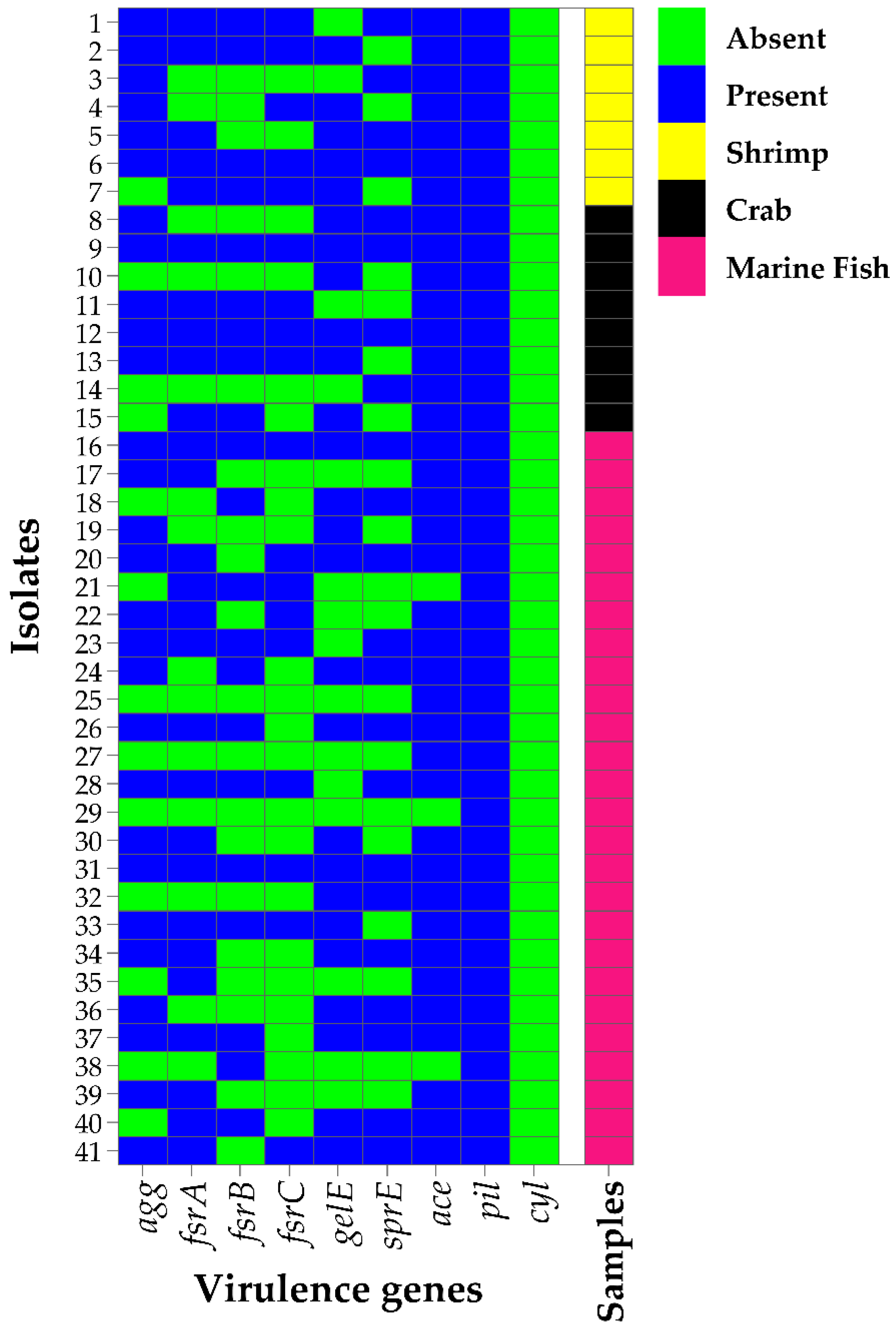
3.3. Prevalence of Virulence Factors in Biofilm-Forming Enterococcus faecium

In PCR screening, all the E. faecium isolates were found to carry at least one of the nine virulence genes tested (Figure 3 and Supplementary Table S1). The highest prevalence of virulence genes was observed for pil (100%), followed by ace (92.7%), agg (68.3%), fsrA (65.9%), gelE (63.4%), sprE (53.7%), fsrB (51.2%), and fsrC (43.9%). However, none of the E. faecium isolates were found to harbor the virulence cyl gene (Figure 3 and Table 3).
In bivariate analysis, a strong and positive significant correlation was observed between virulence genes fsrA and fsrC (Spearman correlation coefficient, ρ = 0.533, p < 0.001), fsrB and fsrC (ρ = 0.470, p < 0.002), fsrA and fsrB (ρ = 0.429, p = 0.005), and ace and agg (ρ = 0.412, p = 0.007). A moderate to lower positive significant correlation was also depicted between agg and fsrA (ρ = 0.394, p = 0.011), agg and fsrC (ρ = 0.392, p = 0.011), ace and gelE (ρ = 0.370, p = 0.017), agg and sprE (ρ = 0.313, p = 0.046), and gelE and sprE (ρ = 0.310, p = 0.049) (Supplementary Table S2).
Moreover, the occurrence rate of all the investigated virulence genes (except pil and cyl) showed a higher trend in the strong biofilm-forming E. faecium isolates than in intermediate and non-biofilm formers, i.e., agg (strong: 88.9% vs. intermediate: 57.1% vs. non: 44.4%), fsrA (88.9% vs. 50% vs. 44.4%), fsrB (77.8% vs. 42.9% vs. 11.1%), fsrC (72.2%, 28.6% vs. 11.1%), gelE (83.3% vs. 57.1% vs. 33.3%), sprE (72.2% vs. 50% vs. 22.2%), and ace (100% vs. 92.9% vs. 77.8%) (Table 3). In the statistical analysis, the percentage of virulence genes, i.e., agg, fsrA, fsrB, fsrC, gelE, and sprE, was significantly (p < 0.05) higher in strong biofilm-forming E. faecium isolates than in intermediate and non-biofilm formers (Table 3).

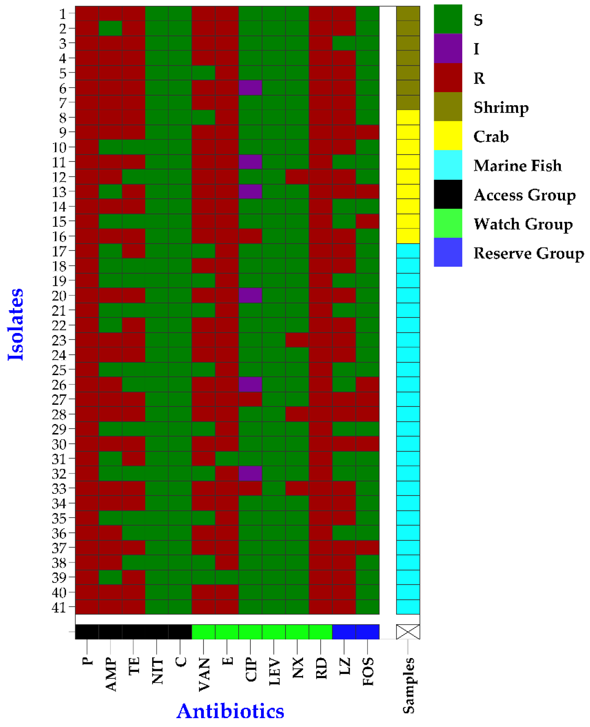
3.4. Antibiogram Profiles of Biofilm-Forming Enterococcus faecium

In AST, E. faecium isolates showed resistance to all three WHO-classified antibiotic groups, i.e., access, watch, and reserve (Figure 4). All the E. faecium isolates were resistant to at least three antibiotics (Figure 4 and Supplementary Table S1). The highest resistance of E. faecium was observed against rifampin and penicillin (100%), followed by erythromycin (95.1%), vancomycin (73.2%), linezolid (70.7%), tetracycline (68.3%), ampicillin (63.4%), fosfomycin (19.5%), norfloxacin (9.8%), and ciprofloxacin (7.3%) (Figure 4 and Table 4). No isolates showed resistance to levofloxacin, nitrofurantoin, and chloramphenicol (Figure 4 and Table 4). Moreover, the beta-lactamase gene blaTEM was found in 61% (25/41, 95% CI: 45.7–74.3%) of the E. faecium isolates (Table 4).
According to bivariate analysis, a strong and positive significant correlation was found between resistance of E. faecium isolates to any of two antibiotics, i.e., tetracycline and linezolid (ρ = 0.483, p = 0.001), tetracycline and ampicillin (ρ = 0.462, p = 0.002), ampicillin and vancomycin (ρ = 0.454, p = 0.003), and vancomycin and tetracycline (ρ = 0.415, p = 0.007) (Supplementary Table S3).
Moreover, a statistically significant correlation (p < 0.05) between the degrees of biofilm formation and the resistance patterns of E. faecium isolates to tetracycline, linezolid, ampicillin, and vancomycin was observed (Table 4). Furthermore, the strong biofilm-forming E. faecium isolates showed higher trends of resistance to most of the antibiotics and resistance genes when compared to intermediate and non-biofilm formers, i.e., ciprofloxacin (strong: 11.1% vs. intermediate: 7.1% vs. non: 0%), tetracycline (94.4% vs. 57.1% vs. 33.3%), fosfomycin (33.3% vs. 14.3% vs. 0%), linezolid (88.9% vs. 64.3% vs. 44.4%), norfloxacin (16.7% vs. 7.7% vs. 0%), ampicillin (94.4% vs. 57.1% vs. 11.1%), vancomycin (100% vs. 85.7% vs. 0%), erythromycin (100% vs. 92.9% vs. 88.9%), and resistance blaTEM gene (66.7% vs. 64.3% vs. 44.4%) (Table 4).

3.5. Phenotypic MDR and MAR Nature in Biofilm-Forming Enterococcus faecium

All the E. faecium isolates (95% CI: 91.4–100%) were phenotypically MDR in nature, and a total of 24 resistance patterns were found. The most prevalent MDR pattern, labeled as pattern number 9 (P, AMP, VAN, E, TE, LZ, RD), was found in the highest number of MDR E. faecium isolates (9/41, 22%, 95% CI: 12.0–36.7%). Additionally, one isolate demonstrated resistance to eight antimicrobial classes (Pattern 1), encompassing a total of ten antibiotics (Table 5). Furthermore, all 41 E. faecium isolates displayed MAR indices greater than 0.2 (MAR index: 0.2–0.8) (Table 5).

4. Discussion

Enterococci, primarily due to their ability to withstand harsh environmental conditions during food production processes and their remarkable adaptability to storage conditions, constitute the predominant bacterial group found in food. Research has documented the occurrence of enterococci in seafood sources. Moreover, biofilm formation and the development of AMR in enterococci limit the treatment options for enterococcal infections. To the best of our knowledge, there is currently no information regarding the distribution and diversity of antibiotic resistance and virulence characteristics in biofilm-producing E. faecium isolates obtained from seafood. This study provides the initial comprehensive analyses of antibiotic resistance and virulence determinants within E. faecium from uncooked seafood.

4.1. Enterococcus faecium Isolated from Seafood

In this study, 27.3% (41/150) of the samples were contaminated with E. faecium, where the majority of the isolates were detected in sea fish samples. Aligned with our study, Ben Said et al. [5] recorded a similar prevalence, detecting E. faecium in 25% of seafood samples. However, the prevalence of E. faecium in this study contrasted with previous reports by Valenzuela et al. [16], Boss et al. [28], and Igbinosa and Beshiru [4], in which the detection rate of E. faecium in seafood samples was lower than the prevalence recorded in the present study. The variation in the prevalence of E. faecium isolates in seafood samples might be due to the geographical location of the studies, environmental conditions, types and numbers of seafood samples collected, and microbial loads in those collected samples or selected areas. The presence of E. faecium in seafood samples is not unusual, as several studies have indicated that seafood can become naturally contaminated from the surroundings in which seafood is typically collected [16,29,30]. The findings of the current study also suggest that seafood samples could potentially come into contact with contaminants during the removal of fish entrails and from environmental sources while being processed and handled by individuals. Moreover, the detection of E. faecium in seafood is regarded as a signal of fecal or environmental pollution, signifying a potential hazard to human health. Over the last two decades, E. faecium has undergone swift evolution into a global nosocomial pathogen, effectively adjusting to the environment within healthcare facilities and giving rise to various human illnesses, including but not limited to neonatal meningitis, endocarditis, bloodstream infections, urinary tract infections, and sepsis among infants [31].

4.2. Biofilm Formation of Enterococcus faecium Isolated from Seafood

Biofilm formation significantly impacts enterococci’s pathogenicity by providing an optimal setting for bacterial growth and facilitating genetic element transfer from one strain to another [32]. Biofilm formation can additionally serve as enduring reservoirs of contamination, resulting in hygienic issues within food items. In this study, the CRA test was used to evaluate the biofilm-forming capabilities of isolated E. faecium from seafood. Although the CRA test is not a sensitive assay like molecular and whole genome sequencing techniques for evaluating biofilm growth, this assay is widely used by researchers because of its reasonable balance between sensitivity and specificity [33]. In the present study, 78.1% (32/41) of the E. faecium isolates were biofilm formers, whereas 43.9% and 34.2% of the isolates were strong and intermediate biofilm formers. Our findings suggest that biofilm-forming E. faecium could be detected in seafood samples due to inadequate post-process cleaning. It is important to know that these biofilm-forming bacteria can readily travel extended distances along the production line, leading to equipment failures and food deterioration, and posing health risks if they infiltrate food batches destined for consumers [34]. Moreover, the occurrence of biofilm-forming E. faecium in seafood has adverse effects on public health, as biofilm formation promotes the emergence of AMR and virulence in bacterial pathogens [35]. Enterococci exhibit a remarkable ability to create biofilms, a distinct strategy for inducing diseases that allows them to endure in hostile surroundings and remain at the infection site for extended periods [36].

4.3. Virulence Profiles of Enterococcus faecium Isolated from Seafood

Bacterial virulence attributes can encompass factors that aid in colonization, such as enhancing bacterial adherence to host cells or invasive elements that promote the invasion of epithelial cells, weakening the immune response. Different cell-wall-incorporated surface proteins contribute to the pathogenic nature of enterococci, including substances that cause aggregation, enterococcal surface proteins, and components that bind to collagen [37]. In this study, each E. faecium isolate contained at least one of the examined virulence genes. Although the virulence gene cyl was absent in all the isolates, other virulence genes were detected in a high number of isolates. Previously, Valenzuela et al. [16], Hammad et al. [15], Chajęcka-Wierzchowska et al. [38], Boss et al. [28], Ben Said et al. [15], and Igbinosa and Beshiru [4] also detected different virulence traits with a different prevalence rate in E. faecium isolates from seafood samples. The substantial presence of virulence genes within E. faecium detected in seafood samples signals a significant concern for human well-being. Our findings also imply that seafood might serve as a notable means for transmitting these virulent E. faecium strains to both humans and the environment. However, this requires further exploration. Furthermore, a significant rise in the presence of virulence genes was noted in E. faecium isolates, demonstrating strong and/or moderate biofilm formation. The present finding indicates that as the extent of biofilm formation in E. faecium isolates intensifies, so does their capacity to initiate infections. Nevertheless, further research is necessary to establish the precise connection between the biofilm-forming aptitude of enterococci isolates and the presence of their virulence genes.

4.4. Antibiotic Resistance Profiles of Enterococcus faecium Isolated from Seafood

The primary concern with enterococci involves their resistance to antibiotics commonly used as treatments for human patients. In addition, employing antimicrobial agents for disease treatment or prevention may apply selective force to both harmful bacteria and symbiotic microorganisms present in the digestive system of seafood. While antibiotic resistance in enterococci isolates has been documented across different origins in Bangladesh [39,40,41,42,43,44,45,46,47,48], there are a lack of data regarding the prevalence of antibiotic resistance in these bacteria specifically in seafood. In this study, all the 41 E. faecium isolates were resistant to ≥3 antimicrobial agents and ≥3 antimicrobial classes (MDR), where a higher resistance was exhibited against penicillin, erythromycin, vancomycin, linezolid, tetracycline, and ampicillin. Moreover, the MAR indices of the isolates ranged between 0.2 and 0.8, and the beta-lactamase gene blaTEM was found in 61% of the isolates. The elevated proportion of multidrug resistance and MAR indices in E. faecium identified in seafood underscores its significance as a potential reservoir of antibiotic resistance that could potentially spread to humans. Previous studies have demonstrated that E. faecium isolated from seafood samples exhibited resistance to various antimicrobial agents [4,5,15,16,28,38].
A standard approach to manage severe enterococcal infections involves combining cell-wall inhibitor antibiotics (penicillin, ampicillin, or vancomycin) with aminoglycosides (streptomycin or gentamicin). However, the concerning aspect is that E. faecium detected in seafood exhibited elevated resistance levels: 100% to penicillin, 63.4% to ampicillin, and 73.2% to vancomycin, posing a significant public health concern. Additionally, it is alarming that E. faecium isolates showed resistance to the reserve group of antibiotics. In this study, 70.7% and 19.5% of the isolates were resistant to critically important antibiotics, linezolid and fosfomycin, respectively. Previously, Chajęcka-Wierzchowska et al. [38] also found that a higher percentage of enterococci (45.7%) isolated from marine samples were resistant to fosfomycin; however, no resistance to linezolid was observed. The occurrence of E. faecium isolates in seafood resistant to linezolid and fosfomycin represents a significant menace to public health. This is because fosfomycin and linezolid are last-resort antibiotics employed to address critical infections arising from MDR enterococci [49,50]. Nonetheless, before arriving at such a crucial inference, additional investigations employing MIC determination and molecular methods should be undertaken. The presence of antibiotic-resistant bacteria in seafood has the potential to pose a public health risk, as they could facilitate the spread of resistance traits along the food chain to other bacteria that hold importance in human clinical contexts. It is conceivable that antibiotic-resistant fecal bacteria originating from domestic sewage or other sources such as animal or fish farming, when released into the sea, could potentially transmit their antibiotic resistance traits to the native fish flora, triggering their propagation and prevalence in the marine ecosystem.
Despite the employment of antibiotics and the host’s immune and inflammatory responses, biofilms persist within the organism owing to biofilm cells’ reduced metabolic activity, quorum sensing, and distinct mechanisms of resilience [51]. Bacteria possessing the capacity to form biofilms exhibited heightened resistance compared to bacteria lacking this potential [52]. In accordance with this statement, strong biofilm-forming E. faecium isolates exhibited significantly greater resistance to the majority of tested antibiotics. This indicates a potential connection between the biofilm-forming capacity of E. faecium isolates and their antimicrobial resistance. The remarkable antibiotic resistance displayed by E. faecium that forms biofilms can be explained by various factors. These include the slower metabolism and growth of biofilm producers, making them naturally less responsive to antibiotics. The composition of the extracellular polymeric substance matrix of the biofilm also restricts antibiotic penetration into its areas. Additionally, the unique physiological attributes of biofilm cells stimulate the activation of multidrug efflux pumps and stress-responsive regulatory systems, nurturing the development of antibiotic resistance [53].

5. Conclusions

Antibiotic-resistant biofilm-forming E. faecium-carrying virulence genes were detected in certain seafood analyzed in this study. This study, therefore, marks the initial investigation highlighting the significance of certain seafood as a potential source of the antibiotic-resistant biofilm-forming pathogenic E. faecium that could affect humans. The findings of this research emphasize the crucial need for rigorous hygiene protocols during the processing, packaging, and storage of seafood in Bangladesh. Moreover, the current study also proposes the adoption of proactive measures to ensure responsible antimicrobial usage across various sectors, including the production of food animals.

Supplementary Materials

The following supporting information can be downloaded at: https://www.mdpi.com/article/10.3390/pathogens12091101/s1, Table S1: Antibiotic resistance, biofilm, and virulence profiles of Enterococcus faecium isolated from seafood in Bangladesh; Table S2: Pearson correlation coefficient to assess the pairs of any of two virulence genes detected in E. faecium isolated from seafood in Bangladesh; Table S3: Pearson correlation coefficients between any of two antibiotics to which E. faecium isolates showed resistance.

Author Contributions

Conceptualization, M.T.R.; methodology, M.A.U. and M.S.I.; software, M.S.I. and M.A.U.; validation, M.T.R.; formal analysis, M.S.I. and M.A.U.; investigation, M.A.U., M.S.I., M.L.R., F.B.F., F.H.N. and Z.F.; resources, M.T.R.; data curation, M.A.U. and M.S.I.; writing—original draft preparation, M.S.I. and M.A.U.; writing—review and editing, M.S.I., J.H. and M.T.R.; visualization, M.S.I. and M.A.U.; supervision, J.H. and M.T.R.; project administration, M.T.R.; funding acquisition, M.T.R. All authors have read and agreed to the published version of the manuscript.

Funding

This research was funded by the Bangladesh Agricultural University Research System (BAURES) (grant No. 2022/12/BAU).

Institutional Review Board Statement

The study procedures received approval from the Animal Welfare and Ethics Committee of Bangladesh Agricultural University (BAU) in Mymensingh, Bangladesh (AWEEC/BAU/2023(25)).

Informed Consent Statement

Not applicable.

Data Availability Statement

All the data are available in the manuscript and Supplementary Material File.

Acknowledgments

The authors would like to thank the Department of Microbiology and Hygiene, Faculty of Veterinary Science, Bangladesh Agricultural University, Mymensingh-2202 for the support during the present research.

Conflicts of Interest

The authors declare no conflict of interest.

References

  1. Islam, M.M.; Shamsuzzaman, M.M.; Mozumder, M.M.H.; Xiangmin, X.; Ming, Y.; Jewel, M.A.S. Exploitation and conservation of coastal and marine fisheries in Bangladesh: Do the fishery laws matter? Mar. Policy 2017, 76, 143–151. [Google Scholar] [CrossRef]
  2. Department of Fisheries (DoF). Yearbook of Fisheries Statistics of Bangladesh, 2021–2022, Fisheries Resources Survey System (FRSS), 2023, Volume 39, Ministry of Fisheries, Bangladesh. Available online: http://fisheries.portal.gov.bd/site/download/42836060-aa5e-491d-8309-cf750886813b (accessed on 2 August 2023).
  3. Novoslavskij, A.; Terentjeva, M.; Eizenberga, I.; Valciņa, O.; Bartkevičs, V.; Bērziņš, A. Major food borne pathogens in fish and fish products: A review. Ann. Microbiol. 2016, 66, 1–15. [Google Scholar] [CrossRef]
  4. Igbinosa, E.O.; Beshiru, A. Antimicrobial resistance, virulence determinants, and biofilm formation of Enterococcus species from ready-to-eat seafood. Front. Microbiol. 2019, 10, 728. [Google Scholar] [CrossRef]
  5. Ben Said, L.; Hamdaoui, M.; Klibi, A.; Ben Slama, K.; Torres, C.; Klibi, N. Diversity of species and antibiotic resistance in enterococci isolated from seafood in Tunisia. Ann. Microbiol. 2017, 67, 135–141. [Google Scholar] [CrossRef]
  6. Ramos, S.; Silva, V.; Dapkevicius, M.D.L.E.; Igrejas, G.; Poeta, P. Enterococci, from harmless bacteria to a pathogen. Microorganisms 2020, 8, 1118. [Google Scholar] [CrossRef]
  7. Torres, C.; Alonso, C.A.; Ruiz-Ripa, L.; León-Sampedro, R.; Del Campo, R.; Coque, T.M. Antimicrobial resistance in Enterococcus spp. of animal origin. Microbiol. Spectr. 2018, 6, 185–227. [Google Scholar] [CrossRef]
  8. ECDC. European Centre for Disease Prevention and Control Publishes Annual Epidemiological Report 2011. Euro. Surveill. 2011, 16, 20012. [Google Scholar]
  9. Costa, O.Y.; Raaijmakers, J.M.; Kuramae, E.E. Microbial extracellular polymeric substances: Ecological function and impact on soil aggregation. Front. Microbiol. 2018, 9, 1636. [Google Scholar] [CrossRef]
  10. Souza, E.L.; Meira, Q.G.; Barbosa, I.D.; Athayde, A.J.; Conceição, M.L.; Siqueira Júnior, J.P. Biofilm formation by Staphylococcus aureus from food contact surfaces in a meat-based broth and sensitivity to sanitizers. Braz. J. Microbiol. 2014, 45, 67–75. [Google Scholar] [CrossRef] [PubMed]
  11. Hashem, Y.A.; Amin, H.M.; Essam, T.M.; Yassin, A.S.; Aziz, R.K. Biofilm formation in enterococci: Genotype-phenotype correlations and inhibition by vancomycin. Sci. Rep. 2017, 7, 5733. [Google Scholar] [CrossRef]
  12. Hancock, L.E.; Gilmore, M.S. Pathogenicity of enterococci. In Gram-Positive Pathogens, 2nd ed.; Fischetti, V.A., Novick, R.P., Ferretti, J.J., Portnoy, D.A., Rood, J.I., Eds.; ASM Press: Washington, DC, USA, 2006; pp. 251–258. [Google Scholar]
  13. Prestinaci, F.; Pezzotti, P.; Pantosti, A. Antimicrobial resistance: A global multifaceted phenomenon. Pathog. Glob. Health 2015, 109, 309–318. [Google Scholar] [CrossRef]
  14. Michael, C.A.; Dominey-Howes, D.; Labbate, M. The Antimicrobial Resistance Crisis: Causes, Consequences, and Management. Front. Public Health 2014, 2, 145. [Google Scholar] [CrossRef]
  15. Hammad, A.M.; Shimamoto, T.; Shimamoto, T. Genetic characterization of antibiotic resistance and virulence factors in Enterococcus spp. From Japanese retail ready-to-eat raw fish. Food Microbiol. 2014, 38, 62–66. [Google Scholar] [CrossRef] [PubMed]
  16. Valenzuela, A.S.; Benomar, N.; Abriouel, H.; Cañamero, M.M.; Gálvez, A. Isolation and identification of Enterococcus faecium from seafoods: Antimicrobial resistance and production of bacteriocin-like substances. Food Microbiol. 2010, 27, 955–961. [Google Scholar] [CrossRef] [PubMed]
  17. Samia, S.; Galib, H.T.; Tanvir, A.S.; Basudeb, C.S.; Walliullah, M.; Tasnia, A.; Sakil, M.M.; Afsana, F.N.; Sadia, K.P.; Kamal, K.D.; et al. Microbiological quality analysis of shrimps collected from local market around Dhaka city. Int. Food Res. J. 2014, 21, 33–38. [Google Scholar]
  18. Facklam, R.R.; da Carvalho, M.G.S.; Teixeira, L.M. History, Taxonomy, Biochemical Characteristics, and Antibiotic Susceptibility Testing of Enterococci. In The Enterococci; American Society of Microbiology: Washington, DC, USA, 2002; pp. 1–54. [Google Scholar]
  19. Dutka-Malen, S.; Evers, S.; Courvalin, P. Detection of glycopeptide resistance genotypes identification to the species level of clinically relevant enterococci by PCR. J. Clin. Microbiol. 1995, 33, 24–27. [Google Scholar] [CrossRef]
  20. Randall, L.P.; Cooles, S.W.; Osborn, M.K.; Piddock, L.J.V.; Woodward, M.J. Antibiotic resistance genes integrons multiple antibiotic resistance in thirty-five serotypes of Salmonella enterica isolated from humans animals in the UK. J. Antimicrob. Chemother. 2004, 53, 208–216. [Google Scholar] [CrossRef]
  21. Queipo-Ortuno, M.I.; De Dios Colmenero, J.; Macias, M.; Bravo, J.M.; Morata, P. Preparation of bacterial DNA template by boiling and effect of immunoglobulin G as an inhibitor in real-time PCR for serum samples from patients with brucellosis. Clin. Vaccine Immunol. 2008, 15, 293–296. [Google Scholar] [CrossRef]
  22. Zheng, J.X.; Bai, B.; Lin, Z.W.; Pu, Z.Y.; Yao, W.M.; Chen, Z.; Li, D.Y.; Deng, X.B.; Deng, Q.W.; Yu, Z.J. Characterization of biofilm formation by Enterococcus faecalis isolates derived from urinary tract infections in China. J. Med. Microbiol. 2018, 67, 60. [Google Scholar] [CrossRef]
  23. M100-S32; Performance Standards for Antimicrobial Susceptibility Testing. Clinical and Laboratory Standards Institute: Wayne, PA, USA, 2022.
  24. Bauer, A.T.; Kirby, W.M.M.; Sherris, J.C.; Turck, M. Antibiotic susceptibility testing by a standardized single disc method. Am. J. Clin. Pathol. 1966, 45, 149–158. [Google Scholar] [CrossRef]
  25. Magiorakos, A.P.; Srinivasan, A.; Carey, R.B.; Carmeli, Y.; Falagas, M.E.; Giske, C.G.; Harbarth, S.; Hindler, J.F.; Kahlmeter, G.; Olsson-Liljequist, B.; et al. Multidrug-resistant, extensively drug-resistant and pandrug-resistant bacteria: An international expert proposal for interim standard definitions for acquired resistance. Clin. Microbiol. Infect. 2012, 18, 268–281. [Google Scholar] [CrossRef]
  26. Krumperman, P.H. Multiple antibiotic resistance indexing of Escherichia coli to identify high-risk sources of fecal contamination of foods. Appl. Environ. Microbiol. 1983, 46, 165–170. [Google Scholar] [CrossRef]
  27. Brown, L.D.; Cai, T.T.; DasGupta, A. Interval estimation for a binomial proportion. Stat. Sci. 2001, 16, 101–133. [Google Scholar] [CrossRef]
  28. Boss, R.; Overesch, G.; Baumgartner, A. Antimicrobial Resistance of Escherichia coli, Enterococci, Pseudomonas aeruginosa, and Staphylococcus aureus from Raw Fish and Seafood Imported into Switzerland. J. Food Protect. 2016, 79, 1240–1246. [Google Scholar] [CrossRef] [PubMed]
  29. Çardak, M.; Özmen Toğay, S.; Ay, M.; Karaalioğlu, O.; Erol, Ö.; Bağcı, U. Antibiotic resistance and virulence genes in Enterococcus species isolated from raw and processed seafood. J. Food Sci. Technol. 2022, 59, 2884–2893. [Google Scholar] [CrossRef] [PubMed]
  30. Chotinantakul, K.; Chansiw, N.; Okada, S. Antimicrobial resistance of Enterococcus spp. isolated from Thai fermented pork in Chiang Rai Province, Thailand. J. Glob. Antimicrob. Resist. 2018, 12, 143–148. [Google Scholar] [CrossRef] [PubMed]
  31. Zhou, X.; Willems, R.J.; Friedrich, A.W.; Rossen, J.W.; Bathoorn, E. Enterococcus faecium: From microbiological insights to practical recommendations for infection control and diagnostics. Antimicrob. Resist. Infect. Control 2020, 9, 130. [Google Scholar] [CrossRef]
  32. Sieńko, A.; Wieczorek, P.; Majewski, P.; Ojdana, D.; Wieczorek, A.; Olszańska, D.; Tryniszewska, E. Comparison of antibiotic resistance and virulence between biofilm-producing and non-producing clinical isolates of Enterococcus faecium. Acta Biochim. Pol. 2015, 62, 859–866. [Google Scholar] [CrossRef] [PubMed]
  33. Hentzer, M.; Riedel, K.; Rasmussen, T.B.; Heydorn, A.; Andersen, J.B.; Parsek, M.R.; Rice, S.A.; Eberl, L.; Molin, S.; Høiby, N.; et al. Inhibition of quorum sensing in Pseudomonas aeruginosa biofilm bacteria by a halogenated furanone compound. Microbiology 2002, 148, 87–102. [Google Scholar] [CrossRef]
  34. Gajewska, J.; Chajęcka-Wierzchowska, W.; Byczkowska-Rostkowska, Z.; Saki, M. Biofilm Formation Capacity and Presence of Virulence Determinants among Enterococcus Species from Milk and Raw Milk Cheeses. Life 2023, 13, 495. [Google Scholar] [CrossRef] [PubMed]
  35. Liu, X.; Yao, H.; Zhao, X.; Ge, C. Biofilm Formation and Control of Foodborne Pathogenic Bacteria. Molecules 2023, 28, 2432. [Google Scholar] [CrossRef]
  36. Tibúrcio, A.A.C.M.; Paiva, A.D.; Pedrosa, A.L.; Rodrigues, W.F.; da Silva, R.B.; Oliveira, A.G. Effect of sub-inhibitory concentrations of antibiotics on biofilm formation and expression of virulence genes in penicillin-resistant, ampicillin-susceptible Enterococcus faecalis. Heliyon 2022, 8, e11154. [Google Scholar] [CrossRef] [PubMed]
  37. Hendrickx, A.P.; Willems, R.J.; Bonten, M.J.; van Schaik, W. LPxTG surface proteins of enterococci. Trends Microbiol. 2009, 17, 423–430. [Google Scholar] [CrossRef]
  38. Roy, K.; Islam, M.S.; Paul, A.; Ievy, S.; Talukder, M.; Sobur, M.A.; Ballah, F.M.; Khan, M.S.R.; Rahman, M.T. Molecular detection and antibiotyping of multi-drug resistant Enterococcus faecium from healthy broiler chickens in Bangladesh. Vet. Med. Sci. 2022, 8, 200–210. [Google Scholar] [CrossRef] [PubMed]
  39. Chajęcka-Wierzchowska, W.; Zadernowska, A.; Łaniewska-Trokenheim, Ł. Virulence factors, antimicrobial resistance and biofilm formation in Enterococcus spp. isolated from retail shrimps. LWT-Food Sci. Technol. 2016, 69, 117–122. [Google Scholar] [CrossRef]
  40. Banik, A.; Mohammad, N.; Akter, T.; Fatema, K.; Abony, M. Prevalence, identification and antibiotic susceptibility of Enterococcus Species isolated from chicken and pigeon meat in Gazipur area of Bangladesh. Open J. Med. Microbiol. 2018, 8, 74–83. [Google Scholar] [CrossRef]
  41. Roy, S.; Aung, M.S.; Paul, S.K.; Ahmed, S.; Haque, N.; Khan, E.R.; Barman, T.K.; Islam, A.; Abedin, S.; Sultana, C.; et al. Drug resistance determinants in clinical isolates of Enterococcus faecalis in Bangladesh: Identification of oxazolidinone resistance gene optrA in ST59 and ST902 lineages. Microorganisms 2020, 8, 1240. [Google Scholar] [CrossRef] [PubMed]
  42. Akter, T.; Foysal, M.J.; Alam, M.; Ehsan, R.; Paul, S.I.; Momtaz, F.; Siddik, M.A.; Tay, A.C.Y.; Fotedar, R.; Gupta, S.K.; et al. Involvement of Enterococcus species in streptococcosis of Nile tilapia in Bangladesh. Aquaculture 2021, 531, 735790. [Google Scholar] [CrossRef]
  43. Islam, M.S.; Paul, A.; Talukder, M.; Roy, K.; Sobur, M.A.; Ievy, S.; Nayeem, M.M.H.; Rahman, S.; Nazir, K.N.H.; Hossain, M.T.; et al. Migratory birds travelling to Bangladesh are potential carriers of multi-drug resistant Enterococcus spp., Salmonella spp., and Vibrio spp. Saudi J. Biol. Sci. 2021, 28, 5963–5970. [Google Scholar] [CrossRef]
  44. Sagor, M.S.; Hossain, M.S.; Islam, T.; Mahmud, M.A.; Miah, M.S.; Karim, M.R.; Giasuddin, M.; Samad, M.A. Phenotypic and genotypic antibiotic resistance and virulence profiling of Enterococcus faecalis isolated from poultry at two major districts in Bangladesh. Pak. Vet. J. 2022, 42, 153–160. [Google Scholar]
  45. Bag, M.A.S.; Arif, M.; Riaz, S.; Khan, M.S.R.; Islam, M.S.; Punom, S.A.; Ali, M.W.; Begum, F.; Islam, M.S.; Rahman, M.T.; et al. Antimicrobial resistance, virulence profiles, and public health significance of Enterococcus faecalis isolated from clinical mastitis of cattle in Bangladesh. BioMed Res. Int. 2022, 2022, 8101866. [Google Scholar] [CrossRef] [PubMed]
  46. Samad, M.A.; Sagor, M.S.; Hossain, M.S.; Karim, M.R.; Mahmud, M.A.; Sarker, M.S.; Shownaw, F.A.; Mia, Z.; Card, R.M.; Agunos, A.; et al. High prevalence of vancomycin non-susceptible and multi-drug resistant enterococci in farmed animals and fresh retail meats in Bangladesh. Vet. Res. Commun. 2022, 46, 811–822. [Google Scholar] [CrossRef] [PubMed]
  47. Akter, T.; Haque, M.N.; Ehsan, R.; Paul, S.I.; Foysal, M.J.; Tay, A.C.Y.; Islam, M.T.; Rahman, M.M. Virulence and antibiotic-resistance genes in Enterococcus faecalis associated with streptococcosis disease in fish. Sci. Rep. 2023, 13, 1551. [Google Scholar] [CrossRef] [PubMed]
  48. Ferdous, F.B.; Islam, M.S.; Ullah, M.A.; Rana, M.L.; Punom, S.A.; Neloy, F.H.; Chowdhury, M.N.U.; Hassan, J.; Siddique, M.P.; Saha, S.; et al. Antimicrobial Resistance Profiles, Virulence Determinants, and Biofilm Formation in Enterococci Isolated from Rhesus Macaques (Macaca mulatta): A Potential Threat for Wildlife in Bangladesh? Animals 2023, 13, 2268. [Google Scholar] [CrossRef] [PubMed]
  49. Fioriti, S.; Coccitto, S.N.; Cedraro, N.; Simoni, S.; Morroni, G.; Brenciani, A.; Mangiaterra, G.; Vignaroli, C.; Vezzulli, L.; Biavasco, F.; et al. Linezolid resistance genes in enterococci isolated from sediment and zooplankton in two Italian coastal areas. Appl. Environ. Microbiol. 2021, 87, e02958-20. [Google Scholar] [CrossRef] [PubMed]
  50. Abbott, I.J.; van Gorp, E.; van der Meijden, A.; Wijma, R.A.; Meletiadis, J.; Roberts, J.A.; Mouton, J.W.; Peleg, A.Y. Oral fosfomycin treatment for enterococcal urinary tract infections in a dynamic in vitro model. Antimicrob. Agents Chemother. 2020, 64, 10–1128. [Google Scholar] [CrossRef] [PubMed]
  51. Høiby, N.; Bjarnsholt, T.; Givskov, M.; Molin, S.; Ciofu, O. Antibiotic resistance of bacterial biofilms. Int. J. Antimicrob. Agents 2010, 35, 322–332. [Google Scholar] [CrossRef] [PubMed]
  52. Uruén, C.; Chopo-Escuin, G.; Tommassen, J.; Mainar-Jaime, R.C.; Arenas, J. Biofilms as promoters of bacterial antibiotic resistance and tolerance. Antibiotics 2020, 10, 3. [Google Scholar] [CrossRef]
  53. Davies, D. Understanding biofilm resistance to antibacterial agents. Nat. Rev. Drug Discov. 2003, 2, 114–122. [Google Scholar] [CrossRef]
Figure 1. The map of the sampling sites during this study. The study area map was prepared using ArcMap.v.10.7 (ArcGIS Enterprise, ESRI, Redlands, CA, USA).
Pathogens 12 01101 g001
Figure 2. PCR amplification of ddlE. faecium gene of Enterococcus faecium. Lane 1: 100 bp DNA Marker; Lane 2: Positive control; Lane 3: Negative control; and Lane 4–13: Representative ddlE. faecium gene of Enterococcus faecium isolates.
Pathogens 12 01101 g002
Figure 3. Heatmap showing the virulence profiles of Enterococcus faecium isolated from raw seafood in Bangladesh.
Pathogens 12 01101 g003
Figure 4. Heatmap exhibiting the antibiogram profiles of Enterococcus faecium isolated from raw seafood in Bangladesh; P = penicillin, AMP = ampicillin, TE = tetracycline, NIT = nitrofurantoin, C = chloramphenicol, VA = vancomycin, E = erythromycin, CIP = ciprofloxacin, LEV = levofloxacin, NOR = norfloxacin, RD = rifampin, LZD = linezolid, FOS = fosfomycin.
Pathogens 12 01101 g004
Table 1. List of primers used for specific detection of Enterococcus faecium isolates and their virulence and antibiotic resistance genes.
Target Organism and DeterminantsTarget GenesPrimer Sequences (5′-3′)Annealing Tm (°C)Size (bp)References
Enterococcus faeciumddlE. faeciumF: GCAAGGCTTCTTAGAGA
R: CATCGTGTAAGCTAACTTC
50550[19]
VirulenceAggF: TCTTGGACACGACCCATGAT
R: AGAAAGAACATCACCACGAGC
58413[11]
fsrAF: CGTTCCGTCTCTCATAGTTA
R: GCAGGATTTGAGGTTGCTAA
53474[11]
fsrBF: TAATCTAGGCTTAGTTCCCAC
R: CTAAATGGCTCTGTCGTCTAG
55428[11]
fsrCF: GTGTTTTTGATTTCGCCAGAGA
R: TATAACAATCCCCAACCGTG
54716[11]
gelEF: GGTGAAGAAGTTACTCTGAC
R: GGTATTGAGTTATGAGGGGC
52704[11]
sprEF: CTGAGGACAGAAGACAAGAAG
R: GGTTTTTCTCACCTGGATAG
53432[11]
AceF: GAATGACCGAGAACGATGGC
R: CTTGATGTTGGCCTGCTTCC
58615[11]
PilF: GAAGAAACCAAAGCACCTAC
R: CTACCTAAGAAAAGAAACGCG
53620[11]
CylF: TGGCGGTATTTTTACTGGAG
R: TGAATCGCTCCATTTCTTC
52186[11]
Antibiotic resistanceblaTEMF: CATTTCCGTGTCGCCCTTAT
R: TCCATAGTTGCCTGACTCCC
56793[20]
Table 2. Prevalence of Enterococcus faecium isolated from different seafoods in Bangladesh.
Type of Samples (N*)n (%) P [95% CI] Qp-Valuen* (%) A [95% CI] B
Shrimp (50)7 (14 a) [7.0–26.1]0.03441 (27.3%) [20.8–34.9]
Crabs (25)8 (32 a,b) [17.2–51.6]
Marine fish (75)26 (34.7 b) [24.9–45.9]
Here, values with different superscripts differ significantly (p < 0.05) within the variable under assessment, N* = category-wise sample number, n = number of sample-wise positive isolates, P = sample-wise prevalence, CI = confidence interval, Q = confidence intervals at 95%, n* = number of overall positive isolates, A = overall prevalence, B = overall confidence interval at 95%.
Table 3. Association in the detection of virulence genes and determination of biofilm formation in E. faecium (n = 41) isolated from raw seafood in Bangladesh.
Virulence GenesVirulence in Different Degrees of Biofilm FormationTotal No. of Positive Isolates (%) [95% CI]p-Value
No. (%) of Strong Biofilm Former (n = 18)No. (%) of Intermediate Biofilm Former (n = 14)No. (%) of Non-Biofilm Former (n = 9)
agg16 (88.9 a)8 (57.1 a,b)4 (44.4 b)28 (68.3) [53.0–80.4]0.035
fsrA16 (88.9 a)7 (50 b)4 (44.4 b)27 (65.9) [50.6–78.4]0.022
fsrB14 (77.8 a)6 (42.9 a,b)1 (11.1 b)21 (51.2) [36.5–65.8]0.004
fsrC13 (72.2 a)4 (28.6 b)1 (11.1 b)18 (43.9) [29.9–59.0]0.004
pil18 (100 a)14 (100 a)9 (100 a)41 (100) [91.4–100]NA
gelE15 (83.3 a)8 (57.1 a,b)3 (33.3 b)26 (63.4) [48.1–76.4]0.033
sprE13 (72.2 a)7 (50 a,b)2 (22.2 b)22 (53.7) [38.8–67.9]0.046
ace18 (100 a)13 (92.9 a)7 (77.8 a)38 (92.7) [80.6–97.5]0.112
cyl0 (0 a)0 (0 a)0 (0 a)0 (0) [0.0–8.6]NA
Here, values with different superscripts differ significantly (p < 0.05) within the variable under assessment, CI = confidence interval, NA = not applied.
Table 4. Association of antibiotic resistance patterns and biofilm formation in E. faecium strains (n = 41) detected in raw seafood in Bangladesh.
CategoriesAntibioticsAntibiotic Resistance in Different Degrees of Biofilm FormationTotal No. of Positive Isolates (%) [95% CI]p-Value
No. (%) of Strong Biofilm Former (n = 18)No. (%) of Intermediate Biofilm Former (n = 14)No. (%) of Non-Biofilm Former (n = 9)
PhenotypicCIP2 (11.1 a)1 (7.1 a)0 (0 a)3 (7.3) [2.5–19.4]0.579
TE17 (94.4 a)8 (57.1 b)3 (33.3 b)28 (68.3) [53.0–80.4]0.003
LEV0 (0 a)0 (0 a)0 (0 a)0 (0) [0.0–8.6]NA
FOS6 (33.3 a)2 (14.3 a)0 (0 a)8 (19.5) [10.2–34.0]0.100
RD18 (100 a)14 (100 a)9 (100 a)41 (100) [91.4–100]NA
P18 (100 a)14 (100 a)9 (100 a)41 (100) [91.4–100]NA
LZD16 (88.9 a)9 (64.3 a,b)4 (44.4 b)29 (70.7) [55.5–82.4]0.046
NOR3 (16.7 a)1 (7.7 a)0 (0 a)4 (9.8) [3.9–22.6]0.374
NIT0 (0 a)0 (0 a)0 (0 a)0 (0) [0.0–8.6]NA
AMP17 (94.4 a)8 (57.1 b)1 (11.1 b)26 (63.4) [48.1–76.4]<0.001
C0 (0 a)0 (0 a)0 (0 a)0 (0) [0.0–8.6]NA
VA18 (100 a)12 (85.7 a)0 (0 a)30 (73.2) [58.1–84.3]<0.001
E18 (100 a)13 (92.9 a)8 (88.9 a)39 (95.1) [83.9–99.1]0.400
GenotypicblaTEM12 (66.7 a)9 (64.3 a)4 (44.4 a)25 (61.0) [45.7–74.3]0.511
Here, values with different superscripts differ significantly (p < 0.05) within the variable under assessment; CIP = ciprofloxacin, TE = tetracycline, LEV = levofloxacin, FOS = fosfomycin, RD = rifampin, P = penicillin, LZD = linezolid, NOR = norfloxacin, NIT = nitrofurantoin, AMP = ampicillin, C = chloramphenicol, VA = vancomycin, E = erythromycin, CI = confidence interval, NA = not applied.
Table 5. Multidrug resistance (MDR) and multiple antibiotic resistance (MAR) profiles of Enterococcus faecium isolates detected in raw seafood samples in Bangladesh.
No. of PatternsAntibiotic Resistance PatternsNo. of Antibiotics (Classes)No. of IsolatesOverall MDR Isolates (%)MAR Index
1P, AMP, VAN, E, TE, CIP, NX, LZ, FOS, RD10 (8)141 (100)0.8
2P, AMP, VAN, E, TE, NX, LZ, FOS, RD9 (8)1 0.7
3P, AMP, VAN, E, TE, CIP, LZ, FOS, RD9 (8)1
4P, AMP, VAN, E, TE, CIP, NX, LZ, RD9 (7)1
5P, AMP, VAN, E, TE, NX, LZ, RD8 (7)1 0.6
6P, AMP, VAN, E, TE, CIP, LZ, RD8 (7)1
7P, AMP, VAN, E, TE, LZ, FOS, RD8 (7)2
8P, VAN, E, TE, LZ, FOS, RD7 (7)1 0.5
9P, AMP, VAN, E, TE, LZ, RD7 (6)9
10P, AMP, VAN, E, NX, LZ, RD7 (6)1
11P, VAN, E, TE, LZ, RD6 (6)3 0.5
12P, VAN, E, TE, FOS, RD6 (6)1
13P, AMP, VAN, E, TE, RD6 (5)3
14P, AMP, E, TE, LZ, RD6 (5)2
15P, AMP, VAN, E, LZ, RD6 (5)1
16P, VAN, E, LZ, RD5 (5)1 0.4
17P, E, TE, LZ, RD5 (5)1
18P, AMP, VAN, TE, RD5 (4)1
19P, AMP, VAN, E, RD5 (4)1
20P, AMP, E, LZ, RD5 (4)1
21P, E, TE, RD4 (4)1 0.3
22P, E, LZ, RD4 (4)1
23P, TE, LZ, RD4 (4)1
24P, E, RD3 (3)4 0.2
Here, MDR = multidrug-resistance, MAR = multiple antibiotic resistance, CIP = ciprofloxacin, TE = tetracycline, LEV = levofloxacin, FOS = fosfomycin, RD = rifampin, P = penicillin, LZD = linezolid, NOR = norfloxacin, NIT = nitrofurantoin, AMP = ampicillin, C = chloramphenicol, VA = vancomycin, E = erythromycin.
Disclaimer/Publisher’s Note: The statements, opinions and data contained in all publications are solely those of the individual author(s) and contributor(s) and not of MDPI and/or the editor(s). MDPI and/or the editor(s) disclaim responsibility for any injury to people or property resulting from any ideas, methods, instructions or products referred to in the content.

