Article

Development of a One-Step Multiplex qPCR Assay for Detection of Methicillin and Vancomycin Drug Resistance Genes in Antibiotic-Resistant Bacteria

by
Jiyoung Lee
1,†,
Eunyoung Baek
1,†,
Hyesun Ahn
2,
Jinyoung Bae
3,
Sangha Kim
4,
Sohyeong Kim
5,6,
Suchan Lee
2,* and
Sunghyun Kim
5,6,*
1
Department of Research & Development, DreamDX Inc., C001, 57, Oryundae-ro, Geumjeong-gu, Busan 46252, Republic of Korea
2
Joint & Arthritis Research Center, Himchan Hospital, 120, Sinmok-ro, Yangcheon-gu, Seoul 07999, Republic of Korea
3
Department of Nano-Bio Convergence Division, Korea Institute of Materials Science, 797 Changwondae-ro, Changwon 51508, Republic of Korea
4
Department of Laboratory Medicine, 158, Gwanjeodong-ro, Seo-gu, Konyang University Hospital, Daejeon 35365, Republic of Korea
5
Department of Clinical Laboratory Science, College of Health Sciences, Catholic University of Pusan, Busan 46252, Republic of Korea
6
Next-Generation Industrial Field-Based Specialist Program for Molecular Diagnostics, Brain Busan 21 Plus Project, Graduate School, Catholic University of Pusan, Busan 46252, Republic of Korea
*
Authors to whom correspondence should be addressed.
These authors contributed equally to this work.
Pathogens 2024, 13(10), 853; https://doi.org/10.3390/pathogens13100853
Submission received: 5 September 2024 / Revised: 25 September 2024 / Accepted: 27 September 2024 / Published: 30 September 2024

Abstract

:
The most common antibiotic-resistant bacteria in Korea are methicillin-resistant Staphylococcus aureus (MRSA) and vancomycin-resistant enterococci (VRE). Pathogen identification in clinical laboratories can be divided into traditional phenotype- and genotype-based methods, both of which are complementary to each other. The genotype-based method using multiplex real-time polymerase chain reaction (PCR) is a rapid and accurate technique that analyzes material at the genetic level by targeting genes simultaneously. Accordingly, we aimed to develop a rapid method for studying the genetic characteristics of antibiotic-resistant bacteria and to provide an experimental guide for the efficient antibiotic resistance gene analysis of mecA detection for MRSA and vanA or vanB detection for VRE using a one-step multiplex qPCR assay at an early stage of infection. As a result, the sensitivity and specificity of the mecA gene for clinical S. aureus isolates, including MRSA and methicillin-susceptible S. aureus, were 97.44% (95% CI, 86.82–99.87%) and 96.15% (95% CI, 87.02–99.32%), respectively. The receiver operating characteristic area under the curve for the diagnosis of MRSA was 0.9798 (*** p < 0.0001). Therefore, the molecular diagnostic method using this newly developed one-step multiplex qPCR assay can provide accurate and rapid results for the treatment of patients with MRSA and VRE infections.

1. Introduction

Antibiotic resistance was considered a serious threat to global public health by the WHO and applies to both developed and underdeveloped countries [1,2]. Higher overall resistance rates among major bacteria were detected in Asian countries including Korea relative to those of Western countries [2,3,4]. The causative agents of the six types of statutory infectious diseases in South Korea are methicillin-resistant Staphylococcus aureus (MRSA), vancomycin-resistant enterococci (VRE), vancomycin-resistant S. aureus (VRSA), multidrug-resistant Pseudomonas aeruginosa (MRPA), multidrug-resistant Acinetobacter baumannii (MRAB), and carbapenem-resistant Enterobacteriaceae (CRE) groups of bacteria [5,6]. In South Korea, several studies have reported that MRSA and VRE infections are the highest among the six major antibiotic-resistant bacterial infections [7,8,9,10].
Currently, clinical laboratories use several AST methods, including disk diffusion and minimum inhibitory concentration (MIC), depending on the equipment and their laboratory test manuals [11,12]. Classical culture-dependent methods have been firmly established in the diagnostic routine, with the main limitation being that results for the most clinically important bacteria are obtained in at least 18–24 or 48 h [12,13]. The diameter should be measured visually or using an automated system, and the results obtained should be interpreted and classified according to the recommended clinical guidelines of the standard in use [14].
Additionally, as an auxiliary molecular diagnostic technology for identifying antibiotic-resistant bacteria, a genotype-based method using real-time polymerase chain rection (PCR) was applied, and this analysis was performed by targeting the mecA, vanA, or vanB genes of MRSA and VRE, respectively [15,16,17,18]. According to these guidelines, MRSA diagnosis is based on the confirmation of oxacillin or cefoxitin antibiotic resistance in S. aureus isolates from clinical specimens and the detection of the methicillin resistance-specific gene (mecA). In addition, VRE is based on the confirmation of vancomycin antibiotic resistance in enterococci isolated from clinical specimens and the detection of vancomycin resistance-specific genes (vanA or vanB) [18,19,20].
The continued emergence of resistance to currently available antimicrobial agents highlights the importance of preventing the transmission and infection of these microorganisms [21]. MRSA and VRE, major hospital infectious strains, have been reported to cause resistance prevalence through the horizontal transfer of resistance genes [22,23,24,25]. MRSA acquires resistance to beta-lactam antibiotics via the mecA gene [26], which is located on the chromosomal mobile genetic element staphylococcal cassette chromosome mec (SCCmec) [27] and is 2.1 kb long [28]. Among the clinically isolated S. aureus, the mecA gene was mainly detected in the MRSA isolates [29]. In South Korea, hospital isolates of VRE are reported to have vancomycin-resistant characteristics by possessing the vanA and vanB genes [25,30,31], of which resistance transmission has been demonstrated [32,33]. In VRE, nine resistance gene types, vanA, vanB, vanC, vanD, vanE, vanG, vanL, vanM, and vanN, which are resistant to glycopeptide antibiotics, have been identified and divided into nine resistance types according to the base sequences of the resistance ligases [25,31,33,34,35]. Among several types, vanA and vanB were the predominant vancomycin resistance gene clusters associated with transposon sequences Tn1546 and Tn1549, respectively [36].
Currently, microbial culture and phenotypic AST play important roles in diagnosis. However, phenotypic AST can be time-consuming, and this is the main limitation of this method, owing to the need for microbial growth. The lag phase is the early stage of microbial growth and a time-limiting factor. A variety of spectral methods are available to enhance the speed of results, but sophisticated microscopy is required for visualizing the cells. Additionally, the appropriate detection of heterogeneity is clinically important but currently difficult with AST. The development of techniques for the potential detection of heterogeneity is crucial, as individual cells within a bacterial population may have different antimicrobial susceptibilities or exhibit different levels of resistance [37]; therefore, it is necessary to fully promote molecular diagnostics to compensate for these shortcomings and enable rapid diagnosis. Many molecular diagnostic tests have been developed to overcome the limitations of microbiological tests, and research and the commercialization of diagnostic kits are currently underway. For rapid diagnosis, a genotypic method that improves the time and cost aspects of the current PCR methods used is needed. Moreover, developing the simultaneous diagnostic detection of MRSA and VRE in this study would reduce processing time, increase specimen throughput [38], and overcome these limitations through a one-step multiplex qPCR assay. In addition, MRSA and VRE, which account for most of the antibiotic resistance in South Korea, have a high multidrug resistance (MDR) rate [39]; thus, the detection efficiency can be increased by a one-step multiplex qPCR assay.
Therefore, the aim of this study is to design a technique to simultaneously detect mecA and vanA or vanB genes specific for MRSA and VRE. We aim to develop a novel one-step multiplex qPCR assay using clinical isolates from patients to screen for antibiotic resistance genes, a rapid molecular diagnostic method that is useful for the simultaneous detection of antibiotic resistance genes and allows for their early detection with sensitivity and specificity [40].

2. Materials and Methods

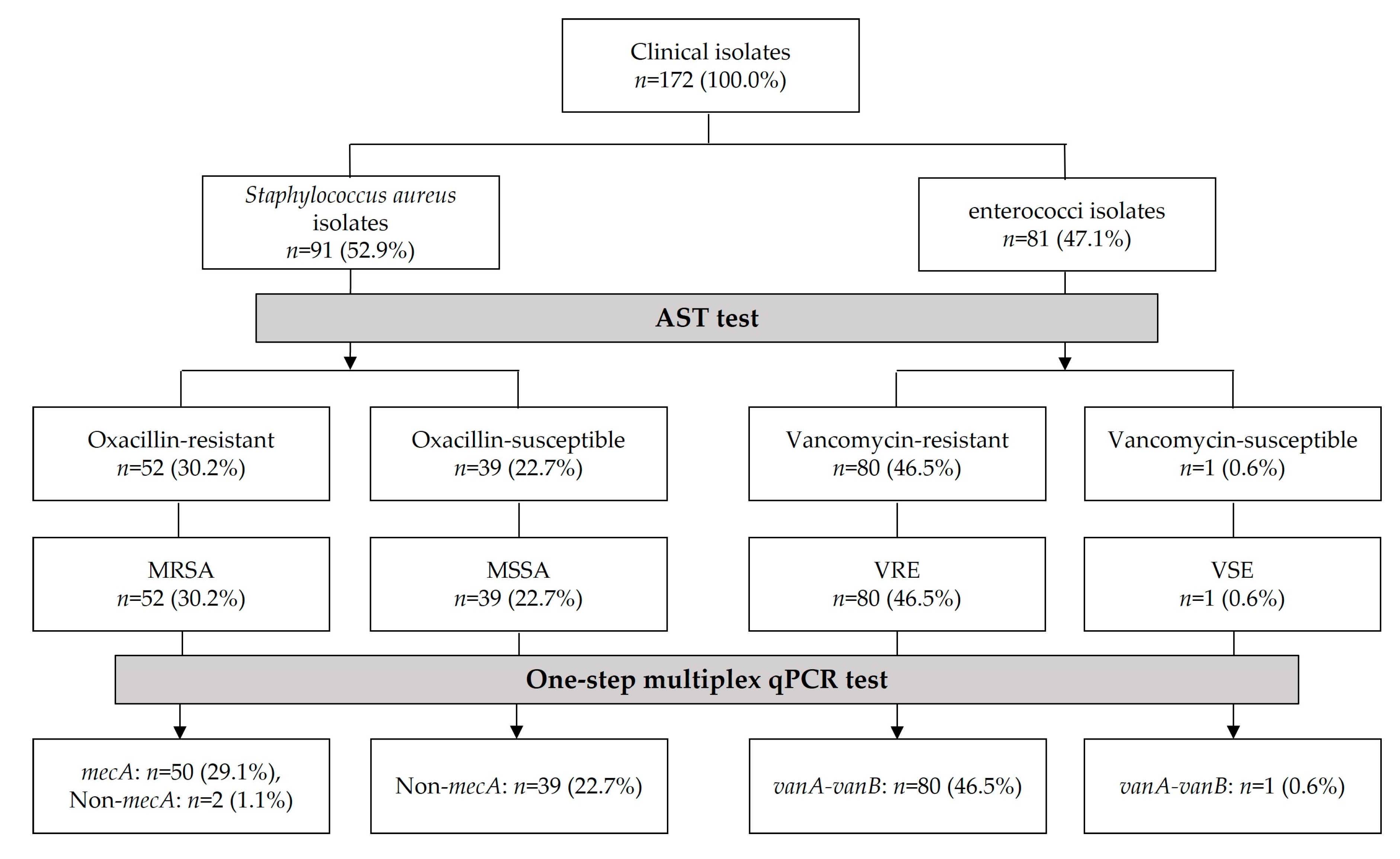
2.1. Clinical Isolates and Reference Strains

The 172 clinical isolates identified by results of the AST included in the study were 52 MRSA isolates and 39 MSSA isolates, collected during the period from August 2015 to June 2018, obtained from blood cultures and 80 VRE isolates and one vancomycin-susceptible enterococci (VSE) isolate, collected during the period January 2022 to December 2022, from the stool and urine cultures of patients. This study was approved by the Institutional Review Board of the Catholic University of Pusan (approval no. CUPIRB-24-01-008). Reference strains were obtained from American Type Culture Collection (ATCC; Manassas, VA, USA), National Culture Collection for Pathogens (NCCP; Cheongju, Chungcheongbuk-do, Republic of Korea), and Korean Collection for Type Cultures (KCTC; Taejon, Republic of Korea; Table S1). All clinical isolates and bacterial reference strains were routinely grown on brain heart infusion (Difco, Le Pont-de-Claix, France) overnight at 37 °C.

2.2. Antimicrobial Susceptibility Testing (AST)

The AST of the clinical isolates was performed using BACTEC FX (Becton Dickinson, Sparks, MD, USA) and interpreted according to the CLSI guidelines [14]. The sample solution was simultaneously inoculated on a blood agar plate (BAP) and MacConkey agar plate for the isolation and incubation of clinical isolates, and cultured in a 35 °C, 5% CO2 incubator for 24 h.

2.3. Genomic DNA Extraction

Genomic DNA from reference strains and clinical isolates was extracted using the boiling method with Chelex-100 Resin (Bio-Rad Laboratories, Hercules, CA, USA) [41,42]. The clinical isolates from patients are routinely cultured on selective agar plates to distinguish between MRSA and VRE. Pure colonies were picked resuspended in BHI broth for the extraction of genomic DNA. Then, 1 mL of the culture broth of the reference strains and isolates were centrifuged at 13,000 rpm for 1 min, then frozen in a deep freezer for 10 min; they were mixed with 200 μL of 5% Chelex-100 solution and boiled at 100 °C for 10 min. The 5% Chelex-100 solution contained 5 g of Chelex-100 resin dissolved in 100 mL TE buffer. The samples were then centrifuged at 3000 rpm for 10 min. Only the upper soluble fraction containing genomic DNA was collected and transferred to a 1.5 mL sterilized microcentrifuge tube and stored at −20 °C. The DNA concentration and quality was estimated using a NanoDrop™ 2000 spectrophotometer (Thermo Fisher Scientific, Waltham, MA, USA).

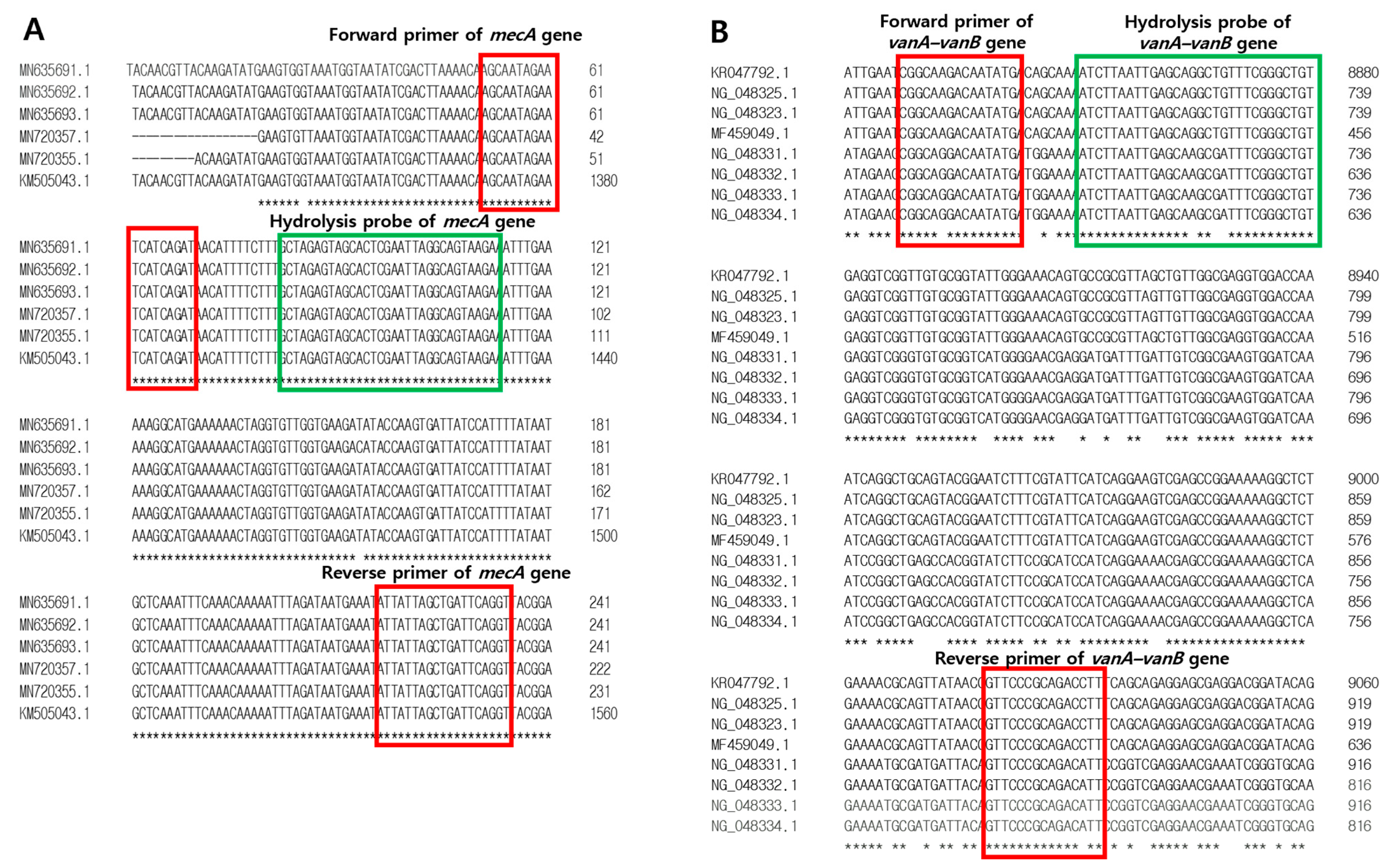
2.4. Development of Primers and Hydrolysis Probes for mecA and vanA-vanB Antibotic Resistance Genes

To detect resistance genes, sequences of mecA, vanA, and vanB were collected from GenBank (www.ncbi.nlm.nih.gov/genbank/ (accessed on 29 August 2024); Table S2). Real-time PCR primers and fluorogenic probes targeting mecA, vanA, and vanB as resistance genes were designed to distinguish them from other bacterial, fungal, and human genomic DNA. The primers amplified approximately 184 and 206 bp of the mecA- and vanA-vanB-specific fragment of resistance genes, respectively. For the one-step multiplex qPCR assay, specific hydrolysis probes for mecA and vanA-vanB genes were designed and labeled with different hydrolysis dyes (Table 1 and Figure 1). To design primer–probes in specific regions for the resistance gene mecA of MRSA and vanA-vanB of VRE, a multiple sequence alignment was performed using Clustal Omega Tools (https://www.ebi.ac.uk/jdispatcher/msa/clustalo; accessed on 20 June 2024; EMBL-EBI, Hinxton, UK) for nucleotide sequences with accession numbers of target genes from the National Center for Biotechnology Information (Table S2). Then, the primer and probe sequences were selected from regions without mismatches, and the secondary structures of the primer and probe, and the possibility of dimer formation and hairpins were evaluated using IDT Oligo analyzer Tools (https://sg.idtdna.com/calc/analyzer; accessed on 20 June 2024; Integrated DNA Technologies, Coralville, IA, USA).

2.5. One-Step Multiplex qPCR Assay

The one-step multiplex qPCR assay was performed using a CFX-96 real-time PCR system (Bio-Rad, Hercules, CA, USA). Amplification was carried out in 20 μL reaction volumes, including 10 μL of 2× Thunderbird probe quantitative PCR mixture (Toyobo, Osaka, Japan), 1 µL of each 10 µM forward primer and reverse primer for mecA, 1 µL of 1 µM hydrolysis probe for mecA, 1 µL of each 10 µM forward primer and reverse primer for vanA-vanB, 1 µL of 1 µM hydrolysis probe for vanA-vanB, 2 µL template DNA, and distilled water. The conditions for the amplification were 95 °C for 10 min, followed by 40 cycles of 95 °C for 15 s, 55 °C for 30 s, and 72 °C for 30 s. Each sample was amplified in a single reaction containing primers and probes specific for two targets, including the mecA gene of MRSA and vanA-vanB gene of VRE. We used different fluorescent dyes as individual targets (Table 1). These probes, with unique emission spectra as fluorescent dyes, can be added to the same PCR amplification reaction for the simultaneous detection of multiple targets in a single assay. Multiplexing tends to significantly hamper the amplification performance for many combinations; therefore, the reagent mixtures and qPCR conditions were carefully evaluated. We set the optimal conditions under which the Ct value and amplification curve of the multiplex were improved compared to those when detection was performed in a separate reaction. Therefore, the qPCR method was developed using the primers and hydrolysis probes designed in this study and was optimized for amplifying two target genes with each fluorescent probe in one tube by adjusting the qPCR cycle conditions (Figure 2).

2.6. Analytical Sensitivity and Specificity

To determine the analytical sensitivity for the detection of mecA and vanA-vanB genes, 10-fold serially diluted synthetic plasmid DNA containing the mecA or vanA-vanB sequence, ranging from 1.0 × 10⁸ to 1.0 × 100 copies/μL, was analyzed. Plasmid DNA genes were synthesized at the Macrogen Institute (Seoul, Republic of Korea), and each sample for the one-step multiplex qPCR assay was analyzed in triplicate. The synthetic plasmid DNA was quantified using the NanoDrop™ 2000 spectrophotometer (Thermo Fisher Scientific) to determine the DNA concentration. The DNA Copy Number and Dilution Calculator web tool (https://www.thermofisher.com/kr/ko/home/brands/thermo-scientific/molecular-biology/molecular-biology-learning-center/molecular-biology-resource-library/thermo-scientific-web-tools/dna-copy-number-calculator.html; accessed on 27 June 2024) was used to calculate the recombinant DNA copy number to dilute the first dilution with a known number of DNA copies. Synthetic plasmid DNA was used to determine specificity for the detection of the mecA and vanA-vanB genes. Cross-reactivity tests were conducted on target and nontarget substances to confirm that no other substances were detected by the probes in the sample. For the one-step multiplex qPCR assay, each sample was analyzed in triplicate.

2.7. Sequencing Analysis

To perform the bacterial 16S rRNA-based molecular analysis, PCR products amplified from clinical isolates were sequenced using mecA and vanA-vanB, and universal primers: F27 (5′-AGAGTTTGATCMTGGCTCAG-3′) and R1492 (5′-TACGGYTACCTTGTTACGACTT-3′) [43]. Sequences were analyzed at the Bionics Institute using Sanger sequencing in an Applied Biosystems 3730XL DNA Analyzer (Thermo Fisher Scientific).

2.8. Statistical Analysis

To analyze the qPCR data, statistical analyses were conducted using Prism 8 (GraphPad Software, San Diego, CA, USA). Receiver operating characteristic (ROC) curves and areas under the curves (AUCs) were used to analyze the DNA of clinical isolates, MRSA, and methicillin-susceptible S. aureus (MSSA), which have the potential to be used as samples for the diagnosis of resistant infections. A p-value < 0.05 was considered statistically significant.

3. Results

3.1. Antimicrobial Susceptibility Testing (AST) Data of Clinical Isolates

AST result data for 172 clinical isolates were provided, and according to the AST profile results, 30.2% (52/172) were confirmed to be resistant to oxacillin, 22.7% (39/172) were confirmed to be susceptible to oxacillin, 46.5% (80/172) were confirmed to be resistant to vancomycin, and 0.6% (1/172) were confirmed to be susceptible to vancomycin (Table 2). The Korea Centers for Disease Control and Prevention (KCDC) operates infectious disease surveillance systems to monitor national disease incidence [44]. Therefore, because VSE isolates are difficult to collect as clinical samples, it was difficult to include additional cases other than the one case included in this study.

3.2. Analytical Sensitivity and Specificity of the One-Step Multiplex qPCR Assay

To confirm the analytical sensitivity and specificity of primers and probes designed in this study, synthetic plasmid DNA was used. The synthetic plasmid gene size of mecA and vanA-vanB were 224 bp and 246 bp, respectively (Table S2). This one-step multiplex qPCR assay amplified the mecA and vanA-vanB target genes from each hydrolysis probe, respectively, and no cross-reactivity was found (Figure 3A). Primers and hydrolysis probes were synthesized at the Macrogen Institute (Seoul, Republic of Korea). The linear standard curve of the real-time qPCR for the mecA and vanA-vanB genes is shown in Figure 3B,C, respectively. The calculation of the E and R2 values for mecA and vanA-vanB genes was performed using each pair of primers and probe. The qPCR results showed that the R2 values for the synthetic plasmid DNA mecA and vanA-vanB were 0.998 and 0.999, respectively, indicating good linearity. The amplification efficiencies for the plasmid of mecA and vanA-vanB genes was at 94.9% and 100.1%, respectively. When the Ct value was verified using the reference strains as positive controls, Ct values of >30 were considered inadequate. In this study, the negative cut-off value was set to ≥30. Therefore, the analytical sensitivity for the target of mecA and vanA-vanB genes were confirmed to be 7.94 × 10² copies/μL and 1.10 × 10³ copies/μL, respectively (Table 3). The plasmid genes synthesized as positive controls were confirmed by the one-step multiplex qPCR assay and by sequencing (Table S3). Additionally, this one-step multiplex qPCR assay amplified the three reference strains of the positive controls but did not amplify the two reference strains of the negative controls, and no cross-reactivity was found (Figure S1 and Table S4).

3.3. Identification of MRSA, MSSA, VRE, and VSE Isolates Using One-Step Multiplex qPCR Assay

To further validate this assay for detecting mecA, vanA, and vanB genes, the DNA extracted from clinical isolates was used as the template DNA. The detection of the target gene for clinical isolates was confirmed using the one-step multiplex qPCR assay, and the results of MRSA, MSSA, VRE, and VSE isolates were 50 (96.2%), 39 (100%), 80 (100%), and 1 (100%), respectively, and were similar to the microbiological culture and AST results. The clinical isolates of MRSA and VRE were confirmed using the mecA primer, and vanA-vanB primers were used for amplifying the DNA between positions 27 and 1492 of bacterial 16S rRNA. Universal primers (27F and 1492R) are widely used for bacterial identification by analyzing the 16S rRNA gene [45,46] (Table 4). The sensitivity and specificity of the mecA gene for clinical S. aureus isolates, including MRSA and MSSA, were 97.44% (95% CI, 86.82–99.87%) and 96.15% (95% CI, 87.02–99.32%), respectively (Figure 4). The ROC AUC for the diagnosis of MRSA was 0.9798 (*** p < 0.0001).

3.4. Comparison of AST and One-Step Multiplex qPCR Assay for Clinical Isolates

Through AST, among 172 clinical isolates, 30.2% (52/172) were identified as MRSA, 22.7% (39/172) as MSSA, 46.5% (80/172) as VRE, and 0.6% (1/172) as VSE. Meanwhile, 29.1% (50/172) were confirmed to be mecA positive, 22.7% (39/172) were confirmed to be mecA negative, 46.5% (80/172) were confirmed to be vanA-vanB positive, and 0.6% (1/172) were confirmed to be vanA-vanB positive by the one-step multiplex qPCR assay (Figure 5). Therefore, overall, the AST results and mecA and vanA-vanB one-step multiplex qPCR analysis for clinical isolates tended to be 98.3% (169/172) consistent, except for three samples.

4. Discussion

Antimicrobial resistance is becoming increasingly serious owing to the use and abuse of a wide range of antibiotics, and the emergence of antibiotic-resistant bacteria poses a major threat to public health [47,48]. The rate of antibiotic resistance remains high; therefore, systematic management is required. In South Korea, the proportion of MRSA and VRE is still high, and the multi-drug resistance (MDR) rate of S. aureus and E. faecium, which are resistant to three or more classes of antibiotics, is also high at 46.9% and 57.9%, respectively [39]. Currently, genotypic molecular methods are used as gold standard diagnostic methods to confirm each antibiotic resistance target gene [49,50,51]. Therefore, this study established an approach showing an improved, simultaneous diagnostic detection over the existing genotypic molecular methods that was developed by enabling the simultaneous diagnostic detection of mecA and vanA-vanB genes. The worldwide incidence of MRSA was reported to be 2.3–69.1% [52] and the global pooled prevalence of MRSA was reported to be 14.69% [48]. According to the European Centre for Disease and Control 2018 report, the population-weighted prevalence of VRE across European countries was 17.3% in 2018 [53]. The overall prevalence of VRE in clinically isolated enterococci in Latin America has been reported to be 31% [53]. In South Korea, MRSA accounts for 60–70% of all isolates from tertiary hospitals [54]. In 2020, the incidence of MRSA in general and nursing hospitals in South Korea was reported to be 47.4% and 88.8%, respectively [55]. The vancomycin resistance rate of E. faecium in 2020 was 38.6% in general hospitals and 94.9% in nursing hospitals in South Korea [55]. Six types of antimicrobial resistant bacteria have been legally declared as the causative agents behind infectious diseases in South Korea and managed since the Infectious Disease Control and Prevention Act was enacted in 2009 [56]. The six types of antibiotic-resistant bacteria in South Korea include MRSA, VRE, VRSA, MRPA, MRAB, and CRE [5,6,10,56]. After the start of the Korea Global AMR Surveillance System (Kor-GLASS) in 2016 in South Korea, methicillin resistance rates of MRSA and vancomycin resistance rates of VRE were reported to be 45.2% and 37.7% in 2021, respectively [57,58]. In clinical laboratories, antibiotic resistance tests are performed to identify antibiotic-resistant bacteria, with culture-based screening being the most conventional gold standard method [49,59,60]. General methods for diagnosing antibiotic-resistant bacteria include phenotypic AST and genotypic molecular methods [49,50,51]. The classical and gold standard phenotypic methods of AST for S. aureus include the broth microdilution and disk diffusion methods [60]. Additionally, traditional methods for detecting VRE identify bacteria through Gram staining, standard biochemical tests, and characterize antimicrobial resistance through disc diffusion, minimum inhibitory concentration, and broth dilution [35,61,62]. Culture-based screening involves the use of a selective medium specific for the target bacteria, culturing it for at least one day, and then identifying the bacteria growing on the medium [12,59,63], which ultimately takes at least 48–72 h [12,64,65,66,67]. However, culture-based methods are still used despite the disadvantage of taking a long time [47,68,69,70].
Genetic methods to detect MRSA and VRE have also recently been used. Genotypic molecular methods include PCR and whole genome sequencing and are performed by targeting resistant genes [19,20,50,52,71]. The molecular methods involved the detection of the methicillin resistance gene (mecA) and vancomycin resistance genes (vanA and vanB) [21,47,72,73]. Molecular-based diagnostic methods have shortened the time required to detect and identify antibiotic-resistant infections [74]. Therefore, it is important to develop a diagnostic method that can efficiently and effectively detect the antibiotic-resistant bacteria that frequently occur in the clinical environment. In this study, clinical isolates were identified as 52 MRSA, 39 MSSA, 80 VRE, and 1 VSE isolates according to AST (Table 2). According to the one-step multiplex qPCR results, 50 of 52 MRSA isolates were confirmed mecA positive, 39 of 39 MSSA isolates were mecA negative, 80 of 80 VRE isolates were positive for vanA-vanB, and 1 of 1 VSE isolate was vanA-vanB positive (Table 4). Therefore, the AST results and one-step multiplex real-time PCR results for clinical isolates were 97.8% (89/91) consistent for 91 S. aureus isolates and 98.8% (80/81) for 81 enterococci isolates (Figure 5).
Recently, molecular diagnostic methods have been used to detect and diagnose infections by targeting the mecA gene in MRSA, and the vanA and vanB genes in VRE [21,40]. Molecular AST methods are based on the direct detection of the antimicrobial resistance genes in bacterial isolates [12,75]. The mecA gene is known to be 2.1 kb in length. This gene is carried by a mec element or staphylococcal cassette chromosome mec (SCCmec) and inserted near the origin of chromosomal replication. Several types of glycopeptide resistance are known in enterococci, and the various types can be genotyped based on sequence differences in the ligase genes. The vanA gene cluster is located on the non-conjugative transposon Tn1546, which may be part of a mobile chromosome or an extrachromosomal element, while the vanB gene cluster is more heterogeneous at the DNA sequence level and the vanB2-operon appears to be linked to the putative conjugative transposon Tn5382/Tn1549 [76]. Compared to traditional PCR analysis, real-time PCR can be processed quickly from sample preparation to results in less than 5 h and provides improved sensitivity and specificity. As real-time PCR is performed in a closed system, the potential for contamination is significantly reduced, and overall costs have decreased as the number of commercially available real-time PCR chemicals and platforms increase [38]. Moreover, for more rapid diagnosis from the existing PCR method, a molecular method that improves time and cost is needed. Accordingly, introducing simultaneous diagnostic detection methods for MRSA and VRE can reduce time and increase specimen throughput [38]. In addition, MRSA and VRE account for most of the antibiotic resistance in South Korea, and because they have a high multidrug resistance rate [39], detection efficiency can be increased by using the one-step multiplex qPCR assay. The one-step multiplex qPCR assay can simultaneously detect each target gene (mecA and vanA-vanB) with different fluorescence compared to the single real-time PCR detection method, which detects only a single target. Accordingly, this one-step multiplex qPCR assay for detecting MRSA and VRE-resistant bacteria uses different fluorescent dyes in the primer and probe design process. This one-step multiplex qPCR assay has clinical utility because it increases specimen throughput and saves time and cost to identify each target [38,77,78,79].
Currently, commercially available kits based on molecular genetic methods target mecA genes for MRSA: the GeneOhm™ MRSA assay (BD Diagnostics, San Diego, CA, USA) [80] and Xpert MRSA kit (Cepheid, Sunnyvale, CA, USA) [81]. Additionally, the targets for VRE are the vanA or vanB genes using the AnyplexTM VanR Real-time Detection kit (VanR) (Seegene, Inc., Seoul, Republic of Korea) [82], and the iNtRON VRE vanA/vanB real-time PCR detection kit (iNtRON Biotechnology, Seongnam, Republic of Korea) [83]. The sensitivity and specificity for MRSA detection were 92.5% and 96.3% for the GeneOhm MRSA assay [80] and 89.5% and 97.3% for the Xpert MRSA assay [81], respectively. In the study, the sensitivity and specificity of qPCR for the diagnosis of MRSA were 97.44% (95% CI, 86.82–99.87%) and 96.15% (95% CI, 87.02–99.32%; Figure 4), which were similar or significantly higher than those in the previous study [80,81]. The sensitivity and specificity for VRE detection were 86.0% and 83.3%, respectively, for the iNtRON vanA/vanB assay [83]. The vanA-vanB primer–hydrolysis probe set developed for this study was designed for the detection of the vanA-vanB gene. In South Korea, because the Korea Centers for Disease Control and Prevention (KCDC) operates an infectious disease surveillance system to monitor nationwide disease incidence [44], VSE is difficult to collect as clinical samples, and only one clinical VSE isolate was included in this study. Another GeneSoC® system kit that targets the mecA gene had an analytical sensitivity of 10 copies/μL, and the detection limit using MRSA bacterial burden is reported to be 103 CFU/Ml [84]. Similarly, in the qPCR method developed in this study, the analytical sensitivity was 7.94 × 10² and 1.10 × 10³ copies/μL targeting mecA and vanA-vanB genes, respectively (Figure 3 and Table 3). However, these available diagnostic kits only detect a single resistance gene of a single resistant bacterium, making it difficult to simultaneously diagnose MRSA and VRE, which are the most common bacteria in South Korea. We aimed to develop a new diagnostic method that can simultaneously detect multiple resistance genes of MRSA and VRE in a single tube and one reaction of the one-step multiplex qPCR assay. Accordingly, our research team developed a molecular diagnostic method to simultaneously detect two types of antibiotic genes, mecA and vanA-vanB genes, to identify antibiotic-resistant bacteria, such as MRSA and VRE. Subsequently, during the development of this study, this one-step multiplex qPCR assay was able to detect multiple target genes using different fluorescent dyes, thus reducing the time, costs, and labor required to diagnose resistant bacterial infections compared to that of other detection methods that only detect a single target gene [77]. Considering the current high incidence of MRSA and VRE among the six resistant bacteria in South Korea, the simultaneous detection of mecA and vanA-vanB genes using this newly developed one-step multiplex qPCR assay is a very useful method for accurately identifying resistant bacteria. Additionally, when mass testing becomes possible at a price similar to that of conventional culture-based methods, it is expected that the number of patients for antibiotic resistance screening tests will expand from specific patients to all hospitalized patients. Therefore, the molecular diagnostic method using this newly developed one-step multiplex qPCR assay can provide accurate and rapid results for the treatment of patients with MRSA and VRE infections. Because this study focused on simultaneously detecting the mecA gene and vanA-vanB gene using different fluorescent probes, the mecA gene possessed by both MRSA and methicillin-resistant coagulase negative staphylococci (MR-CoNS) could be detected, but the limitation was that it could not distinguish between MRSA and MR-CoNS. In the future, the researchers plan to collect MR-CoNS samples to further study the differences between MRSA and MR-CoNS. In addition, among Enterococcus, vanA-vanB resistance genes were be detected for E. faecalis and E. faecium, but species discrimination was not possible. In future studies, more clinical specimens will be collected, and the effectiveness of the mecA and vanA-vanB gene biomarkers developed in this study should be verified through statistical analysis after the additional collection of clinical isolates of MSSA and VSE, as well as MRSA and VRE. As a routine test, culture methods are time-consuming and produce negative results in most cases, leading to the widespread use of antibacterial agents. Although molecular diagnostic research is underway to directly detect pathogens in blood, there are still many difficulties in applying this to routine use [74,85,86]. The molecular diagnostic platform currently offered by DreamDX demonstrates the potential for the simultaneous diagnosis of MRSA and VRE. Therefore, additional tests are needed after collecting samples directly from patients. Performing molecular testing directly on blood without culture has the potential to provide timely and appropriate treatment to patients.

5. Patents

A patent is in process for the primer–probe set for detecting methicillin- and vancomycin-type antibiotic resistance genes: Composition for detecting antibiotic-resistant bacteria and detection method using the same (Patent No. 10-2023-0097961).

Supplementary Materials

The following supporting information can be downloaded at: https://www.mdpi.com/article/10.3390/pathogens13100853/s1, Table S1: Bacterial reference strains; Table S2: Accession numbers and gene synthesis sequences of the mecA and vanA-vanB genes; Table S3: Confirmation for positive control of mecA and vanA-vanB genes by sequencing; Table S4: Results of singleplex, multiplex real-time PCR, and sequencing for reference strains as positive and negative controls in this study; Figure S1: Amplification curve positive control strains, negative control strains, and NTC.

Author Contributions

J.L. and E.B.; data curation, formal analysis, investigation, methodology, validation, visualization, writing—original draft, and writing—review and editing; H.A.; resources, and writing—review and editing; J.B.; resources and writing—review and editing; S.K. (Sangha Kim); resources and writing—review and editing; S.K. (Sohyeong Kim); resources and writing—review and editing; S.K. (Sunghyun Kim) and S.L.; funding acquisition, writing—review and editing, project administration, resources, supervision, and conceptualization. All authors have read and agreed to the published version of the manuscript.

Funding

This work was supported by Busan Techno Park (Busan). Grant numbers and sources of support (including departmental or institutional funding when no extramural funding was received): Busan Techno Park.

Institutional Review Board Statement

This study was approved by the Institutional Review Board of the Catholic University of Pusan (approval no. CUPIRB-24-01-008).

Informed Consent Statement

Patient consent was waived due to the study being declared exempt for use of residual human derivatives.

Data Availability Statement

The original contributions presented in the study are included in the article/Supplementary Material; further inquiries can be directed to the corresponding authors.

Conflicts of Interest

Jiyoung Lee and Eunyoung Baek are employees of DreamDX Inc., a company that aims to commercialize the presented multiplex real-time PCR-based molecular diagnostic assays. Uiseok Byun, Sunghyun Kim, Donghyun Kim, Jiyoung Lee, Eunyoung Baek, Wongeun Je and Sua Lim are the inventors of patent No. 10-2023-0097961. All other authors declare no conflicts of interest. The funders had no role in the design of the study; in the collection, analyses, or interpretation of data; in the writing of the manuscript; or in the decision to publish the results.

References

  1. Antimicrobial Resistance Collaborators. Global burden of bacterial antimicrobial resistance in 2019: A Systematic Analysis. Lancet 2022, 399, 629–655. [Google Scholar] [CrossRef] [PubMed]
  2. Lee, D.K.; Park, J.E.; Kim, K.T.; Jang, D.H.; Song, Y.C.; Ha, N.J. Bacterial contamination and antimicrobial resistance of the surrounding environment influencing health. Korean J. Microbiol. 2014, 50, 101–107. [Google Scholar] [CrossRef]
  3. Kang, C.I.; Song, J.H. Antimicrobial resistance in Asia: Current epidemiology and clinical implications. Infect. Chemother. 2013, 45, 22–31. [Google Scholar] [CrossRef] [PubMed]
  4. Park, S.Y.; Choi, J.H.; Shin, D.W.; Kwon, K.T.; Kim, S.H.; Wi, Y.M.; Kwan, S.K. Conversion to colistin susceptibility by tigecycline exposure in colistin-resistant Klebsiella pneumoniae and Its implications to combination therapy. Int. J. Antimicrob. Agents 2024, 63, 107017. [Google Scholar] [CrossRef] [PubMed]
  5. Lee, H.M.; Yoon, E.J.; Kim, D.K.; Jeong, S.H.; Shim, J.H.; Shin, J.H.; Shin, K.S.; Kim, Y.A.; Uh, Y.U.; Park, C.; et al. Establishment of the South Korean national antimicrobial resistance surveillance system, Kor-GLASS, in 2016. Perspective 2018, 23, 1700734. [Google Scholar] [CrossRef] [PubMed]
  6. Rho, K.H.; Jeong, H.R.; Kim, S.H.; Choi, H.J.; Jung, S.J.; Son, H.J.; Han, S.H.; Choi, J.Y.; Kim, S.W.; Kim, H.B.; et al. The Korean surgical site infection surveillance system report, 2018. Korean. J. Healthc. Assoc. Infect. Control Prev. 2020, 25, 128–136. [Google Scholar] [CrossRef]
  7. Snyder, G.M.; Thom, K.A.; Furuno, J.P.; Perencevich, E.N.; Roghmann, M.C.; Strauss, S.M.; Netzer, G.; Harris, A.D. Detection of methicillin-resistant Staphylococcus aureus and vancomycin-resistant enterococci by healthcare workers on infection control gown and gloves. Infect. Control Hosp. Epidemiol. 2008, 29, 583–589. [Google Scholar] [CrossRef]
  8. Nelwan, E.J.; Andayani, D.; Clarissa, G.; Pramada, T. Vancomycin-resistant Staphylococcus aureus infection post-liposuction in South Korea. Cureus 2021, 13, e14357. [Google Scholar] [CrossRef] [PubMed]
  9. Kim, D.; Yoon, E.J.; Hong, J.S.; Choi, M.H.; Kim, H.S.; Kim, Y.R.; Kim, Y.A.; Uh, Y.; Shin, K.S.; Shin, J.H.; et al. Major bloodstream infection-causing bacterial pathogens and their antimicrobial resistance in South Korea, 2017–2019: Phase I Report from Kor-GLASS. Front. Microbiol. 2022, 12, 799084. [Google Scholar] [CrossRef] [PubMed]
  10. Song, K.H.; Kim, C.J.; Choi, N.K.; Ahn, J.; Choe, P.G.; Park, W.B.; Kim, N.J.; Choi, H.J.; Bae, J.Y.; Kim, E.S.; et al. Clinical and economic burden of bacteremia due to multidrug-resistant organisms in Korea: A prospective case control study. Glob. Antimicrob. Resist. 2022, 31, 379–385. [Google Scholar] [CrossRef]
  11. Humphries, R.M. Update on Susceptibility Testing: Genotypic and Phenotypic Methods. Clin. Lab. Med. 2020, 40, 433–446. [Google Scholar]
  12. Gajic, I.; Kabic, J.; Kekic, D.; Jovicevic, M.; Milenkovic, M.; Mitic Culafic, D.; Trudic, A.; Ranin, L.; Opavski, N. Antimicrobial Susceptibility Testing: A Comprehensive Review of Currently Used Methods. Antibiotics 2022, 11, 427. [Google Scholar] [CrossRef] [PubMed]
  13. Balouiri, M.; Sadiki, M.; Ibnsouda, S.K. Methods for in vitro evaluating antimicrobial activity: A review. J. Pharm. Anal. 2016, 6, 71–79. [Google Scholar] [CrossRef]
  14. CLSI. Performance Standards for Antimicrobial Susceptibility Testing, M100, 32nd ed.; Clinical and Laboratory Standards Institute: Wayne, PA, USA, 2022. [Google Scholar]
  15. Chan, W.S.; Chan, T.M.; Lai, T.W.; Chan, J.F.; Lai, R.W.; Lai, C.K.; Tang, B.S. Complementary use of MALDI-TOF MS and real-time PCR–melt curve analysis for rapid identification of methicillin-resistant staphylococci and VRE. J. Antimicrob. Chemother. 2015, 70, 441–447. [Google Scholar] [CrossRef] [PubMed]
  16. Babiker, A.; Mustapha, M.M.; Pacey, M.P.; Shutt, K.A.; Ezeonwuka, C.D.; Ohm, S.L.; Cooper, V.S.; Marsh, J.W.; Doi, Y.; Harrison, L.H. Use of online tools for antimicrobial resistance prediction by whole-genome sequencing in methicillin-resistant Staphylococcus aureus (MRSA) and vancomycin-resistant enterococci (VRE). J. Glob. Antimicrob. Resist. 2019, 19, 136–143. [Google Scholar] [CrossRef]
  17. He, Y.H.; Ruan, G.J.; Hao, H.; Xue, F.; Ma, Y.K.; Zhu, S.N.; Zheng, B. Real-time PCR for the rapid detection of vanA, vanB and vanM genes. J. Microbiol. Immunol. Infect. 2020, 53, 746–750. [Google Scholar] [CrossRef] [PubMed]
  18. Abdulsahib, S.S. Identification of the respiratory tract infection due to methicillin-resistant Staphylococcus aureu by TaqMan real-time PCR. Sciendo 2021, 9, 86–92. [Google Scholar]
  19. Costa, A.M.; Kay, I.; Palladino, S. Rapid detection of mecA and nuc genes in staphylococci by real-time multiplex polymerase chain reaction. Diagn. Microbiol. Infect. Dis. 2005, 51, 13–17. [Google Scholar] [CrossRef]
  20. Baek, Y.H.; Hong, S.B.; Shin, K.S. Simple and rapid detection of vancomycin-resistance gene from enterococci by loop-mediated isothermal amplification. Biomed. Sci. Lett. 2020, 26, 149–156. [Google Scholar] [CrossRef]
  21. Calfee, D.P. Methicillin-resistant Staphylococcus aureus and vancomycin-resistant enterococci, and other gram-positives in healthcare. Curr. Opin. Infect. Dis. 2012, 25, 385–394. [Google Scholar] [CrossRef]
  22. Palmer, K.L.; Kos, V.N.; Gilmore, M.S. Horizontal gene transfer and the genomics of enterococcal antibiotic resistance. Curr. Opin. Microbiol. 2010, 13, 632–639. [Google Scholar] [CrossRef] [PubMed]
  23. Kriegeskorte, A.; Peters, G. Horizontal gene transfer boosts MRSA spreading. Nat. Med. 2012, 18, 662–663. [Google Scholar] [CrossRef] [PubMed]
  24. Cafini, F.; Thuy, N.T.L.; Román, F.; Prieto, J.; Dubrac, S.; Msadek, T.; Morikawa, K. Methodology for the study of horizontal gene transfer in Staphylococcus aureus. J. Vis. Exp. 2017, 121, e55087. [Google Scholar] [CrossRef]
  25. Moosavian, M.; Ghadri, H.; Zahra, S. Molecular detection of vanA and vanB genes among vancomycin-resistant enterococci in ICU-hospitalized patients in Ahvaz in Southwest of Iran. Infect. Drug Resist. 2018, 11, 2269–2275. [Google Scholar] [CrossRef] [PubMed]
  26. Wielders, C.L.C.; Fluit, A.C.; Brisse, S.; Verhoef, J.; Schmitz, F.J. mecA gene is widely disseminated in Staphylococcus aureus population. J. Clin. Microbiol. 2002, 40, 3970–3975. [Google Scholar] [CrossRef]
  27. Dhungel, S.; Rijal, K.R.; Yadav, B.; Dhungel, B.; Adhikari, N.; Shrestha, U.T.; Adhikari, B.; Banjara, M.R.; Ghimire, P. Methicillin-resistant Staphylococcus aureus (MRSA): Prevalence, antimicrobial susceptibility pattern, and detection of mecA gene among cardiac patients from a tertiary care heart center in Kathmandu, Nepal. Infect. Dis. Res. Treat. 2021, 14, 11786337211037355. [Google Scholar]
  28. Deurenberg, R.H.; Stobberingh, E.E. The evolution of Staphylococcus aureus. Infect. Genet. Evol. 2008, 8, 747–763. [Google Scholar] [CrossRef]
  29. Lee, H.W.; Yoon, J.H.; Sohn, J.H.; Lee, K.H.; Yeh, B.I.; Park, D.W.; Kim, H.W.; Choi, J.W. Detection of mecA gene in clinical isolates of Staphylococcus aureus by multiplex-PCR, and antimicrobial susceptibility of MRSA. J. Microbiol. Biotechnol. 2003, 13, 354–359. [Google Scholar]
  30. Seo, K.S.; Lim, J.Y.; Yoo, H.S.; Bae, W.K.; Park, Y.H. Comparison of vancomycin-resistant enterococci isolates from human, poultry and pigs in Korea. Vet. Microbiol. 2005, 106, 225–233. [Google Scholar] [CrossRef]
  31. Chang, E.; Kim, H.B. It is time to address the isolation policy for patients colonized with vancomycin-resistant enterococci. Korean J. Healthc. Assoc. Infect. Control Prev. 2021, 26, 16–23. [Google Scholar] [CrossRef]
  32. Courvalin, P. Vancomycin resistance in gram-positive cocci. Clin. Infect. Dis. 2006, 42, S25–S34. [Google Scholar] [CrossRef] [PubMed]
  33. Lee, W.G. Resistance mechanism and epidemiology of vancomycin-resistant enterococci. Korean J. Clin. Microbiol. 2008, 11, 71–77. [Google Scholar] [CrossRef]
  34. Arredondo-Alonso, S.; Top, J.; Corander, J.; Willems, R.J.L.; Schürch, A.C. Mode and dynamics of vanA-type vancomycin resistance dissemination in Dutch hospitals. Genome Med. 2021, 13, 9. [Google Scholar] [CrossRef]
  35. Mirzaie, S.; Faghiri, I.; Badouei, M.A.; Madani, S.A. Molecular detection and occurrence of vancomycin resistance genes (van A, B, C1, C2/C3) among Enterococcus species isolated from farm ostriches. Vet. Med. Sci. 2023, 9, 226–233. [Google Scholar] [CrossRef]
  36. Guzman Prieto, A.M.; van Schaik, W.; Rogers, M.R.; Coque, T.M.; Baquero, F.; Corander, J.; Willems, R.J. Global Emergence and Dissemination of Enterococci as Nosocomial Pathogens: Attack of the Clones? Front. Microbiol. 2016, 26, 788. [Google Scholar] [CrossRef]
  37. van Belkum, A.; Burnham, C.A.D.; Rossen, J.W.A.; Mallard, F.; Rochas, O.; Dunne, W.M. Innovative and rapid antimicrobial susceptibility testing systems. Nat. Rev. Microbiol. 2020, 18, 299–311. [Google Scholar] [CrossRef]
  38. Gunson, R.N.; Bennett, S.; Maclean, A.; Carman, W.F. Using multiplex real time PCR in order to streamline a routine diagnostic service. J. Clin. Virol. 2008, 43, 372–375. [Google Scholar] [CrossRef] [PubMed]
  39. Kang, S.; Lee, S.Y.; Seo, S.H.; Lee, J.; Moon, D.C.; Yoo, J.S.; Choi, Y.H. Antimicrobial Resistance of Major Clinical Pathogens Isolated from General Hospitals in Korea: Results from the 2nd Phase (2020–2022) Kor-GLASS. Public Health Wkly. Rep. 2024, 17, 1034–1054. [Google Scholar]
  40. Maurin, M. Real-time PCR as a diagnostic tool for bacterial diseases. Expert Rev. Mol. Diagn. 2012, 12, 731–754. [Google Scholar] [CrossRef] [PubMed]
  41. Singh, U.A.; Kumari, M.; Iyengar, S. Method for improving the quality of genomic DNA obtained from minute quantities of tissue and blood samples using Chelex 100 resin. Biol. Proced. Online 2018, 20, 12. [Google Scholar] [CrossRef]
  42. Hwang, I.; Kim, S.; Jung, T.; Kim, Y.; Kim, S. DNA Mutation Pattern of gyrA and gyrB Genes according to the SCCmec Subtype of Quinolone-resistant Staphylococcus aureus Isolates from Blood Culture. Korean J. Clin. Lab. Sci. (KJCLS) 2024, 56, 115–124. [Google Scholar] [CrossRef]
  43. Weisburg, W.G.; Barns, S.M.; Pelletier, D.A.; Lane, D.J. 16S Ribosomal DNA Amplification for Phylogenetic Study. J. Bacteriol. 1991, 173, 697–703. [Google Scholar] [CrossRef] [PubMed]
  44. Park, S.; Cho, E. National Infectious Diseases Surveillance data of South Korea. Epidemiol. Health 2014, 36, e2014030. [Google Scholar] [CrossRef] [PubMed]
  45. Hou, Q.; Bai, X.; Li, W.; Gao, X.; Zhang, F.; Sun, Z.; Zhang, H. Design of Primers for Evaluation of Lactic Acid Bacteria Populations in Complex Biological Samples. Front. Microbiol. 2018, 9, 2045. [Google Scholar] [CrossRef] [PubMed]
  46. Drancourt, M.; Berger, P.; Raoult, D. Systematic 16S rRNA Gene Sequencing of Atypical Clinical Isolates Identified 27 New Bacterial Species Associated with Humans. J. Clin. Microbiol. 2004, 42, 2197–2202. [Google Scholar] [CrossRef] [PubMed]
  47. Banerjee, R.; Patel, R. Molecular diagnostics for genotypic detection of antibiotic resistance: Current landscape and future directions. JAC Antimicrob. Resist. 2023, 5, dlad018. [Google Scholar] [CrossRef] [PubMed]
  48. Hasanpour, A.H.; Sepidarkish, M.; Mollalo, A.; Ardekani, A.; Almukhtar, M.; Mechaal, A.; Hosseini, S.R.; Bayani, M.; Javanian, M.; Rostami, A. The global prevalence of methicillin-resistant Staphylococcus aureus colonization in residents of elderly care centers: A systematic review and meta-analysis. Antimicrob. Resist. Infect. Control 2023, 12, 4. [Google Scholar] [CrossRef] [PubMed]
  49. Freitas, A.R.; Werner, G. Antibiotic susceptibility testing for therapy and antimicrobial resistance surveillance: Genotype beats phenotype? Future Microbiol. 2022, 17, 1093–1097. [Google Scholar] [CrossRef]
  50. Qodrati, M.; SeyedAlinaghi, S.; Dehghan Manshadi, S.A.; Abdollahi, A.; Dadras, O. Antimicrobial susceptibility testing of Staphylococcus aureus isolates from patients at a tertiary hospital in Tehran, Iran, 2018–2019. Eur. J. Med. Res. 2022, 27, 152. [Google Scholar] [CrossRef]
  51. Wang, W.Y.; Chen, Y.H.; Lee, Y.L.; Chiu, C.F.; Tsao, S.M. Comparative analysis of two commercial automated systems with agar dilution for oxacillin susceptibility and their association with genotypes of invasive Staphylococcus aureus isolates (2011–2021). Infect. Drug Resist. 2024, 17, 1121–1129. [Google Scholar] [CrossRef]
  52. Hassoun, A.; Linden, P.K.; Friedman, B. Incidence, prevalence, and management of MRSA bacteremia across patient populations—A review of recent developments in MRSA management and treatment. Crit. Care 2017, 21, 211. [Google Scholar] [CrossRef] [PubMed]
  53. Rios, R.; Reyes, J.; Carvajal, L.P.; Rincon, S.; Panesso, D.; Echeverri, A.M.; Dinh, A.; Kolokotronis, S.O.; Narechania, A.; Tran, T.T.; et al. Genomic epidemiology of vancomycin-resistant Enterococcus faecium (VREfm) in Latin America: Revisiting the global VRE population structure. Sci. Rep. 2020, 10, 5636. [Google Scholar] [CrossRef] [PubMed]
  54. Kang, G.S.; Jung, Y.H.; Kim, H.S.; Lee, Y.S.; Park, C.; Lee, K.J.; Cha, J.O. Prevalence of major methicillin-resistant Staphylococcus aureus clones in Korea between 2001 and 2008. Ann. Lab. Med. 2016, 36, 536–541. [Google Scholar] [CrossRef] [PubMed]
  55. Song, K.H. Antibiotics for multidrug-resistant gram-positive bacteria. J. Korean Med. Assoc. 2022, 65, 478–489. [Google Scholar] [CrossRef]
  56. Kim, D.; Jeong, S.H. Current status of multidrug-resistant bacteria. J. Korean Med. Assoc. 2022, 65, 468–477. [Google Scholar] [CrossRef]
  57. Kim, D.; Choi, M.H.; Hong, J.S.; Shin, J.H.; Jeong, S.H. Current status and prospects of the national antimicrobial resistance surveillance system, Kor-GLASS. Korean J. Healthc. Assoc. Infect. Control Prev. 2022, 27, 96–103. [Google Scholar] [CrossRef]
  58. Korea Disease Control and Prevention Agency. Korea Home Page. Available online: https://www.korea.kr/archive/expDocView.do?docId=40204&group=S&pageIndex=2&pageUnit=20&startDate=2021-11-23&endDate=2022-11-23&srchKeyword=&srchCode=&codeLevel1=&codeLevel2=&allChkYN=&period= (accessed on 4 September 2024).
  59. McLain, J.E.; Cytryn, E.; Durso, L.M.; Young, S. Culture-based methods for detection of antibiotic resistance in agroecosystems: Advantages, challenges, and gaps in knowledge. J. Environ. Qual. 2016, 45, 432–440. [Google Scholar] [CrossRef]
  60. Sanchini, A. Recent Developments in Phenotypic and Molecular Diagnostic Methods for Antimicrobial Resistance Detection in Staphylococcus aureus: A Narrative Review. Diagnostics 2022, 12, 208. [Google Scholar] [CrossRef] [PubMed]
  61. Tajeddin, E.; Rashidan, M.; Razaghi, M.; Javadi, S.S.S.; Sherafat, S.J.; Alebouyeh, M.; Sarbazi, M.R.; Mansouri, N.; Zali, M.R. The role of the intensive care unit environment and health-care workers in the transmission of bacteria associated with hospital acquired infections. J. Infect. Public Health 2016, 9, 13–23. [Google Scholar] [CrossRef]
  62. Kim, H.J.; Na, S.W.; Alodaini, H.A.; Al-Dosary, M.A.; Nandhakumari, P.; Dyona, L. Prevalence of multidrug-resistant bacteria associated with polymicrobial infections. J. Infect. Public Health 2021, 14, 1864–1869. [Google Scholar] [CrossRef]
  63. Zautner, A.E.; Groß, U.; Emele, M.F.; Hagen, R.M.; Frickmann, H. More pathogenicity or just more pathogens?—on the interpretation problem of multiple pathogen detections with diagnostic multiplex assays. Front. Microbiol. 2017, 8, 1210. [Google Scholar] [CrossRef] [PubMed]
  64. Zirakzadeh, A.; Patel, R. Vancomycin-resistant enterococci: Colonization, infection, detection, and treatment. In Mayo Clinic Proceedings; Elsevier: Amsterdam, The Netherlands, 2006; Volume 81, pp. 529–536. [Google Scholar]
  65. Kim, M.J.; Kang, D.H.; Park, J.I.; Choi, T.Y. Evaluation of ChromID MRSA for the detection of methicillin-resistant Staphylococcus aureus. Korean J. Clin. Microbiol. 2009, 12, 169–173. [Google Scholar] [CrossRef]
  66. van Hal, S.J.; Stark, D.; Lockwood, B.; Marriott, D.; Harkness, J. Methicillin-resistant Staphylococcus aureus (MRSA) detection: Comparison of two molecular methods (IDI-MRSA PCR assay and genotype MRSA direct PCR assay) with three selective MRSA agars (MRSA ID, MRSA elect, and CHROMagar MRSA) for use with infection-control swabs. J. Clin. Microbiol. 2009, 47, 1818–1823. [Google Scholar] [PubMed]
  67. Kim, H.S.; Park, S.B.; Kim, S.H.; Kim, S.H.; Hyun, S.H.; Kim, Y.K. Molecular genetic characteristics of methicillin-resistant Staphylococcus aureus isolated from patients and environment of general hospital intensive care unit in a Chungnam province, Korea. Korean J. Clin. Lab. Sci. 2018, 50, 110–117. [Google Scholar] [CrossRef]
  68. Kurkela, S.; Brown, D.W.G. Molecular diagnostic techniques. Medicine 2009, 37, 535–540. [Google Scholar] [CrossRef] [PubMed]
  69. March-Rosselló, G.A. Rapid methods for detection of bacterial resistance to antibiotics. Enferm. Infecc. Microbiol. Clin. 2017, 35, 182–188. [Google Scholar] [CrossRef] [PubMed]
  70. Yamin, D.; Uskoković, V.; Wakil, A.M.; Goni, M.D.; Shamsuddin, S.H.; Mustafa, F.H.; Alfouzan, W.A.; Alissa, M.; Alshengeti, A.; Almaghrabi, R.H.; et al. Current and future technologies for the detection of antibiotic-resistant bacteria. Diagnostics 2023, 13, 3246. [Google Scholar] [CrossRef]
  71. Vasala, A.; Hytönen, V.P.; Laitinen, O.H. Modern tools for rapid diagnostics of antimicrobial resistance. Front. Cell. Infect. Microbiol. 2020, 10, 308. [Google Scholar] [CrossRef]
  72. Kwak, O.S.; Kwon, M.H.; Jeong, J.H.; Kang, M.I.; Cheun, J.Y.; Lee, G.E.; Kim, Y.K.; Choi, E.G.; Na, M.J.; Kwon, H.U.; et al. Molecular Epidemiology and Antimicrobial Resistance of Methicillin-resistant Staphylococcus aureus Isolated from Nasal Swab at Intensive Care Unit. Tuberc. Respir. Dis. 2008, 65, 91–98. [Google Scholar] [CrossRef]
  73. Giurazza, R.; Mazza, M.C.; Andini, R.; Sansone, P.; Pace, M.C.; Durante-Mangoni, E. Emerging treatment options for multi-drug-resistant bacterial infections. Life 2021, 11, 519. [Google Scholar] [CrossRef]
  74. van den Brand, M.; Peters, R.P.H.; Catsburg, A.; Rubenjan, A.; Broeke, F.J.; van den Dungen, F.A.M.; van Weissenbruch, M.M.; van Furth, A.M.; Kõressaar, T.; Remm, M.; et al. Development of a multiplex real-time PCR assay for the rapid diagnosis of neonatal late onset sepsis. J. Microbiol. Methods 2014, 106, 8–15. [Google Scholar] [CrossRef] [PubMed]
  75. Jacobs, M.R.; Colson, J.D.; Rhoads, D.D. Recent advances in rapid antimicrobial susceptibility testing systems. Expert Rev. Mol. Diagn. 2021, 21, 563–578. [Google Scholar] [CrossRef] [PubMed]
  76. Sundsfjord, A.; Simonsen, G.S.; Haldorsen, B.C.; Haaheim, H.; Hjelmevoll, S.O.; Littauer, P.; Dahl, K.H. Genetic methods for detection of antimicrobial resistance. Apmis 2004, 112, 815–837. [Google Scholar] [CrossRef]
  77. Wada, K.; Mizoguchi, S.; Ito, Y.; Kawada, J.; Yamauchi, Y.; Morishima, T.; Nishiyama, Y.; Kimura, H. Multiplex real-time PCR for the simultaneous detection of herpes simplex virus, human herpesvirus 6, and human herpesvirus 7. Microbiol. Immunol. 2009, 53, 22–29. [Google Scholar] [CrossRef] [PubMed]
  78. Wang, X.; Theodore, M.J.; Mair, R.; Trujillo-Lopez, E.; du Plessis, M.; Wolter, N.; Baughman, A.L.; Hatcher, C.; Vuong, J.; Lott, L.; et al. Clinical Validation of Multiplex Real-Time PCR Assays for Detection of Bacterial Meningitis Pathogens. J. Clin. Microbiol. 2012, 50, 702–708. [Google Scholar] [CrossRef]
  79. Trung, N.T.; Tong, H.V.; Lien, T.T.; Son, T.V.; Huyen, T.T.T.; Quyen, D.T.; Hoan, P.Q.; Meyer, C.G.; Song, L.H. Clinical utility of an optimised multiplex real-time PCR assay for the identification of pathogens causing sepsis in Vietnamese patients. Int. J. Infect. Dis. 2018, 67, 122–128. [Google Scholar] [CrossRef] [PubMed]
  80. Mehta, M.S.; McClure, J.T.; Mangold, K.; Peterson, L.R. Performance of 3 real-time PCR assays for direct detection of Staphylococcus aureus and MRSA from clinical samples. Diagn. Microbiol. Infect. Dis. 2015, 83, 211–215. [Google Scholar] [CrossRef]
  81. Rossney, A.S.; Herra, C.M.; Brennan, G.I.; Morgan, P.M.; O’Connell, B. Evaluation of the Xpert methicillin-resistant Staphylococcus aureus (MRSA) assay using the GeneXpert real-time PCR platform for rapid detection of MRSA from screening specimens. J. Clin. Microbiol. 2008, 46, 3285–3290. [Google Scholar] [CrossRef]
  82. Jung, M.K.; Lee, W.G.; Park, M.H. Evaluation of a newly developed multiplex real-time PCR assay for the detection of vancomycin-resistant enterococci from rectal swabs. Korean J. Clin. Microbiol. 2011, 14, 138–147. [Google Scholar] [CrossRef]
  83. Bae, M.H.; Kim, J.; Sung, H.; Jeong, Y.S.; Kim, M.N. Evaluation of iNtRON VRE vanA/vanB real-time PCR for follow-up surveillance of VRE-infected or colonized patient. Diagn. Microbiol. Infect. Dis. 2013, 77, 292–295. [Google Scholar] [CrossRef] [PubMed]
  84. Chiba, M.; Aoyagi, T.; Yoshida, M.; Katsumi, M.; Fujimaki, S.I.; Ishii, Y.; Tateda, K.; Kaku, M. Evaluation of the performance of GeneSoC®, a novel rapid real-time PCR system, to detect Staphylococcus aureus and methicillin resistance in blood cultures. Infect. Chemother. 2023, 29, 718–721. [Google Scholar] [CrossRef] [PubMed]
  85. Westh, H.; Lisby, G.; Breysse, F.; Böddinghaus, B.; Chomarat, M.; Gant, V.; Goglio, A.; Raglio, A.; Schuster, H.; Stuber, F.; et al. Multiplex real-time PCR and blood culture for identification of bloodstream pathogens in patients with suspected sepsis. Clin. Microbiol. Infect. 2009, 15, 544–551. [Google Scholar] [CrossRef] [PubMed]
  86. Samuel, L. Direct Detection of Pathogens in Bloodstream During Sepsis: Are We There Yet? J. Appl. Lab. Med. 2019, 3, 631–642. [Google Scholar] [CrossRef] [PubMed]
Figure 1. An alignment (5′–3′) of the primers and probe with (A) the mecA gene and (B) the vanA-vanB gene. Forward and reverse primer sequences are shown in red frames and probe sequences are shown in green frames. The “*” character means that the residue or nucleotide is identically conserved.
Pathogens 13 00853 g001
Figure 2. Flow chart for sample processing by the one-step multiplex qPCR assay protocol for MRSA and VRE detection.
Pathogens 13 00853 g002
Figure 3. Results of specificity and analytic sensitivity using synthetic plasmid DNA. (A) Amplification curve of the mecA and vanA-vanB genes. (B) Amplification curves and standard curves of log10 genome versus Ct value of mecA genes. (C) Amplification curves and standard curves of log10 genome versus Ct value of vanA-vanB genes.
Pathogens 13 00853 g003
Figure 4. Results of specificity and sensitivity using the mecA primer–hydrolysis probe set. (A) The Ct value (mean) of mecA gene among the MRSA and MSSA samples. (B) Receiver operating characteristic (ROC) curve analysis to evaluate the diagnostic value of the mecA primer–hydrolysis probe set. Data are presented as mean ± SEM (standard error of the mean). Abbreviations: MRSA, methicillin-resistant S. aureus; MSSA, methicillin-susceptible S. aureus; ROC, receiver operating characteristic.
Pathogens 13 00853 g004
Figure 5. Comparison of the results of AST and multiplex qPCR in clinical isolates.
Pathogens 13 00853 g005
Table 1. The design of primers and probe sequences for mecA and vanA-vanB genes on one-step multiplex qPCR assay.
TargetPrimers and ProbesSequences (5′-3′)Amplicon Size (bp)Length (mer)
mecAForward primerAGCAATAGAATCATCAGAT18419
Reverse primerACCTGAATCAGCTAATAAT19
ProbeFAM-GCTAGAGTAGCACTCGAATTAGGCAGTAAGA-TAMRA31
vanA-vanBForward primerCGGCAGGACAATATGA20616
Reverse primerAATGTCTGCGGGAAC15
ProbeHEX-ATCTTAATTGAGCAAGCGATTTCGGGCTGT-TAMRA30
Table 2. Antimicrobial susceptibility testing results of clinical isolates.
Antimicrobial AgentStandard Interpretation (CLSI) [14]Total
n (%)
Resistant
n (%)
Intermediate
n (%)
Susceptible
n (%)
Oxacillin52 (30.2%)0 (0%)39 (22.7%)172 (100%)
Vancomycin80 (46.5%)0 (0%)1 (0.6%)
Oxacillin is a narrow-spectrum beta-lactam antibiotic of the penicillin class developed by Beecham, and vancomycin is a glycopeptide antibiotic medication used to treat a number of bacterial infections.
Table 3. Mean Ct values for qPCR carried out on serial dilutions of plasmid DNA of mecA and vanA-vanB genes.
Plasmid DNA of Resistance GenePlasmid DNA ContentMean Ct ValueSDLog ValueCopies per Reaction
(copies/μL)
mecA gene2.58 ng9.040.008.021.04 × 108
258 pg12.180.187.111.28 × 107
25.8 pg16.090.115.979.42 × 105
2.58 pg19.530.174.989.49 × 104
258 fg23.150.183.938.48 × 103
25.8 fg26.700.002.907.94 × 102
2.58 fg30.170.091.897.84 × 101
258 ag32.540.321.211.61 × 101
vanA-vanB gene964 pg11.610.118.031.07 × 108
96.4 pg15.150.076.969.22 × 106
9.64 pg18.260.076.031.07 × 106
964 fg21.720.144.999.66 × 104
96.4 fg25.250.053.928.31 × 103
9.64 fg28.170.213.041.10 × 103
964 ag31.140.012.151.40 × 102
96.4 ag35.260.240.90.83 × 100
The copies per reaction of mecA gene were 2.58 ng = 1.04 × 10⁸ copies, 258 pg = 1.28 × 10⁷ copies, 25.8 pg = 9.42 × 10⁵ copies, 2.58 pg = 9.49 × 10⁴ copies, 258 fg = 8.48 × 10³ copies, 25.8 fg = 7.94 × 10² copies, 2.58 fg = 7.84 × 101 copies, and 258 ag = 1.61 × 101 copies. The copies per reaction of vanA-vanB gene were 964 pg = 1.07 × 10⁸ copies, 96.4 pg = 9.22 × 10⁶ copies, 9.64 pg = 1.07 × 10⁶ copies, 964 fg = 9.66 × 10⁴ copies, 96.4 fg = 8.31 × 10³ copies, 9.64 fg = 1.10 × 10³ copies, 964 ag = 1.40 × 10² copies, and 96.4 ag = 0.83 × 100 copies. Abbreviations: SD, standard deviation.
Table 4. Sample types and qPCR results of clinical isolates.
Clinical IsolatesMutiplex qPCR Using mecA and vanA-vanB Primers-Hydrolysis ProbesSequencing Results Using mecA Primer, vanA-vanB Primer, and Universal Primer
mecA Positive
Ct Mean ± SD
(n, %)
mecA Negative
Ct Mean ± SD
(n, %)
vanA-vanB Positive
Ct Mean ± SD
(n, %)
vanA-vanB Negative
Ct Mean ± SD
(n, %)
mecA Positive Using mecA Primer
(n, %)
van Positive Using vanA-vanB Primer
(n, %)
Using Universal Primer
(n, %)
MRSA isolates
(n = 52, 100%)
20.7 ± 2.3
(50, 96.2%)
40.0 ± 0.0
(2, 3.8%)
(0, 0%)40.0 ± 0.0
(52, 100%)
(50, 96.2%)-(52, 100%)
MSSA isolates
(n = 39, 100%)
(0, 0%)40.0 ± 0.2
(39, 100%)
(0, 0%)40.0 ± 0.0
(39, 100%)
---
VRE isolates
(n = 80, 100%)
(0, 0%)39.8 ± 1.2
(80, 100%)
18.3 ± 1.4
(80, 100%)
(0, 0%)-(80, 100%)(80, 100%)
VSE isolate
(n = 1, 100%)
(0, 0%)40.0 ± 0.0
(1, 100%)
16.8 ± 0.0
(1, 100%)
(0, 0%)-(1, 100%)(1, 100%)
Total isolates
(n = 172, 100%)
20.7 ± 2.3
(50, 29.1%)
39.9 ± 1.0
(122, 70.9%)
18.3 ± 1.4
(81, 47.1%)
40.0 ± 0.0
(91, 52.9%)
---
Abbreviations: SD, standard deviation; MRSA, methicillin-resistant Staphylococcus aureus; MSSA, methicillin-susceptible S. aureus; VRE, vancomycin-resistant enterococci; VSE, vancomycin-susceptible enterococci.
Disclaimer/Publisher’s Note: The statements, opinions and data contained in all publications are solely those of the individual author(s) and contributor(s) and not of MDPI and/or the editor(s). MDPI and/or the editor(s) disclaim responsibility for any injury to people or property resulting from any ideas, methods, instructions or products referred to in the content.

