Hard Ticks as Vectors: The Emerging Threat of Tick-Borne Diseases in India

 ,
,
Abstract
:
1. Introduction
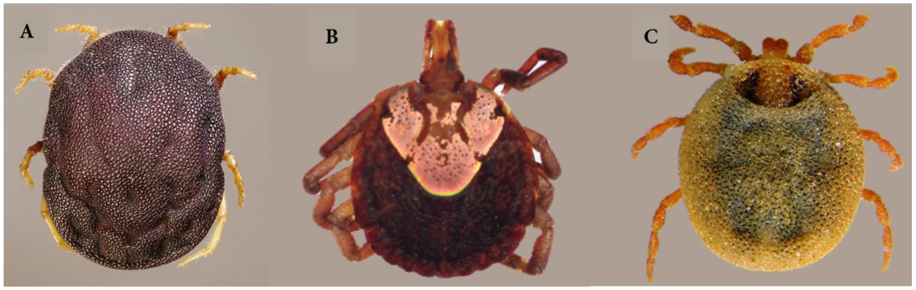
1.1. Tick Vectors: An Introduction
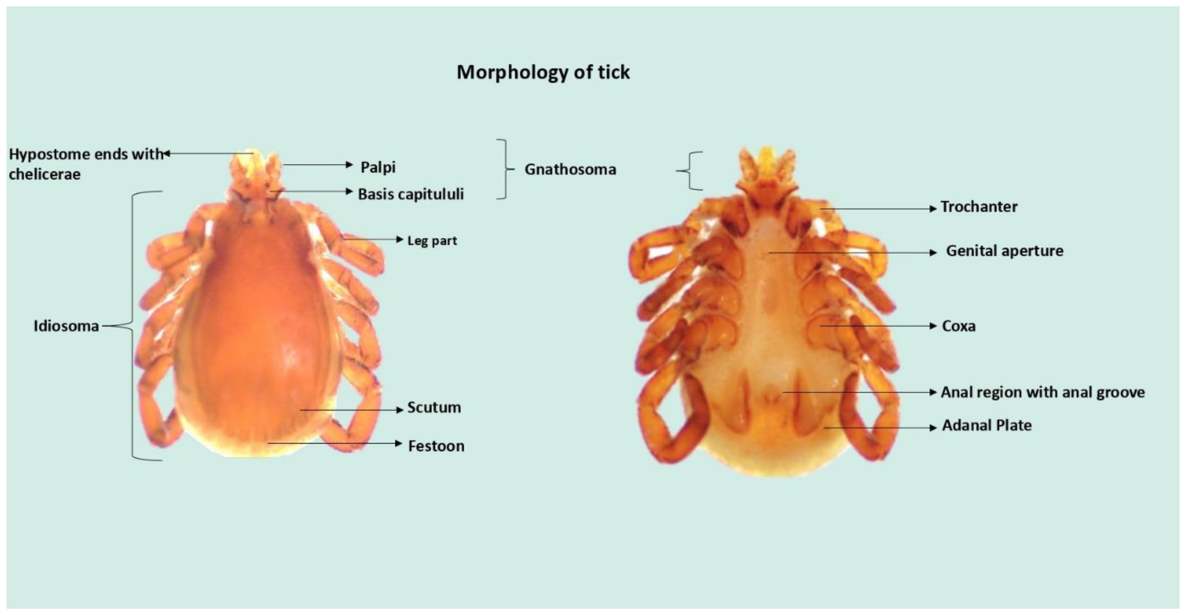
1.2. Structure and Physiology of Hard Ticks
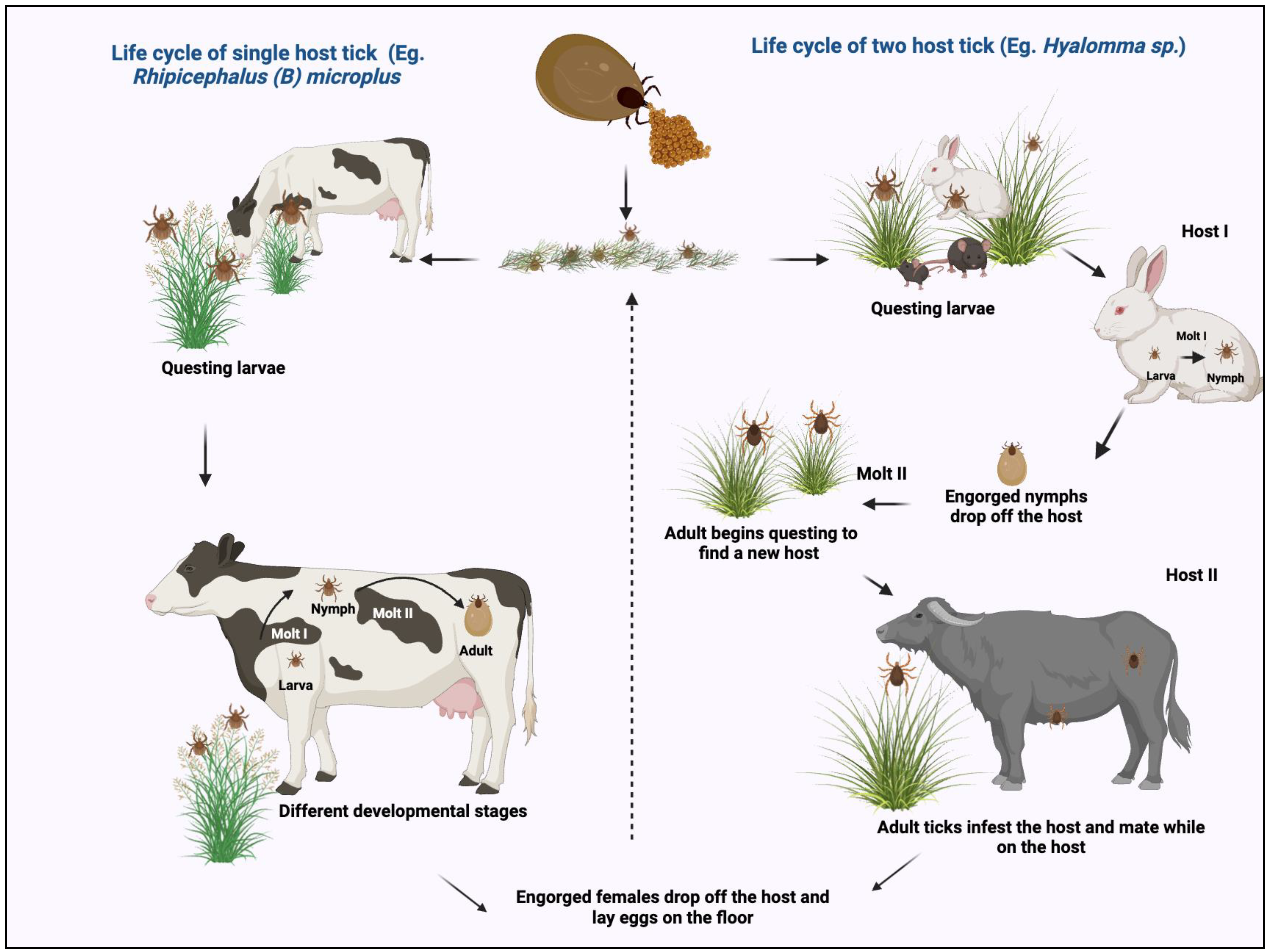
1.3. Life Cycle of Hard Ticks
2. Hard Ticks: An Efficient Vector for TBDs
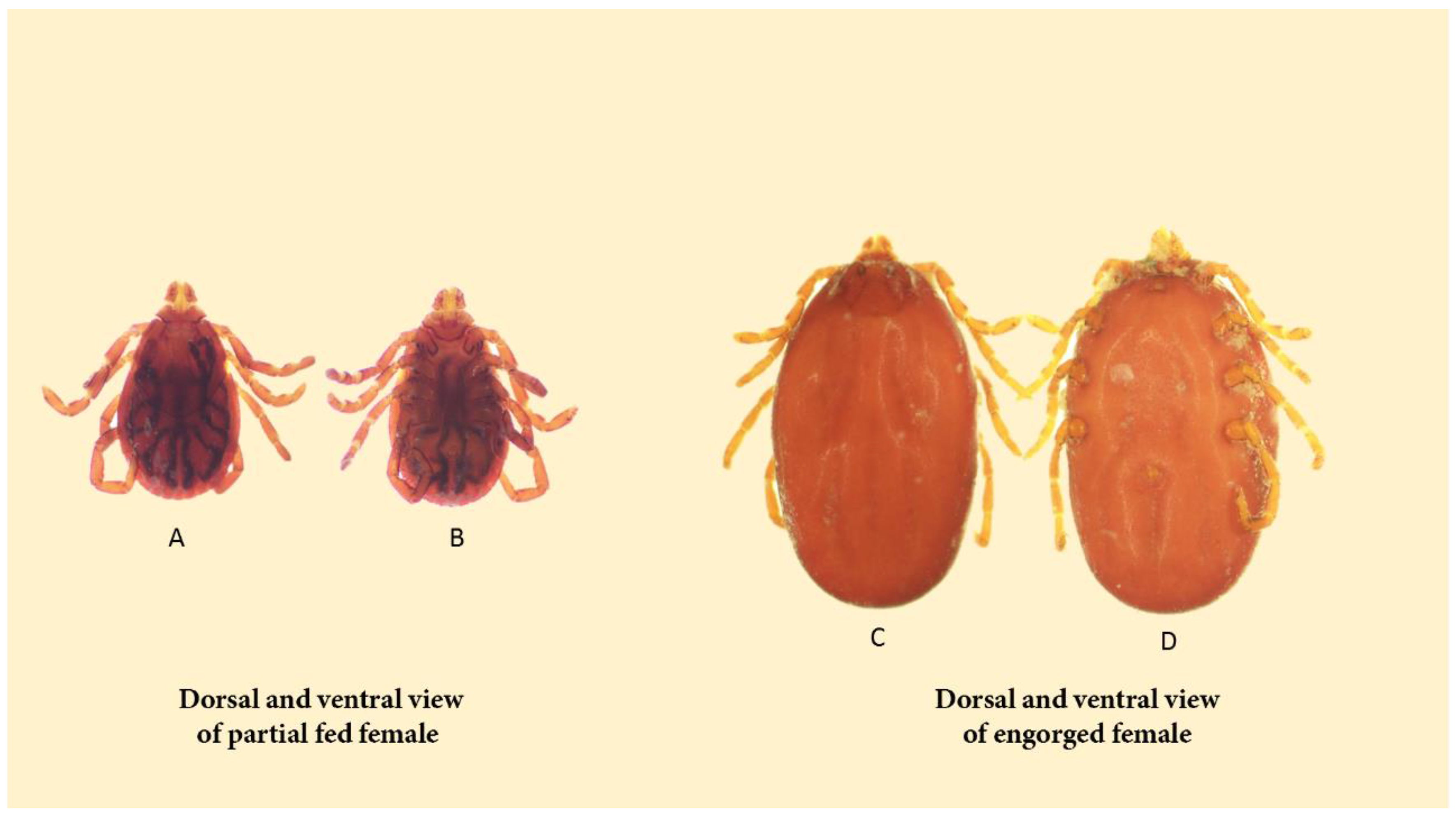
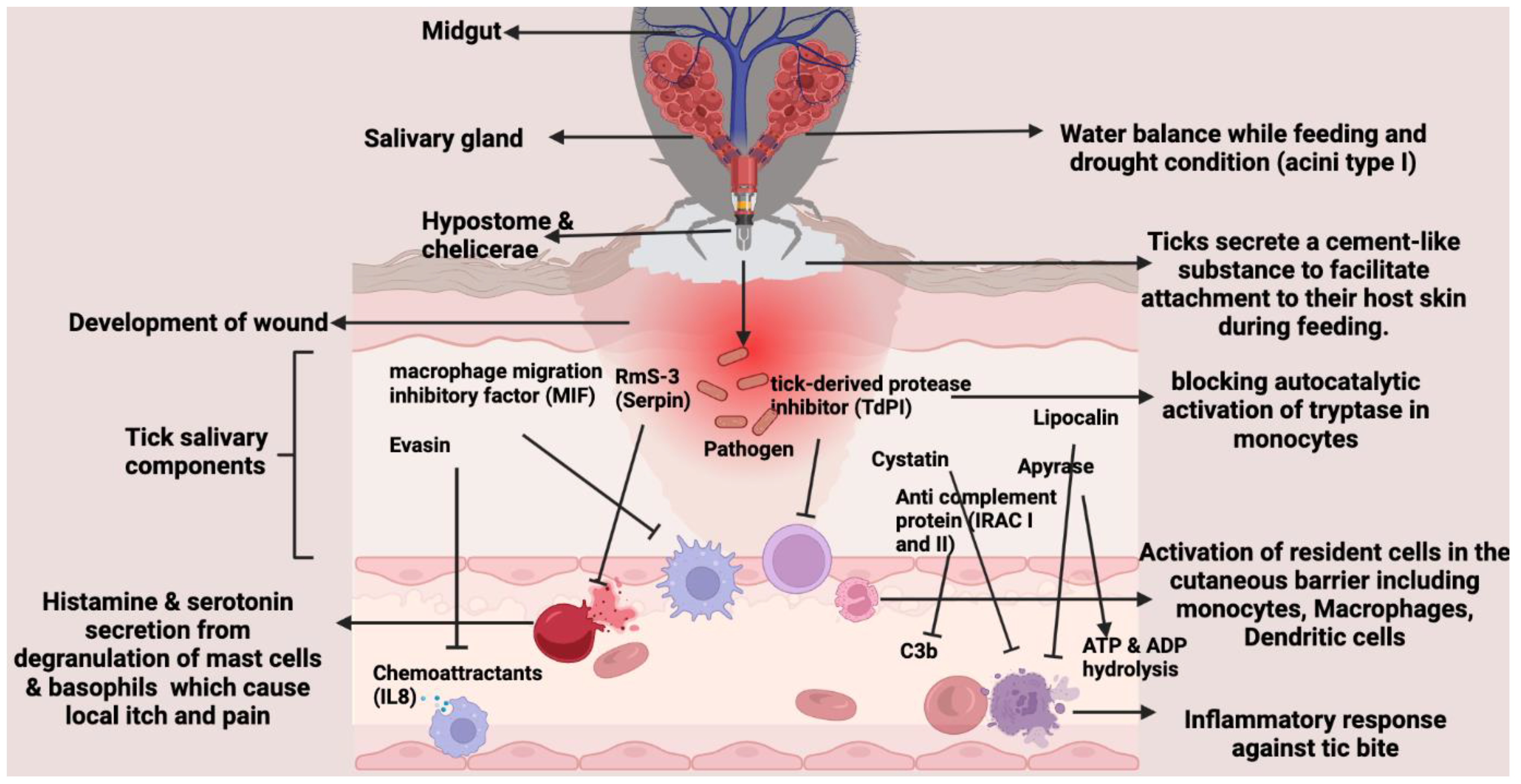
2.1. Blood-Feeding Mechanism of Hard Ticks
2.2. Specialized Function of Salivary Glands in Hard Ticks
3. Distribution and Diversity of Tick Vectors
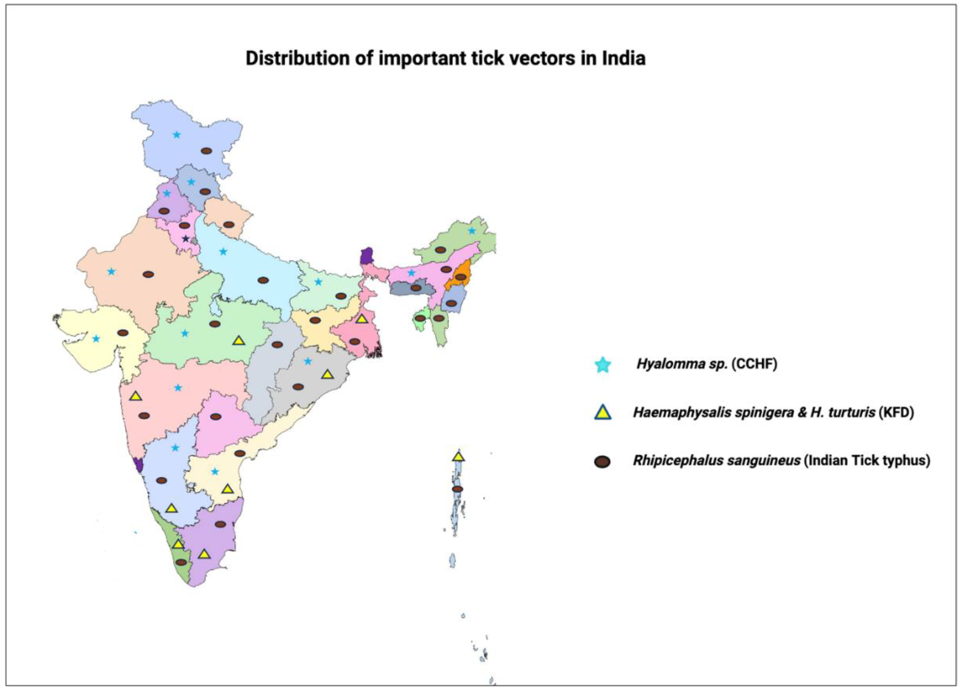
3.1. Distribution of Hard Ticks in India
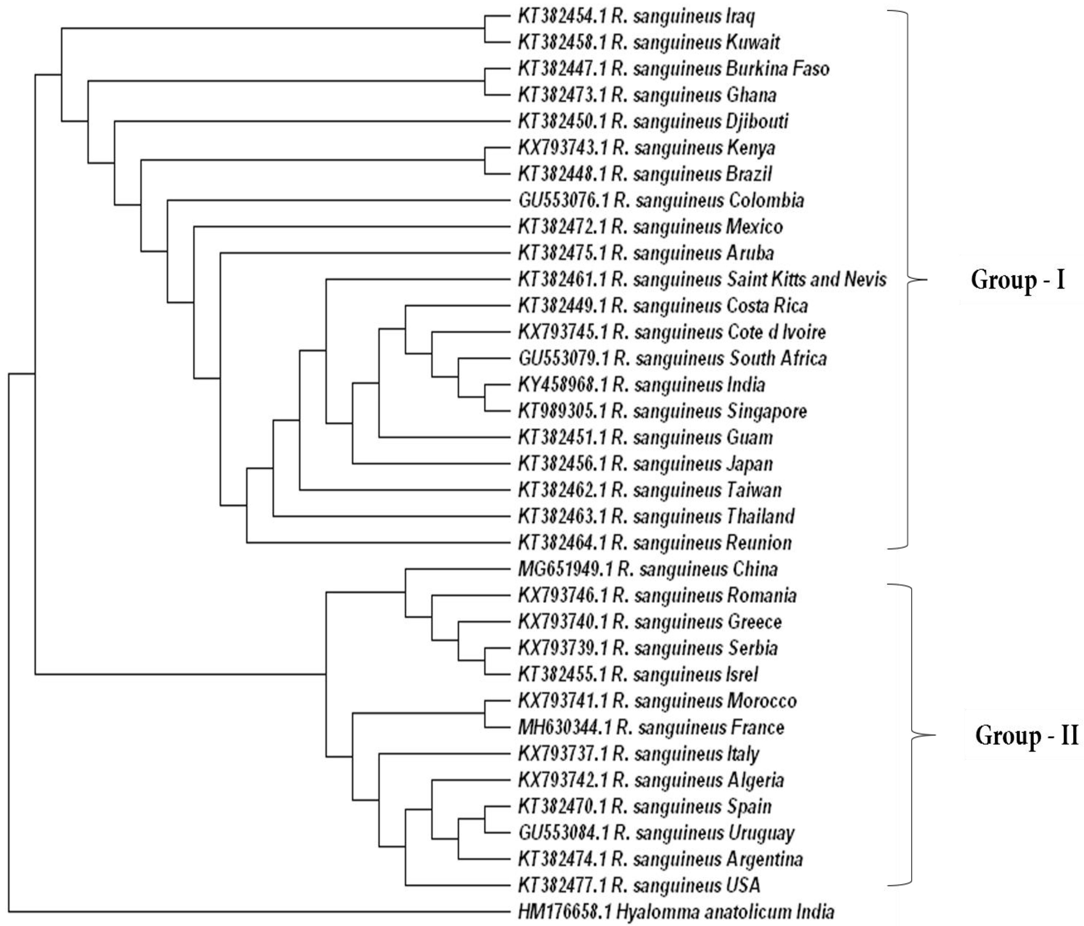
3.2. Genetic Diversity of Ticks
4. Impacts of Climate Change on the Expansion of Hard Ticks
5. Important TBDs in India Transmitted by Hard Ticks
5.1. Kyasanur Forest Disease (KFD)
5.2. Crimean–Congo Hemorrhagic Fever (CCHF)
5.3. Lyme Disease (LD)
5.4. Tick-Associated Rickettsial Pathogens
5.5. Eco-Epidemiology of TBDs
5.6. Molecular Diagnostics and Emerging TBDs
6. Challenges Associated with the TBDs
6.1. Strategies to Control TBDs
6.2. Acaricides and Biological Control
6.3. Acaricide Resistance and Mechanism
6.4. Drugs/Vaccines against TBDs

| Diseases | Tick Species | Distribution | Author |
|---|---|---|---|
| Kyasanur Forest Disease (KFD) | Haemaphysalis spinigera | Karnataka, Tamil Nadu, Kerala, Andhra Pradesh, Orissa, Madhya Pradesh, Meghalaya, Goa, Maharashtra, Gujarat, West Bengal, Andaman and Nicobar Islands | [45,217,224,229,311] |
| Haemaphysalis turturis | Karnataka, Tamil Nadu, Kerala and Uttar Pradesh | [45,217,224,229,311] | |
| Haemaphysalis wellingtoni | Karnataka, Orissa, Andaman and Nicobar Islands and West Bengal | [45,224,311] | |
| Haemaphysalis cuspidata | Sri Lanka and Karnataka | [224,229] | |
| Haemaphysalis aculeata | Karnataka | [229,311] | |
| Haemaphysalis bispinosa | Andhra Pradesh, Arunachal Pradesh, Assam, Bihar, Goa, Gujarat, Himachal Pradesh, Jammu and Kashmir, Karnataka, Madhya Pradesh, Maharashtra, Mizoram, Orissa, Punja, Sikkim, Tamil Nadu, West Bengal | [160,224,311] | |
| Haemaphysalis kyasanurensis | Karnataka | [45,224,311] | |
| Haemaphysalis minuta | Himachal Pradesh, Karnataka, Orissa and Uttar Pradesh | [45,311] | |
| Haemaphysalis kinneri | Karnataka and West Bengal | [45,311] | |
| Haemaphysalis papuanakinneari | Karnataka and other parts of South East Asia | [311] | |
| Ixodes petauristae | Karnataka | [45,225] | |
| Ixodesceylonensis | Karnataka and Tamil Nadu | [45,225] | |
| Rhipicephalus haemaphysaloides | Throughout India | [45,225] | |
| Dermacentor auratus | Arunachal Pradesh, Assam, Bihar, Himachal Pradesh, Jammu and Kashmir, Orissa, Karnataka, Madhya Pradesh, Maharashtra, Punjab, Uttar Pradesh and West Bengal | [45,225] | |
| Hyalomma marginatum isaaci | Arunachal Pradesh, Bihar, Delhi, Gujarat, Himachal Pradesh, Jammu and Kashmir, Karnataka, Maharashtra, Madhya Pradesh, Orissa, Punjab and Uttar Pradesh | [225] | |
| Ornithodorus | Andra Pradesh, Jammu and Kashmir, Himachal Pradesh, Uttar Pradesh, Karnataka, Maharashtra, Madhya Pradesh, Tamil Nadu and Pondicherry | [225,312] | |
| Crimean-Congo haemorrhagic fever (CCHF) | Hyalomma anatolicum | Andhra Pradesh, Assam, Delhi, Gujarat, Haryana, Himachal Pradesh, Jammu and Kashmir, Karnataka, Maharashtra, Madhya Pradesh, Orissa, Punjab and Rajasthan | [17,233,312,313] |
| H. marginatum | Arunachal Pradesh, Bihar, Delhi, Gujarat, Himachal Pradesh, Jammu and Kashmir, Karnataka, Maharashtra, Madhya Pradesh, Orissa, Punjab and Uttar Pradesh | [17,38,160,312,313] | |
| Hyalomma asiaticum | _ | [17,38,160,233] | |
| Hyalomma truncatum | _ | [38,160,233] | |
| Hyalomma rufipes | _ | [38,160,233] | |
| Hyalomma lusitanicum | _ | [233] | |
| Hyalomma excavatum | _ | [233] | |
| Hyalomma dromedarii | Andhra Pradesh, Gujarat, Himachal Pradesh, Jammu and kahmir, Maharashtra, Orissa, Punjab and Uttar Pradesh | [17,38,313] | |
| Rhipicephalus sanguineus | Throughout India | [17,160,233,313] | |
| Ixodes ricinus | Northeastern States of India | [31,163] | |
| Indian Tick Typhus (ITT) | R. sanguineus | Throughout India | [38,61,242,314] |
| Ixodes ricinus | Northeastern States of India | [38,314] | |
| Haemaphysalis indica | Bihar, Gujarat, Himachal Pradesh, Jammu and Kashmir, Karnataka, Maharashtra, Orissa, Rajasthan, Uttar Pradesh and West Bengal | [244] | |
| H. kinneri | Karnataka and West Bengal | [244] | |
| H. spinigera | Karnataka, Tamil Nadu, Kerala, Andhra Pradesh, Orissa, Madhya Pradesh, Meghalaya, Goa, Maharashtra, Gujarat, West Bengal, Andaman and Nicobar Islands | [244] | |
| H. turturis | Karnataka, Tamil Nadu, Kerala and Uttar Pradesh | [244] | |
| Haemaphysalis leachi | Eastern West Pakistan, India, and Ceylon, and the terai of southern Nepal | [244] |
7. Conclusions
Author Contributions
Funding
Acknowledgments
Conflicts of Interest
Abbreviations
| TBD | Tick-borne diseases |
| KFD | Kyasanur Forest Disease |
| CCHF | Crimean–Congo haemorrhagic fever |
| LD | Lyme disease |
| TBP | Tick-Borne Pathogen |
| POWV II | Powassan (POW) virus lineage II |
| SAT | Saliva-Assisted Transmission |
| MIF | Macrophage migration Inhibitory Factor |
| JAK/STAT | Janus kinase (JAK)–signal transducer and activator of transcription (STAT) pathways |
| TBE | Tick-Borne Encephalitis |
| ITT | Indian Tick Typhus |
| PCR | Polymerase Chain Reaction |
References
- Jankielsohn, A. The Importance of Insects in Agricultural Ecosystems. Adv. Entomol. 2018, 6, 62–73. [Google Scholar] [CrossRef]
- Redak, R. Introduction to and Importance of Insects. In Forest Entomology and Pathology; Allison, J.D., Paine, T.D., Slippers, B., Wingfield, M.J., Eds.; Springer: Cham, Switzerland, 2023; Volume 1, pp. 1–17. [Google Scholar] [CrossRef]
- Baxter, R.H.G.; Contet, A.; Krueger, K. Arthropod Innate Immune Systems and Vector-Borne Diseases. Biochemistry 2017, 56, 907–918. [Google Scholar] [CrossRef]
- Khan, A.; Yasin, M.; Aqueel, M.A.; Farooqi, M.A.; Akram, M.I.; Yousuf, H.M.B.; Noor, M.; Maqsood, A. Vector-Borne Disease and Climate Change; IntechOpen eBooks; IntechOpen: London, UK, 2023. [Google Scholar]
- Cupp, E.W. Biology of Ticks. Vet. Clin. N. Am. Small Anim. Pract. 1991, 21, 1–26. [Google Scholar] [CrossRef]
- Brites-Neto, J.; Duarte, K.M.R.; Martins, T.F. Tick-borne infections in human and animal population worldwide. Vet. World 2015, 8, 301–315. [Google Scholar] [CrossRef] [PubMed]
- Yu, Z.; Wang, H.; Wang, T.; Sun, W.; Yang, X.; Liu, J. Tick-borne pathogens and the vector potential of ticks in China. Parasites Vectors 2015, 8, 24. [Google Scholar] [CrossRef] [PubMed]
- Tahir, D.; Meyer, L.; Fourie, J.; Jongejan, F.; Mather, T.; Choumet, V.; Blagburn, B.; Straubinger, R.K.; Varloud, M. Interrupted Blood Feeding in Ticks: Causes and Consequences. Microorganisms 2020, 8, 910. [Google Scholar] [CrossRef]
- Varma, M.R.G. Ticks and mites (Acari). In Medical Insects and Arachnids; Springer: Dordrecht, The Netherlands, 1993; pp. 597–658. [Google Scholar] [CrossRef]
- Cordeiro, F.A.; Amorim, F.G.; Anjolette, F.A.P.; Arantes, E.C. Arachnids of medical importance in Brazil: Main active compounds present in scorpion and spider venoms and tick saliva. J. Venom. Anim. Toxins Incl. Trop. Dis. 2015, 21, 24. [Google Scholar] [CrossRef]
- Francis, S.; Frank, C.; Buchanan, L.; Green, S.; Stennett-Brown, R.; Gordon-Strachan, G.; Rubio-Palis, Y.; Grant, C.; Alexander-Lindo, R.L.; Nwokocha, C.; et al. Challenges in the control of neglected insect vector diseases of human importance in the Anglo-Caribbean. One Health 2021, 13, 100316. [Google Scholar] [CrossRef]
- Kader, S.; Arriaza, R.H.; Khatri, K.; O’malley, A.; Grbic, V.; Grbic, M.; Chruszcz, M. Current status of structural studies of proteins originating from Arachnida. Syst. Appl. Acarol. 2023, 28, 298–308. [Google Scholar] [CrossRef]
- Snodgrass, R.E. Principles of Insect Morphology; Comstock Publishing Associates: Ithaca, NY, USA, 1993. [Google Scholar]
- Randolph, S. Ticks are not Insects: Consequences of Contrasting Vector Biology for Transmission Potential. Parasitol. Today 1998, 14, 186–192. [Google Scholar] [CrossRef] [PubMed]
- Charrier, N.P.; Hermouet, A.; Hervet, C.; Agoulon, A.; Barker, S.C.; Heylen, D.; Toty, C.; McCoy, K.D.; Plantard, O.; Rispe, C. A transcriptome-based phylogenetic study of hard ticks (Ixodidae). Sci. Rep. 2019, 9, 12923. [Google Scholar] [CrossRef] [PubMed]
- Gilbert, L. The Impacts of Climate Change on Ticks and Tick-Borne Disease Risk. Annu. Rev. Entomol. 2021, 66, 373–388. [Google Scholar] [CrossRef]
- Telmadarraiy, Z.; Kooshki, H.; Edalat, H.; Vatandoost, H.; Bakhshi, H.; Faghihi, F.; Hosseini-Chegeni, A.; Oshaghi, M.A. Study on Hard and Soft Ticks of Domestic and Wild Animals in Western Iran. J. Arthropod-Borne Dis. 2023, 16, 225–232. [Google Scholar] [CrossRef]
- Paddock, C.D.; Lane, R.S.; Staples, J.E.; Labruna, M.B. Changing Paradigms for Tick-Borne Diseases in the Americas; National Academies Press (US): Washington, DC, USA, 2016. Available online: www.ncbi.nlm.nih.gov (accessed on 12 December 2023).
- Diarra, A.Z.; Kelly, P.; Davoust, B.; Parola, P. Tick-Borne Diseases of Humans and Animals in West Africa. Pathogens 2023, 12, 1276. [Google Scholar] [CrossRef]
- de la Fuente, J.; Estrada-Pena, A.; Venzal, J.M.; Kocan, K.M.; Sonenshine, D.E. Overview: Ticks as vectors of pathogens that cause disease in humans and animals. Front. Biosci. 2008, 13, 6938–6946. [Google Scholar] [CrossRef]
- Franta, Z.; Frantová, H.; Konvičková, J.; Horn, M.; Sojka, D.; Mareš, M.; Kopáček, P. Dynamics of digestive proteolytic system during blood feeding of the hard tick Ixodes ricinus. Parasites Vectors 2010, 3, 119. [Google Scholar] [CrossRef]
- Starck, J.M.; Mehnert, L.; Biging, A.; Bjarsch, J.; Franz-Guess, S.; Kleeberger, D.; Hörnig, M. Morphological responses to feeding in ticks (Ixodes ricinus). Zool. Lett. 2018, 4, 20. [Google Scholar] [CrossRef]
- Francischetti, I.M.B.; Sa-Nunes, A.; Mans, B.J.; Santos, I.M.; Ribeiro, J.M.C. The role of saliva in tick feeding. Front. Biosci. 2009, 14, 2051–2088. [Google Scholar] [CrossRef] [PubMed]
- Šimo, L.; Kazimirova, M.; Richardson, J.; Bonnet, S.I. The Essential Role of Tick Salivary Glands and Saliva in Tick Feeding and Pathogen Transmission. Front. Cell. Infect. Microbiol. 2017, 7, 281. [Google Scholar] [CrossRef]
- Bouchard, C.; Dibernardo, A.; Koffi, J.; Wood, H.; Leighton, P.A.; Lindsay, L.R. Increased risk of tick-borne diseases with climate and environmental changes. Can. Commun. Dis. Rep. 2019, 45, 83–89. [Google Scholar] [CrossRef]
- Hromníková, D.; Furka, D.; Furka, S.; Santana, J.A.D.; Ravingerová, T.; Klöcklerová, V.; Žitňan, D. Prevention of tick-borne diseases: Challenge to recent medicine. Biologia 2022, 77, 1533–1554. [Google Scholar] [CrossRef] [PubMed]
- Nuttall, P.A. Climate change impacts on ticks and tick-borne infections. Biologia 2021, 77, 1503–1512. [Google Scholar] [CrossRef]
- Stafford, I.K.C.; Williams, S.C.; Molaei, G. Integrated Pest Management in Controlling Ticks and Tick-Associated Diseases. J. Integr. Pest Manag. 2017, 8, 28. [Google Scholar] [CrossRef]
- Eisen, L.; Stafford, K.C. Barriers to Effective Tick Management and Tick-Bite Prevention in the United States (Acari: Ixodidae). J. Med. Entomol. 2021, 58, 1588–1600. [Google Scholar] [CrossRef] [PubMed]
- Rochlin, I.; Toledo, A. Emerging tick-borne pathogens of public health importance: A mini-review. J. Med. Microbiol. 2020, 69, 781–791. [Google Scholar] [CrossRef] [PubMed]
- Zhao, G.-P.; Wang, Y.-X.; Fan, Z.-W.; Ji, Y.; Liu, M.-J.; Zhang, W.-H.; Li, X.-L.; Zhou, S.-X.; Li, H.; Liang, S.; et al. Mapping ticks and tick-borne pathogens in China. Nat. Commun. 2021, 12, 1075. [Google Scholar] [CrossRef] [PubMed]
- Bai, Y.; Li, Y.; Liu, W.; Li, J.; Tian, F.; Liu, L.; Han, X.; Tong, Y. Analysis of the diversity of tick-borne viruses at the border areas in Liaoning Province, China. Front. Microbiol. 2023, 14, 1179156. [Google Scholar] [CrossRef] [PubMed]
- de la Fuente, J.; Estrada-Peña, A.; Rafael, M.; Almazán, C.; Bermúdez, S.; Abdelbaset, A.E.; Kasaija, P.D.; Kabi, F.; Akande, F.A.; Ajagbe, D.O.; et al. Perception of Ticks and Tick-Borne Diseases Worldwide. Pathogens 2023, 12, 1258. [Google Scholar] [CrossRef] [PubMed]
- Chao, L.-L.; Shih, C.-M. First report of human biting activity of Ixodes acutitarsus (Acari: Ixodidae) collected in Taiwan. Exp. Appl. Acarol. 2012, 56, 159–164. [Google Scholar] [CrossRef]
- Estrada-Peña, A.; Jongejan, F. Ticks feeding on humans: A review of records on human-biting Ixodoidea with special reference to pathogen transmission. Exp. Appl. Acarol. 1999, 23, 685–715. [Google Scholar] [CrossRef]
- Ajithkumar, K.G.; Ravindran, R.; Ghosh, S. Dermacentor auratus Supino, 1897 (Acarina, Ixodidae) reported from Wayanad, Kerala. Indian J. Med. Res. 2012, 135, 435–436. [Google Scholar] [PubMed]
- Soundararajan, C.; Nagarajan, K.; Prakash, M.A. Tick infestation in human beings in the Nilgiris and Kancheepuram district of Tamil Nadu, India. J. Parasit. Dis. 2017, 42, 50–54. [Google Scholar] [CrossRef] [PubMed]
- Kumar, B.; Manjunathachar, H.V.; Ghosh, S. A review on Hyalomma species infestations on human and animals and progress on management strategies. Heliyon 2020, 6, e05675. [Google Scholar] [CrossRef] [PubMed]
- Stephen, S.; Sangeetha, B.; Antony, P.X. Seroprevalence of coxiellosis (Q fever) in sheep & goat in Puducherry & neighbouring Tamil Nadu. Indian J. Med. Res. 2014, 140, 785–787. [Google Scholar] [PubMed]
- Jairath, V.; Sehrawat, M.; Jindal, N.; Jain, V.K.; Aggarwal, P. Lyme disease in Haryana, India. Indian J. Dermatol. Venereol. Leprol. 2014, 80, 320–323. [Google Scholar] [CrossRef]
- Negi, T.; Kandari, L.S.; Arunachalam, K. Update on prevalence and distribution pattern of tick-borne diseases among humans in India: A review. Parasitol. Res. 2021, 120, 1523–1539. [Google Scholar] [CrossRef]
- Mourya, D.T.; Yadav, P.D.; Patil, D.Y.; Sahay, R.R.; Rahi, M. Experiences of Indian Council of Medical Research with tick-borne zoonotic infections: Kyasanur Forest disease & Crimean-Congo haemorrhagic fever in India with One Health focus. Indian J. Med. Res. 2021, 153, 339–347. [Google Scholar] [CrossRef] [PubMed]
- Gurav, Y.K.; Yadav, P.D.; Gokhale, M.D.; Chiplunkar, T.R.; Vishwanathan, R.; Patil, D.Y.; Jain, R.; Shete, A.M.; Patil, S.L.; Sarang, G.; et al. Kyasanur Forest Disease Prevalence in Western Ghats Proven and Confirmed by Recent Outbreak in Maharashtra, India, 2016. Vector-Borne Zoonotic Dis. 2018, 18, 164–172. [Google Scholar] [CrossRef]
- Munivenkatappa, A.; Sahay, R.R.; Yadav, P.D.; Viswanathan, R.; Mourya, D.T. Clinical & epidemiological significance of Kyasanur forest disease. Indian J. Med. Res. 2018, 148, 145–150. [Google Scholar] [CrossRef]
- Chakraborty, S.; Andrade, F.C.D.; Ghosh, S.; Uelmen, J.; Ruiz, M.O. Historical Expansion of Kyasanur Forest Disease in India from 1957 to 2017: A Retrospective Analysis. GeoHealth 2019, 3, 44–55. [Google Scholar] [CrossRef]
- Pattnaik, S.; Agrawal, R.; Murmu, J.; Kanungo, S.; Pati, S. Does the rise in cases of Kyasanur forest disease call for the implementation of One Health in India? IJID Reg. 2023, 7, 18–21. [Google Scholar] [CrossRef] [PubMed]
- Yadav, P.D.; Raut, C.G.; Patil, D.Y.; Majumdar, T.D.; Mourya, D.T. Crimean-Congo Hemorrhagic Fever: Current Scenario in India. Proc. Natl. Acad. Sci. India Sect. B Biol. Sci. 2014, 84, 9–18. [Google Scholar] [CrossRef] [PubMed]
- Mourya, D.T.; Yadav, P.D.; Shete, A.M.; Sathe, P.S.; Sarkale, P.C.; Pattnaik, B.; Sharma, G.; Upadhyay, K.J.; Gosavi, S.; Patil, D.Y.; et al. Cross-sectional Serosurvey of Crimean-Congo Hemorrhagic Fever Virus IgG in Livestock, India, 2013–2014. Emerg. Infect. Dis. 2015, 21, 1837–1839. [Google Scholar] [CrossRef] [PubMed]
- Mourya, D.T.; Makwana, D.; Yadav, P.D.; Kelaiya, A. First confirmed case of Crimean-Congo haemorrhagic fever from Sirohi district in Rajasthan State, India. Indian J. Med. Res. 2015, 142, 489–491. [Google Scholar] [CrossRef] [PubMed]
- Yadav, P.D.; Patil, D.Y.; Shete, A.M.; Kokate, P.; Goyal, P.; Jadhav, S.; Sinha, S.; Zawar, D.; Sharma, S.K.; Kapil, A.; et al. Nosocomial infection of CCHF among health care workers in Rajasthan, India. BMC Infect. Dis. 2016, 16, 624. [Google Scholar] [CrossRef]
- Larcombe, S.D.; Kolte, S.W.; Ponnudurai, G.; Kurkure, N.; Magar, S.; Velusamy, R.; Rani, N.; Rubinibala, B.; Rekha, B.; Alagesan, A.; et al. The impact of tick-borne pathogen infection in Indian bovines is determined by host type but not the genotype of Theileria annulata. Infect. Genet. Evol. 2019, 75, 103972. [Google Scholar] [CrossRef] [PubMed]
- Rajput, Z.I.; Hu, S.-H.; Chen, W.-J.; Arijo, A.G.; Xiao, C.-W. Importance of ticks and their chemical and immunological control in livestock. J. Zhejiang Univ. B 2006, 7, 912–921. [Google Scholar] [CrossRef]
- Bhowmick, B.; Han, Q. Understanding Tick Biology and Its Implications in Anti-tick and Transmission Blocking Vaccines against Tick-Borne Pathogens. Front. Vet. Sci. 2020, 7, 319. [Google Scholar] [CrossRef]
- Hofmeester, T.R.; Rowcliffe, J.M.; Jansen, P.A. Quantifying the Availability of Vertebrate Hosts to Ticks: A Camera-Trapping Approach. Front. Vet. Sci. 2017, 4, 115. [Google Scholar] [CrossRef]
- De La Fuente, J.; Antunes, S.; Bonnet, S.; Cabezas-Cruz, A.; Domingos, A.G.; Estrada-Peña, A.; Johnson, N.; Kocan, K.M.; Mansfield, K.L.; Nijhof, A.M.; et al. Tick-Pathogen Interactions and Vector Competence: Identification of Molecular Drivers for Tick-Borne Diseases. Front. Cell. Infect. Microbiol. 2017, 7, 114. [Google Scholar] [CrossRef]
- Gomez-Chamorro, A.; Hodžić, A.; King, K.C.; Cabezas-Cruz, A. Ecological and evolutionary perspectives on tick-borne pathogen co-infections. Curr. Res. Parasitol. Vector-Borne Dis. 2021, 1, 100049. [Google Scholar] [CrossRef]
- Hoffman, T.; Olsen, B.; Lundkvist, Å. The Biological and Ecological Features of Northbound Migratory Birds, Ticks, and Tick-Borne Microorganisms in the African–Western Palearctic. Microorganisms 2023, 11, 158. [Google Scholar] [CrossRef] [PubMed]
- Voyiatzaki, C.; Papailia, S.I.; Venetikou, M.S.; Pouris, J.; Tsoumani, M.E.; Papageorgiou, E.G. Climate Changes Exacerbate the Spread of Ixodes ricinus and the Occurrence of Lyme Borreliosis and Tick-Borne Encephalitis in Europe—How Climate Models Are Used as a Risk Assessment Approach for Tick-Borne Diseases. Int. J. Environ. Res. Public Health 2022, 19, 6516. [Google Scholar] [CrossRef] [PubMed]
- Nava, S.; Guglielmone, A.A.; Mangold, A.J. An overview of systematics and evolution of ticks. Front. Biosci. 2009, 14, 2857–2877. [Google Scholar] [CrossRef] [PubMed]
- Leung, T.L.F. Fossils of parasites: What can the fossil record tell us about the evolution of parasitism? Biol. Rev. 2017, 92, 410–430. [Google Scholar] [CrossRef] [PubMed]
- Parola, P.; Fenollar, F.; Badiaga, S.; Brouqui, P.; Raoult, D. First Documentation of Rickettsia conorii Infection (Strain Indian Tick Typhus) in a Traveler. Emerg. Infect. Dis. 2001, 7, 909–910. [Google Scholar] [CrossRef] [PubMed]
- Latif, A.A.; Putterill, J.F.; de Klerk, D.G.; Pienaar, R.; Mans, B.J. Nuttalliella namaqua (Ixodoidea: Nuttalliellidae): First Description of the Male, Immature Stages and Re-Description of the Female. PLoS ONE 2012, 7, e41651. [Google Scholar] [CrossRef] [PubMed]
- Mans, B.J.; de Klerk, D.; Pienaar, R.; de Castro, M.H.; Latif, A.A. The Mitochondrial Genomes of Nuttalliella namaqua (Ixodoidea: Nuttalliellidae) and Argas africolumbae (Ixodoidae: Argasidae): Estimation of Divergence Dates for the Major Tick Lineages and Reconstruction of Ancestral Blood-Feeding Characters. PLoS ONE 2012, 7, e49461. [Google Scholar] [CrossRef] [PubMed]
- Chhillar, S.; Chhilar, J.S.; Kaur, H. Investigations on Some Hard Ticks (Acari: Ixodidae) Infesting Domestic Buffalo and Cattle from Haryana, India. J. Entomol. Zool. Stud. 2014, 2, 99–104. [Google Scholar]
- Ranganathan, K.; Renu, G.; Ayyanar, E.; Veeramanoharan, R.; Paulraj, P.S. Species composition of hard ticks (Acari: Ixodidae) on domestic animals and their public health importance in Tamil Nadu, South India. Acarol. Stud. 2021, 3, 16–21. [Google Scholar] [CrossRef]
- Wikimedia Commons Ornithodoros Adult Soft-Tick.jpg. 2020. Available online: https://commons.wikimedia.org/w/index.php?title=File:Ornithodoros_adult_soft-tick.jpg&oldid=493571749 (accessed on 17 December 2023).
- Oliver, J.H. Biology and Systematics of Ticks (Acari:Ixodida). Annu. Rev. Ecol. Syst. 1989, 20, 397–430. [Google Scholar] [CrossRef]
- Couvreur, B.; Beaufays, J.; Charon, C.; Lahaye, K.; Gensale, F.; Denis, V.; Charloteaux, B.; Decrem, Y.; Prévôt, P.-P.; Brossard, M.; et al. Variability and Action Mechanism of a Family of Anticomplement Proteins in Ixodes ricinus. PLoS ONE 2008, 3, e1400. [Google Scholar] [CrossRef] [PubMed]
- Vargas-Sandoval, M.; Priego-Santander, A.G.; Larrazábal, A.; Sosa-Gutiérrez, C.G.; Lara-Chávez, B.; Avila-Val, T. Potential Species Distribution and Richness of Ixodidae Ticks Associated with Wild Vertebrates from Michoacán, Mexico. J. Geogr. Inf. Syst. 2014, 6, 467–477. [Google Scholar] [CrossRef]
- Nicholson, W.L.; Sonenshine, D.E.; Noden, B.H.; Brown, R.N. Ticks (Ixodida). Med. Vet. Entomol. 2019, 2019, 603–672. [Google Scholar] [CrossRef]
- Pospelova-Shtrom, M.V. On the system of classification of ticks of the family Argasidae. Acarologia 1969, 11, 1–22. [Google Scholar] [PubMed]
- Sándor, A.D.; Mihalca, A.D.; Domşa, C.; Péter, Á.; Hornok, S. Argasid Ticks of Palearctic Bats: Distribution, Host Selection, and Zoonotic Importance. Front. Vet. Sci. 2021, 8, 684737. [Google Scholar] [CrossRef] [PubMed]
- Chen, Z.; Liu, J. A review of argasid ticks and associated pathogens of China. Front. Vet. Sci. 2022, 9, 865664. [Google Scholar] [CrossRef] [PubMed]
- Estrada-Pena, A.; Mangold, A.; Nava, S.; Venzal, J.; Labruna, M.; Guglielmone, A. A review of the systematics of the tick family Argasidae (Ixodida). Acarologia 2010, 50, 317–333. [Google Scholar] [CrossRef]
- Lafri, I.; Benredjem, W.; Neffah-Baaziz, F.; Lalout, R.; Abdelouahed, K.; Gassen, B.; Bakhouch, S.; Chergui, M.; Karakellah, M.; Adjmi-Hamoudi, H.; et al. Inventory and update on argasid ticks and associated pathogens in Algeria. New Microbes New Infect. 2018, 23, 110–114. [Google Scholar] [CrossRef]
- Trevisan, G.; Cinco, M.; Trevisini, S.; di Meo, N.; Ruscio, M.; Forgione, P.; Bonin, S. Borreliae Part 2: Borrelia Relapsing Fever Group and Unclassified Borrelia. Biology 2021, 10, 1117. [Google Scholar] [CrossRef]
- Keirans, J.E.; Clifford, C.M.; Hoogstraal, H.; Easton, E.R. Discovery of Nuttalliella namaqua Bedford (Acarina: Ixodoidea: Nuttalliellidae) in Tanzania and Redescription of the Female Based on Scanning Electron Microcopy. Ann. Entomol. Soc. Am. 1976, 69, 926–932. [Google Scholar] [CrossRef]
- Roshdy, M.A.; Hoogstraal, H.; Banaja, A.A.; Shoura, S.M. Nuttalliella namaqua (Ixodoidea: Nuttalliellidae): Spiracle structure and surface morphology. Z. Parasitenkd. 1983, 69, 817–821. [Google Scholar] [CrossRef]
- Jin, K.; Koh, Y.-J.; Ahn, S.K.; Cho, J.; Lim, J.; Song, J.; Lee, J.; Gong, Y.W.; Kwon, M.J.; Kwon, H.W.; et al. Hard Ticks as Vectors Tested Negative for Severe Fever with Thrombocytopenia Syndrome in Ganghwa-do, Korea during 2019–2020. Korean J. Parasitol. 2021, 59, 281–289. [Google Scholar] [CrossRef]
- Arthur, D.R. The morphology of the British Prostriata, with particular reference to Ixodes hexagonus Leach. III. Parasitology 1956, 46, 261–307. [Google Scholar] [CrossRef]
- Feldman-Muhsam, B.; Borut, S. Copulation in Ixodid Ticks. J. Parasitol. 1971, 57, 630. [Google Scholar] [CrossRef]
- Anderson, J.F. The natural history of ticks. Med. Clin. N. Am. 2002, 86, 205–218. [Google Scholar] [CrossRef]
- Chitimia-Dobler, L.; Dunlop, J.A.; Pfeffer, T.; Würzinger, F.; Handschuh, S.; Mans, B.J. Hard ticks in Burmese amber with Australasian affinities. Parasitology 2023, 150, 157–171. [Google Scholar] [CrossRef] [PubMed]
- Sonenshine, D.E.; Šimo, L. Biology and molecular biology of Ixodes scapularis. In Lyme Disease and Relapsing Fever Spirochetes: Genomics, Molecular Biology, Host Interactions and Disease Pathogenesis; Radolf, J.D., Samuels, D.S., Eds.; Caister Academic Press: Poole, UK, 2021; pp. 339–366. [Google Scholar] [CrossRef]
- Yamaguti, N.; Tipton, V.J.; Keegan, H.L.; Toshioka, S. Ticks of Japan, Korea, and the Ryukyu Islands. Brigh. Young Univ. Sci. Bull. Biol. Ser. 1971, 15, 1. [Google Scholar]
- Bowman, A.S.; Sauer, J.R. Tick salivary glands: Function, physiology and future. Parasitology 2005, 129, S67–S81. [Google Scholar] [CrossRef]
- Szlendak, E.; Oliver, J.H. Anatomy of synganglia, including their neurosecretory regions, in unfed, virgin female Ixodes scapularis say (Acari: Ixodidae). J. Morphol. 1992, 213, 349–364. [Google Scholar] [CrossRef]
- Borges, L.M.F.; Li, A.Y.; Olafson, P.U.; Renthal, R.; Bauchan, G.R.; Lohmeyer, K.H.; de León, A.A.P. Neuronal projections from the Haller’s organ and palp sensilla to the synganglion of Amblyomma americanum. Rev. Bras. Parasitol. Veterinária 2016, 25, 217–224. [Google Scholar] [CrossRef] [PubMed]
- Fielden, L.J.; Duncan, F.D.; Rechav, Y.; Crewe, R.M. Respiratory Gas Exchange in the Tick Amblyomma hebraeum (Acari: Ixodidae). J. Med. Entomol. 1994, 31, 30–35. [Google Scholar] [CrossRef] [PubMed]
- Sonenshine, D.E.; Roe, R.M. Biology of Ticks; Oxford University Press: New York, NY, USA, 2014. [Google Scholar]
- Feitosa, A.P.S.; Alves, L.C.; Chaves, M.M.; Veras, D.L.; Silva, E.M.; Aliança, A.S.S.; França, I.R.S.; Gonçalves, G.G.A.; Lima-Filho, J.L.; Brayner, F.A. Hemocytes of Rhipicephalus sanguineus (Acari: Ixodidae): Characterization, Population Abundance, and Ultrastructural Changes Following Challenge with Leishmania infantum. J. Med. Entomol. 2015, 52, 1193–1202. [Google Scholar] [CrossRef]
- Patton, T.G.; Dietrich, G.; Brandt, K.; Dolan, M.C.; Piesman, J.; Gilmore, R.D., Jr. Saliva, Salivary Gland, and Hemolymph Collection from Ixodes scapularis Ticks. J. Vis. Exp. JoVE 2012, 60, e3894. [Google Scholar] [CrossRef] [PubMed]
- Kubiak, K.; Dziekońska-Rynko, J. Seasonal activity of the common European tick, Ixodes ricinus (Linnaeus, 1758), in the forested areas of the city of Olsztyn and its surroundings. Wiad Parazytol. 2006, 52, 59–64. [Google Scholar]
- McCoy, K.D.; Léger, E.; Dietrich, M. Host specialization in ticks and transmission of tick-borne diseases: A review. Front. Cell. Infect. Microbiol. 2013, 3, 57. [Google Scholar] [CrossRef] [PubMed]
- Maxime Madder, M.; Horak, I.; Stoltsz, H. Ticks: Tick Identification; AfriVIP: Pretoria, South Africa, 2018. [Google Scholar]
- Gray, J.S.; Dautel, H.; Estrada-Peña, A.; Kahl, O.; Lindgren, E. Effects of climate change on ticks and tick-borne diseases in Europe. Interdiscip. Perspect. Infect. Dis. 2009, 2009, 593232. [Google Scholar] [CrossRef] [PubMed]
- Sauer, J.R.; McSwain, J.L.; Bowman, A.S.; Essenberg, R.C. Tick Salivary Gland Physiology. Annu. Rev. Entomol. 1995, 40, 245–267. [Google Scholar] [CrossRef]
- Estrada-Peña, A.; Gray, J.S.; Kahl, O.; Lane, R.S.; Nijhof, A.M. Research on the ecology of ticks and tick-borne pathogens—Methodological principles and caveats. Front. Cell. Infect. Microbiol. 2013, 3, 29. [Google Scholar] [CrossRef]
- Maldonado-Ruiz, L.P.; Park, Y.; Zurek, L. Liquid water intake of the lone star tick, Amblyomma americanum: Implications for tick survival and management. Sci. Rep. 2020, 10, 6000. [Google Scholar] [CrossRef]
- Yoder, J.A.; Benoit, J.B.; Rellinger, E.J.; Tank, J.L. Developmental profiles in tick water balance with a focus on the new Rocky Mountain spotted fever vector, Rhipicephalus sanguineus. Med. Vet. Entomol. 2006, 20, 365–372. [Google Scholar] [CrossRef] [PubMed]
- Knülle, W.; Rudolph, D. Humidity Relationships and Water Balance of Ticks. In Physiology of Ticks; Pergamon: Oxford, UK, 1982; pp. 43–70. [Google Scholar] [CrossRef]
- Rahlenbeck, S.; Fingerle, V.; Doggett, S. Prevention of tick-borne diseases: An overview. Br. J. Gen. Pract. 2016, 66, 492–494. [Google Scholar] [CrossRef] [PubMed]
- Nejash, A. Review of Important Cattle Tick and Its Control in Ethiopia. Open Access Libr. J. 2016, 3, 69073. [Google Scholar] [CrossRef]
- Couper, L.I.; Yang, Y.; Yang, X.F.; Swei, A. Comparative vector competence of North American Lyme disease vectors. Parasites Vectors 2020, 13, 29. [Google Scholar] [CrossRef] [PubMed]
- Sharma, R.; Cozens, D.W.; Armstrong, P.M.; Brackney, D.E. Vector competence of human-biting ticks Ixodes scapularis, Amblyomma americanum and Dermacentor variabilis for Powassan virus. Parasites Vectors 2021, 14, 466. [Google Scholar] [CrossRef] [PubMed]
- Ochanda, H.; Young, A.S.; Medley, G.F.; Perry, B.D. Vector competence of 7 Rhipicephalid tick stocks in transmitting 2 Theileria parva parasite stocks from Kenya and Zimbabwe. Parasitology 1998, 116, 539–545. [Google Scholar] [CrossRef] [PubMed]
- Migné, C.V.; de Seixas, H.B.; Heckmann, A.; Galon, C.; Jaafar, F.M.; Monsion, B.; Attoui, H.; Moutailler, S. Evaluation of Vector Competence of Ixodes Ticks for Kemerovo Virus. Viruses 2022, 14, 1102. [Google Scholar] [CrossRef] [PubMed]
- Eisen, L. Vector competence studies with hard ticks and Borrelia burgdorferi sensu lato spirochetes: A review. Ticks Tick-Borne Dis. 2020, 11, 101359. [Google Scholar] [CrossRef] [PubMed]
- Madison-Antenucci, S.; Kramer, L.D.; Gebhardt, L.L.; Kauffman, E. Emerging Tick-Borne Diseases. Clin. Microbiol. Rev. 2020, 33, e00083-18. [Google Scholar] [CrossRef]
- Cutler, S.J.; Vayssier-Taussat, M.; Estrada-Peña, A.; Potkonjak, A.; Mihalca, A.D.; Zeller, H. Tick-borne diseases and co-infection: Current considerations. Ticks Tick-Borne Dis. 2021, 12, 101607. [Google Scholar] [CrossRef]
- Maqbool, M.; Sajid, M.S.; Saqib, M.; Anjum, F.R.; Tayyab, M.H.; Rizwan, H.M.; Rashid, M.I.; Rashid, I.; Iqbal, A.; Siddique, R.M.; et al. Potential Mechanisms of Transmission of Tick-Borne Viruses at the Virus-Tick Interface. Front. Microbiol. 2022, 13, 846884. [Google Scholar] [CrossRef] [PubMed]
- Richter, D.; Allgöwer, R.; Matuschka, F.-R. Co-feeding Transmission and Its Contribution to the Perpetuation of the Lyme Disease Spirochete Borrelia afzelii. Emerg. Infect. Dis. 2002, 8, 1421–1425. [Google Scholar] [CrossRef] [PubMed]
- Nah, K.; Magpantay, F.M.G.; Bede-Fazekas, Á.; Röst, G.; Trájer, A.J.; Wu, X.; Zhang, X.; Wu, J. Assessing systemic and non-systemic transmission risk of tick-borne encephalitis virus in Hungary. PLoS ONE 2019, 14, e0217206. [Google Scholar] [CrossRef] [PubMed]
- Wu, X.; Gao, D.; Song, Z.; Wu, J. Modelling triatomine bug population and Trypanosoma rangeli transmission dynamics: Co-feeding, pathogenic effect and linkage with chagas disease. Math. Biosci. 2020, 324, 108326. [Google Scholar] [CrossRef]
- Voordouw, M.J. Co-feeding transmission in Lyme disease pathogens. Parasitology 2015, 142, 290–302. [Google Scholar] [CrossRef] [PubMed]
- González, J.; González, M.G.; Valcárcel, F.; Sánchez, M.; Martín-Hernández, R.; Tercero, J.M.; Olmeda, A.S. Transstadial Transmission from Nymph to Adult of Coxiella burnetii by Naturally Infected Hyalomma lusitanicum. Pathogens 2020, 9, 884. [Google Scholar] [CrossRef] [PubMed]
- Hauck, D.; Jordan, D.; Springer, A.; Schunack, B.; Pachnicke, S.; Fingerle, V.; Strube, C. Transovarial transmission of Borrelia spp., Rickettsia spp. And Anaplasma phagocytophilum in Ixodes ricinus under field conditions extrapolated from DNA detection in questing larvae. Parasites Vectors 2020, 13, 176. [Google Scholar] [CrossRef] [PubMed]
- Dutra, D.d.A.; Poulin, R.; Ferreira, F.C. Evolutionary consequences of vector-borne transmission: How using vectors shapes host, vector and pathogen evolution. Parasitology 2022, 149, 1667–1678. [Google Scholar] [CrossRef] [PubMed]
- Beard, D.; Stannard, H.J.; Old, J.M. Morphological identification of ticks and molecular detection of tick-borne pathogens from bare-nosed wombats (Vombatus ursinus). Parasites Vectors 2021, 14, 60. [Google Scholar] [CrossRef]
- Bonnet, S.I.; Binetruy, F.; Hernández-Jarguín, A.M.; Duron, O. The Tick Microbiome: Why Non-pathogenic Microorganisms Matter in Tick Biology and Pathogen Transmission. Front. Cell. Infect. Microbiol. 2017, 7, 236. [Google Scholar] [CrossRef]
- Vancová, M.; Bílý, T.; Šimo, L.; Touš, J.; Horodyský, P.; Růžek, D.; Novobilský, A.; Salát, J.; Strnad, M.; Sonenshine, D.E.; et al. Three-dimensional reconstruction of the feeding apparatus of the tick Ixodes ricinus (Acari: Ixodidae): A new insight into the mechanism of blood-feeding. Sci. Rep. 2020, 10, 165. [Google Scholar] [CrossRef]
- Shepherd, J.G. Mating, Sperm Transfer and Oviposition in Soft Ticks (Acari: Argasidae), a Review. Pathogens 2023, 12, 582. [Google Scholar] [CrossRef] [PubMed]
- Carter, C.; Yambem, O.; Carlson, T.; Hickling, G.J.; Collins, K.; Jacewicz, M.; Tsao, J.W. Male tick bite: A rare cause of adult tick paralysis. Neurol. Neuroimmunol. Neuroinflamm. 2016, 3, e243. [Google Scholar] [CrossRef] [PubMed]
- Tan, A.W.; Francischetti, I.M.; Slovak, M.; Kini, R.M.; Ribeiro, J.M. Sexual differences in the sialomes of the zebra tick, Rhipicephalus pulchellus. J. Proteom. 2015, 117, 120–144. [Google Scholar] [CrossRef] [PubMed]
- Lu, S.; Martins, L.A.; Kotál, J.; Ribeiro, J.M.C.; Tirloni, L. A longitudinal transcriptomic analysis from unfed to post-engorgement midguts of adult female Ixodes scapularis. Sci. Rep. 2023, 13, 11360. [Google Scholar] [CrossRef]
- Bartíková, P.; Kazimírová, M.; Štibrániová, I. Ticks and the effects of their saliva on growth factors involved in skin wound healing. J. Venom Res. 2020, 10, 45–52. [Google Scholar]
- Saleh, M.N.; Allen, K.E.; Lineberry, M.W.; Little, S.E.; Reichard, M.V. Ticks infesting dogs and cats in North America: Biology, geographic distribution, and pathogen transmission. Vet. Parasitol. 2021, 294, 109392. [Google Scholar] [CrossRef]
- Suppan, J.; Engel, B.; Marchetti-Deschmann, M.; Nürnberger, S. Tick attachment cement—Reviewing the mysteries of a biological skin plug system. Biol. Rev. 2018, 93, 1056–1076. [Google Scholar] [CrossRef]
- Villar, M.; Pacheco, I.; Merino, O.; Contreras, M.; Mateos-Hernández, L.; Prado, E.; Barros-Picanço, D.K.; Lima-Barbero, J.F.; Artigas-Jerónimo, S.; Alberdi, P.; et al. Tick and Host Derived Compounds Detected in the Cement Complex Substance. Biomolecules 2020, 10, 555. [Google Scholar] [CrossRef]
- Waladde, S.; Young, A.; Morzaria, S. Artificial feeding of ixodid ticks. Parasitol. Today 1996, 12, 272–278. [Google Scholar] [CrossRef]
- Nava, S.; Venzal, J.; González-Acuña, D.; Martins, T.F.; Guglielmone, A.A. Ticks of the Southern Cone of America: Diagnosis, Distribution, and Hosts with Taxonomy, Ecology and Sanitary Importance; Academic Press: Cambridge, MA, USA, 2017. [Google Scholar]
- Kitsou, C.; Foor, S.D.; Dutta, S.; Bista, S.; Pal, U. Tick gut barriers impacting tick–microbe interactions and pathogen persistence. Mol. Microbiol. 2021, 116, 1241–1248. [Google Scholar] [CrossRef]
- Kitsou, C.; Fikrig, E.; Pal, U. Tick host immunity: Vector immunomodulation and acquired tick resistance. Trends Immunol. 2021, 42, 554–574. [Google Scholar] [CrossRef] [PubMed]
- Mateos-Hernandéz, L.; Defaye, B.; Vancová, M.; Hajdusek, O.; Sima, R.; Park, Y.; Attoui, H.; Šimo, L. Cholinergic axons regulate type I acini in salivary glands of Ixodes ricinus and Ixodes scapularis ticks. Sci. Rep. 2020, 10, 16054. [Google Scholar] [CrossRef] [PubMed]
- Neelakanta, G.; Sultana, H. Tick Saliva and Salivary Glands: What Do We Know So Far on Their Role in Arthropod Blood Feeding and Pathogen Transmission. Front. Cell. Infect. Microbiol. 2022, 11, 816547. [Google Scholar] [CrossRef] [PubMed]
- Ghannam, M.G.; Singh, P. Anatomy, Head and Neck, Salivary Glands. [Updated 2023 May 29]. In StatPearls; StatPearls Publishing: Treasure Island, FL, USA, 2023. Available online: https://www.ncbi.nlm.nih.gov/books/NBK538325/ (accessed on 20 December 2023).
- Kazimírová, M.; Štibrániová, I. Tick salivary compounds: Their role in modulation of host defences and pathogen transmission. Front. Cell. Infect. Microbiol. 2013, 3, 43. [Google Scholar] [CrossRef] [PubMed]
- Bensaoud, C.; Aounallah, H.; Sciani, J.M.; Faria, F.; Chudzinski-Tavassi, A.M.; Bouattour, A.; M’ghirbi, Y. Proteomic informed by transcriptomic for salivary glands components of the camel tick Hyalomma dromedarii. BMC Genom. 2019, 20, 675. [Google Scholar] [CrossRef] [PubMed]
- Ogden, N.H.; Mechai, S.; Margos, G. Changing geographic ranges of ticks and tick-borne pathogens: Drivers, mechanisms and consequences for pathogen diversity. Front. Cell. Infect. Microbiol. 2013, 3, 46. [Google Scholar] [CrossRef] [PubMed]
- Hawiger, J. Formation and regulation of platelet and fibrin hemostatic plug. Hum. Pathol. 1987, 18, 111–122. [Google Scholar] [CrossRef]
- Chmelar, J.; Calvo, E.; Pedra, J.H.; Francischetti, I.M.; Kotsyfakis, M. Tick salivary secretion as a source of antihemostatics. J. Proteom. 2012, 75, 3842–3854. [Google Scholar] [CrossRef]
- Narasimhan, S.; Kurokawa, C.; DeBlasio, M.; Matias, J.; Sajid, A.; Pal, U.; Lynn, G.; Fikrig, E. Acquired tick resistance: The trail is hot. Parasite Immunol. 2021, 43, e12808. [Google Scholar] [CrossRef]
- Fogaça, A.C.; Sousa, G.; Pavanelo, D.B.; Esteves, E.; Martins, L.A.; Urbanová, V.; Kopáček, P.; Daffre, S. Tick Immune System: What Is Known, the Interconnections, the Gaps, and the Challenges. Front. Immunol. 2021, 12, 628054. [Google Scholar] [CrossRef] [PubMed]
- Brossard, M.; Wikel, S.K. Tick immunobiology. Parasitology 2004, 129, S161–S176. [Google Scholar] [CrossRef]
- Bhusal, R.P.; Eaton, J.R.; Chowdhury, S.T.; Power, C.A.; Proudfoot, A.E.; Stone, M.J.; Bhattacharya, S. Evasins: Tick Salivary Proteins that Inhibit Mammalian Chemokines. Trends Biochem. Sci. 2020, 45, 108–122. [Google Scholar] [CrossRef] [PubMed]
- Bowen, C.J.; Jaworski, D.C.; Wasala, N.B.; Coons, L.B. Macrophage migration inhibitory factor expression and protein localization in Amblyomma americanum (Ixodidae). Exp. Appl. Acarol. 2010, 50, 343–352. [Google Scholar] [CrossRef] [PubMed]
- Aounallah, H.; Bensaoud, C.; M’ghirbi, Y.; Faria, F.; Chmelar, J.; Kotsyfakis, M. Tick Salivary Compounds for Targeted Immunomodulatory Therapy. Front. Immunol. 2020, 11, 583845. [Google Scholar] [CrossRef]
- Mans, B.J.; Gaspar, A.R.; Louw, A.I.; Neitz, A.W. Apyrase activity and platelet aggregation inhibitors in the tick Ornithodoros savignyi (Acari: Argasidae). Exp. Appl. Acarol. 1998, 22, 353–366. [Google Scholar] [CrossRef]
- Liu, L.; Dai, J.; Zhao, Y.O.; Narasimhan, S.; Yang, Y.; Zhang, L.; Fikrig, E. Ixodes scapularis JAK-STAT pathway regulates tick antimicrobial peptides, thereby controlling the agent of human granulocytic anaplasmosis. J. Infect. Dis. 2012, 206, 1233–1241. [Google Scholar] [CrossRef]
- Greay, T.L.; Oskam, C.L.; Gofton, A.W.; Rees, R.L.; Ryan, U.M.; Irwin, P.J. A survey of ticks (Acari: Ixodidae) of companion animals in Australia. Parasites Vectors 2016, 9, 207. [Google Scholar] [CrossRef]
- Dehhaghi, M.; Panahi, H.K.S.; Holmes, E.C.; Hudson, B.J.; Schloeffel, R.; Guillemin, G.J. Human Tick-Borne Diseases in Australia. Front. Cell. Infect. Microbiol. 2019, 9, 3. [Google Scholar] [CrossRef]
- Ledwaba, M.B.; Nozipho, K.; Tembe, D.; Onyiche, T.E.; Chaisi, M.E. Distribution and prevalence of ticks and tick-borne pathogens of wild animals in South Africa: A systematic review. Curr. Res. Parasitol. Vector-Borne Dis. 2022, 2, 100088. [Google Scholar] [CrossRef]
- Makwarela, T.G.; Nyangiwe, N.; Masebe, T.; Mbizeni, S.; Nesengani, L.T.; Djikeng, A.; Mapholi, N.O. Tick Diversity and Distribution of Hard (Ixodidae) Cattle Ticks in South Africa. Microbiol. Res. 2023, 14, 42–59. [Google Scholar] [CrossRef]
- Keve, G.; Sándor, A.D.; Hornok, S. Hard ticks (Acari: Ixodidae) associated with birds in Europe: Review of literature data. Front. Vet. Sci. 2022, 9, 928756. [Google Scholar] [CrossRef] [PubMed]
- Zhang, Y.; Zhang, X.; Liu, J. Ticks (Acari: Ixodoidea) in China: Geographical distribution, host diversity, and specificity. Arch. Insect Biochem. Physiol. 2019, 102, e21544. [Google Scholar] [CrossRef] [PubMed]
- Sharif, M. A Revision of the Indian Ixodidae with Special Reference to the Collection in the Indian Museum. Rec. Zool. Surv. India 1928, 30, 217–344. [Google Scholar] [CrossRef]
- Sen, P. A checklist and host list of Ixodidae (ticks) occurring in India. Indian J. Vet. Sci. Anim. Husbandary 1938, 8, 133–149. [Google Scholar]
- Jagannath, M.S.; Alwar, V.S.; Lalitha, C.M. Ixodid ticks of domestic stock in Tamil Nadu. Indian J. Anim. Sci. 1973, 43, 119–124. [Google Scholar]
- Miranpuri, G.S.; Naithani, R.C. A check list of Indian ticks (Ixodoidea: Acarina); Indian Veterinary Research Institute: Izatnagar, India, 1978; 50p. [Google Scholar]
- Geevarghese, G.; Fernandes, S.; Kulkarni, S.M. A checklist of Indian ticks (acari: Ixodoidea). Indian J. Anim. Sci. 1997, 67, 566–574. [Google Scholar]
- Gupta, S. Tick diversity in India and its impact on livestock production system. In Faunal Diversity in India; Narendra Publishing House: Delhi, India, 2015. [Google Scholar]
- Prakasan, K.; Ramani, N. Tick parasites of domestic animals of Kerala, South India. Asian J. Anim. Vet. Adv. 2007, 2, 74–80. [Google Scholar] [CrossRef]
- Shyma, K.P.; Stanley, B.; Ray, D.; Ghosh, S. Prevalence of ticks in northern region of Kerala. J. Vet. Parasitol. 2007, 27, 55–56. [Google Scholar]
- Nimisha, M.; Devassy, J.K.; Pradeep, R.K.; Pakideery, V.; Sruthi, M.K.; Pious, A.; Kurbet, P.S.; Amrutha, B.M.; Chandrasekhar, L.; Deepa, C.K.; et al. Ticks and accompanying pathogens of domestic and wild animals of Kerala, South India. Exp. Appl. Acarol. 2019, 79, 137–155. [Google Scholar] [CrossRef]
- Balasubramanian, R.; Yadav, P.D.; Sahina, S.; Nadh, V.A. Distribution and prevalence of ticks on livestock population in endemic area of Kyasanur forest disease in Western Ghats of Kerala, South India. J. Parasit. Dis. 2019, 43, 256–262. [Google Scholar] [CrossRef] [PubMed]
- Sadanandane, C.; Gokhale, M.D.; Elango, A.; Yadav, P.; Mourya, D.T.; Jambulingam, P. Prevalence and spatial distribution of Ixodid tick populations in the forest fringes of Western Ghats reported with human cases of Kyasanur forest disease and monkey deaths in South India. Exp. Appl. Acarol. 2018, 75, 135–142. [Google Scholar] [CrossRef] [PubMed]
- Vathsala, M.; Mohan, P.; Sacikumar; Ramessh, S. Survey of tick species distribution in sheep and goats in Tamil Nadu, India. Small Rumin. Res. 2008, 74, 238–242. [Google Scholar] [CrossRef]
- Kumar, K.; Balakrishnan, N.; Sharma, A.K. Studies on the Vertical Distribution of Ticks of Domestic Animals and Their Public Health Importance in Nilgiri Hills and Adjoining Areas of Tamil Nadu State (India). Int. J. Zool. 2014, 2014, 359812. [Google Scholar] [CrossRef]
- Anish, R.; Venu, R.; Rayulu, V.; Jacob, S.S.; Srilatha, C.; Surya, U.; Pradeep, B.; Prasad, T. Prevalence and diversity of ixodid tick fauna in domestic animals of Andhra Pradesh state, India. J. Entomol. Zool. Stud. 2020, 8, 2346–2351. [Google Scholar] [CrossRef]
- Kandi, S.; Chennuru, S.; Chitichoti, J.; Metta, M.; Krovvidi, S. Morphological and molecular characterization of ticks infesting cattle and buffaloes in different agro-climatic zones in Andhra Pradesh, India, and factors associated with high tick prevalence. Int. J. Acarol. 2022, 48, 192–200. [Google Scholar] [CrossRef]
- Jadhao, S.G.; Sanyal, P.K.; Borkar, S.D.; Chigure, G.M.; Jadhav, N.D.; Shirsikar, P.M.; Kumar, S. Prevalence of ixodid ticks infesting in cattle of Chhattisgarh state, an east-central part of India. Int. J. Trop. Insect Sci. 2020, 40, 951–954. [Google Scholar] [CrossRef]
- Dehuri, M.; Panda, M.R.; Mohanty, B.; Hembram, A.; Mahapatra, T.; Sahu, A. Ixodid ticks infesting cattle and associated risk factors in coastal districts of Odisha. J. Entomol. Zool. Stud. 2017, 5, 129–132. [Google Scholar]
- Thakur, P.S.; Ia, R. Genetic Analysis of Ticks from Livestock of Akola District Maharashtra India. Biosci. Biotechnol. Res. Commun. 2019, 12, 1110–1114. [Google Scholar] [CrossRef]
- Khan, V.; Zala, D.B.; Joshi, K.M. Occurrence of Hyalomma, (Acari: Ixodidae) Koch, 1844 on domestic animal in the Union Territory of Dadra & Nagar Haveli, Indian. J. Parasit. Dis. 2016, 40, 543–545. [Google Scholar] [CrossRef]
- Oza, J.; Bhatt, D.; Patel, K.; Trivedi, J. Study of Prevalence of tick Hyalomma excavatum (Acari: Ixodidae) on Bubalus bubalis in Patan District, Gujarat state, India. J. Biol. Stud. 2020, 3, 69–78. [Google Scholar] [CrossRef]
- Sanyal, A.K.; De, S.K. Status of Ticks (Acari: Metastigmata) of Rajasthan. Rec. Zool. Surv. India 2005, 104, 129. [Google Scholar] [CrossRef]
- Kumar, S.; Singh, A.; Cossio-Bayugar, R.; Moradi-Asl, E.; Singh, D.; Chaubey, A.K. Diversity and Seasonal Distribution of Hard Ticks in Livestock Animal Population from Western part of Uttar Pradesh in India. Acta Sci. Vet. Sci. 2023, 5, 73–84. [Google Scholar] [CrossRef]
- Araya-Anchetta, A.; Busch, J.D.; Scoles, G.A.; Wagner, D.M. Thirty years of tick population genetics: A comprehensive review. Infect. Genet. Evol. 2015, 29, 164–179. [Google Scholar] [CrossRef] [PubMed]
- Andreotti, R.; De León, A.A.P.; Dowd, S.E.; Guerrero, F.D.; Bendele, K.G.; Scoles, G.A. Assessment of bacterial diversity in the cattle tick Rhipicephalus (Boophilus) microplus through tag-encoded pyrosequencing. BMC Microbiol. 2011, 11, 6. [Google Scholar] [CrossRef] [PubMed]
- Salgotra, R.K.; Chauhan, B.S. Genetic Diversity, Conservation, and Utilization of Plant Genetic Resources. Genes 2023, 14, 174. [Google Scholar] [CrossRef] [PubMed]
- Dowling, D.K.; Wolff, J.N. Evolutionary genetics of the mitochondrial genome: Insights from Drosophila. Genetics 2023, 224, iyad036. [Google Scholar] [CrossRef] [PubMed]
- Tian, J.; Hou, X.; Ge, M.; Xu, H.; Yu, B.; Liu, J.; Shao, R.; Holmes, E.C.; Lei, C.; Shi, M. The diversity and evolutionary relationships of ticks and tick-borne bacteria collected in China. Parasites Vectors 2022, 15, 352. [Google Scholar] [CrossRef] [PubMed]
- Guglielmone, A.A.; Nava, S.; Mastropaolo, M.; Mangold, A.J. Distribution and genetic variation of Amblyomma triste (Acari: Ixodidae) in Argentina. Ticks Tick-Borne Dis. 2013, 4, 386–390. [Google Scholar] [CrossRef]
- Páez-Triana, L.; Muñoz, M.; Herrera, G.; Moreno-Pérez, D.A.; Tafur-Gómez, G.A.; Montenegro, D.; Patarroyo, M.A.; Paniz-Mondolfi, A.; Ramírez, J.D. Genetic diversity and population structure of Rhipicephalus sanguineus sensu lato across different regions of Colombia. Parasites Vectors 2021, 14, 424. [Google Scholar] [CrossRef]
- Sungirai, M.; Baron, S.; Van der Merwe, N.A.; Moyo, D.Z.; De Clercq, P.; Maritz-Olivier, C.; Madder, M. Population structure and genetic diversity of Rhipicephalus microplus in Zimbabwe. Acta Trop. 2018, 180, 42–46. [Google Scholar] [CrossRef]
- Weaver, S.C.; Forrester, N.L.; Liu, J.; Vasilakis, N. Population bottlenecks and founder effects: Implications for mosquito-borne arboviral emergence. Nat. Rev. Microbiol. 2021, 19, 184–195. [Google Scholar] [CrossRef]
- Lampo, M.; Rangel, Y.; Mata, A. Population genetic structure of a three-host tick, Amblyomma dissimile, in eastern Venezuela. J. Parasitol. 1998, 84, 1137. [Google Scholar] [CrossRef]
- Regilme, M.A.F.; Sato, M.; Tamura, T.; Arai, R.; Sato, M.O.; Ikeda, S.; Gamboa, M.; Monaghan, M.T.; Watanabe, K. Comparative population genetic structure of two ixodid tick species (Acari:Ixodidae) (Ixodes ovatus and Haemaphysalis flava) in Niigata prefecture, Japan. Infect. Genet. Evol. 2021, 94, 104999. [Google Scholar] [CrossRef]
- Wei, S.; Zhang, Q.; Tang, S.; Liao, W. Genetic and ecophysiological evidence that hybridization facilitated lineage diversification in yellow Camellia (Theaceae) species: A case study of natural hybridization between C. micrantha and C. flavida. BMC Plant Biol. 2023, 23, 154. [Google Scholar] [CrossRef]
- Kovalev, S.; Golovljova, I.; Mukhacheva, T. Natural hybridization between Ixodes ricinus and Ixodes persulcatus ticks evidenced by molecular genetics methods. Ticks Tick-Borne Dis. 2016, 7, 113–118. [Google Scholar] [CrossRef]
- Kovalev, S.Y.; Mikhaylishcheva, M.S.; Mukhacheva, T.A. Natural hybridization of the ticks Ixodes persulcatus and Ixodes pavlovskyi in their sympatric populations in Western Siberia. Infect. Genet. Evol. 2015, 32, 388–395. [Google Scholar] [CrossRef]
- Belova, O.A.; Polienko, A.E.; Averianova, A.D.; Karganova, G.G. Hybrids of Ixodes ricinus and Ixodes persulcatus ticks effectively acquire and transmit tick-borne encephalitis virus. Front. Cell. Infect. Microbiol. 2023, 13, 1104484. [Google Scholar] [CrossRef]
- Kaba, T. Geographical distribution of ixodid ticks and tick-borne pathogens of domestic animals in Ethiopia: A systematic review. Parasites Vectors 2022, 15, 108. [Google Scholar] [CrossRef]
- Brennan, R.N.; Boychuck, S.; Washkwich, A.J.; John-Alder, H.; Fonseca, D.M. Tick abundance and diversity are substantially lower in thinned vs. unthinned forests in the New Jersey Pinelands National Reserve, USA. Ticks Tick-Borne Dis. 2023, 14, 102106. [Google Scholar] [CrossRef] [PubMed]
- Babayani, N.D.; Makati, A. Predictive Analytics of Cattle Host and Environmental and Micro-Climate Factors for Tick Distribution and Abundance at the Livestock–Wildlife Interface in the Lower Okavango Delta of Botswana. Front. Vet. Sci. 2021, 8, 698395. [Google Scholar] [CrossRef]
- Paul, R.E.L.; Cote, M.; Le Naour, E.; Bonnet, S.I. Environmental factors influencing tick densities over seven years in a French suburban forest. Parasites Vectors 2016, 9, 309. [Google Scholar] [CrossRef]
- Dantas-Torres, F. Climate change, biodiversity, ticks and tick-borne diseases: The butterfly effect. Int. J. Parasitol. Parasites Wildl. 2015, 4, 452–461. [Google Scholar] [CrossRef]
- Institute of Medicine (US) Committee on Lyme Disease and Other Tick-Borne Diseases: The State of the Science. 4, Emerging Infections, Tick Biology, and Host–Vector Interactions. In Critical Needs and Gaps in Understanding Prevention, Amelioration, and Resolution of Lyme and Other Tick-Borne Diseases: The Short-Term and Long-Term Outcomes: Workshop Report; National Academies Press (US): Washington, DC, USA, 2011. Available online: https://www.ncbi.nlm.nih.gov/books/NBK57022/ (accessed on 23 November 2023).
- O’neill, X.; White, A.; Gortázar, C.; Ruiz-Fons, F. The Impact of Host Abundance on the Epidemiology of Tick-Borne Infection. Bull. Math. Biol. 2023, 85, 30. [Google Scholar] [CrossRef]
- Fecchio, A.; Martins, T.F.; Bell, J.A.; De La Torre, G.M.; Pinho, J.B.; Weckstein, J.D.; Tkach, V.V.; Labruna, M.B.; Dias, R.I. Low host specificity and lack of parasite avoidance by immature ticks in Brazilian birds. Parasitol. Res. 2020, 119, 2039–2045. [Google Scholar] [CrossRef]
- Sonenshine, D.E. Range Expansion of Tick Disease Vectors in North America: Implications for Spread of Tick-Borne Disease. Int. J. Environ. Res. Public Health 2018, 15, 478. [Google Scholar] [CrossRef]
- Molaei, G.; Little, E.A.; Williams, S.C.; Stafford, K.C. Bracing for the Worst—Range Expansion of the Lone Star Tick in the Northeastern United States. N. Engl. J. Med. 2019, 381, 2189–2192. [Google Scholar] [CrossRef]
- Tokarevich, N.K.; Tronin, A.A.; Blinova, O.V.; Buzinov, R.V.; Boltenkov, V.P.; Yurasova, E.D.; Nurse, J. The impact of climate change on the expansion of Ixodes persulcatus habitat and the incidence of tick-borne encephalitis in the north of European Russia. Glob. Health Action 2011, 4, 8448. [Google Scholar] [CrossRef]
- Ma, B.; Ma, X.Y.; Chen, H.B.; Zhang, Y.; Li, L.H. Effects of environmental factors on the distribution of suitable habitats of Ixodes ovatus in China. Chin. J. Schistosomiasis Control 2021, 33, 281–286. [Google Scholar] [CrossRef]
- Wallace, D.; Ratti, V.; Kodali, A.; Winter, J.M.; Ayres, M.P.; Chipman, J.W.; Aoki, C.F.; Osterberg, E.C.; Silvanic, C.; Partridge, T.F.; et al. Effect of Rising Temperature on Lyme Disease: Ixodes scapularis Population Dynamics and Borrelia burgdorferi Transmission and Prevalence. Can. J. Infect. Dis. Med. Microbiol. 2019, 2019, 9817930. [Google Scholar] [CrossRef]
- Cunze, S.; Glock, G.; Kochmann, J.; Klimpel, S. Ticks on the move—Climate change-induced range shifts of three tick species in Europe: Current and future habitat suitability for Ixodes ricinus in comparison with Dermacentor reticulatus and Dermacentor marginatus. Parasitol. Res. 2022, 121, 2241–2252. [Google Scholar] [CrossRef]
- Ogden, N.H.; Ben Beard, C.; Ginsberg, H.S.; Tsao, J.I. Possible Effects of Climate Change on Ixodid Ticks and the Pathogens They Transmit: Predictions and Observations. J. Med. Entomol. 2021, 58, 1536–1545. [Google Scholar] [CrossRef]
- Strona, G. Past, present and future of host–parasite co-extinctions. Int. J. Parasitol. Parasites Wildl. 2015, 4, 431–441. [Google Scholar] [CrossRef]
- Nabi, G.; Wang, Y.; Lü, L.; Jiang, C.; Ahmad, S.; Wu, Y.; Li, D. Bats and birds as viral reservoirs: A physiological and ecological perspective. Sci. Total Environ. 2021, 754, 142372. [Google Scholar] [CrossRef]
- Rahman, T.; Sobur, A.; Islam, S.; Ievy, S.; Hossain, J.; El Zowalaty, M.E.; Rahman, A.T.; Ashour, H.M. Zoonotic Diseases: Etiology, Impact, and Control. Microorganisms 2020, 8, 1405. [Google Scholar] [CrossRef]
- Egan, S.L.; Egan, S.L.; Taylor, C.L.; Taylor, C.L.; Banks, P.B.; Banks, P.B.; Northover, A.S.; Northover, A.S.; Ahlstrom, L.A.; Ahlstrom, L.A.; et al. The bacterial biome of ticks and their wildlife hosts at the urban–wildland interface. Microb. Genom. 2021, 7, 000730. [Google Scholar] [CrossRef]
- Paulraj, P.S.; Renu, G.; Ranganathan, K.; Veeramanoharan, R.; Kumar, A. Ectoparasites Diversity on Rodents and Shrews at Scrub Typhus Endemic Vellore District of Tamil Nadu, India. J. Arthropod-Borne Dis. 2022, 16, 51–60. [Google Scholar] [CrossRef]
- Kruse, H.; Kirkemo, A.-M.; Handeland, K. Wildlife as Source of Zoonotic Infections. Emerg. Infect. Dis. 2004, 10, 2067–2072. [Google Scholar] [CrossRef]
- Titcomb, G.; Allan, B.F.; Ainsworth, T.; Henson, L.; Hedlund, T.; Pringle, R.M.; Palmer, T.M.; Njoroge, L.; Campana, M.G.; Fleischer, R.C.; et al. Interacting effects of wildlife loss and climate on ticks and tick-borne disease. Proc. R. Soc. B Biol. Sci. 2017, 284, 20170475. [Google Scholar] [CrossRef] [PubMed]
- Chepkwony, R.; Castagna, C.; Heitkönig, I.; van Bommel, S.; van Langevelde, F. Associations between monthly rainfall and mortality in cattle due to East Coast fever, anaplasmosis and babesiosis. Parasitology 2020, 147, 1743–1751. [Google Scholar] [CrossRef] [PubMed]
- Pattnaik, P. Kyasanur forest disease: An epidemiological view in India. Rev. Med. Virol. 2006, 16, 151–165. [Google Scholar] [CrossRef]
- Rajaiah, P. Kyasanur Forest Disease in India: Innovative options for intervention. Hum. Vaccines Immunother. 2019, 15, 2243–2248. [Google Scholar] [CrossRef]
- Chanda, M.M.; Kharkwal, P.; Dhuria, M.; Prajapathi, A.; Yogisharadhya, R.; Shome, B.R.; Shivachandra, S.B. Quantifying the influence of climate, host and change in land-use patterns on occurrence of Crimean Congo Hemorrhagic Fever (CCHF) and development of spatial risk map for India. One Health 2023, 17, 100609. [Google Scholar] [CrossRef]
- Mourya, D.T.; Yadav, P.D.; Gurav, Y.K.; Pardeshi, P.G.; Shete, A.M.; Jain, R.; Raval, D.D.; Upadhyay, K.J.; Patil, D.Y. Crimean Congo hemorrhagic fever serosurvey in humans for identifying high-risk populations and high-risk areas in the endemic state of Gujarat, India. BMC Infect. Dis. 2019, 19, 104. [Google Scholar] [CrossRef] [PubMed]
- Dash, N.; Gonttumukkula, V.; Samyanathan, P.; Rajangam, M.; Biswal, M.; Verma, S. Indian Tick Typhus Presenting as Gangrene: A Case Report. Pediatr. Infect. Dis. J. 2023, 42, e249–e250. [Google Scholar] [CrossRef]
- Vinayaraj, E.; Gupta, N.; Sreenath, K.; Thakur, C.K.; Gulati, S.; Anand, V.; Tripathi, M.; Bhatia, R.; Vibha, D.; Dash, D.; et al. Clinical and laboratory evidence of Lyme disease in North India, 2016–2019. Travel Med. Infect. Dis. 2021, 43, 102134. [Google Scholar] [CrossRef]
- Balasubramanian, R.; Fournier, P.-E.; Ganesan, P.S.; Menon, T. Q fever endocarditis in India: A report of two cases. Indian J. Med. Microbiol. 2022, 40, 315–316. [Google Scholar] [CrossRef]
- Paramanandham, K.; Mohankumar, A.; Suresh, K.P.; Jacob, S.S.; Roy, P. Prevalence of Anaplasma species in India and the World in dairy animals: A systematic review and meta-analysis. Res. Vet. Sci. 2019, 123, 159–170. [Google Scholar] [CrossRef]
- Shah, S.Z.; Jabbar, B.; Ahmed, N.; Rehman, A.; Nasir, H.; Nadeem, S.; Jabbar, I.; Rahman, Z.U.; Azam, S. Epidemiology, Pathogenesis, and Control of a Tick-Borne Disease-Kyasanur Forest Disease: Current Status and Future Directions. Front. Cell. Infect. Microbiol. 2018, 8, 149. [Google Scholar] [CrossRef] [PubMed]
- Holbrook, M.R. Kyasanur forest disease. Antivir. Res. 2012, 96, 353–362. [Google Scholar] [CrossRef] [PubMed]
- Yadav, P.D.; Patil, S.; Jadhav, S.M.; Nyayanit, D.A.; Kumar, V.; Jain, S.; Sampath, J.; Mourya, D.T.; Cherian, S.S. Phylogeography of Kyasanur Forest Disease virus in India (1957–2017) reveals evolution and spread in the Western Ghats region. Sci. Rep. 2020, 10, 1966. [Google Scholar] [CrossRef]
- Pramanik, M.; Singh, P.; Dhiman, R.C. Identification of bio-climatic determinants and potential risk areas for Kyasanur forest disease in Southern India using MaxEnt modelling approach. BMC Infect. Dis. 2021, 21, 1226. [Google Scholar] [CrossRef]
- Bhatia, B.; Feldmann, H.; Marzi, A. Kyasanur Forest Disease and Alkhurma Hemorrhagic Fever Virus—Two Neglected Zoonotic Pathogens. Microorganisms 2020, 8, 1406. [Google Scholar] [CrossRef]
- Balasubramanian, R.; Yadav, P.D.; Sahina, S.; Nadh, V.A. The species distribution of ticks & the prevalence of Kyasanur forest disease virus in questing nymphal ticks from Western Ghats of Kerala, South India. Indian J. Med. Res. 2021, 154, 743–749. [Google Scholar] [CrossRef]
- Papa, A.; Sidira, P.; Larichev, V.; Gavrilova, L.; Kuzmina, K.; Mousavi-Jazi, M.; Mirazimi, A.; Ströher, U.; Nichol, S. Crimean-Congo hemorrhagic fever virus, Greece. Emerg. Infect. Dis. 2014, 20, 288–290. [Google Scholar] [CrossRef]
- Hawman, D.W.; Feldmann, H. Crimean–Congo haemorrhagic fever virus. Nat. Rev. Microbiol. 2023, 21, 463–477. [Google Scholar] [CrossRef]
- Patel, A.A.; Dalal, Y.D.; Parikh, A.; Gandhi, R.; Shah, A.; Patel, A.A. Crimean-Congo Hemorrhagic Fever: An Emerging Viral Infection in India, Revisited and Lessons Learned. Cureus 2023, 15, e43315. [Google Scholar] [CrossRef]
- Sharma, S.N.; Singh, R.; Balakrishnan, N.; Kumawat, R.; Singh, S.K. Vectors of Crimean-Congo Hemorrhagic Fever (CCHF): Prevention and its Control. J. Commun. Dis. 2020, 52, 22–26. [Google Scholar] [CrossRef]
- Ergönül, Ö. Crimean-Congo haemorrhagic fever. Lancet Infect. Dis. 2006, 6, 203–214. [Google Scholar] [CrossRef] [PubMed]
- Coburn, J.; Garcia, B.; Hu, L.T.; Jewett, M.W.; Kraiczy, P.; Norris, S.J.; Skare, J.; Garcia, O. Lyme Disease Pathogenesis. Curr. Issues Mol. Biol. 2021, 42, 473–518. [Google Scholar] [CrossRef] [PubMed]
- Hamer, S.A.; Tsao, J.I.; Walker, E.D.; Hickling, G.J. Invasion of the Lyme Disease Vector Ixodes scapularis: Implications for Borrelia burgdorferi Endemicity. Ecohealth 2010, 7, 47–63. [Google Scholar] [CrossRef]
- Shapiro, E.D. Borrelia burgdorferi (Lyme Disease). Pediatr. Rev. 2014, 35, 500–509. [Google Scholar] [CrossRef]
- Anguita, J.; Hedrick, M.N.; Fikrig, E. Adaptation of Borrelia burgdorferi in the tick and the mammalian host. FEMS Microbiol. Rev. 2003, 27, 493–504. [Google Scholar] [CrossRef] [PubMed]
- Nováková, M.; Šmajs, D. Rickettsial Endosymbionts of Ticks; IntechOpen: London, UK, 2019. [Google Scholar] [CrossRef]
- Tomassone, L.; Portillo, A.; Nováková, M.; de Sousa, R.; Oteo, J.A. Neglected aspects of tick-borne rickettsioses. Parasites Vectors 2018, 11, 263. [Google Scholar] [CrossRef]
- Rahi, M.; Gupte, M.D.; Bhargava, A.; Varghese, G.M.; Arora, R. DHR-ICMR Guidelines for diagnosis & management of Rickettsial diseases in India. Indian J. Med. Res. 2015, 141, 417–422. [Google Scholar] [CrossRef]
- Sahni, A.; Fang, R.; Sahni, S.K.; Walker, D.H. Pathogenesis of Rickettsial Diseases: Pathogenic and Immune Mechanisms of an Endotheliotropic Infection. Annu. Rev. Pathol. Mech. Dis. 2019, 14, 127–152. [Google Scholar] [CrossRef] [PubMed]
- Hulmani, M.; Alekya, P.; Kumar, V. Indian Tick Typhus Presenting as Purpura Fulminans with Review on Rickettsial Infections. Indian J. Dermatol. 2017, 62, 1–6. [Google Scholar] [CrossRef] [PubMed]
- Sharma, A.; Mishra, B. Rickettsial disease existence in India: Resurgence in outbreaks with the advent of 20th century. Indian J. Health Sci. Biomed. Res. (KLEU) 2020, 13, 5. [Google Scholar] [CrossRef]
- Krishnamoorthi, S.; Goel, S.; Kaur, J.; Bisht, K.; Biswal, M. A Review of Rickettsial Diseases Other Than Scrub Typhus in India. Trop. Med. Infect. Dis. 2023, 8, 280. [Google Scholar] [CrossRef]
- Matei, I.A.; Estrada-Peña, A.; Cutler, S.J.; Vayssier-Taussat, M.; Varela-Castro, L.; Potkonjak, A.; Zeller, H.; Mihalca, A.D. A review on the eco-epidemiology and clinical management of human granulocytic anaplasmosis and its agent in Europe. Parasites Vectors 2019, 12, 599. [Google Scholar] [CrossRef]
- Diuk-Wasser, M.A.; VanAcker, M.C.; Fernandez, M.P. Impact of Land Use Changes and Habitat Fragmentation on the Eco-epidemiology of Tick-Borne Diseases. J. Med. Entomol. 2021, 58, 1546–1564. [Google Scholar] [CrossRef]
- March, D.; Susser, E. The eco- in eco-epidemiology. Leuk. Res. 2006, 35, 1379–1383. [Google Scholar] [CrossRef] [PubMed]
- Bain, L.E.; Awah, P.K. Eco–epidemiology: Challenges and opportunities for tomorrow’s epidemiologists. Pan Afr. Med. J. 2014, 17, 317. [Google Scholar] [CrossRef] [PubMed]
- Dhiman, R.C. Emerging Vector-Borne Zoonoses: Eco-Epidemiology and Public Health Implications in India. Front. Public Health 2014, 2, 168. [Google Scholar] [CrossRef] [PubMed]
- MacDonald, A.J.; McComb, S.; Sambado, S. Linking Lyme disease ecology and epidemiology: Reservoir host identity, not richness, determines tick infection and human disease in California. Environ. Res. Lett. 2022, 17, 114041. [Google Scholar] [CrossRef]
- Estrada-Peña, A.; Ayllón, N.; de la Fuente, J. Impact of Climate Trends on Tick-Borne Pathogen Transmission. Front. Physiol. 2012, 3, 64. [Google Scholar] [CrossRef]
- Pfäffle, M.; Littwin, N.; Muders, S.V.; Petney, T.N. The ecology of tick-borne diseases. Int. J. Parasitol. 2013, 43, 1059–1077. [Google Scholar] [CrossRef]
- Estrada-Peña, A.; Jameson, L.; Medlock, J.; Vatansever, Z.; Tishkova, F. Unraveling the Ecological Complexities of Tick-Associated Crimean-Congo Hemorrhagic Fever Virus Transmission: A Gap Analysis for the Western Palearctic. Vector-Borne Zoonotic Dis. 2012, 12, 743–752. [Google Scholar] [CrossRef]
- Murhekar, M.V.; Kasabi, G.S.; Mehendale, S.M.; Mourya, D.T.; Yadav, P.D.; Tandale, B.V. On the transmission pattern of Kyasanur Forest disease (KFD) in India. Infect. Dis. Poverty 2015, 4, 37. [Google Scholar] [CrossRef]
- Alcon-Chino, M.E.T.; De-Simone, S.G. Recent Advances in the Immunologic Method Applied to Tick-Borne Diseases in Brazil. Pathogens 2022, 11, 870. [Google Scholar] [CrossRef]
- Kularatne, S.A.; Gawarammana, I.B. Validity of the Weil-Felix test in the diagnosis of acute rickettsial infections in Sri Lanka. Trans. R. Soc. Trop. Med. Hyg. 2009, 103, 423–424. [Google Scholar] [CrossRef]
- Shriyan, A.S.A. An Atypical Presentation of Rocky Mountain spotted fever (RMSF)—A Case Report. J. Clin. Diagn. Res. 2010, 4, 2546–2549. [Google Scholar]
- Garcia, K.; Weakley, M.; Do, T.; Mir, S. Current and Future Molecular Diagnostics of Tick-Borne Diseases in Cattle. Vet. Sci. 2022, 9, 241. [Google Scholar] [CrossRef]
- Guillemi, E.C.; Tomassone, L.; Farber, M.D. Tick-borne Rickettsiales: Molecular tools for the study of an emergent group of pathogens. J. Microbiol. Methods 2015, 119, 87–97. [Google Scholar] [CrossRef]
- Liu, Q.; Jin, X.; Cheng, J.; Zhou, H.; Zhang, Y.; Dai, Y. Advances in the application of molecular diagnostic techniques for the detection of infectious disease pathogens (Review). Mol. Med. Rep. 2023, 27, 104. [Google Scholar] [CrossRef]
- Amin, I.; Idrees, M.; Awan, Z.; Shahid, M.; Afzal, S.; Hussain, A. PCR could be a method of choice for identification of both pulmonary and extra-pulmonary tuberculosis. BMC Res. Notes 2011, 4, 332. [Google Scholar] [CrossRef]
- Navarro, E.; Serrano-Heras, G.; Castaño, M.; Solera, J. Real-time PCR detection chemistry. Clin. Chim. Acta 2015, 439, 231–250. [Google Scholar] [CrossRef]
- Tokarz, R.; Lipkin, W.I. Discovery and Surveillance of Tick-Borne Pathogens. J. Med. Entomol. 2021, 58, 1525–1535. [Google Scholar] [CrossRef]
- Radzi, S.F.M.; Rückert, C.; Sam, S.-S.; Teoh, B.-T.; Jee, P.-F.; Phoon, W.-H.; Abubakar, S.; Zandi, K. Detection of Langat virus by TaqMan real-time one-step qRT-PCR method. Sci. Rep. 2015, 5, 14007. [Google Scholar] [CrossRef]
- Modarelli, J.J.; Ferro, P.J.; de León, A.A.P.; Esteve-Gasent, M.D. TickPath Layerplex: Adaptation of a real-time PCR methodology for the simultaneous detection and molecular surveillance of tick-borne pathogens. Sci. Rep. 2019, 9, 6950. [Google Scholar] [CrossRef]
- Zakham, F.; Korhonen, E.M.; Puonti, P.T.; Castrén, R.S.; Uusitalo, R.; Smura, T.; Kant, R.; Vapalahti, O.; Sironen, T.; Kinnunen, P.M. Molecular detection of pathogens from ticks collected from dogs and cats at veterinary clinics in Finland. Parasites Vectors 2023, 16, 327. [Google Scholar] [CrossRef]
- Pratt, G.W.; Platt, M.; Velez, A.; Rao, L.V. Utility of Whole Blood Real-Time PCR Testing for the Diagnosis of Early Lyme Disease. Am. J. Clin. Pathol. 2022, 158, 327–330. [Google Scholar] [CrossRef]
- Sukhiashvili, R.; Zhgenti, E.; Khmaladze, E.; Burjanadze, I.; Imnadze, P.; Jiang, J.; St John, H.; Farris, C.M.; Gallagher, T.; Obiso, R.J.; et al. Identification and distribution of nine tick-borne spotted fever group Rickettsiae in the Country of Georgia. Ticks Tick-Borne Dis. 2020, 11, 101470. [Google Scholar] [CrossRef]
- Radolf, J.D.; Strle, K.; Lemieux, J.E.; Strle, F. Lyme Disease in Humans. Curr. Issues Mol. Biol. 2021, 42, 333–384. [Google Scholar] [CrossRef]
- Tiffin, H.S.; Rajotte, E.G.; Sakamoto, J.M.; Machtinger, E.T. Tick Control in a Connected World: Challenges, Solutions, and Public Policy from a United States Border Perspective. Trop. Med. Infect. Dis. 2022, 7, 388. [Google Scholar] [CrossRef]
- Sparagano, O.; Földvári, G.; Derdáková, M.; Kazimírová, M. New challenges posed by ticks and tick-borne diseases. Biologia 2022, 77, 1497–1501. [Google Scholar] [CrossRef]
- Narurkar, R.; Mamorska-Dyga, A.; Nelson, J.C.; Liu, D. Autoimmune hemolytic anemia associated with babesiosis. Biomark. Res. 2017, 5, 14. [Google Scholar] [CrossRef]
- Singh, K.; Kumar, S.; Sharma, A.K.; Jacob, S.; RamVerma, M.; Singh, N.K.; Shakya, M.; Sankar, M.; Ghosh, S. Economic impact of predominant ticks and tick-borne diseases on Indian dairy production systems. Exp. Parasitol. 2022, 243, 108408. [Google Scholar] [CrossRef]
- Sharma, S.R.; Karim, S. Tick Saliva and the Alpha-Gal Syndrome: Finding a Needle in a Haystack. Front. Cell. Infect. Microbiol. 2021, 11, 680264. [Google Scholar] [CrossRef]
- Barkema, H.W.; Von Keyserlingk, M.A.G.; Kastelic, J.P.; Lam, T.J.G.M.; Luby, C.; Roy, J.-P.; Leblanc, S.J.; Keefe, G.P.; Kelton, D.F. Invited review: Changes in the dairy industry affecting dairy cattle health and welfare. J. Dairy Sci. 2015, 98, 7426–7445. [Google Scholar] [CrossRef]
- Mandli, J.T.; Lee, X.; Bron, G.M.; Paskewitz, S.M. Integrated Tick Management in South Central Wisconsin: Impact of Invasive Vegetation Removal and Host-Targeted Acaricides on the Density of Questing Ixodes scapularis (Acari: Ixodidae) Nymphs. J. Med. Entomol. 2021, 58, 2358–2367. [Google Scholar] [CrossRef]
- Wróbel, B.; Zielewicz, W.; Staniak, M. Challenges of Pasture Feeding Systems—Opportunities and Constraints. Agriculture 2023, 13, 974. [Google Scholar] [CrossRef]
- Hue, T.; Wang, H.-H.; Grant, W.E.; Teel, P.D.; de Leon, A.A.P. Implementation Research for Integrated Tick Control of Rhipicephalus australis (Acari: Ixodidae) Through the Pasture and Cattle Management Method in New Caledonia. J. Integr. Pest Manag. 2022, 13, 26. [Google Scholar] [CrossRef]
- Schulze, T.L.; Eisen, L.; Russell, K.; Jordan, R.A. Community-based integrated tick management programs: Cost and feasibility scenarios. J. Med. Entomol. 2023, 60, 1048–1060. [Google Scholar] [CrossRef]
- Stafford, K.C. Tick Management Handbook: An Integrated Guide for Homeowners, Pest Control Operators, and Public Health Officials for the Prevention of Tick-Associated Disease; The Connecticut Agricultural Experiment Station: New Heaven, CT, USA, 2007.
- de León, A.A.P.; Teel, P.D.; Auclair, A.N.; Messenger, M.T.; Guerrero, F.D.; Schuster, G.; Miller, R.J. Integrated Strategy for Sustainable Cattle Fever Tick Eradication in USA is Required to Mitigate the Impact of Global Change. Front. Physiol. 2012, 3, 195. [Google Scholar] [CrossRef]
- Obaid, M.K.; Islam, N.; Alouffi, A.; Khan, A.Z.; Vaz, I.d.S.; Tanaka, T.; Ali, A. Acaricides Resistance in Ticks: Selection, Diagnosis, Mechanisms, and Mitigation. Front. Cell. Infect. Microbiol. 2022, 12, 941831. [Google Scholar] [CrossRef]
- Chizyuka, H.G.; Mulilo, J.B. Methods currently used for the control of multi-host ticks: Their validity and proposals for future control strategies. Parassitologia 1990, 32, 127–132. [Google Scholar]
- Adenubi, O.; Fasina, F.; McGaw, L.; Eloff, J.; Naidoo, V. Plant extracts to control ticks of veterinary and medical importance: A review. S. Afr. J. Bot. 2016, 105, 178–193. [Google Scholar] [CrossRef]
- Kwenti, T.E. Biological Control of Parasites; InTech: London, UK, 2017. [Google Scholar] [CrossRef]
- Ostfeld, R.S.; Brunner, J.L. Climate change and Ixodes tick-borne diseases of humans. Philos. Trans. R. Soc. Lond. Ser. B Biol. Sci. 2015, 370, 20140051. [Google Scholar] [CrossRef] [PubMed]
- Garris, G.I. Control of Ticks. Vet. Clin. N. Am. Small Anim. Pract. 1991, 21, 173–183. [Google Scholar] [CrossRef] [PubMed]
- Alonso-Díaz, M.A.; Fernández-Salas, A. Entomopathogenic Fungi for Tick Control in Cattle Livestock from Mexico. Front. Fungal Biol. 2021, 2, 657694. [Google Scholar] [CrossRef]
- Dzemo, W.D.; Thekisoe, O.; Vudriko, P. Development of acaricide resistance in tick populations of cattle: A systematic review and meta-analysis. Heliyon 2022, 8, e08718. [Google Scholar] [CrossRef]
- Waldman, J.; Klafke, G.M.; Tirloni, L.; Logullo, C.; da Silva Vaz, I., Jr. Putative target sites in synganglion for novel ixodid tick control strategies. Ticks Tick-Borne Dis. 2023, 14, 102123. [Google Scholar] [CrossRef]
- Nagar, G.; Upadhaya, D.; Sharma, A.K.; Kumar, R.; Fular, A.; Ghosh, S. Association between overexpression of cytochrome P450 genes and deltamethrin resistance in Rhipicephalus microplus. Ticks Tick-Borne Dis. 2021, 12, 101610. [Google Scholar] [CrossRef]
- Gupta, S.; Sangwan, N.; Sangwan, A.K.; Mann, S.; Gupta, S.; Kumar, A.; Kumar, S. Understanding the resistance mechanisms of Rhipicephalus microplus ticks to synthetic pyrethroids and organophosphates in south-west regions of Haryana, North India. Pestic. Biochem. Physiol. 2023, 196, 105634. [Google Scholar] [CrossRef]
- Chitombo, L.; Lebani, K.; Sungirai, M. Acaricide resistance in Rhipicephalus appendiculatus ticks collected from different farming systems in Zimbabwe. Trop. Anim. Health Prod. 2021, 53, 431. [Google Scholar] [CrossRef]
- Li, A.Y.; Chen, A.C.; Miller, R.J.; Davey, R.B.; George, J.E. Acaricide resistance and synergism between permethrin and amitraz against susceptible and resistant strains of Boophilus microplus (Acari: Ixodidae). Pest Manag. Sci. 2007, 63, 882–889. [Google Scholar] [CrossRef]
- Donovan, B.J.; Weber, D.J.; Rublein, J.C.; Raasch, R.H. Treatment of Tick-Borne Diseases. Ann. Pharmacother. 2002, 36, 1590–1597. [Google Scholar] [CrossRef]
- Sanchez-Vicente, S.; Tokarz, R. Tick-Borne Co-Infections: Challenges in Molecular and Serologic Diagnoses. Pathogens 2023, 12, 1371. [Google Scholar] [CrossRef] [PubMed]
- Sarli, M.; Miró, M.V.; Rossner, M.V.; Nava, S.; Lifschitz, A. Successive treatments with ivermectin (3.15%) to control the tick Rhipicephalus (Boophilus) microplus in cattle: Pharmacokinetic and efficacy assessment. Ticks Tick-Borne Dis. 2021, 13, 101848. [Google Scholar] [CrossRef] [PubMed]
- Gonzales, J.; Muniz, R.; Farias, A.; Goncalves, L.; Rew, R. Therapeutic and persistent efficacy of doramectin against Boophilus microplus in cattle. Vet. Parasitol. 1993, 49, 107–119. [Google Scholar] [CrossRef]
- Abbas, M.N.; Jmel, M.A.; Mekki, I.; Dijkgraaf, I.; Kotsyfakis, M. Recent Advances in Tick Antigen Discovery and Anti-Tick Vaccine Development. Int. J. Mol. Sci. 2023, 24, 4969. [Google Scholar] [CrossRef]
- Bragazzi, N.L.; Gianfredi, V.; Villarini, M.; Rosselli, R.; Nasr, A.; Hussein, A.; Martini, M.; Behzadifar, M. Vaccines Meet Big Data: State-of-the-Art and Future Prospects. From the Classical 3Is (“Isolate–Inactivate–Inject”) Vaccinology 1.0 to Vaccinology 3.0, Vaccinomics, and Beyond: A Historical Overview. Front. Public Health 2018, 6, 62. [Google Scholar] [CrossRef]
- Domnich, A.; Panatto, D.; Arbuzova, E.K.; Signori, A.; Avio, U.; Gasparini, R.; Amicizia, D. Immunogenicity against Far Eastern and Siberian subtypes of tick-borne encephalitis (TBE) virus elicited by the currently available vaccines based on the European subtype: Systematic review and meta-analysis. Hum. Vaccines Immunother. 2014, 10, 2819–2833. [Google Scholar] [CrossRef]
- Zavadska, D.; Anca, I.; Andre, F.; Bakir, M.; Chlibek, R.; Čižman, M.; Ivaskeviciene, I.; Mangarov, A.; Mészner, Z.; Pokorn, M.; et al. Recommendations for tick-borne encephalitis vaccination from the Central European Vaccination Awareness Group (CEVAG). Hum. Vaccines Immunother. 2013, 9, 362–374. [Google Scholar] [CrossRef]
- Angulo, F.J.; Zhang, P.; Halsby, K.; Kelly, P.; Pilz, A.; Madhava, H.; Moïsi, J.C.; Jodar, L. A systematic literature review of the effectiveness of tick-borne encephalitis vaccines in Europe. Vaccine 2023, 41, 6914–6921. [Google Scholar] [CrossRef]
- Oliveira, A.; Selvaraj, K.; Tripathy, J.P.; Betodkar, U.; Cacodcar, J.; Wadkar, A. Kyasanur Forest Disease vaccination coverage and its perceived barriers in Goa, India—A mixed methods operational research. PLoS ONE 2019, 14, e0226141. [Google Scholar] [CrossRef]
- Kasabi, G.S.; Murhekar, M.V.; Sandhya, V.K.; Raghunandan, R.; Kiran, S.K.; Channabasappa, G.H.; Mehendale, S.M. Coverage and Effectiveness of Kyasanur Forest Disease (KFD) Vaccine in Karnataka, South India, 2005–2010. PLoS Neglected Trop. Dis. 2013, 7, e2025. [Google Scholar] [CrossRef]
- Bhatia, B.; Tang-Huau, T.-L.; Feldmann, F.; Hanley, P.W.; Rosenke, R.; Shaia, C.; Marzi, A.; Feldmann, H. Single-dose VSV-based vaccine protects against Kyasanur Forest disease in nonhuman primates. Sci. Adv. 2023, 9, eadj1428. [Google Scholar] [CrossRef]
- Valle, M.R.; Mèndez, L.; Valdez, M.; Redondo, M.; Espinosa, C.M.; Vargas, M.; Cruz, R.L.; Barrios, H.P.; Seoane, G.; Ramirez, E.S.; et al. Integrated control of Boophilus microplus ticks in Cuba based on vaccination with the anti-tick vaccine GavacTM. Exp. Appl. Acarol. 2004, 34, 375–382. [Google Scholar] [CrossRef]
- Ndawula, C. From Bench to Field: A Guide to Formulating and Evaluating Anti-Tick Vaccines Delving beyond Efficacy to Effectiveness. Vaccines 2021, 9, 1185. [Google Scholar] [CrossRef]
- Kasaija, P.D.; Contreras, M.; Kirunda, H.; Nanteza, A.; Kabi, F.; Mugerwa, S.; de la Fuente, J. Inspiring Anti-Tick Vaccine Research, Development and Deployment in Tropical Africa for the Control of Cattle Ticks: Review and Insights. Vaccines 2022, 11, 99. [Google Scholar] [CrossRef]
- Sharma, S.N.; Kumawat, R.; Singh, S.K. Kyasanur Forest Disease: Vector Surveillance and its Control. J. Commun. Dis. 2019, 51, 38–44. [Google Scholar] [CrossRef]
- Mostafavizadeh, K.; Ataei, B.; Rostami, M.; Salehi, H.; Karimi, I.; Javadi, A.A.; Sherkat, R.; Emami, A.R.; Khademi, M.R.; Chinikar, M.; et al. Crime congo hemorhagic fever epidemy: A preliminary report of Isfahan province in Iran. J. Res. Med. Sci. 2002, 7, 78–79. [Google Scholar]
- Bonnet, S.I.; Vourc’h, G.; Raffetin, A.; Falchi, A.; Figoni, J.; Fite, J.; Hoch, T.; Moutailler, S.; Quillery, E. The control of Hyalomma ticks, vectors of the Crimean–Congo hemorrhagic fever virus: Where are we now and where are we going? PLoS Neglected Trop. Dis. 2022, 16, e0010846. [Google Scholar] [CrossRef]
- Kumar, K.; Jain, S.; Kimar, A.; Sharma, A. Outbreak Indian Tick Typhus amongst residents of Deol village, District, Kangra, Himachal Pradesh (INDIA). Int. J. Med. Public Health 2011, 1, 67–71. [Google Scholar] [CrossRef]


Disclaimer/Publisher’s Note: The statements, opinions and data contained in all publications are solely those of the individual author(s) and contributor(s) and not of MDPI and/or the editor(s). MDPI and/or the editor(s) disclaim responsibility for any injury to people or property resulting from any ideas, methods, instructions or products referred to in the content. |
© 2024 by the authors. Licensee MDPI, Basel, Switzerland. This article is an open access article distributed under the terms and conditions of the Creative Commons Attribution (CC BY) license (https://creativecommons.org/licenses/by/4.0/).
Share and Cite
Perumalsamy, N.; Sharma, R.; Subramanian, M.; Nagarajan, S.A. Hard Ticks as Vectors: The Emerging Threat of Tick-Borne Diseases in India. Pathogens 2024, 13, 556. https://doi.org/10.3390/pathogens13070556
Perumalsamy N, Sharma R, Subramanian M, Nagarajan SA. Hard Ticks as Vectors: The Emerging Threat of Tick-Borne Diseases in India. Pathogens. 2024; 13(7):556. https://doi.org/10.3390/pathogens13070556
Chicago/Turabian StylePerumalsamy, Nandhini, Rohit Sharma, Muthukumaravel Subramanian, and Shriram Ananganallur Nagarajan. 2024. "Hard Ticks as Vectors: The Emerging Threat of Tick-Borne Diseases in India" Pathogens 13, no. 7: 556. https://doi.org/10.3390/pathogens13070556