Review

Grade C Molar-Incisor Pattern Periodontitis in Young Adults: What Have We Learned So Far?

by
Manuela Maria Viana Miguel
1 and
Luciana Macchion Shaddox
1,2,*
1
Center for Oral Health Research, College of Dentistry, University of Kentucky, Lexington, KY 40508, USA
2
Department of Oral Health Practice, Periodontology Division, College of Dentistry, University of Kentucky, Lexington, KY 40508, USA
*
Author to whom correspondence should be addressed.
Pathogens 2024, 13(7), 580; https://doi.org/10.3390/pathogens13070580
Submission received: 8 June 2024 / Revised: 26 June 2024 / Accepted: 9 July 2024 / Published: 12 July 2024
(This article belongs to the Special Issue Aggregatibacter actinomycetemcomitans)

Abstract

:
Grade C molar-incisor pattern periodontitis (C-MIP) is a disease that affects specific teeth with an early onset and aggressive progression. It occurs in systemically healthy patients, mostly African descendants, at an early age, with familial involvement, minimal biofilm accumulation, and minor inflammation. Severe and rapidly progressive bone loss is observed around the first molars and incisors. This clinical condition has been usually diagnosed in children and young adults with permanent dentition under 30 years of age. However, this disease can also affect the primary dentition, which is not as frequently discussed in the literature. Radiographic records have shown that most patients diagnosed in the permanent dentition already presented disease signs in the primary dentition. A hyperresponsive immunological profile is observed in local (gingival crevicular fluid-GCF) and systemic environments. Siblings have also displayed a heightened inflammatory profile even without clinical signs of disease. A. actinomycetemcomitans has been classified as a key pathogen in C-MIP in both dentitions. Scaling and root planning associated with systemic antibiotics is the current gold standard to treat C-MIP, leading to GCF biomarker reduction, some systemic inflammatory response modulation and microbiome profile changes to a healthy-site profile. Further studies should focus on other possible disease-contributing risk factors.

1. Clinical Overview

Periodontitis is a biofilm-dependent condition characterized by a shift in the resident microbiota, leading to increased host response to its challenge [1]. As a result of this dysbiosis and host immunological profile, destruction of the tooth-supporting apparatus [2] can clinically be detected in different progression and severity patterns. According to the 1999 classification of periodontal diseases and conditions [3], Localized Aggressive Periodontitis (LAP) was characterized to occur in systemically healthy patients at an early age with familial involvement, minimal biofilm accumulation, and low gingival margin inflammation, severe bone loss along with rapid clinical attachment loss (CAL) progression, specifically in first molars and incisors. According to the new periodontitis classification based on stages and grades, LAP was then named Grade C molar-incisor pattern periodontitis (C-MIP) [4]. Epidemiological data have provided evidence that early onset aggressive periodontal condition oscillates in different populations, where higher prevalences can be observed in African descendants (up to 6% of prevalence) and the lowest in Caucasians (less than 1%) [5,6]. Along with the African continent, South America comprises a prevalence of 0.3–5% of the disease [5,7] which may be correlated to locations with a mixed-race population. This pattern suggests a specific geographic distribution of the disease. This clinical condition has been frequently diagnosed in older children and young adults in permanent dentition under 30–35 years of age [6,8]. However, it is important to highlight that primary dentition can also be affected by Grade C Periodontitis [9,10,11,12]. The lack of periodontal assessment in pediatric dental examination may overlook this disease’s initial development, jeopardizing the actual timing of onset, and may contribute to the development of the disease in the permanent dentition, as suggested before [13,14,15,16]. In general, Grade C periodontitis in primary dentition can mostly be detected in primary molars and may be diagnosed late at severe stages or by early exfoliation due to disproportional bone loss occurring around these teeth in relation to the rate of physiological apical root resorption [10,16]. In addition, external and internal root resorptions in primary dentition are conditions that have also been reported in this disease [10,16] (Figure 1A). Early diagnosis in primary dentition displays a pivotal role in treatment success, along with strict periodontal maintenance follow-up to prevent the development of the disease in the permanent dentition (Figure 1B) [9,15,16,17]. Gender influence in LAP/C-MIP prevalence has been studied; however, inconsistent evidence has been reported [18]. Literature has shown similar [19] or higher [20] disease prevalence in females. However, due to some differences found in the inflammatory response in this disease between sexes [18], sex hormones may be the key to a better understanding of how gender may impact this disease (see further discussion below regarding host response).

2. Immunological Aspects

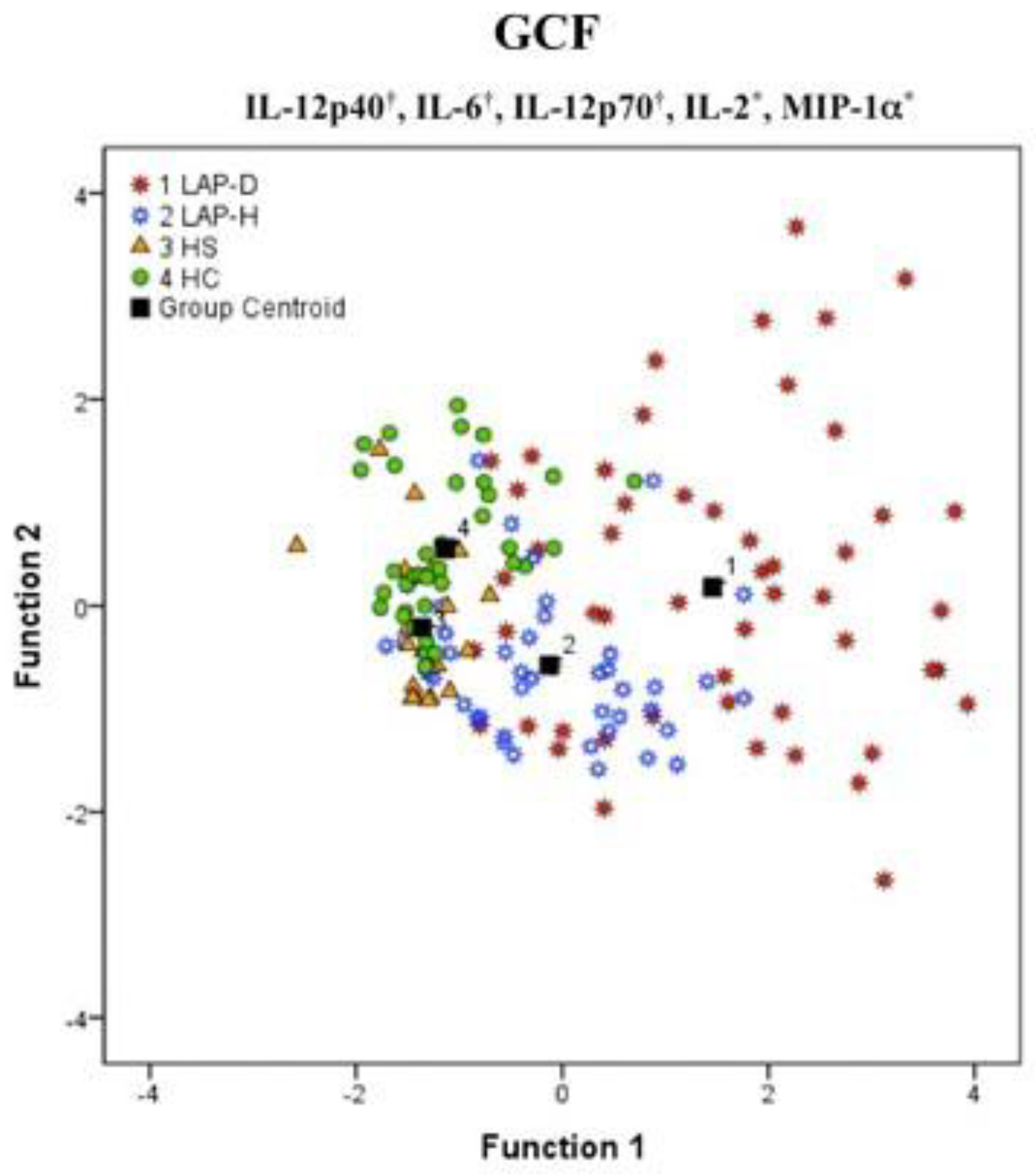
Local inflammation. Focusing on host immunological response complexity to a low biofilm aggregation, Branco-de-Almeida et al. [21] and Shaddox et al. [22] reported high levels of IL-12p40, GMCSF, TNF-α, IL-6, IL-12p70, IL-2, INFγ, IL1β, and MIP-1α gingival crevicular fluid (GCF) of LAP patients diseased sites when compared to health sites from the same patients [22] and/or health siblings and healthy unrelated controls [21,22] Interestingly, a significant differential profile of these markers was observed among these sites, where most differences fall between diseased and healthy sites, however, the healthy sites from healthy individuals tended to cluster more closely together (Figure 2). For instance, higher GCF concentrations of Eotaxin, GM-CSF, IL-10, IL-12p70, and IL-2 were observed in health sites from LAP patients when compared to their health siblings [21]. Additionally, healthy siblings of LAP subjects displayed an increase in IFN-γ and OPG without any clinical evidence of disease [21]. Interestingly, Martins et al. [23] showed similarity of some biomarker’s expression (IL1β, ICAM-1, and GMCSF) either in C-MIP or generalized aggressive forms of periodontitis (GAgP; CAL > 4 mm affecting at least three permanent teeth other than the first molars and incisors), at baseline to health controls. A current systematic review and meta-analysis [24] confirm that most of the cited biomarkers above were highly expressed in the GCF of C-MIP patients; however, the literature still needs more evidence to use this approach as a diagnosis tool. Some of these biomarkers have a pivotal role in stimulating cells’ environment towards osteoclastic activity and sustaining proinflammatory profile by chemotaxis. Thus, these differentiated local levels of inflammatory markers may contribute to local microbial dysbiosis and possible site breakdown in these individuals (see microbial discussion below).
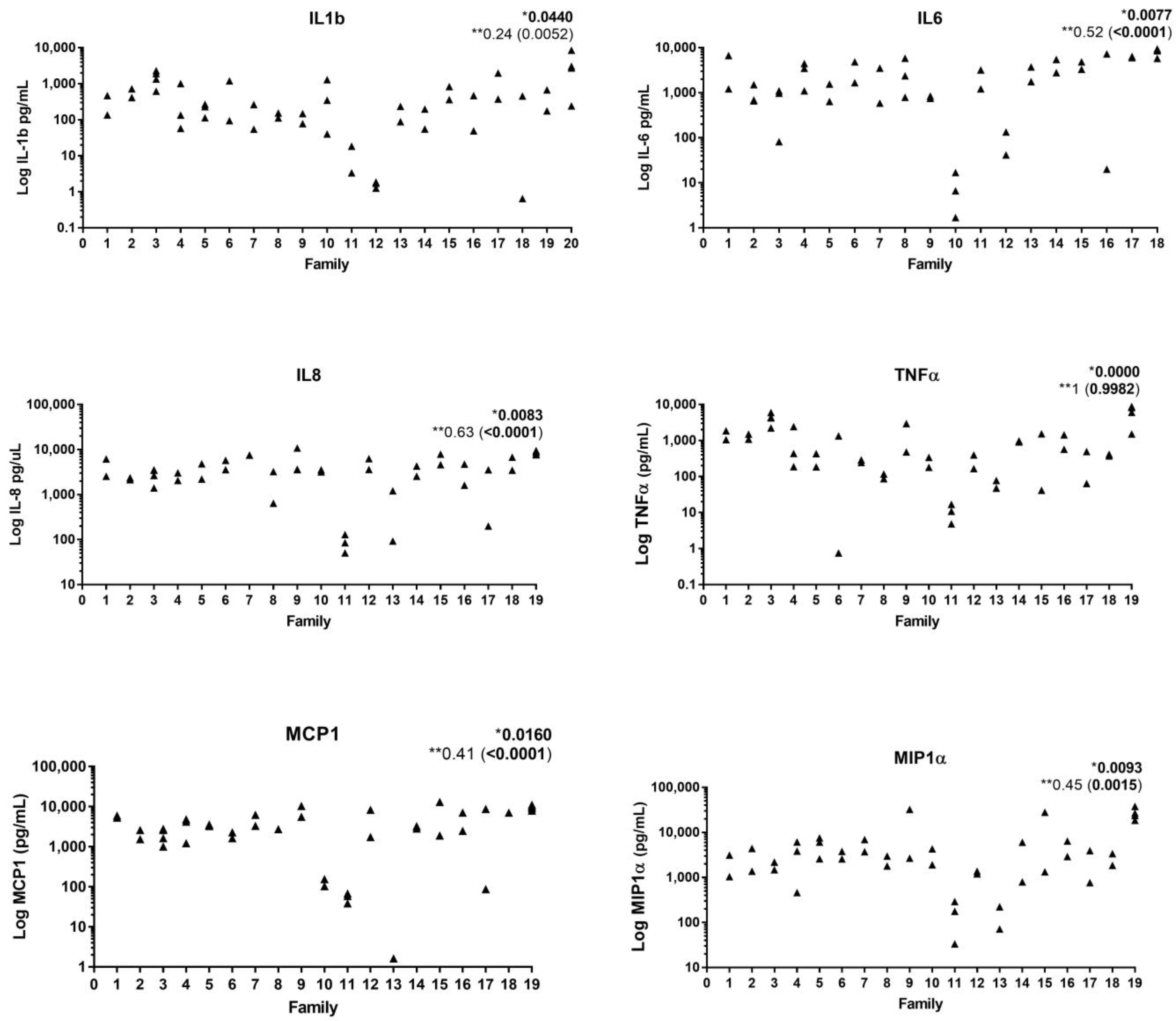
Systemic inflammation. Peripheral blood from C-MIP stimulated either with LPS or biofilm from healthy and diseased sites has shown a hyperinflammatory responsiveness profile with increased levels of G-CSF, IFNγ, IL-10, IL12(p40), IL1β, IL-6, IL-8, MCP-1, MIP-1α, and TNFα when compared healthy control patients [25,26]. Interestingly, the similar response, regardless of the specific biofilm used for stimulation, highlights the role of host response in this unique disease. Tabaa et al. [27] have shown a similar pattern of proinflammatory response (IL-6, IL-8, IL-10, MIP1α, MCP1, and IL-1β) among affected siblings after peripheral blood LPS stimulation (Figure 3). Similarly, both E. coli and P. gingivalis LPS-stimulated peripheral blood from healthy siblings of LAP subjects has shown a tendency of hyperresponsiveness with IL2, IL12p40, TNFα, IFNγ, and IL6 elevated levels compared to unrelated health controls [25]. In fact, healthy siblings of LAP individuals tend to show a high peripheral response to diseased biofilms, showing increased levels of IL-6 and INFγ. In fact, even in response to healthy biofilm stimulation, this group had a higher concentration of INFγ compared to unrelated healthy patients [26]. This evidence collectively can point to two reasonable hypotheses. First, there seems to be a familial aggregation associated with this hyper-inflammatory response to bacterial stimuli in families with this disease [27]. Second, it can be hypothesized that inflammatory response could potentially precede or exacerbate bacterial dysbiosis in susceptible individuals in this clinical condition since both healthy and pathogenic biofilms have been shown to elicit a hyper-inflammatory host response in these individuals [26]. In addition, healthy sites in diseased patients display biomarkers featuring a differentiated and proinflammatory local profile [26].
The first line of defense in individuals who suffer from an aggressive form of periodontitis can be compromised due to either neutrophil hyperactivity [28] or dysfunction [29,30]. Van Dyke et al. [29] showed a deficiency in neutrophil chemotaxis in 26 out of 32 patients diagnosed with C-MIP/LAP compared to age and sex-matched healthy controls. This pattern remains even in those whose disease has already been treated. According to the same study, 7 out of 10 patients displayed neutrophil chemotaxis deficiency after 6 months to 2 years post-therapy [29]. Thus, it seems that a genetic determination may be associated with this neutrophil role. Van Dyke et al. [30] have shown neutrophil dysfunction in 90% of healthy siblings under 12 years old featuring a familial aggregation. This deficiency can be considered a predictor of aggressive forms of periodontitis since some of the healthy siblings detected with neutrophils at the beginning of the mentioned studies developed the disease during the study [30]. Focusing on humoral response, high levels of immunoglobulin G2 (IgG2 antibody) have also been observed in LAP patients [31]. Studies are controversial if this immunological response is specific to A. actinomycetemcomitans [32,33]; however, a racial influence was observed in this immunoglobulin levels [34], where higher concentrations of IgG2 serum was observed in African American descendants diagnosed with LAP when compared to Caucasians [34].
Gender and host response. Although the literature is still controversial about gender influence in C-MIP, local and systemic inflammatory biomarkers are differently expressed in males and females. Tavakoli et al. [18] reported increased GCF levels of TNFα, IFNγ, MCP1, and MIP-1α in diseased sites of males. Interestingly, healthy sites in male patients diagnosed with LAP/C-MIP also presented high levels of IFNγ and a tendency to higher G-CSF levels as well. On the other hand, peripheral blood from C-MIP females stimulated with P. gingivalis e E. coli LPS showed an increase in systemic biomarkers, such as Eotaxin, IFNγ, and GM-CSF, compared to C-MIP males [18]. Considering the age range of disease initiation in the permanent dentition, sex hormones may indeed have an impact on both the local and systemic inflammatory response. In fact, high testosterone levels in males have been correlated to periodontitis prevalence and severity [35], while estrogen reduction in a woman’s lifetime may increase the risk of periodontal issues due to its protective role [36]. However, based on the results above regarding systemic responses, it can be inferred that females diagnosed with C-MIP have a hyperresponsive tendency to biofilm challenge (LPS), which may impact faster periodontal apparatus loss [18]. Thus, hormonal imbalances during puberty in both males and females and their impact on C-MIP predisposition or initiation certainly warrant further investigation.
Genetic influence on host response. Genetic aspects could also have an impact on the host inflammatory response [37,38,39]. Toll-like receptors (TLR), e.g., TLR-2 and TLR-4, have been associated with LAP/C-MIP hyper-inflammatory response [25]. This transmembrane glycoprotein pathway has shown more than a two-fold-increase in specific gene expression correlated to inflammation, such as TICAM-1 (TRIF), FOS, IRAK1, TLR2, and CCL2 in LAP/C-MIP patients compared to healthy unrelated controls [40]. IRAK1 displayed a significantly increased expression in these subjects, which may be explained by IL-1β increase in these patients [41]. Epigenetic regulation plays an important role in host immunological response via the TLR pathway. According to Shaddox et al. [42], up- (e.g., MYD88, MAP3K7, RIPK2, IL6R) and downregulation (e.g., FADD, PPARA, IRAK1BP1) of several genes can be explained by DNA methylation in specific sites. For example, methylation in FADD position 5 was positively correlated with LPS-stimulated levels of IL-6 and TNF-α systemically. Conversely, MYD88 methylation in position 4 presented a negative correlation with IL-10, IP-10, and MCP-1 [42].
Several gene single nucleotide polymorphisms (SNP) can influence immunological patterns and susceptibility in aggressive forms of periodontitis. IL-1α (rs1800587) and IL-1-β (rs1143634) polymorphisms have been correlated to generalized aggressive periodontitis [43]. SNP in the Lactoferrin gene, the iron-binding protein with antimicrobial activity related to host first-line defense (G-allele), was detected in young African Americans [44] and Taiwanese [45] diagnosed with aggressive forms of periodontitis. Similarly, cathepsin C SNP (rs3888798), a cysteine protease involved in neutrophil maturation and its activity in immune response, was also associated with this disease, along with lower PMN function, enhancing the risk of aggressive periodontitis development [46]. Several reviews of the literature evaluating the role of genetics in aggressive periodontitis have been published [37,47,48,49,50,51]. Given the several genes potentially associated with the disease and its inflammatory pathways, it is likely that several genetic polymorphisms are associated with susceptibility to this disease. There is also some evidence that indicates a possible genetic predisposition to specific microbial colonization [52,53]. Thus, comprehensive genomic studies with a high number of diseased individuals from different populations need to be conducted to clarify specific genes and their role in this disease.
Finally, some studies have investigated the roles of small RNAs, known as microRNAs, or miRNAs, in periodontitis [40,54,55] and other inflammatory diseases [56]. In our cohort of LAP individuals, several miRNAs, including miR-9-5p, 155-5p, and 147a, all presented elevated expression (2-fold up-regulated) in our LAP/C-MIP cohort [40]. Interestingly, increased miR-9-5p levels were previously found in inflamed gingival tissue from patients diagnosed with periodontitis [54] and also in neutrophils and monocytes post-TLR2-4 activation [57], and both miR155-5 and 147a have also been found elevated post-LPS stimulation, promoting further inflammatory cascade [58,59,60]. There seems to be an important role for miRNA regulation in TLR responsiveness in this disease, which deserves to be further explored by comprehensive next-generation sequencing approaches at different disease stages.

3. Microbiological Aspects

Literature has shown a microbial profile associated with localized early-onset periodontitis [61,62,63,64,65]. Microorganisms such as A. actinomycetemcomitans, T. lecithinolyticum, and T. forsythia have been strongly associated with this condition [62,64]. Moreover, studies have shown that P. gingivalis, P. intermedia, T. denticola, C. gracilis, E. nodatum, and F. nucleatum are highly correlated with active disease and its prevalence [63,66]. A. actinomycetemcomitans has been classified as a key pathogen in young adults diagnosed with LAP/C-MIP [63,64,65,66,67,68,69] and even with sites with progressive bone loss [70,71]. This microorganism has different mechanisms of action including direct and indirect ways of influence in host response [72,73,74]. Seven serotypes can be linked with this species (a–g) based on membrane polysaccharides surface. A particular JP2 Genotype from serotype “b” has been highly associated with a severe form of periodontal disease [12,75,76] and in sites with progressive bone loss as well [76,77]. In the beginning, this species was identified in subjects from North and West Africa. Due to the transatlantic slave trade, new colonization was spread out in North and South America [75], which could explain the higher prevalence of this disease in African descendants in both these regions. In fact, JP2 clone has been reported in aggressive periodontitis in both North [78] and South American populations [67]. One of the main virulence factors from this genotype/serotype “b” is the expressive release of leukotoxin (LtxA), intensifying leukotoxic and cell death in high doses [79]. Not only does LtxA depreciate leukocyte activity, but it also increases lysosomal release in macrophages reducing its phagocytic profile [80].
Tissue invasion is a well-known propriety from A. actinomycetemcomitans, and it has already been identified in periodontal tissues from young adults [81], even at a very young age [82]. Several virulence factors in relation to this action have been described in in vitro and in vivo analysis [83]. Outer membrane protein 100 (Omp100) present in A. actinomycetemcomitans membrane surface composition has the capacity to bind in human epithelial cells [84]. After its adhesion, other Omp protein class groups act specifically in tissue invasion. Omp29 allows microorganism diffusion into epithelial cells, enhancing permeability due to F-actin rearrangement via FAK signaling cascade [85]. In the same way, cytolethal distending toxin (CDT) produced by this bacterium has the capacity to impact the cell cycle, leading to epithelial layer organization disruption due to the dissolution of cell junctions (e.g., cadherin) [86]. This scenario results in soft tissue collapse, allowing microorganisms and their toxins to make intimate contact with the connective tissue below [86].
Early A. actinomycetemcomitans acquisition can be correlated with C-MIP in primary and mixed dentition [64,71,75,87]. A longitudinal study reported that individuals containing A. actinomycetemcomitans, S. parasanguinis, and F. alocis are more prone to develop LAP/C-MIP disease [71]. In fact, a higher risk for bone loss was observed when the three microorganisms coexisted [71]. Jensen et al. have shown that healthy Moroccan toddlers between 7–10 years presented a higher CAL in mixed dentition when the JP2 genotype of A. actinomycetemcomitans was detected in their biofilm at incisor and molars [12]. Indeed, studies have reported a higher risk of bone loss when either non- or JP2 genotype is part of their microbiota in advance [76,77]. Aberg et al. [77] evaluated a 2-year progression of CAL based on the presence of JP2 and non-JP2 genotypes of A. actinomycetemcomitans in 500 adolescents (from 10 to 19 years of age). After the longitudinal follow-up, it was possible to predict a higher progression of CAL ≥ 3 mm when JP2 (OR = 14.3) and non-JP2 genotypes (OR = 3.4) were detected in subgingival biofilm. Moreover, an increasing risk of disease development followed by CAL ≥ 3 mm (RR = 7.3) was detected in healthy individuals at baseline with JP2 identification in subgingival biofilm. This evidence emphasizes the role of specific microorganisms in aggressive forms of periodontitis [77].
One of the possible explanations for this precocious colonization is family aggregation. Monteiro et al. [88] have reported a vertical pathogenic microbiome transmission between parents diagnosed with grade C periodontitis and their progeny. In fact, offspring’ subgingival biofilm from parents diagnosed with periodontal disease has shown exclusive microorganisms such as Filifactor alocis, Porphyromonas gingivalis, Streptococcus parasanguinis, Fusobacterium nucleatum subsp. nucleatum when compared to progeny from parents who do not suffer from grade C periodontitis. Moreover, even after biofilm maintenance, several periodontopathogenic species remain stable and in higher abundance in offspring from periodontal diagnosed families, including A. actinomycetemcomitans, P. gingivalis, T. denticola, and T. forsythia [88].
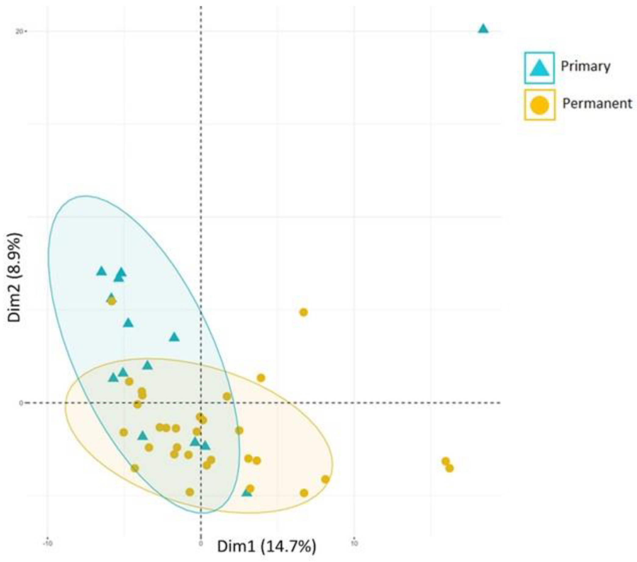
Microbiome profiles in diagnosed C-MIP subjects can distinguish between primary and permanent dentition. According to a recent study (Koo et al. [89]), a partial overlap was observed between the two types of dentitions in disease conditions with the detection of distinguished microorganisms in primary and permanent affected sites (Figure 4). Capnocytophaga ochracea, Leptotrichia buccalis, goodfellowii, and Sneathia sanguinegens were found more frequently in primary teeth-affected sites when compared to the permanent ones. On the other hand, Filifactor alocis, Tannerella forsythia, and Synergistetes sp were elevated at C-MIP permanent sites, whereas A. actinomycetemcomitans, Campylobacter, Fusobacterium nucleatum, and Gemella morbillorum were identified in both primary and permanent affected sites characterizing the overlapping between them. In fact, A. actinomycetemcomitans was highly abundant in both affected dentitions (85% and 71%, respectively), corroborating its important role in this disease, both in primary and permanent dentitions. It seems that a more mature microbiome is developed in permanent diseased sites. However, the identification of some periodontopathogens, such as Campylobacter gracilis and F. nucletum ss nucleatum were also observed early in the primary dentition [89], which has been reported by the literature in early-onset periodontitis [90,91]. Thus, despite their low abundance, primary dentition already exhibits an identification of microorganisms that potentially increase the risk of disease development in the latest detention. Moreover, the high abundance of A. actinomycetemcomitans, regardless of dentition, highlights this species’ role in C-MIP disease.
An important point to consider is intrafamilial transmission and its role in the acquisition of periodontopathogens. It has been shown that individuals from the same family harbored the same biotype and serotype of A. actinomycetemcomitans [92], and Christersson also found that members of the same family with LAP also harbored the same biotype and serotype of A. actinomycetemcomitans [93]. Haubek et al. also reported the same isolates of JP2 clones in African LAP families and suggested that the strong familial aggregation of this disease was due to intrafamilial transmission of this virulent strain [94]. The most likely route of transmission is via the saliva and could be vertical (parent to child) or horizontal (partner to partner or sibling to sibling) [95,96]. Transmission theories have been reinforced by sequencing studies of specific strains [97,98,99]. However, it is possible that transient transmission does not lead to persistent colonization of the organism, as this will also depend on the host, bacterium characteristics, and the abundance of the transferred species, among others. There may also be genetic influence of colonization patterns, as the periodontal flora of identical twins has been shown to be more similar than that of fraternal ones [100,101].

4. Diseases Initial Time-Point

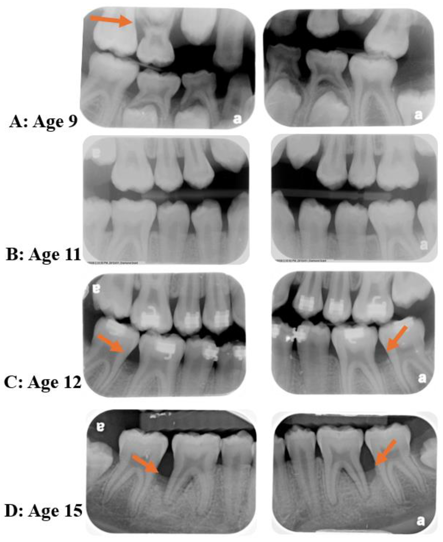
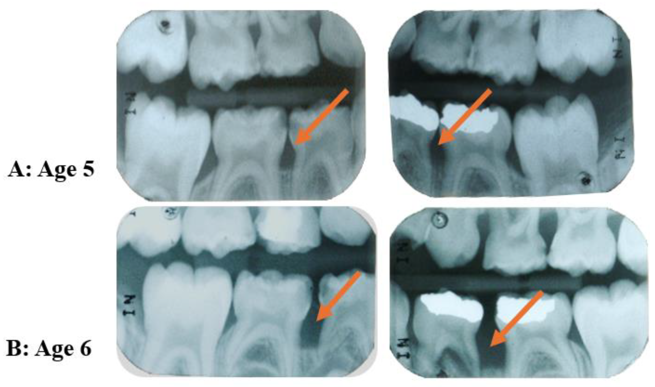
In reviewing the topics discussed above, it is possible to question whether C-MIP actually starts in the primary dentition. If left untreated, the persistent presence of the pathogen A. actinomycetemcomitans within tissues [93,102,103] may allow this species to recolonize the pockets around permanent dentition and within sites of hyper-inflammatory host predisposition, may lead to rapid periodontal destruction in these individuals, as studies show that treatment of this disease early in the primary dentition seems to reduce disease recurrence in the permanent dentition [15,16,17]. This microorganism can act in tissue breakdown in indirect and direct ways jeopardizing the host first-line response (PMN recruitment) all together with microorganism selectivity in the subgingival environment, respectively [72]. Other more recent retrospective analyses [10,16] were carried out evaluating radiograph bone loss patterns, external/internal root resorption, enlarged chamber pulp, and exfoliation patterns in primary dentition records from patients diagnosed with LAP/C-MIP in permanent teeth. The first study [16] had access to 39 patient’s radiographic records (20 C-MIP diagnosed and 19 healthy siblings). Ninety percent of those who had C-MIP in the permanent dentition also presented signs of radiographic bone loss around their first and second primary molars in the retrospective evaluation (Figure 5). Moreover, six of the 19 siblings who were healthy in their permanent dentition presented bone loss in primary radiographic records retrospectively, and two of them went on to develop the disease in the study follow-up [16] (Figure 6). Likewise, the second study [10] evaluated 49 periapical radiographs in 33 patients who presented the disease in the primary dentition. The first primary molar was the most affected teeth, followed by the second primary molar [10] (Figure 7). The authors suggest there seems to be a pattern of progression from the first to the second primary molar, and this could potentially lead to colonization of the first permanent molar if left untreated. Thus, it seems reasonable to infer that it is possible that C-MIP may indeed start in the primary dentition and, if left undiagnosed and untreated, may lead to disease in the permanent dentition. Thus, the role of pediatric dentists in the diagnosis and management of this disease early becomes essential for the proper treatment of this disease and prevention of possible breakdown of the permanent dentition [104]. The Academy of Pediatric Dentistry’s latest recommendation for the diagnosis of periodontal disease in children is to perform a full mouth periodontal examination as soon as the first permanent molar erupts, around 6 years of age [105], or earlier if clinical signs of disease or radiographic bone loss are detected or suspected.

5. Treatment Aspects

Scaling and root planning (SRP) associated with systemic antibiotic therapy (Amoxicillin and Metronidazole) is the current gold standard for treating C-MIP [62,106,107,108,109]. According to most clinical studies, this therapy can provide significant probing depth (PD) reduction, clinical attachment level (CAL) gain, along with positive modulation in host proinflammatory profile (GCF), maintaining microbial compatibility with health sites in the short and long term (Figure 8) [62,106,107,108,109]. However, it is important to highlight that although patients respond well to this therapy, patients who present a high LPS responsiveness may not achieve equal outcomes to the ones presenting a lower responsiveness, even under the same clinical approach [110].
Branco de Almeida et al. [109] and Miller et al. [16] showed great PD reduction and clinical attachment gain in diagnosed LAP/C-MIP patients when a regimen of full mouth debridement associated with systemic antibiotic therapy (ABX at baseline: Metronidazole-250 mg and Amoxicillin-500 mg, 3× per day for 7 days) immediately after the clinical debridement was the treatment of choice. Positive clinical parameter reductions were observed in the short-term (6 months) and maintained in the long-term (2 years), while Miller et al. [16] showed this reduction in clinical parameters was maintained for up to 4 years post-treatment. Moreover, several biomarkers in diseased and health sites from LAP/C-MIP patients were reduced in at least one time point when compared to baseline levels [109]. Most of these decreased biomarkers have been characterized as disease features in discriminative analysis [21]. Thus, it seems that this type of approach can lead to clinical stability and local host modulation in the long term. Despite the positive and sustained modulation in GCF biomarkers after the non-surgical periodontal therapy combined with ABX, serum biomarkers modulation does not provide the same outcome [111]. Limited cytokines/chemokines are reduced in some post-treatment time points, while others seem to rebound, especially after 6 months [112]. In fact, some studies have shown that serum inflammatory biomarkers persist at higher rates compared to diagnosed patients even after treatment [113] or return to their baseline levels after long-term follow-up [114]. Comparing C-MIP and GAgP response after 1-year long-term follow under the ABX approach, it is possible to observe similarities between both disease types of outcomes regarding serum and GCF levels [23].
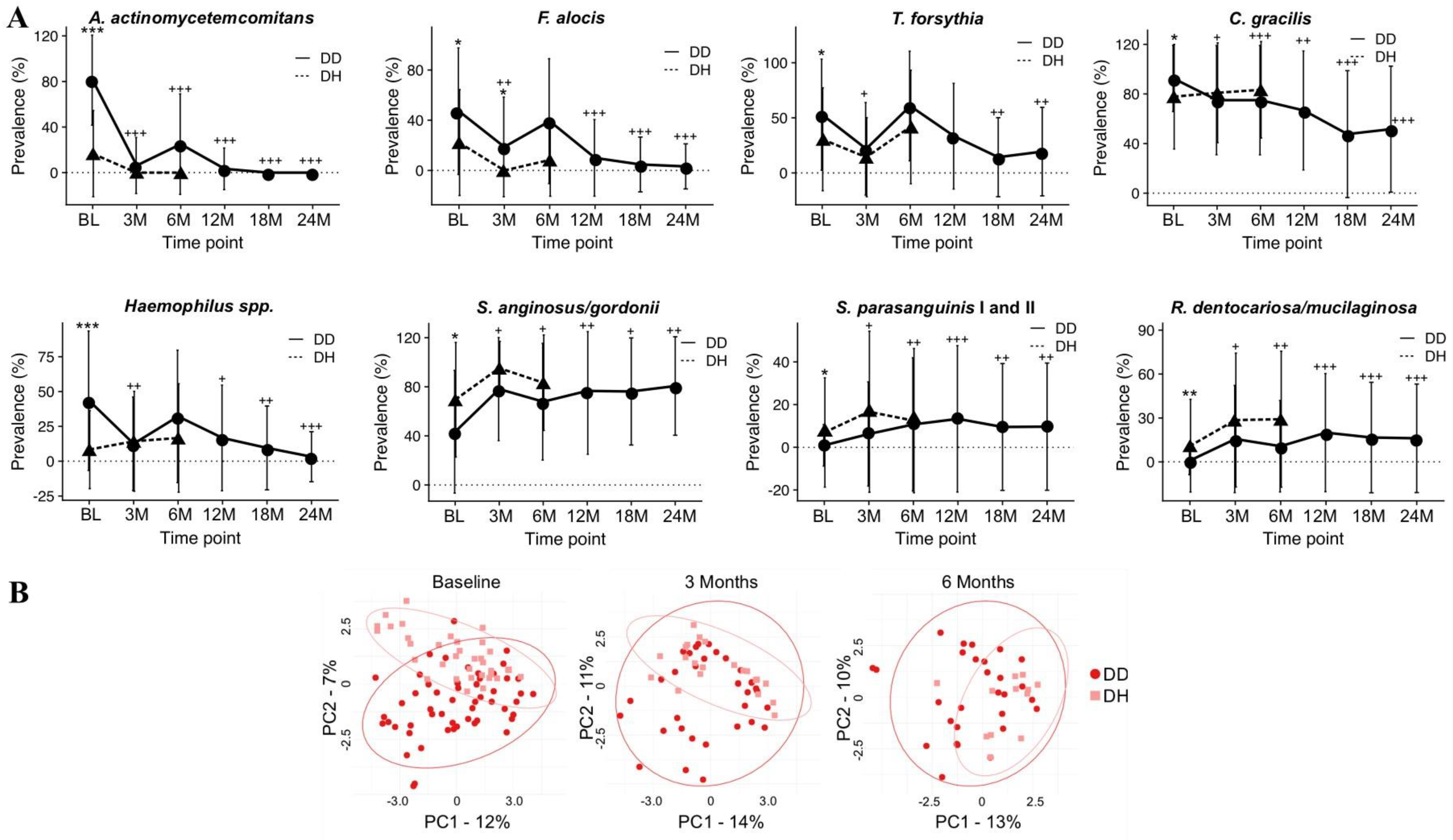
From a microbiological perspective, the same clinical approach (SRP+ABX) can modify host microbiota, reducing putative species correlated to C-MIP disease [114]. Velsko et al. [62] have shown that this protocol (debridement with a 7-day course of ABX) maintained a healthy microbial environment in a 2-year long-term, decreasing A. actinomycetemcomitans, F. alocis, T. forsythia, and C. gracilis while health-associated species, such S. anginosus/gordonii and S. parasanguinis, increased (Figure 9A). A statistical difference in community profile could be observed between healthy and diseased sites before therapy, and a closer cluster (more similar profile) between health and disease profiles could be observed in C-MIP patients post-treatment compared to baseline (Figure 9B) [62]. Burgess et al. [78] also reported a drastic reduction in the initially high prevalence of JP2 genotype post-treatment, and this significant reduction was maintained for 12 months post-treatment, with one course of ABX at baseline and proper periodontal maintenance. In diseased sites, JP2 genotype prevalence was reduced to 3.23% (1/31 site detection) after 1 year of treatment compared to baseline, while no detection was observed in the health sites of African American patients diagnosed with LAP/C-MIP [78]. A different regiment of ABX (Metronidazole-400 mg and Amoxicilin-500 mg, 3× per day for 14 days) along with SRP also decreased JP2 genotype in LAP/C-MIP patients after 1 year. Additionally, serum IgG against Omp29 was also decreased, which is one of the virulence factors correlated to tissue invasion [106].
Considering dentition type under SRP+ABX clinical approach, Merchant et al. [9] have shown a better clinical attachment gain in primary dentition at 3, 6, and 12 months compared to the permanent dentition, although both dentitions presented a significant reduction in all clinical parameters post-treatment. These results were encouraging regarding early treatment of disease and indicated that younger patients may be more likely to have disease resolution [9]. This can be associated with a few conditions, such as low inflammatory rates in younger children [115], as well as precocious disease diagnosis and intervention under a less complex dysbiotic environment [89]. Depending on the amount of bone level compromised due to disease progression, primary tooth extraction may be an alternative, especially in cases with extensive mobility, pain/abscess, or loss of function impact on quality of life [116].
There is no study confirming disease prevention in permanent teeth in patients diagnosed and treated in the primary dentition. However, the few longitudinal studies following patients diagnosed with LAP/C-MIP in primary dentition indicate a favorable response to treatment and a low incidence of disease in permanent dentition with proper maintenance. According to Mros and Berglundh [15], no bone loss was detected in 7 out of 13 subjects previously diagnosed with the disease in early dentition under proper periodontal maintenance. Miller et al. did not report a recurrence of the disease in permanent dentition in a long-term follow-up of the Florida cohort [16]. Despite the small sample size, low rates of disease recurrence were also observed by Bimstein [17] and Merchant et al. [9]. Thus, it seems reasonable to reinforce periodontal screening in pediatric dental appointments focusing on early diagnosis, especially when cases of aggressive periodontitis are reported in the family.

6. Conclusions

C-MIP is an oral disease more prevalent in African descendants and young systemically healthy adults (under 35 years old) that affects the tooth-supporting apparatus of very specific teeth and features rapid alveolar bone loss. Most cases are associated with low biofilm accumulation, bone loss affecting first molars and incisors, along with familial aggregation. Although less studied, this disease can also be diagnosed in the primary dentition, which may lead to more successful treatment and possibly prevent the occurrence of the disease in the permanent dentition. Studies evaluating primary dentition records have shown that most patients diagnosed in the permanent dentition had disease signs (i.e., bone loss) in the primary dentition.
A heightened inflammatory response to bacteria can be observed in these patients, and healthy siblings also present a tendency of high inflammatory response even without a clinical diagnosis, which may be a result of a genetic predisposition to the disease, given the similar patterns of host response seen in families. The immunological response to biofilm is high regardless of the stimuli used. Periodontal therapy has been shown to successfully manage the local inflammatory profile and clinical response to treatment; however, the modulation of serum biomarkers is not as consistent, and some markers seem to remain high or rebound in the long term despite clinical response remaining positive.
A. actinomycetemcomitans is a key pathogen either in primary or permanent dentition affected with this disease, characterized by special tools for tissue invasion and more virulent genotypes (e.g., JP2). A. actinomycetemcomitans early acquisition was also reported as a possible predictor to disease initiation. The colonization of A. actinomycetemcomitans in primary dentition can be explained by familial aggregation and possible pathogenic transmission within families. Similar subgingival biofilm composition between parents diagnosed with an aggressive type of periodontitis and their offspring has been reported in the literature. In fact, the bacterial community in these children seems to be resistant even after periodontal maintenance. After periodontal therapy, periodontopathogens are significantly reduced, and this profile can be maintained for a long period with proper supportive periodontal therapy.
SRP + ABX is the gold standard for treating C-MIP, supported by several studies and systematic reviews. Most studies report amoxicillin and metronidazole as the regimen of choice as adjuncts to SRP/full mouth debridement. The disease affecting both primary and permanent dentition presents a favorable clinical response to treatment. However, higher gains in clinical attachment levels seem to happen in younger children. This may be explained by a lower inflammatory profile in young children [115] and a less mature microbial community compared to permanent dentition [89]

7. Future Directions

There are still many gaps to be investigated in C-MIP, such as sex hormones (androgens, estrogens, and progestogens) and other cofactors during childhood development that may influence the disease incidence. Moreover, epigenetic studies and other comprehensive analyses using integrative -omics analysis (e.g., proteomic, metagenomic, genomics) should be carried out focusing on better understanding the biomolecular features of this disease in different time points and in different populations. In addition, there is not yet a group of feature inflammatory biomarkers to control diagnosis, progression, and disease severity over time. A better understanding and correlation between serum, GCF, and saliva biomarkers may guide research and clinical practitioners to a more assertive approach to this disease. Despite the great success under SRP+ABX therapy, host modulation, e.g., pro-resolving lipid mediators, may be an alternative option in future studies [117,118,119], given the increase in ABX resistance and the heightened inflammatory response seen in these individuals. Considering the current classification system, it is important to include clinical characteristics that have been discussed and published in the past for this disease in different stages so that studies with specific inclusion criteria are conducted, and knowledge continues to be developed for this particular disease in the future. Moreover, given the fact that some evidence indicates a possible disease initiation in the primary dentition, periodontal screening during pediatric appointments remains crucial for early disease diagnosis and treatment and possible prevention of disease at later stages.

Author Contributions

Conceptualization, L.M.S.; Methodology, L.M.S. and M.M.V.M.; Writing—Original Draft Preparation, L.M.S. and M.M.V.M.; Writing—Review and Editing, L.M.S. and M.M.V.M.; Supervision, L.M.S.; Funding Acquisition, L.M.S. All authors have read and agreed to the published version of the manuscript.

Funding

National Institutes of Health: R01DE019456 and 1U01DE031223.

Institutional Review Board Statement

Not applicable.

Informed Consent Statement

Not applicable.

Data Availability Statement

Not applicable.

Conflicts of Interest

The authors declare no conflicts of interest.

References

  1. Scannapieco, F.A.; Dongari-Bagtzoglou, A. Dysbiosis Revisited: Understanding the Role of the Oral Microbiome in the Pathogenesis of Gingivitis and Periodontitis: A Critical Assessment. J. Periodontol. 2021, 92, 1071–1078. [Google Scholar] [CrossRef] [PubMed]
  2. Ebersole, J.L.; Dawson, D.R.; Morford, L.A.; Peyyala, R.; Miller, C.S.; Gonzaléz, O.A. Periodontal Disease Immunology: “double Indemnity” in Protecting the Host. Periodontology 2000 2013, 62, 163–202. [Google Scholar] [CrossRef] [PubMed]
  3. Armitage, G.C. Development of a Classification System for Periodontal Diseases and Conditions. Ann. Periodontol. 1999, 4, 1–6. [Google Scholar] [CrossRef] [PubMed]
  4. Papapanou, P.N.; Sanz, M.; Buduneli, N.; Dietrich, T.; Feres, M.; Fine, D.H.; Flemmig, T.F.; Garcia, R.; Giannobile, W.V.; Graziani, F.; et al. Periodontitis: Consensus Report of Workgroup 2 of the 2017 World Workshop on the Classification of Periodontal and Peri-Implant Diseases and Conditions. J. Periodontol. 2018, 89 (Suppl. S1), S173–S182. [Google Scholar] [CrossRef] [PubMed]
  5. Susin, C.; Haas, A.N.; Albandar, J.M. Epidemiology and Demographics of Aggressive Periodontitis. Periodontology 2000 2014, 65, 27–45. [Google Scholar] [CrossRef] [PubMed]
  6. Albandar, J.M.; Tinoco, E.M.B. Global Epidemiology of Periodontal Diseases in Children and Young Persons. Periodontology 2000 2002, 29, 153–176. [Google Scholar] [CrossRef] [PubMed]
  7. Bouziane, A.; Hamdoun, R.; Abouqal, R.; Ennibi, O. Global Prevalence of Aggressive Periodontitis: A Systematic Review and Meta-analysis. J. Clin. Periodontol. 2020, 47, 406–428. [Google Scholar] [CrossRef] [PubMed]
  8. Lang, N.; Bartold, P.M.; Cullinan, M.; Jeffcoat, M.; Mombelli, A.; Murakami, S.; Page, R.; Papapanou, P.; Tonetti, M.; Van Dyke, T. Consensus Report: Aggressive Periodontitis. Ann. Periodontol. 1999, 4, 53. [Google Scholar] [CrossRef]
  9. Merchant, S.N.; Vovk, A.; Kalash, D.; Hovencamp, N.; Aukhil, I.; Harrison, P.; Zapert, E.; Bidwell, J.; Varnado, P.; Shaddox, L.M. Localized Aggressive Periodontitis Treatment Response in Primary and Permanent Dentitions. J. Periodontol. 2014, 85, 1722–1729. [Google Scholar] [CrossRef]
  10. Miller, K.; Treloar, T.; Guelmann, M.; Rody, W.J.; Shaddox, L.M. Clinical Characteristics of Localized Aggressive Periodontitis in Primary Dentition. J. Clin. Pediatr. Dent. 2018, 42, 95–102. [Google Scholar] [CrossRef]
  11. Bimstein, E. Radiographic Description of the Distribution of Aggressive Periodontitis in Primary Teeth. J. Clin. Pediatr. Dent. 2018, 42, 91–94. [Google Scholar] [CrossRef] [PubMed]
  12. Jensen, A.B.; Ennibi, O.K.; Ismaili, Z.; Poulsen, K.; Haubek, D. The JP2 Genotype of Aggregatibacter Actinomycetemcomitans and Marginal Periodontitis in the Mixed Dentition. J. Clin. Periodontol. 2016, 43, 19–25. [Google Scholar] [CrossRef] [PubMed]
  13. Cogen, R.B.; Wright, J.T.; Tate, A.L. Destructive Periodontal Disease in Healthy Children. J. Periodontol. 1992, 63, 761–765. [Google Scholar] [CrossRef] [PubMed]
  14. Mandell, R.L.; Siegal, M.D.; Umland, E. Localized Juvenile Periodontitis of the Primary Dentition. ASDC J. Dent. Child. 1986, 53, 193–196. [Google Scholar] [PubMed]
  15. Mros, S.T.; Berglundh, T. Aggressive Periodontitis in Children: A 14–19-Year Follow-Up. J. Clin. Periodontol. 2010, 37, 283–287. [Google Scholar] [CrossRef] [PubMed]
  16. Miller, K.A.F.S.; Branco-de-Almeida, L.S.; Wolf, S.; Hovencamp, N.; Treloar, T.; Harrison, P.; Aukhil, I.; Gong, Y.; Shaddox, L.M. Long-Term Clinical Response to Treatment and Maintenance of Localized Aggressive Periodontitis: A Cohort Study. J. Clin. Periodontol. 2017, 44, 158–168. [Google Scholar] [CrossRef] [PubMed]
  17. Bimstein, E. Seven-Year Follow-up of 10 Children with Periodontitis. Pediatr. Dent. 2003, 25, 389–396. [Google Scholar]
  18. Tavakoli, T.T.; Gholami, F.; Huang, H.; Gonçalves, P.F.; Villasante-Tezanos, A.; Aukhil, I.; de Oliveira, R.C.G.; Hovencamp, N.; Wallet, S.; Ioannidou, E.; et al. Gender Differences in Immunological Response of African-American Juveniles with Grade C Molar Incisor Pattern Periodontitis. J. Periodontol. 2022, 93, 392–402. [Google Scholar] [CrossRef] [PubMed]
  19. Melvin, W.L.; Sandifer, J.B.; Gray, J.L. The Prevalence and Sex Ratio of Juvenile Periodontitis in a Young Racially Mixed Population. J. Periodontol. 1991, 62, 330–334. [Google Scholar] [CrossRef]
  20. Nassar, M.M.; Afifi, O.; Deprez, R.D. The Prevalence of Localized Juvenile Periodontitis in Saudi Subjects. J. Periodontol. 1994, 65, 698–701. [Google Scholar] [CrossRef]
  21. Branco-De-Almeida, L.S.; Cruz-Almeida, Y.; Gonzalez-Marrero, Y.; Huang, H.; Aukhil, I.; Harrison, P.; Wallet, S.M.; Shaddox, L.M. Local and Plasma Biomarker Profiles in Localized Aggressive Periodontitis. JDR Clin. Trans. Res. 2017, 2, 258–268. [Google Scholar] [CrossRef] [PubMed]
  22. Shaddox, L.M.; Wiedey, J.; Calderon, N.L.; Magnusson, I.; Bimstein, E.; Bidwell, J.A.; Zapert, E.F.; Aukhil, I.; Wallet, S.M. Local Inflammatory Markers and Systemic Endotoxin in Aggressive Periodontitis. J. Dent. Res. 2011, 90, 1140–1144. [Google Scholar] [CrossRef] [PubMed]
  23. Martins, E.S.; César-Neto, J.B.; Albuquerque-Souza, E.; Rebeis, E.S.; Holzhausen, M.; Pannuti, C.M.; Mayer, M.P.A.; Saraiva, L. One-Year Follow-up of the Immune Profile in Serum and Selected Sites of Generalized and Localized Aggressive Periodontitis. Cytokine 2019, 116, 27–37. [Google Scholar] [CrossRef] [PubMed]
  24. Alamri, M.M.; Antonoglou, G.N.; Proctor, G.; Balsa-Castro, C.; Tomás, I.; Nibali, L. Biomarkers for Diagnosis of Stage III, Grade C with Molar Incisor Pattern Periodontitis in Children and Young Adults: A Systematic Review and Meta-Analysis. Clin. Oral Investig. 2023, 27, 4929–4955. [Google Scholar] [CrossRef] [PubMed]
  25. Shaddox, L.; Wiedey, J.; Bimstein, E.; Magnuson, I.; Clare-Salzler, M.; Aukhil, I.; Wallet, S.M. Hyper-Responsive Phenotype in Localized Aggressive Periodontitis. J. Dent. Res. 2010, 89, 143–148. [Google Scholar] [CrossRef] [PubMed]
  26. Shaddox, L.M.; Spencer, W.P.; Velsko, I.M.; Al-Kassab, H.; Huang, H.; Calderon, N.; Aukhil, I.; Wallet, S.M. Localized Aggressive Periodontitis Immune Response to Healthy and Diseased Subgingival Plaque. J. Clin. Periodontol. 2016, 43, 746–753. [Google Scholar] [CrossRef] [PubMed]
  27. Tabaa, M.; Adatowovor, R.; Shabila, A.; Morford, L.; Dawson, D.; Harrison, P.; Aukhil, I.; Huang, H.; Stromberg, A.; Goncalves, J.; et al. Pattern of Grade C Molar-Incisor Pattern Periodontitis in Families. J. Periodontol. 2023, 94, 811–822. [Google Scholar] [CrossRef]
  28. Kantarci, A.; Oyaizu, K.; Van Dyke, T.E. Neutrophil-Mediated Tissue Injury in Periodontal Disease Pathogenesis: Findings from Localized Aggressive Periodontitis. J. Periodontol. 2003, 74, 66–75. [Google Scholar] [CrossRef] [PubMed]
  29. Van Dyke, T.E.; Horoszewicz, H.U.; Cianciola, L.J.; Genco, R.J. Neutrophil Chemotaxis Dysfunction in Human Periodontitis. Infect. Immun. 1980, 27, 124–132. [Google Scholar] [CrossRef]
  30. Van Dyke, T.E.; Schweinebraten, M.; Cianciola, L.J.; Offenbacher, S.; Genco, R.J. Neutrophil Chemotaxis in Families with Localized Juvenile Periodontitis. J. Periodontal Res. 1985, 20, 503–514. [Google Scholar] [CrossRef]
  31. Schenkein, H.A.; Barbour, S.E.; Tew, J.G. Cytokines and Inflammatory Factors Regulating Immunoglobulin Production in Aggressive Periodontitis. Periodontology 2000 2007, 45, 113–127. [Google Scholar] [CrossRef] [PubMed]
  32. Wilson, M.E.; Hamilton, R.G. Immunoglobulin G Subclass Response of Localized Juvenile Periodontitis Patients to Actinobacillus Actinomycetemcomitans Y4 Lipopolysaccharide. Infect. Immun. 1992, 60, 1806–1812. [Google Scholar] [CrossRef] [PubMed]
  33. Ling, T.Y.; Sims, T.J.; Chen, H.A.; Whitney, C.W.; Moncla, B.J.; Engel, L.D.; Page, R.C. Titer and Subclass Distribution of Serum IgG Antibody Reactive with Actinobacillus Actinomycetemcomitans in Localized Juvenile Periodontitis. J. Clin. Immunol. 1993, 13, 101–112. [Google Scholar] [CrossRef] [PubMed]
  34. Lu, H.; Wang, M.; Gunsolley, J.C.; Schenkein, H.A.; Tew, J.G. Serum Immunoglobulin G Subclass Concentrations in Periodontally Healthy and Diseased Individuals. Infect. Immun. 1994, 62, 1677–1682. [Google Scholar] [CrossRef] [PubMed]
  35. Steffens, J.P.; Wang, X.; Starr, J.R.; Spolidorio, L.C.; Van Dyke, T.E.; Kantarci, A. Associations Between Sex Hormone Levels and Periodontitis in Men: Results From NHANES III. J. Periodontol. 2015, 86, 1116–1125. [Google Scholar] [CrossRef] [PubMed]
  36. Boyapati, R.; Cherukuri, S.A.; Bodduru, R.; Kiranmaye, A. Influence of Female Sex Hormones in Different Stages of Women on Periodontium. J. Midlife Health 2021, 12, 263–266. [Google Scholar] [CrossRef] [PubMed]
  37. Shaddox, L.M.; Morford, L.A.; Nibali, L. Periodontal Health and Disease: The Contribution of Genetics. Periodontology 2000 2021, 85, 161–181. [Google Scholar] [CrossRef] [PubMed]
  38. Gonçalves, P.F.; Harris, T.H.; Elmariah, T.; Aukhil, I.; Wallace, M.R.; Shaddox, L.M. Genetic Polymorphisms and Periodontal Disease in Populations of African Descent: A Review. J. Periodontal Res. 2018, 53, 164–173. [Google Scholar] [CrossRef]
  39. Harris, T.H.; Wallace, M.R.; Huang, H.; Li, H.; Mohiuddeen, A.; Gong, Y.; Kompotiati, T.; Harrison, P.; Aukhil, I.; Shaddox, L.M. Association of P2RX7 Functional Variants with Localized Aggressive Periodontitis. J. Periodontal Res. 2020, 55, 32–40. [Google Scholar] [CrossRef]
  40. Gonçalves Fernandes, J.; Morford, L.A.; Harrison, P.L.; Kompotiati, T.; Huang, H.; Aukhil, I.; Wallet, S.M.; Macchion Shaddox, L. Dysregulation of Genes and MicroRNAs in Localized Aggressive Periodontitis. J. Clin. Periodontol. 2020, 47, 1317–1325. [Google Scholar] [CrossRef]
  41. Vollmer, S.; Strickson, S.; Zhang, T.; Gray, N.; Lee, K.L.; Rao, V.R.; Cohen, P. The Mechanism of Activation of IRAK1 and IRAK4 by Interleukin-1 and Toll-like Receptor Agonists. Biochem. J. 2017, 474, 2027–2038. [Google Scholar] [CrossRef] [PubMed]
  42. Shaddox, L.M.; Mullersman, A.F.; Huang, H.; Wallet, S.M.; Langaee, T.; Aukhil, I. Epigenetic Regulation of Inflammation in Localized Aggressive Periodontitis. Clin. Epigenetics 2017, 9, 94. [Google Scholar] [CrossRef] [PubMed]
  43. Diehl, S.R.; Wang, Y.; Brooks, C.N.; Burmeister, J.A.; Califano, J.V.; Wang, S.; Schenkein, H.A. Linkage Disequilibrium of Interleukin-1 Genetic Polymorphisms with Early-Onset Periodontitis. J. Periodontol. 1999, 70, 418–430. [Google Scholar] [CrossRef] [PubMed]
  44. Jordan, W.J.; Eskdale, J.; Lennon, G.P.; Pestoff, R.; Wu, L.; Fine, D.H.; Gallagher, G. A Non-Conservative, Coding Single-Nucleotide Polymorphism in the N-Terminal Region of Lactoferrin Is Associated with Aggressive Periodontitis in an African-American, but Not a Caucasian Population. Genes Immun. 2005, 6, 632–635. [Google Scholar] [CrossRef]
  45. Wu, Y.M.; Juo, S.H.; Ho, Y.P.; Ho, K.Y.; Yang, Y.H.; Tsai, C.C. Association between Lactoferrin Gene Polymorphisms and Aggressive Periodontitis among Taiwanese Patients. J. Periodontal Res. 2009, 44, 418–424. [Google Scholar] [CrossRef] [PubMed]
  46. Noack, B.; Görgens, H.; Hempel, U.; Fanghänel, J.; Hoffmann, T.; Ziegler, A.; Schackert, H.K. Cathepsin C Gene Variants in Aggressive Periodontitis. J. Dent. Res. 2008, 87, 958–963. [Google Scholar] [CrossRef] [PubMed]
  47. Vieira, A.R.; Albandar, J.M. Role of Genetic Factors in the Pathogenesis of Aggressive Periodontitis. Periodontology 2000 2014, 65, 92–106. [Google Scholar] [CrossRef] [PubMed]
  48. Kinane, D.F.; Hart, T.C. Genes and Gene Polymorphisms Associated with Periodontal Disease. Crit. Rev. Oral Biol. Med. 2003, 14, 430–449. [Google Scholar] [CrossRef] [PubMed]
  49. Meng, H.; Xu, L.; Li, Q.; Han, J.; Zhao, Y. Determinants of Host Susceptibility in Aggressive Periodontitis. Periodontology 2000 2007, 43, 133–159. [Google Scholar] [CrossRef]
  50. de Carvalho, F.M.; Tinoco, E.M.B.; Govil, M.; Marazita, M.L.; Vieira, A.R. Aggressive Periodontitis Is Likely Influenced by a Few Small Effect Genes. J. Clin. Periodontol. 2009, 36, 468–473. [Google Scholar] [CrossRef]
  51. Nibali, L. Aggressive Periodontitis: Microbes and Host Response, Who to Blame? Virulence 2015, 6, 223–228. [Google Scholar] [CrossRef]
  52. Nibali, L.; Henderson, B.; Tariq Sadiq, S.; Donos, N. Genetic Dysbiosis: The Role of Microbial Insults in Chronic Inflammatory Diseases. J. Oral Microbiol. 2014, 6, 22962. [Google Scholar] [CrossRef]
  53. Zoheir, N.; Kurushima, Y.; Lin, G.-H.; Nibali, L. Periodontal Infectogenomics: A Systematic Review Update of Associations between Host Genetic Variants and Subgingival Microbial Detection. Clin. Oral Investig. 2022, 26, 2209–2221. [Google Scholar] [CrossRef] [PubMed]
  54. Lee, Y.H.; Na, H.S.; Jeong, S.Y.; Jeong, S.H.; Park, H.R.; Chung, J. Comparison of Inflammatory MicroRNA Expression in Healthy and Periodontitis Tissues. Biocell 2011, 35, 43–49. [Google Scholar] [CrossRef] [PubMed]
  55. Li, Y.; Liu, X.; Du, A.; Zhu, X.; Yu, B. MiR-203 Accelerates Apoptosis and Inflammation Induced by LPS via Targeting NFIL3 in Cardiomyocytes. J. Cell. Biochem. 2019, 120, 6605–6613. [Google Scholar] [CrossRef]
  56. Luan, X.; Zhou, X.; Naqvi, A.; Francis, M.; Foyle, D.; Nares, S.; Diekwisch, T.G.H. MicroRNAs and Immunity in Periodontal Health and Disease. Int. J. Oral Sci. 2018, 10, 24. [Google Scholar] [CrossRef] [PubMed]
  57. Bazzoni, F.; Rossato, M.; Fabbri, M.; Gaudiosi, D.; Mirolo, M.; Mori, L.; Tamassia, N.; Mantovani, A.; Cassatella, M.A.; Locati, M. Induction and Regulatory Function of MiR-9 in Human Monocytes and Neutrophils Exposed to Proinflammatory Signals. Proc. Natl. Acad. Sci. USA 2009, 106, 5282–5287. [Google Scholar] [CrossRef]
  58. Tang, B.; Xiao, B.; Liu, Z.; Li, N.; Zhu, E.-D.; Li, B.-S.; Xie, Q.-H.; Zhuang, Y.; Zou, Q.-M.; Mao, X.-H. Identification of MyD88 as a Novel Target of MiR-155, Involved in Negative Regulation of Helicobacter Pylori-Induced Inflammation. FEBS Lett. 2010, 584, 1481–1486. [Google Scholar] [CrossRef]
  59. Tili, E.; Michaille, J.-J.; Cimino, A.; Costinean, S.; Dumitru, C.D.; Adair, B.; Fabbri, M.; Alder, H.; Liu, C.G.; Calin, G.A.; et al. Modulation of MiR-155 and MiR-125b Levels Following Lipopolysaccharide/TNF-Alpha Stimulation and Their Possible Roles in Regulating the Response to Endotoxin Shock. J. Immunol. 2007, 179, 5082–5089. [Google Scholar] [CrossRef]
  60. Liu, G.; Friggeri, A.; Yang, Y.; Park, Y.-J.; Tsuruta, Y.; Abraham, E. MiR-147, a MicroRNA That Is Induced upon Toll-like Receptor Stimulation, Regulates Murine Macrophage Inflammatory Responses. Proc. Natl. Acad. Sci. USA 2009, 106, 15819–15824. [Google Scholar] [CrossRef]
  61. Amado, P.P.P.; Kawamoto, D.; Albuquerque-Souza, E.; Franco, D.C.; Saraiva, L.; Casarin, R.C.V.; Horliana, A.C.R.T.; Mayer, M.P.A. Oral and Fecal Microbiome in Molar-Incisor Pattern Periodontitis. Front. Cell. Infect. Microbiol. 2020, 10, 583761. [Google Scholar] [CrossRef] [PubMed]
  62. Velsko, I.M.; Harrison, P.; Chalmers, N.; Barb, J.; Huang, H.; Aukhil, I.; Shaddox, L. Grade C Molar-Incisor Pattern Periodontitis Subgingival Microbial Profile before and after Treatment. J. Oral Microbiol. 2020, 12, 1814674. [Google Scholar] [CrossRef] [PubMed]
  63. Darby, I.; Curtis, M. Microbiology of Periodontal Disease in Children and Young Adults. Periodontology 2000 2001, 26, 33–53. [Google Scholar] [CrossRef] [PubMed]
  64. Shaddox, L.M.; Huang, H.; Lin, T.; Hou, W.; Harrison, P.L.; Aukhil, I.; Walker, C.B.; Klepac-Ceraj, V.; Paster, B.J. Microbiological Characterization in Children with Aggressive Periodontitis. J. Dent. Res. 2012, 91, 927–933. [Google Scholar] [CrossRef]
  65. Könönen, E.; Müller, H. Microbiology of Aggressive Periodontitis. Periodontology 2000 2014, 65, 46–78. [Google Scholar] [CrossRef] [PubMed]
  66. Faveri, M.; Figueiredo, L.C.; Duarte, P.M.; Mestnik, M.J.; Mayer, M.P.A.; Feres, M. Microbiological Profile of Untreated Subjects with Localized Aggressive Periodontitis. J. Clin. Periodontol. 2009, 36, 739–749. [Google Scholar] [CrossRef]
  67. Cortelli, J.R.; Cortelli, S.C.; Jordan, S.; Haraszthy, V.I.; Zambon, J.J. Prevalence of Periodontal Pathogens in Brazilians with Aggressive or Chronic Periodontitis. J. Clin. Periodontol. 2005, 32, 860–866. [Google Scholar] [CrossRef]
  68. Slots, J.; Ting, M. Actinobacillus Actinomycetemcomitans and Porphyromonas Gingivalis in Human Periodontal Disease: Occurrence and Treatment. Periodontology 2000 1999, 20, 82–121. [Google Scholar] [CrossRef]
  69. Åberg, C.H.; Kelk, P.; Johansson, A. Aggregatibacter Actinomycetemcomitans: Virulence of Its Leukotoxin and Association with Aggressive Periodontitis. Virulence 2015, 6, 188–195. [Google Scholar] [CrossRef]
  70. Fine, D.H.; Markowitz, K.; Furgang, D.; Fairlie, K.; Ferrandiz, J.; Nasri, C.; McKiernan, M.; Gunsolley, J. Aggregatibacter Actinomycetemcomitans and Its Relationship to Initiation of Localized Aggressive Periodontitis: Longitudinal Cohort Study of Initially Healthy Adolescents. J. Clin. Microbiol. 2007, 45, 3859–3869. [Google Scholar] [CrossRef]
  71. Fine, D.H.; Markowitz, K.; Fairlie, K.; Tischio-Bereski, D.; Ferrendiz, J.; Furgang, D.; Paster, B.J.; Dewhirst, F.E. A Consortium of Aggregatibacter Actinomycetemcomitans, Streptococcus Parasanguinis, and Filifactor Alocis Is Present in Sites Prior to Bone Loss in a Longitudinal Study of Localized Aggressive Periodontitis. J. Clin. Microbiol. 2013, 51, 2850–2861. [Google Scholar] [CrossRef] [PubMed]
  72. Fine, D.H.; Patil, A.G.; Velusamy, S.K. Aggregatibacter Actinomycetemcomitans (Aa) Under the Radar: Myths and Misunderstandings of Aa and Its Role in Aggressive Periodontitis. Front. Immunol. 2019, 10, 728. [Google Scholar] [CrossRef] [PubMed]
  73. Herbert, B.A.; Novince, C.M.; Kirkwood, K.L. Aggregatibacter Actinomycetemcomitans, a Potent Immunoregulator of the Periodontal Host Defense System and Alveolar Bone Homeostasis. Mol. Oral Microbiol. 2016, 31, 207–227. [Google Scholar] [CrossRef] [PubMed]
  74. Raja, M. Aggregatibacter Actinomycetemcomitans—A Tooth Killer? J. Clin. Diagn. Res. 2014, 8, ZE13–6. [Google Scholar] [CrossRef] [PubMed]
  75. Haubek, D.; Poulsen, K.; Kilian, M. Microevolution and Patterns of Dissemination of the JP2 Clone of Aggregatibacter (Actinobacillus) Actinomycetemcomitans. Infect. Immun. 2007, 75, 3080–3088. [Google Scholar] [CrossRef] [PubMed]
  76. Haubek, D.; Ennibi, O.-K.; Poulsen, K.; Vaeth, M.; Poulsen, S.; Kilian, M. Risk of Aggressive Periodontitis in Adolescent Carriers of the JP2 Clone of Aggregatibacter (Actinobacillus) Actinomycetemcomitans in Morocco: A Prospective Longitudinal Cohort Study. Lancet 2008, 371, 237–242. [Google Scholar] [CrossRef] [PubMed]
  77. Höglund Åberg, C.; Kwamin, F.; Claesson, R.; Dahlén, G.; Johansson, A.; Haubek, D. Progression of Attachment Loss Is Strongly Associated with Presence of the JP2 Genotype of Aggregatibacter Actinomycetemcomitans: A Prospective Cohort Study of a Young Adolescent Population. J. Clin. Periodontol. 2014, 41, 232–241. [Google Scholar] [CrossRef] [PubMed]
  78. Burgess, D.K.; Huang, H.; Harrison, P.; Kompotiati, T.; Aukhil, I.; Shaddox, L.M. Non-Surgical Therapy Reduces Presence of JP2 Clone in Localized Aggressive Periodontitis. J. Periodontol. 2017, 88, 1263–1270. [Google Scholar] [CrossRef] [PubMed]
  79. Höglund Åberg, C.; Haubek, D.; Kwamin, F.; Johansson, A.; Claesson, R. Leukotoxic Activity of Aggregatibacter Actinomycetemcomitans and Periodontal Attachment Loss. PLoS ONE 2014, 9, e104095. [Google Scholar] [CrossRef]
  80. Johansson, A.; Claesson, R.; Hänström, L.; Sandström, G.; Kalfas, S. Polymorphonuclear Leukocyte Degranulation Induced by Leukotoxin from Actinobacillus Actinomycetemcomitans. J. Periodontal Res. 2000, 35, 85–92. [Google Scholar] [CrossRef]
  81. Saglie, F.R.; Marfany, A.; Camargo, P. Intragingival Occurrence of Actinobacillus Actinomycetemcomitans and Bacteroides Gingivalis in Active Destructive Periodontal Lesions. J. Periodontol. 1988, 59, 259–265. [Google Scholar] [CrossRef] [PubMed]
  82. Lamell, C.W.; Griffen, A.L.; McClellan, D.L.; Leys, E.J. Acquisition and Colonization Stability of Actinobacillus Actinomycetemcomitans and Porphyromonas Gingivalis in Children. J. Clin. Microbiol. 2000, 38, 1196–1199. [Google Scholar] [CrossRef] [PubMed]
  83. Oscarsson, J.; Claesson, R.; Lindholm, M.; Åberg, C.H.; Johansson, A. Tools of Aggregatibacter Actinomycetemcomitans to Evade the Host Response. J. Clin. Med. 2019, 8, 1079. [Google Scholar] [CrossRef] [PubMed]
  84. Asakawa, R.; Komatsuzawa, H.; Kawai, T.; Yamada, S.; Goncalves, R.B.; Izumi, S.; Fujiwara, T.; Nakano, Y.; Suzuki, N.; Uchida, Y.; et al. Outer Membrane Protein 100, a Versatile Virulence Factor of Actinobacillus Actinomycetemcomitans. Mol. Microbiol. 2003, 50, 1125–1139. [Google Scholar] [CrossRef] [PubMed]
  85. Kajiya, M.; Komatsuzawa, H.; Papantonakis, A.; Seki, M.; Makihira, S.; Ouhara, K.; Kusumoto, Y.; Murakami, S.; Taubman, M.A.; Kawai, T. Aggregatibacter Actinomycetemcomitans Omp29 Is Associated with Bacterial Entry to Gingival Epithelial Cells by F-Actin Rearrangement. PLoS ONE 2011, 6, e18287. [Google Scholar] [CrossRef] [PubMed]
  86. DiRienzo, J.M. Breaking the Gingival Epithelial Barrier: Role of the Aggregatibacter Actinomycetemcomitans Cytolethal Distending Toxin in Oral Infectious Disease. Cells 2014, 3, 476–499. [Google Scholar] [CrossRef] [PubMed]
  87. Alaluusua, S.; Saarela, M.; Jousimies-Somer, H.; Asikainen, S. Ribotyping Shows Intrafamilial Similarity in Actinobacillus Actinomycetemcomitans Isolates. Oral Microbiol. Immunol. 1993, 8, 225–229. [Google Scholar] [CrossRef] [PubMed]
  88. Monteiro, M.F.; Altabtbaei, K.; Kumar, P.S.; Casati, M.Z.; Ruiz, K.G.S.; Sallum, E.A.; Nociti-Junior, F.H.; Casarin, R.C.V. Parents with Periodontitis Impact the Subgingival Colonization of Their Offspring. Sci. Rep. 2021, 11, 1357. [Google Scholar] [CrossRef]
  89. Koo, S.S.; Fernandes, J.G.; Li, L.; Huang, H.; Aukhil, I.; Harrison, P.; Diaz, P.I.; Shaddox, L.M. Evaluation of Microbiome in Primary and Permanent Dentition in Grade C Periodontitis in Young Individuals. J. Periodontol. 2024; Epub ahead of print. [Google Scholar] [CrossRef]
  90. Dibart, S.; Chapple, I.L.; Skobe, Z.; Shusterman, S.; Nedleman, H.L. Microbiological Findings in Prepubertal Periodontitis. A Case Report. J. Periodontol. 1998, 69, 1172–1175. [Google Scholar] [CrossRef]
  91. Yoshida-Minami, I.; Kishimoto, K.; Suzuki, A.; Fujiwara, T.; Shintani, S.; Morisaki, I.; Sobue, S.; Miyamoto, M.; Nagai, A.; Kurihara, H. Clinical, Microbiological and Host Defense Parameters Associated with a Case of Localized Prepubertal Periodontitis. J. Clin. Periodontol. 1995, 22, 56–62. [Google Scholar] [CrossRef]
  92. Zambon, J.J.; Christersson, L.A.; Slots, J. Actinobacillus Actinomycetemcomitans in Human Periodontal Disease. Prevalence in Patient Groups and Distribution of Biotypes and Serotypes within Families. J. Periodontol. 1983, 54, 707–711. [Google Scholar] [CrossRef] [PubMed]
  93. Christersson, L.A. Actinobacillus Actinomycetemcomitans and Localized Juvenile Periodontitis. Clinical, Microbiologic and Histologic Studies. Swed. Dent. J. Suppl. 1993, 90, 1–46. [Google Scholar] [PubMed]
  94. Haubek, D.; Poulsen, K.; Westergaard, J.; Dahlèn, G.; Kilian, M. Highly Toxic Clone of Actinobacillus Actinomycetemcomitans in Geographically Widespread Cases of Juvenile Periodontitis in Adolescents of African Origin. J. Clin. Microbiol. 1996, 34, 1576–1578. [Google Scholar] [CrossRef] [PubMed]
  95. van Steenbergen, T.J.; van der Velden, U.; Abbas, F.; de Graaff, J. Microbiological and Clinical Monitoring of Non-Localized Juvenile Periodontitis in Young Adults: A Report of 11 Cases. J. Periodontol. 1993, 64, 40–47. [Google Scholar] [CrossRef] [PubMed]
  96. Sixou, M.; Duffaut-Lagarrigue, D.; Lodter, J.P. [The Distribution and Prevalence of Haemophilus Actinomycetemcomitans in the Oral Cavity]. J. Biol. Buccale 1991, 19, 221–228. [Google Scholar] [PubMed]
  97. Saarela, M.; Mättö, J.; Asikainen, S.; Jousimies-Somer, H.; Torkko, H.; Pyhälä, L.; Stucki, A.M.; Hannula, J.; Hölttä, P.; Alaluusua, S. Clonal Diversity OfActinobacillus Actinomycetemcomitans, Porphyromonas GingivalisandPrevotella Intermedia/Nigrescensin Two Families: CLINICAL MICROBIOLOGY. Anaerobe 1996, 2, 19–27. [Google Scholar] [CrossRef]
  98. Petit, M.D.; van Steenbergen, T.J.; Scholte, L.M.; van der Velden, U.; de Graaff, J. Epidemiology and Transmission of Porphyromonas Gingivalis and Actinobacillus Actinomycetemcomitans among Children and Their Family Members. A Report of 4 Surveys. J. Clin. Periodontol. 1993, 20, 641–650. [Google Scholar] [CrossRef] [PubMed]
  99. DiRienzo, J.M.; Slots, J.; Sixou, M.; Sol, M.A.; Harmon, R.; McKay, T.L. Specific Genetic Variants of Actinobacillus Actinomycetemcomitans Correlate with Disease and Health in a Regional Population of Families with Localized Juvenile Periodontitis. Infect. Immun. 1994, 62, 3058–3065. [Google Scholar] [CrossRef] [PubMed]
  100. Hodge, P.; Michalowicz, B. Genetic Predisposition to Periodontitis in Children and Young Adults. Periodontology 2000 2001, 26, 113–134. [Google Scholar] [CrossRef] [PubMed]
  101. Moore, W.E.; Burmeister, J.A.; Brooks, C.N.; Ranney, R.R.; Hinkelmann, K.H.; Schieken, R.M.; Moore, L. V Investigation of the Influences of Puberty, Genetics, and Environment on the Composition of Subgingival Periodontal Floras. Infect. Immun. 1993, 61, 2891–2898. [Google Scholar] [CrossRef]
  102. Wennström, J.L.; Dahlén, G.; Svensson, J.; Nyman, S. Actinobacillus Actinomycetemcomitans, Bacteroides Gingivalis and Bacteroides Intermedius: Predictors of Attachment Loss? Oral Microbiol. Immunol. 1987, 2, 158–163. [Google Scholar] [CrossRef] [PubMed]
  103. Christersson, L.A.; Albini, B.; Zambon, J.J.; Wikesjö, U.M.E.; Genco, R.J. Tissue Localization of Actinobacillus Actinomycetemcomitans in Human Periodontitis. J. Periodontol. 1987, 58, 529–539. [Google Scholar] [CrossRef] [PubMed]
  104. Clerehugh, V.; Tugnait, A. Diagnosis and Management of Periodontal Diseases in Children and Adolescents. Periodontology 2000 2001, 26, 146–168. [Google Scholar] [CrossRef] [PubMed]
  105. American Academy of Pediatric Dentistry. Periodontal Diseases of Children and Adolescents. J. Periodontol. 2003, 74, 1696–1704. [Google Scholar] [CrossRef] [PubMed]
  106. Rebeis, E.S.; Albuquerque-Souza, E.; Paulino da Silva, M.; Giudicissi, M.; Mayer, M.P.A.; Saraiva, L. Effect of Periodontal Treatment on Aggregatibacter Actinomycetemcomitans Colonization and Serum IgG Levels against A. Actinomycetemcomitans Serotypes and Omp29 of Aggressive Periodontitis Patients. Oral Dis. 2019, 25, 569–579. [Google Scholar] [CrossRef] [PubMed]
  107. Rabelo, C.C.; Feres, M.; Gonçalves, C.; Figueiredo, L.C.; Faveri, M.; Tu, Y.-K.; Chambrone, L. Systemic Antibiotics in the Treatment of Aggressive Periodontitis. A Systematic Review and a Bayesian Network Meta-Analysis. J. Clin. Periodontol. 2015, 42, 647–657. [Google Scholar] [CrossRef] [PubMed]
  108. Keestra, J.A.J.; Grosjean, I.; Coucke, W.; Quirynen, M.; Teughels, W. Non-Surgical Periodontal Therapy with Systemic Antibiotics in Patients with Untreated Chronic Periodontitis: A Systematic Review and Meta-Analysis. J. Periodontal Res. 2015, 50, 294–314. [Google Scholar] [CrossRef] [PubMed]
  109. Branco-de-Almeida, L.S.; Cruz-Almeida, Y.; Gonzalez-Marrero, Y.; Kudsi, R.; de Oliveira, I.C.V.; Dolia, B.; Huang, H.; Aukhil, I.; Harrison, P.; Shaddox, L.M. Treatment of Localized Aggressive Periodontitis Alters Local Host Immunoinflammatory Profiles: A Long-Term Evaluation. J. Clin. Periodontol. 2021, 48, 237–248. [Google Scholar] [CrossRef] [PubMed]
  110. Allin, N.; Cruz-Almeida, Y.; Velsko, I.; Vovk, A.; Hovemcamp, N.; Harrison, P.; Huang, H.; Aukhil, I.; Wallet, S.M.; Shaddox, L.M. Inflammatory Response Influences Treatment of Localized Aggressive Periodontitis. J. Dent. Res. 2016, 95, 635–641. [Google Scholar] [CrossRef]
  111. Branco-de-Almeida, L.S.; Velsko, I.M.; de Oliveira, I.C.V.; de Oliveira, R.C.G.; Shaddox, L.M. Impact of Treatment on Host Responses in Young Individuals with Periodontitis. J. Dent. Res. 2023, 102, 473–488. [Google Scholar] [CrossRef]
  112. Shaddox, L.M.; Gonçalves, P.F.; Vovk, A.; Allin, N.; Huang, H.; Hou, W.; Aukhil, I.; Wallet, S.M. LPS-Induced Inflammatory Response after Therapy of Aggressive Periodontitis. J. Dent. Res. 2013, 92, 702–708. [Google Scholar] [CrossRef] [PubMed]
  113. Cifcibasi, E.; Koyuncuoglu, C.; Ciblak, M.; Badur, S.; Kasali, K.; Firatli, E.; Cintan, S. Evaluation of Local and Systemic Levels of Interleukin-17, Interleukin-23, and Myeloperoxidase in Response to Periodontal Therapy in Patients with Generalized Aggressive Periodontitis. Inflammation 2015, 38, 1959–1968. [Google Scholar] [CrossRef] [PubMed]
  114. Tinoco, E.M.B.; Beldi, M.I.; Campedelli, F.; Lana, M.; Loureiro, C.A.; Bellini, H.T.; Rams, T.E.; Tinoco, N.M.B.; Gjermo, P.; Preus, H.R. Clinical and Microbiological Effects of Adjunctive Antibiotics in Treatment of Localized Juvenile Periodontitis. A Controlled Clinical Trial. J. Periodontol. 1998, 69, 1355–1363. [Google Scholar] [CrossRef] [PubMed]
  115. Shaddox, L.; Hu, J.; Vovk, A.; Huang, H.; Wallet, S.; Harrison, P.; Aukhil, I.L. Hyper-Inflammatory Response between Primary and Permanent Dentition with Aggressive Periodontitis. Int. Assoc. Dent. Res.-J. Den. Res. 2012, 91, 941. [Google Scholar]
  116. Suzuki, J.; Okada, M.; Wang, Y.; Nii, N.; Miura, K.; Kozai, K. Localized Aggressive Periodontitis in Primary Dentition: A Case Report. J. Periodontol. 2003, 74, 1060–1066. [Google Scholar] [CrossRef] [PubMed]
  117. Castro dos Santos, N.C.; Furukawa, M.V.; Oliveira-Cardoso, I.; Cortelli, J.R.; Feres, M.; Van Dyke, T.; Rovai, E.S. Does the Use of Omega-3 Fatty Acids as an Adjunct to Non-surgical Periodontal Therapy Provide Additional Benefits in the Treatment of Periodontitis? A Systematic Review and Meta-analysis. J. Periodontal Res. 2022, 57, 435–447. [Google Scholar] [CrossRef] [PubMed]
  118. Castro dos Santos, N.C.; Andere, N.M.R.B.; Araujo, C.F.; de Marco, A.C.; Kantarci, A.; Van Dyke, T.E.; Santamaria, M.P. Omega-3 PUFA and Aspirin as Adjuncts to Periodontal Debridement in Patients with Periodontitis and Type 2 Diabetes Mellitus: Randomized Clinical Trial. J. Periodontol. 2020, 91, 1318–1327. [Google Scholar] [CrossRef]
  119. El-Sharkawy, H.; Aboelsaad, N.; Eliwa, M.; Darweesh, M.; Alshahat, M.; Kantarci, A.; Hasturk, H.; Van Dyke, T.E. Adjunctive Treatment of Chronic Periodontitis With Daily Dietary Supplementation With Omega-3 Fatty Acids and Low-Dose Aspirin. J. Periodontol. 2010, 81, 1635–1643. [Google Scholar] [CrossRef]
Figure 1. Clinical Case: 8-year-old African American female diagnosed with C-MIP in primary dentition. Patient smiles with low gingival margin inflammation; however, probing depth > 5 mm in the first molar. In the X-ray, severe bone loss in the lower first primary molars (orange arrow and circle) along with internal and external resorption of the lower left primary first molar in the primary dentition (orange arrow) (A). Permanent dentition in healthy conditions following therapy (SRP+ABX) in the primary dentition (B). (Source of the image [10]).
Pathogens 13 00580 g001
Figure 2. Discriminatory gingival crevicular fluid (GCF) in localized aggressive periodontitis (LAP) subjects, their healthy siblings (HS), and unrelated healthy controls (HC) displaying four different group centroids (1−4). The separation between LAP diseased sites (LAP−D) and LAP healthy sites (LAP−H) is clearly observed in the graphic along with a closely related profile (although statistically different) for HS sites and HC sites by a biomarker group of IL−12p−40, IL−6, IL−12p70, IL−2, and MIP−1α (Wilks’s lambda < 0.001 in canonical functions 1 and 2). * Significant biomarker in function 1. Significant biomarker in function 2. (Source of the image [21]).
Pathogens 13 00580 g002
Figure 3. Significant correlations of host response to bacterial lipopolysaccharides within and among families with C-MIP. * Overall covariance p-value among families; ** Within families intraclass correlation coefficient followed by within family p-value in parenthesis. (Source of the image [27]).
Pathogens 13 00580 g003
Figure 4. Beta-diversity analysis of C-MIP sites in children affected in the primary and permanent dentitions based on detection levels for all taxa in the HOMIM microarray. Principal component 1 (PC1, x-axis, 14.7%) vs. principal component 2 (PC2, y-axis, 8.9%) account for 23.6% of the total data variability. Ellipses represent 90% of the data variability within each group. Primary and Permanent groups differed when compared (PERMANOVA p < 0.01) and after adjusting for probing depth (PD) (PERMANOVA p < 0.05). (Source of the image [89]).
Pathogens 13 00580 g004
Figure 5. Clinical Case: The patient was referred to periodontal treatment at age 15 with significant vertical bone loss on #19 and 30. Retrospective radiographic analyses of the permanent and primary dentitions show evidence of bone loss in the upper (orange arrow) and lower primary molars at age 9 (A), healthy permanent dentition at age 11 (B), and beginning of bone defects in permanent dentition at age 12 and 15 (C,D); #19 and 30 distal surfaces) (Source of the image [10]).
Pathogens 13 00580 g005
Figure 6. Retrospective evaluation of radiographs of a sibling of a C-MIP patient. Childhood radiographic records show initial bone loss at the first primary molars at five years old (A). The disease progressed fast and spread to the second primary molars at age 6 (orange arrows) (B). (Source of the image [16]).
Pathogens 13 00580 g006
Figure 7. Seven-year-old African American male diagnosed with C-MIP in primary dentition. Severe bone loss on lower left primary first molar (orange arrow) (A). Severe bone loss of lower right first and second primary molars along with external root resorption (orange arrows) (B). (Source of the image [10]).
Pathogens 13 00580 g007
Figure 8. Female patient diagnosed with C-MIP presenting bone loss in the incisors (25) and 1st permanent molars (baseline-upper). After SRP + ABX treatment, bone fill and stability were achieved at 2 years of follow-up (bottom).
Pathogens 13 00580 g008
Figure 9. Prevalence of disease- and healthy-associated species after treatment (mechanical debridement with systemic antibiotics −metronidazole 250 mg + amoxicillin 500 mg per 7 days) of C/MIP patients over time (A). Principal coordinates analysis (PCoA) shows visibly separated clusters at baseline disease (DD) and healthy (DH) sites than after treatment with an overlap in bacterial profiles of DD and DH sites at 6 months (p < 0.05). Ellipses show 95% confidence intervals (B). Values are mean ± SD. * p < 0.05, ** p < 0.01, *** p < 0.001 between DD and DH. + p < 0.05, ++ p < 0.01, +++ p < 0.001 in DD compared to baseline. (Source of the image [62]).
Pathogens 13 00580 g009
Disclaimer/Publisher’s Note: The statements, opinions and data contained in all publications are solely those of the individual author(s) and contributor(s) and not of MDPI and/or the editor(s). MDPI and/or the editor(s) disclaim responsibility for any injury to people or property resulting from any ideas, methods, instructions or products referred to in the content.