Article

Genomic Insight into Vibrio Isolates from Fresh Raw Mussels and Ready-to-Eat Stuffed Mussels

1
Department of Food Hygiene and Technology, Faculty of Veterinary Medicine, Bursa Uludag University, Bursa 16059, Türkiye
2
Department of Aquatic Animal Disease, Faculty of Veterinary Medicine, Bursa Uludag University, Bursa 16059, Türkiye
3
Department of Molecular Biology and Genetics, Faculty of Arts and Science, Yildiz Technical University, Istanbul 34220, Türkiye
4
Department of Histology and Embryology, Faculty of Veterinary Medicine, Bursa Uludag University, Bursa 16059, Türkiye
5
Department of Blue Biotechnologies and Sustainable Mariculture, The Leon H. Charney School of Marine Sciences, University of Haifa, Haifa 3498838, Israel
*
Authors to whom correspondence should be addressed.
Pathogens 2025, 14(1), 52; https://doi.org/10.3390/pathogens14010052
Submission received: 15 November 2024 / Revised: 28 December 2024 / Accepted: 7 January 2025 / Published: 10 January 2025
(This article belongs to the Special Issue Diagnosis, Immunopathogenesis and Control of Bacterial Infections)

Abstract

:
Consuming raw or undercooked mussels can lead to gastroenteritis and septicemia due to Vibrio contamination. This study analyzed the prevalence, density, species diversity, and molecular traits of Vibrio spp. in 48 fresh raw wild mussels (FRMs) and 48 ready-to-eat stuffed mussels (RTE-SMs) through genome analysis, assessing health risks. The results showed Vibrio prevalence rates of 12.5% in FRMs and 4.2% in RTE-SMs, with V. alginolyticus as the most common species (46.7%). It was determined that the seasonal distribution of Vibrio spp. prevalence in the samples was higher in the summer months. The genome sizes of the Vibrio spp. ranged from approximately 3.9 to 6.1 Mb, with the GC contents varying between 41.9% and 50.4%. A total of 22 virulence factor (VF) classes and up to six antimicrobial resistance (AMR) genes were detected in different Vibrio species. The presence of nine different biosynthetic gene clusters (BGCs), 27 prophage regions, and eight CRISPR/Cas systems in 15 Vibrio strains provides information about their potential pathogenicity, survival strategies, and adaptation to different habitats. Overall, this study provides a comprehensive understanding of the genomic diversity of Vibrio spp. isolated from FRM and RTE-SM samples, shedding light on the prevalence, pathogenicity, and toxicity mechanisms of Vibrio-induced gastroenteritis.

1. Introduction

Marine mussels, which are widespread in many areas in the world, have significant commercial value, and one of the most important mussel species is Mytilus galloprovincialis (the Mediterranean mussel), which also lives in the marine environments of Türkiye [1]. The amount of Mediterranean mussels produced in Türkiye in 2023 was reported as 2526.7 tons [2]. According to the World Bank’s 2023 data, 5136 kg of mussels was exported to 26 countries as live, fresh, and chilled [3], most of which were used for domestic consumption. Stuffed mussels, especially in Türkiye and other Mediterranean countries, are among the most consumed traditional foods, and they are usually sold by street stalls in Türkiye. In the production of stuffed mussels, shells of daily harvested fresh raw mussels (Mytilus galloprovincialis) are cleaned by scraping them with a knife. The shells are opened with a knife, and any beards and physical contaminants are removed and washed away. A pre-prepared mixture of pre-cooked rice, vegetable oil, salt, onions, herbs, and spices is stuffed manually into each shell, including mussel meat. Then, the shells are closed tightly before being cooked by means of steaming (to an internal temperature of ≥72 °C) [4,5].
Members of the genus Vibrio, commonly found in marine environments, are Gram-negative, rod-shaped, motile, facultative anaerobic pathogens [6,7,8,9]. The consumption of contaminated raw or undercooked seafood, especially bivalves, and exposure to contaminated water can cause vibriosis in humans [6,10]. V. parahaemolyticus, V. cholerae, V. vulnificus, V. alginolyticus, and V. furnissii are Vibrio spp. that have been identified as the causative agents of foodborne infections [9,11,12]. Some species that have not been reported to cause foodborne infections to date—such as V. jasicida (formerly named V. harveyi), V. barjaei, V. rumoiensis, V. diabolicus, and V. owensii—have significant implications for ecological, human, and animal health due to their widespread presence in marine environments and their potential to cause aquatic and zoonotic diseases [13,14,15,16,17]. They can threaten the food chain by infecting aquatic organisms and transferring pathogenic genes to other bacterial species [18]. From an ecological perspective, these species play a crucial role in nutrient cycling, organic matter decomposition, and maintaining microbial biodiversity in marine ecosystems. For instance, V. barjaei is a member of the Mediterranei clade, often found in association with marine bivalves such as clams, demonstrating its role in marine bivalve microbiomes [14]. Similarly, V. rumoiensis, known for its high catalase activity, thrives in oxidative environments such as wastewater from fishery processing plants, highlighting its potential for bioremediation and wastewater treatment applications [13]. Regarding animal health, V. owensii has been identified as a severe pathogen for aquaculture species, particularly the ornate spiny lobster (Panulirus ornatus), in which it causes larval mortality rates of up to 89% within 72 h post-infection. This infection is vectored by live feed organisms such as Artemia, further exacerbating its spread in aquaculture systems. The capacity of V. owensii to colonize the hepatopancreas and cause systemic infections underscores its threat to aquaculture sustainability and the necessity for effective control strategies [19]. In terms of human health, several Vibrio species have zoonotic potential. V. diabolicus has been linked to human infections, as isolates from human samples have been identified alongside environmental isolates, raising concerns about its pathogenic potential [17]. V. jasicida and V. rotiferianus have been isolated from shellfish in the United Kingdom for the first time, signaling the potential risk of seafood-borne infections, especially as warming sea-surface temperatures promote the growth and survival of Vibrio species. Additionally, V. jasicida is considered to be an emerging aquaculture pathogen, which could pose dual threats to aquatic animal health and human health through seafood consumption [20].
Marine mussels, as filter feeders, are continually exposed to a wide array of microorganisms, including potentially pathogenic bacteria that can threaten their health. Mussels have the ability to collect and concentrate bacteria from the marine environment [21]. The bacterial and chemical composition of mussels is affected by many factors, such as the characteristics of the geographical region where they are located, climate, temperature, depth, and environmental pollution [22]. Mussels are highly sensitive to water pollution and serve as key water quality indicators [23]. Like other bivalve mollusks, such as oysters, they are considered to be a common source of vibriosis, a foodborne illness [21].
Alongside external microbial exposure, mussels also maintain complex interactions with their internal microbiota. Vibrio species are naturally present within the microbiota of healthy mussels and oysters, accumulating within their tissues and body fluids, such as hemolymph. Although Vibrio species are part of the normal microbiota in these shellfish, many are pathogenic to other animals and humans. The US Centers for Disease Control and Prevention (CDC) lists the three most important species of Vibrio causing human infections in the United States of America (USA) as V. parahemolyticus, V. fulnificus, and V. alginolyticus. It is known that 80,000 cases of vibriosis occur each year in the USA, and 52,000 of these cases occur because of the consumption of contaminated food [24]. As a result of infection caused by the foodborne pathogen Vibrio spp., the pathogenesis table is characterized by septicemia and gastroenteritis [21]. Marine environments, which constitute the natural habitats of Vibrio spp., serve as the reservoirs of various genes associated with pathogenesis and antimicrobial resistance (AMR). It is also known that marine environments play a role in the transfer of bacterial species to humans through the food chain [25]. AMR genes in Vibrio spp. have been a significant public health problem for decades [26].
Considering the widespread consumption of mussels and mussel-derived food in the diet worldwide, determining the contamination of Vibrio spp. in foods through deep genomic analysis is important for public health. The aims of this study were (I) to determine the prevalence, species diversity, and genomic characteristics of Vibrio spp. in samples of fresh raw mussels and ready-to-eat stuffed mussels via whole-genome analysis, and (II) to determine potential virulence mechanisms and risk factors of Vibrio species contaminating mussels and mussel products.

2. Materials and Methods

2.1. Sampling

During the 12 months between June 2022 and May 2023, fresh raw wild mussels (FRMs) (n = 48) and ready-to-eat stuffed mussels (RTE-SMs) (n = 48) were obtained monthly from four companies harvesting and processing mussels from the Marmara Sea in Türkiye. The locations of harvesting for these companies were as follows: R-1: Balikesir (40°34′41.8″ N 27°35′37.8″ E), R-2: Mudanya (40°22′59.8″ N 28°52′45.3″ E), R-3: Gemlik (40°28′18.2″ N 28°54′28.3″ E), and R-4: Istanbul (40°59′31.1″ N 29°00′46.1″ E). While freshly and daily prepared RTE-SMs are sold in restaurants under a cold chain, the RTE-SMs used in this study were obtained on a day-to-day basis from street vendors exposed to open-air conditions around the second half of the day. All of the samples (each sample containing 25 mussels) were packed in sterile bags and transferred to the laboratory below 4 °C within three hours [27]. At each sampling time, the air temperature at each sales point (SP) and the seawater temperature in each region where the mussels were harvested were measured using a handheld probe (Model 85, YSI Inc., Yellow Springs, OH, USA) [28].

2.2. Isolation

For the isolation and selection of Vibrio spp., the inter-shell contents of each sample (n = 25) were mixed, and 10 g of the mixture from each sample was transferred to sterile stomacher bags (Seward Medical, London, UK). Then, 90 mL of sterile Maximum Recovery Diluent (MRD, Oxoid, Thermo Fisher, Milano, Italy) was added and homogenized with a stomacher (Seward™ Stomacher™ Model 400C Circulator Lab Blender, Fisher Scientific, Waltham, MA, USA). Subsequently, a loopful of each homogenate was streaked onto the four different media, i.e., thiosulfate citrate bile salt sucrose agar (TCBS; Oxoid, Basingstoke, UK), marine agar (MA, BD Difco, Franklin Lakes, NJ, USA), tryptic soy agar (TSA; Oxoid, Thermo Fisher, Madrid, Spain) supplemented with 2% NaCl [29], and Pseudomonas isolation agar (BD Diagnostic Systems, Heidelberg, Germany) [30]. After incubation at 30 °C for 72 h under aerobic conditions, potential Vibrio spp. isolates were selected. All the selected isolates were sub-cultured in tryptic soy broth (TSB, Oxoid, Thermo Fisher, Madrid, Spain) and on TSA at 30 °C for 72 h. All the isolates were then confirmed by means of standard biochemical tests and stored at −80 °C for further analysis.

2.3. MALDI-TOF MS Identification

The species of the selected isolates were first determined by means of MALDI-TOF MS (matrix-assisted laser desorption/ionization time-of-flight mass spectrometry; Bruker Scientific LLC, Billerica, MA, USA) in accordance with the manufacturer’s instructions. Briefly, isolates were streaked onto BHI agar plates and incubated at 30 °C overnight. A fresh single colony was picked with a 1 μL inoculation loop and transferred directly to the 96-well MALDI target plate in a thin layer, overlaid with 1 μL of HCCA matrix solution (α-cyano-4-hydroxycinnamic acid dissolved in 50% acetonitrile [ACN], 47.5% deionized [DI] water, and 2.5% trifluoroacetic acid [TFA]), and air-dried at room temperature [24]. The spectra were then acquired and compared using BioTyper 3.1 software (Bruker Daltonics, Bremen, Germany). According to the manufacturer’s interpretation criteria, identification scores of ≥2.0 were considered for reliable identification at the species level.

2.4. Genome Sequencing, Assembly, and Identification

The genomic DNA of the Vibrio isolates was extracted using a QIAamp DNA mini kit (Qiagen, Hilden, Germany) according to the manufacturer’s instructions. The amount and purity of the DNA in each sample were measured at 260 nm and 260/280 nm wavelengths with a spectrophotometer (Multiskan Go, Thermo, Waltham, MA, USA). For comprehensive genome-based analyses, the genomes of the Vibrio strains were sequenced using the Oxford Nanopore ligation sequencing kit (SQK-NBD114-24, Oxford Nanopore Technologies, Oxford, UK) on the PromethION platform. Sequencing libraries were loaded onto a PromethION Flow Cell and sequenced for 24 h using the P2 Solo sequencer (ONT) following the manufacturer’s instructions.
The ONT Fast5 files were converted to FASTQ files using the Guppy basecaller (v6.1.7) in high-accuracy (HAC) mode to ensure optimal read quality. The resulting FASTQ files were uploaded to the BV-BRC (Bacterial and Viral Bioinformatics Resource Center) online server (https://www.bv-brc.org/ (accessed on 13 October 2024)) for further processing. The BV-BRC server conducted normalization and quality filtering using BBNorm (v38.90) to balance the sequencing depth and reduce redundancy, thereby mitigating errors during assembly caused by over-represented reads. Genome assembly was conducted using the Flye assembler (v2.9.1-b1780) after normalization. The assembly was further refined through two rounds of polishing using Racon (v1.4.20) and alignment correction with Minimap2 (v2.17-r974) to improve the accuracy and continuity of the contigs. Contigs were filtered by length and coverage, with a minimum length of 1000 bases and a coverage threshold of 5×, resulting in 5 high-quality contigs. Assembly quality was assessed using Quast (v5.2.0), which provided key metrics such as N50, total contig length, genome completeness, and number of contigs. Species identification of the strains was achieved using the TYGS (Type Strain Genome Server, https://tygs.dsmz.de/ (accessed on 13 October 2024)) [31], and genome annotations were performed using the NCBI Prokaryotic Genome Automatic Annotation system, as well as the Rapid Annotations Using Subsystems Technology (RAST) server (https://rast.nmpdr.org/ (accessed on 13 October 2024)) using the RASTtk pipeline [32,33]. The final draft genome sequences were deposited in the NCBI GenBank database following the completion of genome assembly, annotation, and species identification, in order to facilitate accessibility for comparative genomic studies and ensure compliance with data sharing guidelines. Separate phylogenomic trees were constructed using the BV-BRC for each species using the genome sequences of V. alginolyticus, V. diabolicus, V. jasicida, V. furnissii, and V. owensii, selected based on diverse host, country, and year data available in GenBank. The BV-BRC bacterial genome tree pipeline followed the methodology outlined by the codon tree method, which utilizes single-copy protein-coding genes conserved across the selected genomes. Genome sequences were curated to ensure that high-quality and complete genomes were included in the analysis. The pipeline identified conserved single-copy genes across all the Vibrio genomes, which were then aligned using MUSCLE. The aligned sequences were concatenated into a supermatrix, enabling the phylogenomic signal from multiple genes to be considered simultaneously. A maximum-likelihood phylogenomic tree was constructed using IQ-TREE, with the best-fit model of sequence evolution selected using ModelFinder (v1.6.12). A bootstrap analysis with 1000 replicates was performed to assess the robustness of the tree. The resulting phylogenomic tree was visualized using FigTree (v1.4.4), and evolutionary relationships between pathogenic and non-pathogenic strains, as well as among different Vibrio species, were inferred based on the tree’s topology [34].

2.5. Genome Analysis

The analyses of antibiotic resistance genes and virulence genes were conducted using the Resistance Gene Identifier (RGI) integrated within the Comprehensive Antibiotic Resistance Database (https://card.mcmaster.ca/analyze/rgi (accessed on 13 October 2024)) and the Virulence Factor Database (VFDB) (http://www.mgc.ac.cn/cgi-bin/VFs/v5/main.cgi (accessed on 13 October 2024)), respectively [35,36]. Moreover, the antiSMASH server (https://antismash.secondarymetabolites.org/ (accessed on 13 October 2024)) [37] was employed to identify the bioactive secondary metabolite gene clusters. Additionally, prophages were predicted from the genomes using the PHASTEST (PHAge Search Tool with Enhanced Sequence Translation) web server (https://phastest.ca/ (accessed on 13 October 2024)). The intact, questionable, and incomplete prophage sequences were defined by the score values of >90, 70 to 90, and <70, respectively [38]. The CRISPRCasFinder (https://crisprcas.i2bc.paris-saclay.fr/CrisprCasFinder/Index (accessed on 13 October 2024)) server was employed to identify clustered regularly interspaced short palindromic repeat (CRISPR)/CRISPR-associated (cas) gene sequences. The pathogenic potential of the Vibrio strains was preliminarily assessed using PathogenFinder (https://cge.food.dtu.dk/services/PathogenFinder/; accessed on 13 October 2024). This tool provides genomic-based predictions but is limited in its capacity to determine definitive pathogenicity [39]. In addition, the prediction, clustering, and visualization of genomic islands—i.e., the clusters of genes of probable horizontal transfer origin—and associated antimicrobial resistance genes across the genomes were achieved using IslandCompare, available at https://islandcompare.ca/ (accessed on 13 October 2024) [40].

3. Results

The data highlight significant seasonal fluctuations in seawater temperatures, potentially influencing mussels’ biology and associated microbial communities. The summer months (June to August) exhibited the highest temperatures across all the regions, with R-1 recording a peak temperature of 34.8 °C in August, significantly higher than that of the other regions during the same period. Table 1 presents a comprehensive analysis of the seawater temperatures across the four different mussel-harvesting regions in the Marmara Sea, Türkiye, spanning a full year from June 2022 to May 2023.
Table 1 also highlights the air temperature variations at the SPs of the RTE-SM samples across the four regions over a year, from June 2022 to May 2023, in the Marmara Sea, Türkiye. Each SP exhibited unique air temperature trends. There was a clear pattern of seasonal temperature fluctuations, with peaks during summer and dips during winter (December to February). SP-1 had consistently higher temperatures throughout the year than other SPs, while SP-4 experienced significantly lower temperatures during winter, dropping to 2.6 °C in February.
Among the 96 samples (48 FRM and 48 RTE-SM), 6 (12.5%) of the 48 FRM samples and 2 (4.2%) of the 48 RTE-SM samples were contaminated with Vibrio spp. (Table 2). Based on the biochemical characteristics and colony type on isolation media and their sensitivity to a vibriostatic agent (O/129), the isolates were assumed to be Vibrio spp. A total of 15 Vibrio isolates belonging to seven species (V. alginolyticus [n = 7], V. diabolicus [n = 2], V. rumoiensis [n = 2], V. jasicida [n = 1, formerly known as V. harveyi], V. furnissii [n = 1], V. barjai [n = 1], and V. owensii [n = 1]) were identified in nine different samples. V. alginolyticus was the most prevalent species (46.7%), identified in the FRM and RTE-SM samples across various months, regions, and sales points. Although the genus-specific identification of Vibrio strains with MALDI-TOF MS was 93.3% (14/15), the species-specific identification was 66.7% (10/15), consistent with NGS. The results revealed that Vibrio spp. varied between the FRM and RTE-SM samples and across the regions in the Marmara Sea, Türkiye. A pattern emerged where higher seawater temperatures, especially in the summer months, were correlated with the increased detection of Vibrio spp. in the FRM and RTE-SM samples.

3.1. Sequence Analysis
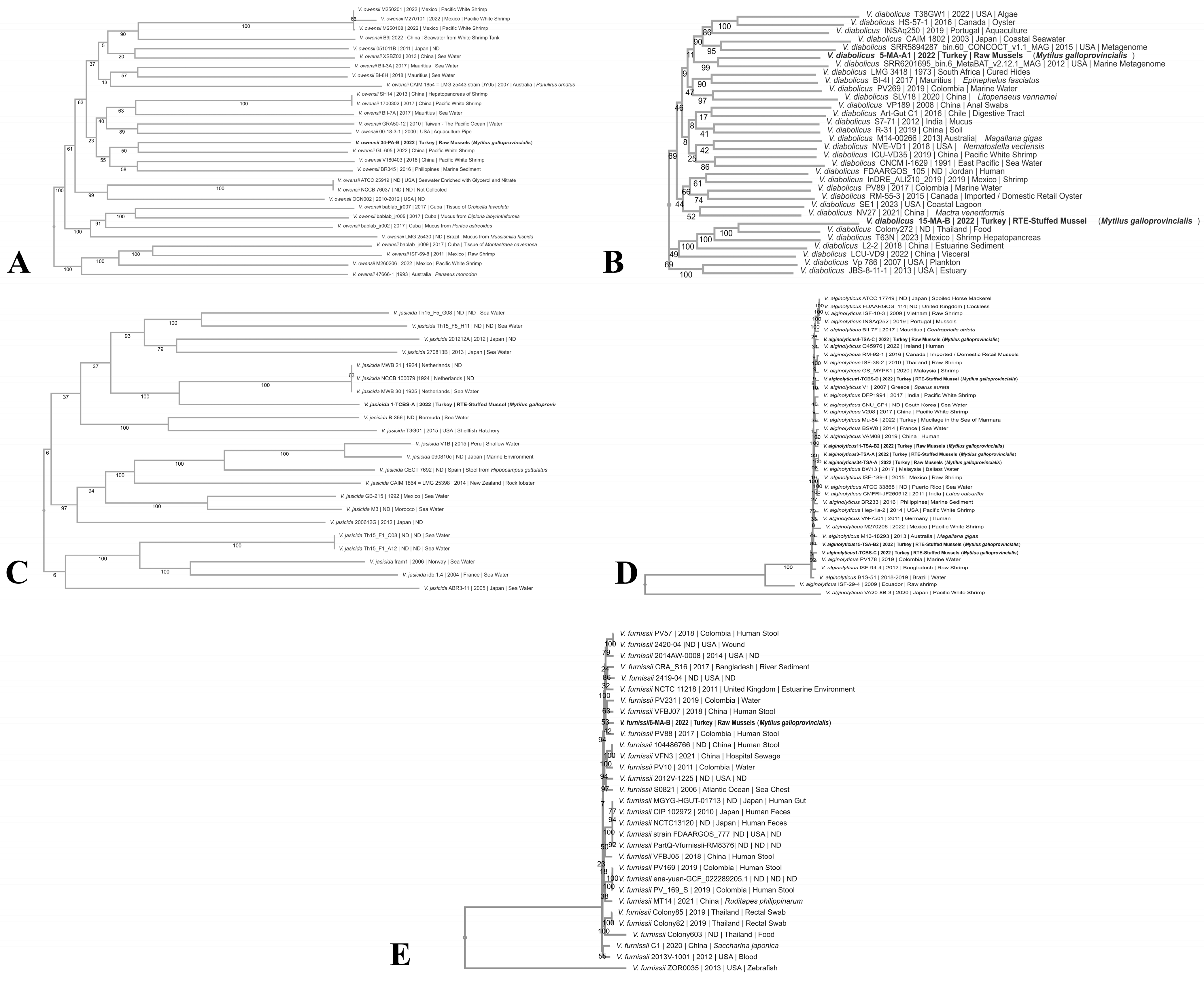
The draft genome sequences of the Vibrio strains were obtained. The genome sizes of the strains ranged from approximately 3.9 to 6.1 Mb, with the GC contents varying between 41.9% and 50.4%. The general characteristics of the genomes and identification results are presented in Table 3.

3.2. Phylogenomics

The phylogenomic relationships of V. alginolyticus, V. diabolicus, V. furnissii, V. owensii, and V. jasicida were examined using publicly available genome data; totals of 37, 33, 31, 31, and 24 genomes, respectively, were included for these species, with all the genomes providing sufficient data for analysis except for one genome of V. jasicida, which was excluded due to incomplete information. The analysis revealed significant genomic diversity among the species. While V. owensii, V. diabolicus, and V. jasicida exhibited heterogeneous genome similarity with strains from the NCBI database, V. alginolyticus and V. furnissii were positioned in well-defined phylogroups, closely related to strains available in the database.
The phylogenetic analysis identified species-specific relationships, shedding light on the evolutionary connections of these strains. V. owensii strain 34-PA-B was clustered with strains previously reported from Pacific white shrimp and marine sediment in China and the Philippines (Figure 1A). V. diabolicus strains 5-MA-A1 and 15-MA-B were located in distinct phylogenetic clusters, closely related to strains from the marine metagenome of the USA and food samples from Thailand (Figure 1B). The V. jasicida strain was grouped near strains isolated from seawater in the Netherlands, suggesting a potential geographical link or shared environmental niche (Figure 1C). The seven strains of V. alginolyticus exhibited broad genomic diversity, forming distinct clusters with isolates from marine water, raw shrimp, Pacific white shrimp, humans, seawater, and mussels from regions such as Ireland, Malaysia, Mexico, Colombia, and Bangladesh (Figure 1D). This wide distribution highlights the adaptive potential of V. alginolyticus to diverse ecological niches. In contrast, V. furnissii strains demonstrated a closer genetic relationship with strains from human stool, hospital sewage, water, and estuarine environments in China, Colombia, the UK, and Bangladesh (Figure 1E), indicating potential public health significance.
These findings highlight the diverse evolutionary trajectories of Vibrio species, emphasizing the genetic links between the isolates from seafood, human-associated environments, and aquatic habitats. The clustering of strains from geographically distant regions suggests the potential for global dissemination, possibly facilitated by the movement of seafood products or the natural dispersal of marine organisms.

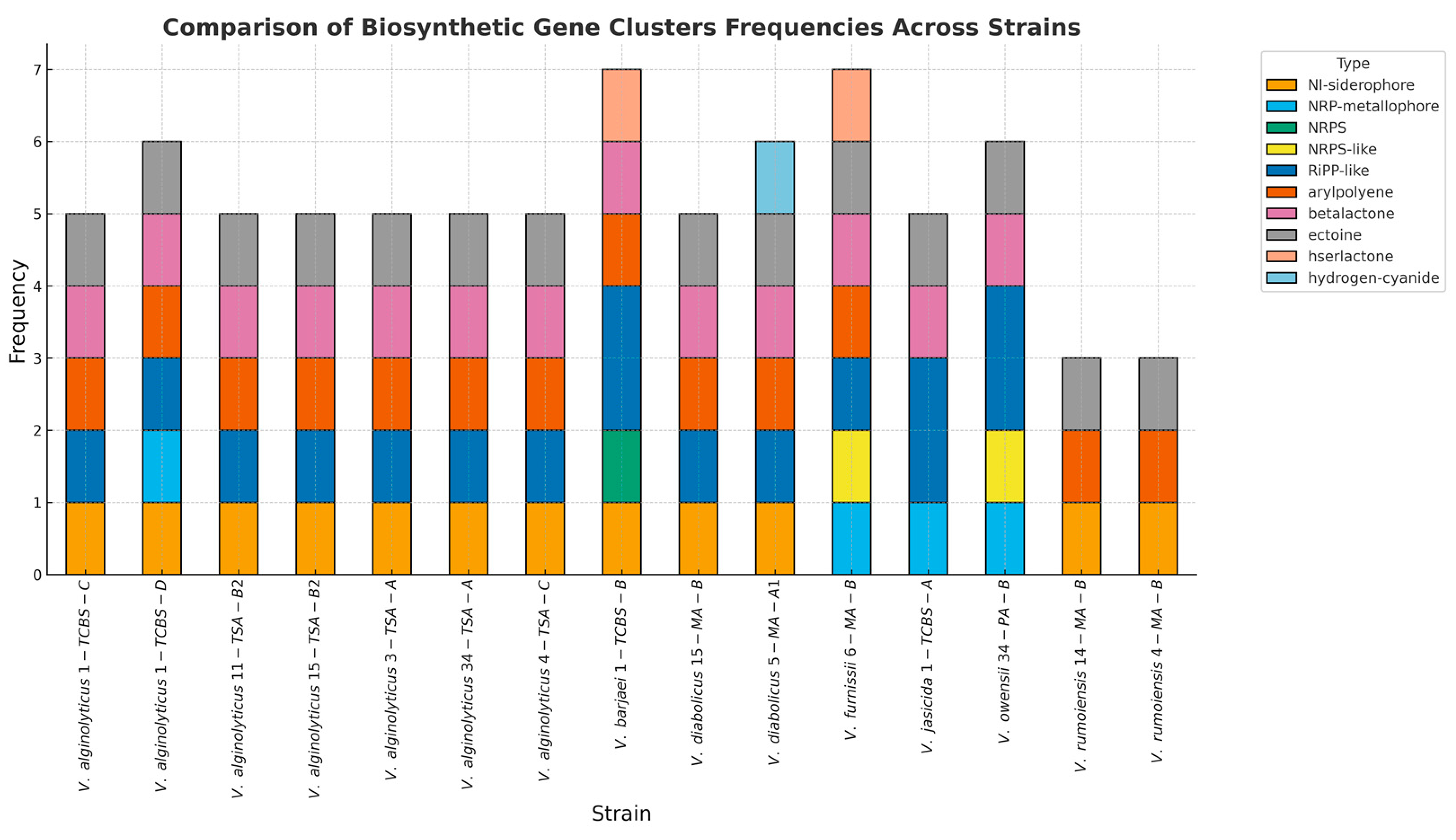
3.3. Biosynthetic Gene Clusters (BGCs) and Prophage

A total of nine distinct biosynthetic gene cluster (BGC) types were identified in the Vibrio genomes, including ectoine, non-ribosomal peptide synthetase (NRPS)/NRPS-like fragment, non-ribosomal peptide metallophore (NRP-metallophore), aryl polyene (APE), beta-lactone, ribosomally synthesized and post-translationally modified peptide product (RiPP-like), NI-siderophores, homoserine lactones (hserlactones), and hydrogen cyanide (HCN). The number of BGCs varied across the 15 Vibrio strains, ranging from three to eight BGCs per strain, with V. rumoiensis 4-MA-B having the lowest count (three BGCs) and V. furnissii 6-MA-B having the highest (eight BGCs). On average, each strain contained approximately six BGCs. The analysis revealed that NI-siderophore clusters were present in 12 of the 15 strains, showing similarity rates of 62% and 87% with aerobactin and other gene clusters, and a 100% similarity to vibrioferrin and related gene clusters. These findings, as illustrated in Figure 2, highlight the diversity and functional potential of BGCs in Vibrio strains, suggesting their role in survival, virulence, and adaptation to environmental conditions.
Prophage sequences and viral elements embedded in Vibrio genomes, which may influence bacterial virulence, diversity, and evolution, are summarized in Table S1. The analysis identified 27 prophage regions with lengths ranging from 5.4 to 63.6 Kb, of which 18 regions were classified as intact. The highest number of phage regions (four regions) was observed in the V. alginolyticus 4-TSA-C strain, while only one phage region was detected in each of the seven strains. These findings highlight the potential role of prophage elements in shaping the genomic diversity and functional potential of Vibrio species.

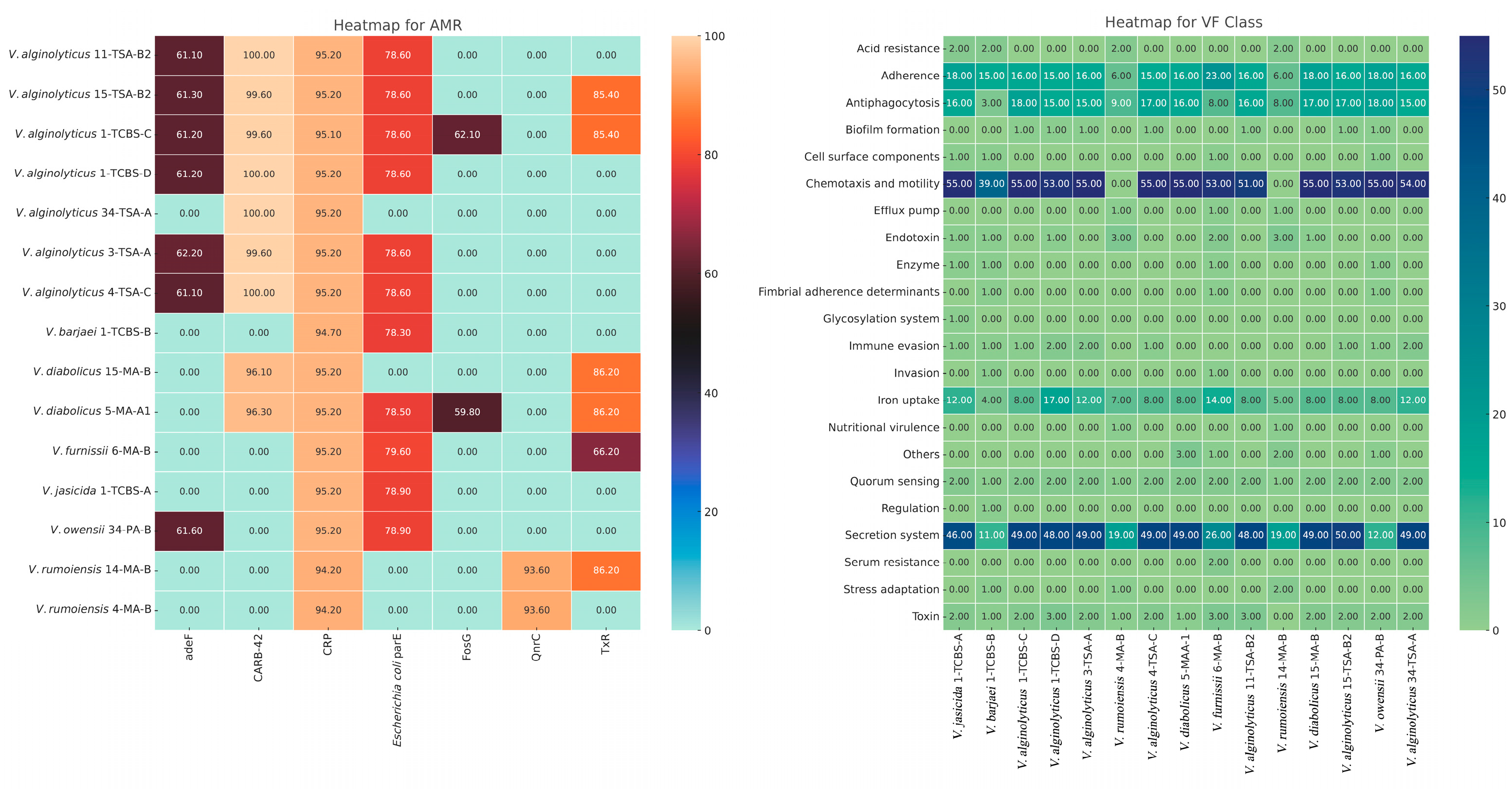
3.4. Antimicrobial Resistance (AMR) and Virulence Factor (VF) Genes

Figure 3 and Table S2 show antimicrobial resistance (AMR) genes in the genomes of the Vibrio species isolated from the FRMs and RTE-SMs utilizing the Comprehensive Antibiotic Resistance Database (CARD). The cyclic AMP receptor protein gene (CRP), which controls multifactorial fluoroquinolone susceptibility [41], was detected in all the Vibrio strains, with homology levels ranging from 94.29% to 95.24%. Notably, a D476N single-nucleotide polymorphism (SNP) was found in the Escherichia coli parE gene in 73.3% of the strains, with homology values between 78.34% and 79.62%, suggesting a potential mechanism for quinolone resistance. The highest number of AMR genes was detected in the V. alginolyticus strains isolated from the RTE-SM samples, specifically in V. alginolyticus 1-TCBS-C (TxR, adeF, FosG, blaCARB-42, CRP, and E. coli parE) and V. alginolyticus 15-TSA-B2 (adeF, CRP, TxR, qacG, blaCARB-42, and E. coli parE). In contrast, the lowest number of AMR genes was observed in the V. rumoiensis 4-MA-B (CRP and qnrC) and V. alginolyticus 34-TSA-A (blaCARB-42 and CRP) strains isolated from the FRM samples. The detection of qnrC in V. rumoiensis is significant, as it confers resistance to quinolones, a critical class of antibiotics. Resistance to tetracyclines was prevalent across the strains. The presence of the TxR and adeF genes, which are associated with tetracycline resistance, was observed in 10 strains. Additionally, the FosG gene, providing resistance to phosphonic acid derivatives, was identified in V. alginolyticus 1-TCBS-C and V. diabolicus 5-MA-A1. A noteworthy finding was the detection of the qacG gene, which encodes resistance to quaternary ammonium compounds (QACs), in V. alginolyticus 15-TSA-B2. This gene is linked to resistance against disinfectants and sanitizers, posing a significant concern for food processing environments. Beta-lactamase resistance genes, particularly blaCARB-42 and blaCARB-56, were identified in several Vibrio strains. All the V. alginolyticus strains carried the blaCARB-42 gene, with homology reaching 100% in some isolates, demonstrating resistance to penicillins and other β-lactam antibiotics. The highest prevalence of blaCARB-42 was observed in the V. alginolyticus strains, whereas blaCARB-56 was detected in V. diabolicus 5-MA-A1. The presence of these resistance genes indicates a strong capacity for resistance to β-lactams, which are crucial for clinical treatment. Overall, the presence of multiple AMR genes, including those encoding resistance to fluoroquinolones, tetracyclines, phosphonic acids, quaternary ammonium compounds, and β-lactams, highlights the multidrug resistance potential of these Vibrio species.
VF genes across 22 different VF classes, including adherence, anti-phagocytosis, chemotaxis and motility, enzyme, iron uptake, quorum sensing (QS), secretion system, toxin, acid resistance, biofilm formation, cell surface components, efflux pump, endotoxin, fimbrial adherence determinants, glycosylation system, immune evasion, invasion, nutritional virulence, others (O-antigen [Yersinia]), regulation, serum resistance, and stress adaptation, were detected, with notable differences in their presence across different Vibrio species (Table S3). Accessory colonization factors (acfA, acfB, acfC, and acfD) were not detected in our strains, although they were present in V. cholerae O1 biovar El Tor strain N16961 and V. cholerae O395. The QS molecule autoinducer-2 (AI-2; encoded by luxS) was detected in all the strains except for V. barjaei 1-TCBS-B. The other QS molecule, cholerae autoinducer-1 (CAI-1, encoded by cqsA), was not detected in two V. rumeniensis strains (4-MA-B and 14-MA-B) in this study. Our Vibrio strains were also closely related to those found in other bacterial taxa, such as Escherichia, Aeromonas, Pseudomonas, Haemophilus, Yersinia, Burkholderia, Klebsiella, Streptococcus, Acinetobacter, Shigella, Coxiella, Helicobacter, Mycobacterium, Neisseria, Salmonella, Campylobacter, and Francisella. The heatmap shows the distribution of VF gene counts across bacterial isolates. Rows correspond to the VF classes, while columns represent the bacterial isolates. The intensity of color indicates gene abundance, where darker red signifies higher counts, while blue represents lower counts. The hierarchical clustering of the bacterial isolates revealed distinct groupings, with isolates such as V. diabolicus and V. alginolyticus clustering together, while V. rumoiensis displayed a more distinct virulence profile (Figure 3).

3.5. Potential Human Pathogenicity

The pathogenic potential of the Vibrio strains was determined using the PathogenFinder tool (v1.1.) (Table 4 and Table S4). All the strains except for V. barjaei 1-TCBS-B and V. rumoiensis 4-MA-B were predicted as potential human pathogens. Based on these results, 13 Vibrio strains were predicted as potential human pathogens, with probabilities ranging from 0.535 to 0.864. V. barjaei 1-TCBS-B, from an RTE-SM sample, was categorized as a non-human pathogen, with a lower probability of 0.457. The highest probabilities of being human pathogens were attributed to the RTE-SM isolate V. diabolicus 15-MA-B (86.4%, with 49 pathogenic families) and the FRM strains V. diabolicus 5-MA-A1 (86.1%, with 49 pathogenic families), V. alginolyticus 11-TSA-B2 (84.9%, with 50 pathogenic families), and V. alginolyticus 4-TSA-C (84.9%, with 44 pathogenic families). The RAST annotation also confirmed that the genomes of the Vibrio strains encode genes for type I, II, III, IV, V, VI, and VII secretion systems, all of which play critical roles in VF delivery and interbacterial competition.
The analysis of CRISPR/Cas systems in V. rumoiensis, V. diabolicus, and V. owensii strains revealed the presence of CRISPR elements, but notably, no Cas (CRISPR-associated) genes were detected in any of the analyzed strains (Table S5). Only four Vibrio strains carried one or three CRISPR arrays, and in total, eight CRISPRs were identified, but no Cas genes. This finding is significant, as the functionality of the CRISPR system is typically associated with the presence of both CRISPR arrays and Cas genes, which together form the adaptive immune system of bacteria against phages and mobile genetic elements. CRISPR/Cas systems generally include a CRISPR array and Cas genes arranged in one or more operons. However, a significant proportion of CRISPR arrays are not adjacent to Cas genes [42], so we scrutinized the annotation of the genomes for the CRISPR-associated genes on the RAST tool.
The genome of V. owensii 34-PA-B has a CRISPR-associated protein of the Csx3 family with a 1224 bp length. The genome of V. rumoiensis 14-MA-B encodes a Csy3 family Cas protein (1026 bp), while the genome of V. diabolicus 5-MA-A1 also harbors a Csy4 family Cas protein (597 bp) adjacent to a Csy3 family protein (1044 bp). However, no CRISPR-associated proteins were detected in the genome of V. diabolicus 15-MA-B. One CRISPR array of 28 bp, consisting of two spacers, was identified in the genome of V. rumoiensis strain 4-MA-B. The genomes of V. rumoiensis 14-MA-B and V. owensii 34-PA-B showed multiple CRISPR arrays with varying spacer lengths and sequences, indicating active CRISPR systems potentially driven by diverse phage exposure. The CRISPR arrays found in the genomes of V. diabolicus 15-MA-B and V. owensii 34-PA-B had unique spacers, which are crucial for specifying the targets of the CRISPR defense mechanism. On the other hand, considering the low evidence level of CRISPR array detection in the CRISPRFinder tool and the absence of any Cas proteins, the result for V. diabolicus 15-MA-B might be evaluated as a false positive.
The CRISPR sequences identified in V. rumoiensis, V. diabolicus, and V. owensii displayed diverse consensus sequences. In V. rumoiensis 4-MA-B, two CRISPR elements were identified at positions 446,529–446,676 and 417,405–417,672, with consensus repeat sequences of “CTTACTAGCCACACACCCGATAGCACAC” at both loci. In V. rumoiensis 14-MA-B, the repeat sequences were “TTTCCTAGCTGCCTATTCGGCAGGTCAC” and “CGAGGTATTTTTGCAAACACGGA”, while in V. diabolicus 15-MA-B, the repeats were “TTTTGGAACAATAAAGTTTGTACAC” and “GTCATTCCGAGGAGCCTAAGCGA-CATCAGGAATCT”. Notably, V. owensii 34-PA-B had repeat sequences “GGCGGGTTCCGCGCTGGTTCCCGAGGCGGGGTCC” and “TTTAACCAAGATA-TAGGCCATTGGGATAC” at two distinct genomic loci. These diverse repeat sequences highlight the variability of CRISPR elements across Vibrio species.
Without Cas genes, the CRISPR arrays are unlikely to acquire new spacers, but the existing spacers may still play a role in bacterial regulatory processes. CRISPR arrays have been implicated in gene regulation, possibly acting as non-coding RNAs (ncRNAs) that influence transcription. In this context, CRISPRs may form secondary RNA structures that interact with regulatory proteins or small RNAs, thereby affecting gene expression.
Identifying genomic islands that encode mostly VFs, AMR genes, and other adaptation components is an effective way to use a population-based approach to genomic epidemiology and characterization [40]. The IslandCompare tool was used to detect and compare genomic islands found in the genomes of V. alginolyticus, V. diabolicus, and V. rumoiensis strains. Notably, V. alginolyticus strains showed considerable variation in the size, quantity, position, and identity of genomic islands encoded in their genomes (Figure 4 and Table S6).

4. Discussion

The main findings of this study were as follows: (i) Out of the 96 samples analyzed, 6 FRM samples (12.5%) and 2 RTE-SM samples (4.2%) were contaminated with Vibrio spp. (ii) A total of 15 Vibrio isolates from nine samples were identified, belonging to seven species. The most prevalent species was V. alginolyticus, which constituted 46.7% of the strains. We observed regional and seasonal variations in the prevalence of Vibrio spp., with a notable increase in contamination during the warmer summer months. (iii) Phylogenetic trees were constructed for five Vibrio species using genomic data from 33 to 37 genomes, depending on the species. The alignment of amino acids for phylogenetic analysis ranged from 252,823 to 379,159 amino acids. (iv) A genomic analysis identified nine different types of BGCs related to secondary metabolite production. The highest number of BGCs was eight, found in a V. furnissii strain. (v) Across the Vibrio isolates, a diverse array of AMR genes was detected. The CRP gene was found in all the strains, indicating a potential for broad-spectrum antimicrobial resistance. (vi) A prophage analysis within the Vibrio genomes revealed 27 prophage regions, 18 of which were intact. The presence of multiple VFs and secretion systems underscores the pathogenic potential of these strains.
Although Vibrio parahaemolyticus and Vibrio cholerae are well-known foodborne pathogenic Vibrio species, they were not detected in the FRM or RTE-SM samples in this study. In contrast to our findings, V. parahaemolyticus was reported in street food samples at a prevalence of 16.49% [43] and in fresh raw shellfish at a prevalence of 19.9% [44]. Additionally, contrary to our observations, V. cholerae was identified in FRM and RTE food samples in previous reports [45,46,47,48,49]. The most probable reason for the absence of V. parahaemolyticus and V. cholerae in our samples, despite their detection in previous studies, lies in the highly complex genetic similarity within the Vibrio genus. V. cholerae is monitored by the World Health Organization (WHO), and strict measures are taken to prevent the spread of cholera. The absence of V. cholerae could indicate that the mussels’ habitat was not contaminated by fecal matter or wastewater, which are common sources of this pathogen. This could reflect better water management or reduced anthropogenic impact in the sampling regions. The dominance of other Vibrio species, such as V. alginolyticus, in the sampled regions could also inhibit the growth and survival of V. cholerae due to competition for resources [50].
Our findings indicate a correlation between warmer months (June, July, and August) and an increased prevalence of Vibrio species, consistent with the elevated seawater and air temperatures during this period. Higher temperatures in the environment, particularly in seawater, seem to promote the growth and spread of Vibrio spp., as reported in previous studies [51,52]. For instance, in June, with seawater temperatures reaching 28.0 °C in R-1, we observed the presence of strains such as V. alginolyticus and V. furnissii in the FRM samples, supporting the idea that thermotolerant Vibrio species may contribute significantly to foodborne illnesses in summer [53]. V. alginolyticus, known for its resilience in warm conditions (up to 48 °C), was prevalent at multiple sampling points during these warmer months [54,55]. A study reported an 82.61% infection rate in sea bream during summer, which dropped to 30.23% in autumn [56]. Similarly, Mahmoud et al. (2022) [57] observed the highest incidence of V. alginolyticus in sea bass during summer (63.33%), followed by a much lower rate in winter (17.65%). This trend may be attributed to increased water temperatures during the summer, which create optimal conditions for the growth and proliferation of V. alginolyticus. Environmental factors, such as water quality and the presence of heavy metals, also play a critical role in the prevalence of V. alginolyticus. Elevated levels of heavy metals, including cadmium (Cd), lead (Pb), and nickel (Ni), have been linked to reduced fish immunity, making fish more susceptible to Vibrio infections [58,59]. In particular, V. alginolyticus has been found to have a positive correlation (r = 0.69) with higher water temperatures and increased heavy metal concentrations [60]. This suggests that elevated heavy metal concentrations may act as a stressor, weakening the immune systems of fish and providing V. alginolyticus with a competitive advantage over other microbial species. In addition, the concentration of iron (Fe) in the water has also been implicated in the higher prevalence of V. alginolyticus. Elevated iron levels recorded during the winter have been linked to increased V. alginolyticus infections. Iron is a key factor in bacterial growth and virulence, as it is an essential cofactor for many bacterial enzymes and metabolic pathways. As a result, increased iron availability may enhance the growth and colonization potential of V. alginolyticus, giving it an advantage over other species in the aquatic microbiota [61]. Collectively, the greater proportion of V. alginolyticus in our study may be attributed to a combination of seasonal temperature fluctuations, water quality parameters, and the bioavailability of heavy metals, including iron. These environmental factors create a favorable niche for Vibrio species. Furthermore, fish species, immunity status, and site-specific conditions likely contribute to the observed differences in the prevalence of Vibrio species between studies. Our findings are consistent with those of earlier reports highlighting the positive association between elevated temperatures, water quality, and Vibrio infections in aquaculture environments.
The consistent detection of V. alginolyticus and V. diabolicus in the RTE-SM samples from SP-3 in August, when temperatures were notably high (seawater at 25.4 °C and air peaking at 31.7 °C), suggests potential cross-contamination from raw mussels or inadequate cooking. Cross-contamination may occur through contact with raw mussel juices or improper cooking temperatures [62,63]. These findings underscore the importance of maintaining strict temperature controls during seafood preservation, keeping cold products below 5 °C and hot products above 60 °C, particularly in the summer months, to prevent Vibrio proliferation and ensure food safety [64,65,66].
Unlike previous studies that utilized sequence analyses at lengths of approximately 1.5–5 Kb, our study employed WGS with reads extending up to 6.1 Mb. Identifying Vibrio species at the genome level reduces the risk of the misidentification of closely related species, and it may also explain the different results between our study and previous studies. On the other hand, previous studies have determined that the genome lengths of Vibrio species have a wide range of 2.9–6.7 Mb, while their GC contents have a wide range of 38.0–57.2% [67]. The genome characteristics of the Vibrio species in our study are consistent with these studies, and these results also show that Vibrio genomes have high diversity. The phylogenetic analysis based on genome comparison showed that V. owensii, V. diabolicus, and V. jasicida are more heterogeneous than the V. alginolyticus and V. furnissii strains. While V. owensii, V. diabolicus, V. jasicida, and V. alginolyticus have been commonly found in mussels, marine water, and marine sediment, V. funissii is commonly reported from human stool, wound, and rectal swab samples in different countries. The genetic comparison shows a wide species distribution from different countries, from the USA to Thailand.
Several types of BGCs were identified in the Vibrio genomes, including those responsible for ectoine synthesis, NRPS/NRPS-like, APEs, beta-lactones, and NI-siderophores. These BGCs are recognized for their role in bacterial survival, virulence, biofilm formation, and utility in drug development [68,69,70,71,72,73]. All the Vibrio strains, except for V. barjaei 1-TCBS-B and V. owensii 34-PA-B, showed a significant presence of ectoine clusters, critical for the osmotic stress response in saline environments [74]. Notably, V. alginolyticus strains also exhibited a high incidence of siderophore-related clusters, enhancing their iron acquisition capabilities, which are vital in iron-depleted marine environments [75]. APE, a secondary metabolite thought to function in host immune system evasion, is widely found in Gram-negative pathogenic bacteria species [76]. APE BGCs were identified in all the strains except for V. alginolyticus 34-TSA-A, suggesting that they have protective properties against oxidative stress and are thought to play a role in biofilm formation [69]. Consistent with the findings in [77], the NRPS cluster detected in V. jasicida strain 1-TCBS-A in this study demonstrates the capacity of this strain to produce diverse and complex peptides that may have antibiotic properties. The antiSMASH analysis also detected homoserine lactone (hserlactone) clusters—which are generally used in the QS mechanism [78], which mainly regulates biofilm formation in microorganisms [79]—in the V. barjaei 1-TCBS-B and V. furnissii 6-MA-B strains.
Prophages can confer some features to their bacterial hosts, such as toxin production, resistance to environmental stresses, and enhanced virulence, which could impact the ecological fitness of Vibrio strains and their interactions with host organisms [80]. Certain prophages, such as PHAGE_Vibrio_VFJ_NC_021562 (in V. alginolyticus 4-TSA-C, V. furnissii 6-MA-B, and V. alginolyticus 11-TSA-B2) and PHAGE_Entero_DE3_NC_042057 (in V. alginolyticus 3-TSA-A and V. alginolyticus 34-TSA-A), were recurrent across multiple V. alginolyticus strains, suggesting a widespread distribution of these phage types within these Vibrio species. Consistent with our study [81], PHAGE_Vibrio_VFJ_NC_021562 was identified in many V. parahaemolyticus strains, but contrary to our results, they could not identify PHAGE_Entero_DE3_NC_042057, which had been previously identified in E. coli strains in only one study [82], in any Vibrio strain. These commonalities might reflect shared ecological niches or similar selective pressures. Identifying these prophages provides valuable insights into the genomic architecture and evolutionary dynamics of Vibrio species.
In light of the results that we obtained, numerous antimicrobial resistance mechanisms associated with Vibrio isolates were identified. One of the most prevalent mechanisms among pathogenic bacteria is the efflux pump system, which plays a significant role in both intrinsic and acquired antibiotic resistance in bacteria [83,84]; moreover, it is instrumental in regulating the internal environment by expelling toxic substances, quorum-sensing molecules, biofilm formation molecules, and bacterial VFs [85]. Another identified resistance mechanism was antibiotic target modification, which has become an increasingly common resistance strategy among pathogenic bacteria [86]. This resistance mechanism arises either as a result of genetic modifications within the bacteria or through enzymatic activities [86,87]. The resistance–nodulation–cell division (RND) efflux pump genes (CRP and adeF) were prevalent, suggesting a common resistance to multiple antibiotic classes, such as macrolides, fluoroquinolones, and penams [88]. In contrast to our findings, [89] reported that the qnr resistance gene, responsible for quinolone resistance, was the most common in 42 Vibrio strains isolated from shrimp. The presence of genes (blaCARB-42) responsible for β-lactamase resistance has been reported, especially in V. alginolyticus strains [90,91]. In the present study, as in other studies, the blaCARB-42 gene was detected in all the V. alginolyticus strains. The V. alginolyticus 15-TSA-B2 strain isolated from the RTE-SMs may have acquired resistance to disinfectants due to overuse/abuse and misuse in the RTE-SM production line [92]. V. furnissii strain 6-MA-B was also closely related to those found in other bacterial taxa, such as Escherichia, Aeromonas, Pseudomonas, P. aeruginosa, Haemophilus, Klebsiella, Salmonella, Klebsiella, Salmonella, Burkholderia, and Yersinia. Contrary to a study conducted in Italy [93], which showed the presence of aminoglycoside resistance genes (aacC2 and aadA), a β-lactamase resistance gene (blaTEM), a quinolone resistance gene (qnrS), a tetracycline resistance gene (tetD), and a glycopeptide resistance gene (vanB) in 24–66% of the Vibrio strains isolated from mussels (Mytilus galloprovincialis), these genes were not detected in our Vibrio strains.
Compared to other studies conducted in different parts of the world, this study reveals some important similarities and differences related to the presence of virulence genes within Vibrio species isolated from various seafood samples. A study from Egypt reported that the collagenase gene was present in all strains of V. alginolyticus isolated from seafood. Similarly, the genomes of all the Vibrio strains in the present study, except for V. barjaei 1-TCBS-B and V. furnissii 6-MA-B, encoded microbial collagenase genes. In parallel to the previous study, no strain in either investigation transported the gene for thermostable direct hemolysin, tdh [94].
Similarly, a study from South China identified 11 virulence genes, such as hflK, chiA, and flaC, among the Vibrio strains isolated from marine fish samples. For the present work, we did not determine the presence of the hflK and chiA genes in any of our strains; however, the flaC gene was present in V. barjaei 1-TCBS-B and V. furnissii 6-MA-B [95]. In Malaysia, [96] reported the occurrence of the chiA, luxR, and vhpA genes at 66.7% in Vibrio strains isolated from marine fish. However, these genes were not detected in our strains. In a previous study, 98% of Vibrio strains were positive for the tlh gene coding for thermolabile hemolysin, while in our study, the corresponding value was 80%; while 68% of their strains contained the flaC gene, only 13.3% of our strains showed its presence. Agreement with that Malaysian study is seen in the absence of the genes tdh or trh in any of our strains. The gene hlyA was found only in the V. furnissii 6-MA-B strain; likewise, it was not found in their Vibrio strains. The tox gene was not detected in either study. In Japan [97], the genes tlh, VPI, ompW, toxR, and ompU were detected in Vibrio strains; none of these were identified in our strains. Similarly, although the hlyA gene was detected in 3.9% of their isolates, we detected it in only one strain (6.7%). Furthermore, the tcpA, ctxA, ctxB, and tdh genes were detected at 0% in their research, concurrent with the present study’s findings.
The accessory colonization factor, which is encoded by the acfA, acfB, acfC, and acfD genes, is required for efficient intestinal colonization and biofilm formation and found in V. cholerae O1 biovar El Tor strain N1696 and V. cholerae strain O395 [98]; it was not detected in any Vibrio strain in our study. The VFDB analysis also identified the presence of mannose-sensitive hemagglutinin (MSHA type IV pilus), which is crucial for DNA uptake for horizontal gene transfer [99], initial attachment, and the colonization of host cells [100]. This VF was detected in all the Vibrio species, highlighting its widespread distribution among pathogenic strains. None of the Vibrio strains obtained from the samples showed the presence of well-known toxin genes such as ctxA and ctxB (encoding cholera toxin), vvhA (encoding hemolysin/cytolysin), tdh (encoding thermostable direct hemolysin), or rtxA, rtxB, rtxC, and rtxD (encoding RTX toxin). However, the thermolabile hemolysin (encoded by tlh) was detected in all the strains except for a few—e.g., V. barjaei 1-TCBS-B, V. rumoiensis 4-MA-B, and 14-MA-B—indicating a common virulence mechanism that may contribute to the pathogenicity of these species in causing the lysis of red blood cells [101] and diarrheal diseases. Similarly to our study, TLH has been detected in V. alginolyticus, V. diabolicus, V. harveyi, and V. parahaemolyticus strains in previous studies [102,103,104]. Capsular polysaccharide (CPS) VF genes were found in all the strains, underscoring their role in evading host immune responses [105]. The CPS-related wzc gene was the most common among the strains (93.3%), but the CPS-associated hp1 and wbfT genes were not detected in any isolates. Iron is critical for bacterial growth and virulence [106]. The widespread presence of the genes associated with iron uptake, such as enterobactin receptors (except in V. jasicida 1-TCBS-A, V. barjaei 1-TCBS-B, and V. owensii 34-PA-B) and heme receptors (except in V. barjaei 1-TCBS-B, V. rumoiensis 4-MA-B, and V. rumoiensis 14-MA-B), indicates that these Vibrio species have robust mechanisms for acquiring iron from the environment [107,108], which is essential for their survival and virulence. The absence of any flagella VF-associated genes in the V. rumoiensis 4-MA-B and 14-MA-B strains suggests possible differences in the motility and, hence, the infectivity of these strains. All the Vibrio genomes also contained factors of the type II secretion system. The widespread presence of genes encoding the extracellular protein secretion (EPS) type II secretion system across all the strains suggests a fundamental role for this system in the pathogenicity of Vibrio species, likely involved in the secretion of toxins and other effectors [109].
According to the results from PathogenFinder, potentially pathogenic Vibrio species are associated with different environmental niches [110] in the Marmara Sea of Türkiye. These predictions underscore the potential health risks posed by Vibrio strains in the FRM and RTE-SM samples, particularly those with high pathogenic potential, such as the V. diabolicus 5-MA-A1 and 15-MA-B strains. PathogenFinder served as a useful preliminary tool for estimating the pathogenic potential of the Vibrio strains based on genomic data. However, it should be emphasized that the tool has limitations, as it does not account for critical determinants of pathogenicity, such as VFs, sequence types (STs), serotypes, or host-specific traits. To address these limitations, complementary analyses, including the identification of VFs, AMR genes, and secretion systems, were performed in this study. These additional datasets strengthen the understanding of the potential pathogenicity of the studied Vibrio strains. Future research integrating genomic predictions with experimental validations will be necessary to fully assess their pathogenic risks.
Bacterial CRISPR/Cas systems, found in many bacteria, including Vibrio spp., provide adaptive immunity by defending against bacteriophages, plasmids, and other invaders [111]. Although CRISPR/Cas systems have been reported to be commonly found in Vibrio species [112,113,114], they were detected to a lesser extent in the present study. The results suggest that the four Vibrio strains (26.7%) possess a genetic defense mechanism against phage attack or invasion by foreign DNA—a crucial characteristic of these strains. Detecting CRISPR/Cas systems in the Vibrio species underscores the evolutionary arms race between these bacteria and their phage adversaries. The presence of these systems not only provides insights into the microbial immune mechanisms but also impacts the evolutionary dynamics of these bacteria, influencing their pathogenicity and survival strategies in marine environments and the food chain [114]. Previous studies [115,116] have demonstrated that “orphan” CRISPR arrays—those lacking Cas genes—can regulate gene expression through RNA interference-like mechanisms, such as base pairing with mRNA transcripts. Another explanation is that Cas genes may be located elsewhere in the genome or carried by mobile genetic elements, such as prophages or plasmids. Given that prophage sequences were detected in these strains, it is possible that the Cas genes required for the functionality of the CRISPR system are encoded within the phage-related regions. In some cases, the CRISPR system is co-opted by phages to serve as a defense mechanism against other phages, creating a phenomenon known as “CRISPR–phage warfare”. Therefore, the absence of Cas genes within the immediate vicinity of CRISPR arrays does not necessarily imply that the CRISPR system is non-functional. While no Cas genes were identified in the V. rumoiensis, V. diabolicus, or V. owensii strains, the detection of CRISPR arrays suggests that these elements may play regulatory roles or act as residual components of a previously active CRISPR/Cas system.
In gastroenteritis cases caused by Vibrio species, various VFs have been identified, including genes that encode toxins and other pathogenic mechanisms. While previous studies have often focused on thermostable direct hemolysin [117], our research has detected a range of virulence-associated genes, such as tlh, V. cholerae cytolysin, aerolysin (AerA), cytotoxic enterotoxin (Act), and the phytotoxins coronatine and phaseolotoxin. This finding underlines the diverse and complex mechanisms through which Vibrio species can cause disease. Additionally, non-O1/non-O139 V. cholerae serogroups exhibit T3SS-dependent virulence, which is essential for intestinal colonization and the onset of diarrhea. Documented outbreaks linked to these strains—such as those from the Chester River, USA—feature a composite genotype consisting of several virulence genes, including hlyA, stn, sto, hap, rtxA, nanH, vcsC, vspD, vcsN, vcsV, vasA, vasK, and vasH. The presence of these genes in environmental isolates underscores their potential to cause significant illness, revealing the adaptability of Vibrio strains in acquiring VFs [118]. The present study further highlights this complexity, as we detected many of these virulence genes in the non-O1/non-O139 V. cholerae isolates from mussel samples. The mechanisms through which Vibrio species can cause gastroenteritis involve a complex interplay of enzymes, toxins, and secretion systems. The discovery of a “virulence cocktail” in mussels suggests that consuming a single mussel harboring multiple Vibrio species with diverse virulence genes could lead to complex gastroenteritis symptoms and, potentially, to other severe health conditions. This underscores the importance of understanding and monitoring the full spectrum of VFs present in seafood in order to better assess public health risks.

5. Conclusions

The findings of this study provide valuable insights for identifying the genetic diversity and population structure of Vibrio spp. isolated from the FRM and RTE-SM samples. Various possible pathogenic Vibrio spp. were present in mussel-derived foodstuffs in this study, and most showed a detectable seasonal trend that makes these bacteria a potential food safety concern. The frequent detection of V. alginolyticus in several samples, especially during the warmer seasons, might suggest that higher environmental temperatures may increase the prevalence of diseases caused by pathogenic Vibrio species. Numerous strains, especially V. alginolyticus, carried VFs and AMR genes, indicating public health implications for seafood consumption. The analyses of BGCs, prophages, and the CRISPR/Cas system facilitated an extrapolation of the survival methods and the pathogenic capabilities of Vibrio strains. This study provides a foundation for future research, contributing valuable data on the ecological and genetic features of Vibrio spp., with significant implications for public health and marine microbiology.
To avoid gastrointestinal diseases from raw or RTE mussel products contaminated with Vibrio spp. and other seaborne pathogens, measures should include enforcing strict seawater quality standards, applying depuration processes, educating consumers and food handlers about the risks, promoting proper cooking methods, ensuring hygiene during handling and processing, maintaining proper service and sales conditions (particularly cold chain management), and increasing research on the AMR genes and VFs of these pathogens. These actions can significantly reduce health risks and improve public health outcomes.

Supplementary Materials

The following supporting information can be downloaded at https://www.mdpi.com/article/10.3390/pathogens14010052/s1, Table S1: Prophages detected in the genome of the Vibrio strains, Table S2: Antimicrobial resistance (AMR) genes detected in the Vibrio genomes using the Comprehensive Antibiotic Resistance Database (https://card.mcmaster.ca/analyze/rgi (accessed on 13 October 2024)), Table S3: Virulence factor-related genes predicted in the Vibrio genomes using the Virulence Factor Database (VFDB) (http://www.mgc.ac.cn/cgi-bin/VFs/v5/main.cgi (accessed on 13 October 2024)), Table S4: Matched pathogenic families in the genomes of Vibrio strains in PathogenFinder server (https://cge.food.dtu.dk/services/PathogenFinder/ (accessed on 13 October 2024)), Table S5: CRISPR-associated protein (Cas)-coding genes with CRISPR sequences detected on the CRISPRCasFinder (https://crisprcas.i2bc.paris-saclay.fr/CrisprCasFinder/Index (accessed on 13 October 2024)) server, Table S6: Genomic islands detected by the IslandCompare web tool (https://islandcompare.ca/ (accessed on 13 October 2024)).

Author Contributions

Conceptualization, A.Y., I.B.S. and M.D.; methodology, A.Y., I.B.S., H.A. and M.D.; formal analysis, A.Y., I.B.S., H.A., D.M. and M.D.; investigation, A.Y., I.B.S., N.A., G.T., F.G. and M.D.; resources, A.Y., I.B.S. and M.D.; data curation, A.Y., I.B.S., S.G. and M.D.; writing—original draft preparation, A.Y., I.B.S., S.G. and M.D.; writing—review and editing, A.Y., I.B.S., M.D., H.A., N.A., G.T., D.M., S.G. and F.G.; visualization, A.Y., I.B.S. and M.D.; supervision, A.Y., I.B.S. and M.D.; project administration, A.Y.; funding acquisition, A.Y., I.B.S., M.D. and S.G. All authors have read and agreed to the published version of the manuscript.

Funding

This research was funded by the Bursa Uludag University, Scientific Research Project Association Research Grant, grant number TGA-2024-1841. A part of the APC was funded by the Bursa Uludag University, Scientific Research Project Association Research Grant, grant number TGA-2024-1841, and the other part was funded by the authors.

Institutional Review Board Statement

Not applicable.

Informed Consent Statement

Not applicable.

Data Availability Statement

All the data available are included in the manuscript.

Acknowledgments

We would like to express our appreciation to Nedret Guclu for her valuable assistance during the laboratory work. We would also like to extend our gratitude to Bursa Uludag University, Scientific Research Project Association Research Grant, grant number TGA-2024-1759 for providing the equipment and consumable materials used in this research.

Conflicts of Interest

The authors declare no conflicts of interest.

References

  1. Ayvaz, Z. Geleneksel bir ürün olarak “Midye Dolma” ve gelecek önerileri. Ziraat Mühendisliği 2018, 366, 21–27. [Google Scholar] [CrossRef]
  2. Turkish Statistical Institute (TURKSTAT). Annual Statistical Report 2023. Available online: https://data.tuik.gov.tr/Bulten/Index?p=Su-Urunleri-2023-53702 (accessed on 16 December 2024).
  3. World Bank. Turkey Export Data for Product 030731 in 2023. Available online: https://wits.worldbank.org/trade/comtrade/en/country/TUR/year/2023/tradeflow/Exports/partner/ALL/product/030731 (accessed on 16 December 2024).
  4. Kahraman, Y.D.; Berik, N. Phenotypic and genotypic antibiotic resistance of Staphylococcus warneri and Staphylococcus pasteuri isolated from stuffed mussels. Aquat. Sci. Eng. 2024, 39, 172–178. [Google Scholar] [CrossRef]
  5. Kisla, D.; Uzgun, Y. Microbiological evaluation of stuffed mussels. J. Food Prot. 2008, 71, 616–620. [Google Scholar] [CrossRef] [PubMed]
  6. Baker-Austin, C.; Trinanes, J.; Gonzalez-Escalona, N.; Martinez-Urtaza, J. Non-cholera Vibrios: The microbial barometer of climate change. Trends Microbiol. 2017, 25, 76–84. [Google Scholar] [CrossRef]
  7. Destoumieux-Garzón, D.; Canesi, L.; Oyanedel, D.; Travers, M.-A.; Charrière, G.M.; Pruzzo, C.; Vezzulli, L. Vibrio–bivalve interactions in health and disease. Environ. Microbiol. 2020, 22, 4323–4341. [Google Scholar] [CrossRef] [PubMed]
  8. Martins, V.G.P.; Nascimento, J.D.S.; Martins, F.M.D.S.; Vigoder, H.C. Vibriosis and its impact on microbiological food safety. Food Sci. Technol. 2022, 42, 1–6. [Google Scholar] [CrossRef]
  9. Sampaio, A.; Silva, V.; Poeta, P.; Aonofriesei, F. Vibrio spp.: Life strategies, ecology, and risks in a changing environment. Diversity 2022, 14, 97. [Google Scholar] [CrossRef]
  10. European Food Safety Authority (EFSA). Vibrio Bacteria in Seafood: Increased Risk Due to Climate Change and Antimicrobial Resistance. EFSA. 2024. Available online: https://www.efsa.europa.eu/en/news/vibrio-bacteria-seafood-increased-risk-due-climate-change-and-antimicrobial-resistance (accessed on 16 October 2024).
  11. Derber, C.; Coudron, P.; Tarr, C.; Gladney, L.; Turnsek, M.; Shankaran, S.; Wong, E. Vibrio furnissii: An unusual cause of bacteremia and skin lesions after ingestion of seafood. J. Clin. Microbiol. 2011, 49, 2348–2349. [Google Scholar] [CrossRef]
  12. Zhou, K.; Tian, K.Y.; Liu, X.Q.; Liu, W.; Zhang, X.Y.; Liu, J.Y.; Sun, F. Characteristic and otopathogenic analysis of a Vibrio alginolyticus strain responsible for chronic otitis externa in China. Front. Microbiol. 2021, 12, 750642. [Google Scholar] [CrossRef]
  13. Yumoto, I.; Iwata, H.; Sawabe, T.; Ueno, K.; Ichise, N.; Matsuyama, H.; Okuyama, H.; Kawasaki, K. Characterization of a facultatively psychrophilic bacterium, Vibrio rumoiensis sp. nov., that exhibits high catalase activity. Appl. Environ. Microbiol. 1999, 65, 67–72. [Google Scholar] [CrossRef] [PubMed]
  14. Dubert, J.; Balboa, S.; Regueira, M.; González-Castillo, A.; Gómez-Gil, B.; Romalde, J.L. Vibrio barjaei sp. nov., a new species of the Mediterranei clade isolated in a shellfish hatchery. Syst. Appl. Microbiol. 2016, 39, 553–556. [Google Scholar] [CrossRef]
  15. Liu, L.; Xiao, J.; Zhang, M.; Zhu, W.; Xia, X.; Dai, X.; Pan, Y.; Yan, S.; Wang, Y. A Vibrio owensii strain as the causative agent of AHPND in cultured shrimp, Litopenaeus vannamei. J. Invertebr. Pathol. 2018, 153, 156–164. [Google Scholar] [CrossRef] [PubMed]
  16. Zhang, X.H.; He, X.; Austin, B. Vibrio harveyi: A serious pathogen of fish and invertebrates in mariculture. Mar. Life Sci. Technol. 2020, 2, 231–245. [Google Scholar] [CrossRef] [PubMed]
  17. Song, J.; Liu, X.; Wu, C.; Zhang, Y.; Fan, K.; Zhang, X.; Wei, Y. Isolation, identification and pathogenesis study of Vibrio diabolicus. Aquaculture 2021, 533, 736043. [Google Scholar] [CrossRef]
  18. Deng, Y.; Xu, H.; Su, Y.; Liu, S.; Xu, L.; Guo, Z.; Wu, J.; Cheng, C.; Feng, J. Horizontal gene transfer contributes to virulence and antibiotic resistance of Vibrio harveyi 345 based on complete genome sequence analysis. BMC Genomics 2019, 20, 761. [Google Scholar] [CrossRef] [PubMed]
  19. Goulden, E.F.; Hall, M.R.; Bourne, D.G.; Pereg, L.L.; Høj, L. Pathogenicity and infection cycle of Vibrio owensii in larviculture of the Ornate Spiny Lobster (Panulirus ornatus). Appl. Environ. Microbiol. 2012, 78, 2841–2849. [Google Scholar] [CrossRef]
  20. Harrison, J.; Nelson, K.; Morcrette, H.; Morcrette, C.; Preston, J.; Helmer, L.; Titball, R.W.; Butler, C.S.; Wagley, S. The increased prevalence of Vibrio species and the first reporting of Vibrio jasicida and Vibrio rotiferianus at UK shellfish sites. Water Res. 2022, 211, 117942. [Google Scholar] [CrossRef]
  21. Food and Agriculture Organization of the United Nations (FAO); World Health Organization (WHO). Available online: https://openknowledge.fao.org/server/api/core/bitstreams/2b4234e8-a0e1-45b6-8574-e1560e6405cf/content (accessed on 17 October 2024).
  22. Mititelu, M.; Neacșu, S.M.; Oprea, E.; Dumitrescu, D.E.; Nedelescu, M.; Drăgănescu, D.; Nicolescu, T.O.; Roșca, A.C.; Ghica, M. Black Sea mussels qualitative and quantitative chemical analysis: Nutritional benefits and possible risks through consumption. Nutrients 2022, 14, 964. [Google Scholar] [CrossRef]
  23. Bakshi, B.; Bouchard, R.W., Jr.; Dietz, R.; Hornbach, D.; Monson, P.; Sietman, B.; Wasley, D. Freshwater mussels, ecosystem services, and clean water regulation in Minnesota: Formulating an effective conservation strategy. Water 2023, 15, 2560. [Google Scholar] [CrossRef]
  24. Centers for Disease Control and Prevention (CDC). Available online: https://www.cdc.gov/vibrio/about/index.html (accessed on 17 October 2024).
  25. Kumarage, P.M.; De Silva, L.A.D.S.; Heo, G.J. Aquatic environments: A potential source of antimicrobial-resistant Vibrio spp. J. Appl. Microbiol. 2022, 133, 2267–2279. [Google Scholar] [CrossRef] [PubMed]
  26. Stephen, J.; Lekshmi, M.; Ammini, P.; Kumar, S.H.; Varela, M.F. Membrane efflux pumps of pathogenic Vibrio species: Role in antimicrobial resistance and virulence. Microorganisms 2022, 10, 382. [Google Scholar] [CrossRef] [PubMed]
  27. Yibar, A.; Saticioglu, I.B.; Ajmi, N.; Duman, M. Molecular characterization and antibacterial resistance determination of Escherichia coli isolated from fresh raw mussels and ready-to-eat stuffed mussels: A major public health concern. Pathogens 2024, 13, 532. [Google Scholar] [CrossRef] [PubMed]
  28. Vural, P.; Yildiz, H.; Acarli, S. Growth and survival performances of Mediterranean mussel (Mytilus galloprovincialis, Lamarck, 1819) on different depths in Cardak lagoon, Dardanelles. Mar. Sci. Technol. Bull. 2015, 4, 7–12. [Google Scholar]
  29. Pretto, T. Vibrio harveyi group. In Diagnostic Manual for the Main Pathogens in European Seabass and Gilthead Seabream Aquaculture; Zrncic, S., Ed.; International Centre for Advanced Mediterranean Agronomic Studies (CIHEAM): Zaragoza, Spain, 2020; pp. 75–82. [Google Scholar]
  30. Vázquez-Rosas-Landa, M.; Ponce-Soto, G.Y.; Aguirre-Liguori, J.A.; Thakur, S.; Scheinvar, E.; Barrera-Redondo, J.; Ibarra-Laclette, E.; Guttman, D.S.; Eguiarte, L.E.; Souza, V. Population genomics of Vibrionaceae isolated from an endangered oasis reveals local adaptation after an environmental perturbation. BMC Genom. 2020, 21, 418. [Google Scholar] [CrossRef] [PubMed]
  31. Meier-Kolthoff, J.P.; Göker, M. TYGS is an automated high-throughput platform for state-of-the-art genome-based taxonomy. Nat. Commun. 2019, 10, 2182. [Google Scholar] [CrossRef] [PubMed]
  32. Tatusova, T.; DiCuccio, M.; Badretdin, A.; Chetvernin, V.; Nawrocki, E.P.; Zaslavsky, L.; Ostell, J. NCBI prokaryotic genome annotation pipeline. Nucleic Acids Res. 2016, 44, 6614–6624. [Google Scholar] [CrossRef] [PubMed]
  33. Brettin, T.; Davis, J.J.; Disz, T.; Edwards, R.A.; Gerdes, S.; Olsen, G.J.; Xia, F. RASTtk: A modular and extensible implementation of the RAST algorithm for building custom annotation pipelines and annotating batches of genomes. Sci. Rep. 2015, 5, 8365. [Google Scholar] [CrossRef] [PubMed]
  34. Olson, R.D.; Assaf, R.; Brettin, T.; Conrad, N.; Cucinell, C.; Davis, J.J.; Dempsey, D.M.; Dickerman, A.; Dietrich, E.M.; Kenyon, R.W.; et al. Introducing the Bacterial and Viral Bioinformatics Resource Center (BV-BRC): A resource combining PATRIC, IRD, and ViPR. Nucleic Acids Res. 2023, 51, D678–D689. [Google Scholar] [CrossRef] [PubMed]
  35. Alcock, B.P.; Raphenya, A.R.; Lau, T.T.Y.; Tsang, K.K.; Bouchard, M.; Edalatmand, A.; Huynh, W.; Nguyen, A.V.; Cheng, A.A.; Liu, S.; et al. CARD 2020: Antibiotic resistome surveillance with the comprehensive antibiotic resistance database. Nucleic Acids Res. 2020, 48, D517–D525. [Google Scholar] [CrossRef] [PubMed]
  36. Liu, B.; Zheng, D.; Zhou, S.; Chen, L.; Yang, J. VFDB 2022: A general classification scheme for bacterial virulence factors. Nucleic Acids Res. 2022, 50, D912–D917. [Google Scholar] [CrossRef] [PubMed]
  37. Blin, K.; Shaw, S.; Augustijn, H.E.; Reitz, Z.L.; Biermann, F.; Alanjary, M.; Fetter, A.; Terlouw, B.R.; Metcalf, W.W.; Helfrich, E.J.N.; et al. AntiSMASH 7.0: New and improved predictions for detection, regulation, chemical structures and visualisation. Nucleic Acids Res. 2023, 51, W46–W50. [Google Scholar] [CrossRef] [PubMed]
  38. Wishart, D.S.; Han, S.; Saha, S.; Oler, E.; Peters, H.; Grant, J.R.; Stothard, P.; Gautam, V. PHASTEST: Faster than PHASTER, better than PHAST. Nucleic Acids Res. 2023, 51, W443–W450. [Google Scholar] [CrossRef]
  39. Cosentino, S.; Voldby Larsen, M.; Møller Aarestrup, F.; Lund, O. PathogenFinder—Distinguishing friend from foe using bacterial whole genome sequence data. PLoS ONE 2013, 8, e77302. [Google Scholar] [CrossRef]
  40. Bertelli, C.; Gray, K.L.; Woods, N.; Lim, A.C.; Tilley, K.E.; Winsor, G.L.; Brinkman, F.S.L. Enabling genomic island prediction and comparison in multiple genomes to investigate bacterial evolution and outbreaks. Microb. Genom. 2022, 8, 000818. [Google Scholar] [CrossRef] [PubMed]
  41. Kary, S.C.; Yoneda, J.R.K.; Olshefsky, S.C.; Stewart, L.A.; West, S.B.; Cameron, A.D.S. The global regulatory cyclic AMP receptor protein (CRP) controls multifactorial fluoroquinolone susceptibility in Salmonella enterica Serovar Typhimurium. Antimicrob. Agents Chemother. 2017, 61, e01666-17. [Google Scholar] [CrossRef]
  42. Shmakov, S.A.; Utkina, I.; Wolf, Y.I.; Makarova, K.S.; Severinov, K.V.; Koonin, E.V. CRISPR Arrays Away from Cas Genes. CRISPR J. 2020, 3, 535–549. [Google Scholar] [CrossRef] [PubMed]
  43. Beshiru, A.; Igbinosa, E.O. Surveillance of Vibrio parahaemolyticus pathogens recovered from ready-to-eat foods. Sci. Rep. 2023, 13, 4186. [Google Scholar] [CrossRef] [PubMed]
  44. Ashrafudoulla, M.; Na, K.W.; Hossain, M.I.; Mizan, M.F.R.; Nahar, S.; Toushik, S.H.; Roy, P.K.; Park, S.H.; Ha, S. Molecular and pathogenic characterization of Vibrio parahaemolyticus isolated from seafood. Mar. Pollut. Bull. 2021, 172, 112927. [Google Scholar] [CrossRef] [PubMed]
  45. Ahmed, O.M.; Amin, H.F. Detection and survival of Vibrio species in shrimp (Penaeus indicus) and mussel (Mytilus galloprovincialis) at landing and after processing at seafood markets in Suez, Egypt. J. Food Dairy Sci. 2018, 9, 411–417. [Google Scholar] [CrossRef]
  46. Tang, J.-Y.-H.; Farhana Sakinah, M.R.; Nakaguchi, Y.; Nishibuchi, M.; Chai, L.-C.; New, C.Y.; Son, R. Detection of Vibrio cholerae in street food (satar and otak-otak) by Loop-Mediated Isothermal Amplification (LAMP), multiplex Polymerase Chain Reaction (mPCR) and plating methods. Food Res. 2018, 2, 447–452. [Google Scholar] [CrossRef]
  47. Nuñal, S.N.; Jane M Monaya, K.; Rose T Mueda, C.; Mae Santander-De Leon, S. Microbiological quality of oysters and mussels along its market supply chain. J. Food Prot. 2023, 86, 100063. [Google Scholar] [CrossRef] [PubMed]
  48. Preeprem, S.; Aksonkird, T.; Nuidate, T.; Hajimasalaeh, W.; Hajiwangoh, Z.; Mittraparp-arthorn, P. Characterization and genetic relationships of Vibrio spp. isolated from seafood in retail markets, Yala, Thailand. Trends Sci. 2023, 20, 5962. [Google Scholar] [CrossRef]
  49. Kering, K.; Wang, Y.; Mbae, C.; Mugo, M.; Ongadi, B.; Odityo, G.; Muturi, P.; Yakubu, H.; Liu, P.; Durry, S.; et al. Pathways of exposure to Vibrio cholerae in an urban informal settlement in Nairobi, Kenya. PLOS Glob. Public Health 2024, 4, e0002880. [Google Scholar] [CrossRef]
  50. Zohra, T.; Ikram, A.; Salman, M.; Amir, A.; Saeed, A.; Ashraf, Z.; Ahad, A. Wastewater-based environmental surveillance of toxigenic Vibrio cholerae in Pakistan. PLoS ONE 2021, 16, e0257414. [Google Scholar] [CrossRef] [PubMed]
  51. Galbraith, P.S.; Larouche, P.; Chasse, J.; Petrie, B. Sea-surface temperature in relation to air temperature in the Gulf of St. Lawrence: Interdecadal variability and long term trends. Deep-Sea Res. Part II Top. Stud. Oceanogr. 2012, 77–80, 10–20. [Google Scholar] [CrossRef]
  52. Feng, X.; Haines, K.; de Boisseson, E. Coupling of surface air and sea surface temperatures in the CERA-20C re-analysis. Q. J. R. Meteorol. Soc. 2018, 144, 195–207. [Google Scholar] [CrossRef]
  53. Baker-Austin, C.; Trinanes, J.A.; Taylor, N.G.; Hartnell, R.; Siitonen, A. Emerging Vibrio risk at high latitudes in response to ocean warming. Nat. Clim. Chang. 2013, 3, 73–77. [Google Scholar] [CrossRef]
  54. Gu, D.; Guo, M.; Yang, M.; Zhang, Y.; Zhou, X.; Wang, Q. A ςe-mediated temperature gauge controls a switch from luxr-mediated virulence gene expression to thermal stress adaptation in Vibrio alginolyticus. PLoS Pathog. 2016, 12, e1005645. [Google Scholar] [CrossRef]
  55. Sheikh, H.I.; Najiah, M.; Fadhlina, A.; Laith, A.A.; Nor, M.M.; Jalal, K.C.A.; Kasan, N.A. Temperature upshift mostly but not always enhances the growth of Vibrio species: A systematic review. Front. Mar. Sci. 2022, 9, 959830. [Google Scholar] [CrossRef]
  56. Abd El Tawab, A.; Ibrahim, A.M.; Sittien, A. Phenotypic and genotypic characterization of Vibrio species isolated from marine fishes. Benha Vet. Med. J. 2018, 34, 79–93. [Google Scholar] [CrossRef]
  57. Mahmoud, S.; El-Bouhy, Z.; Hassanin, M.; Fadel, A.H. Vibrio alginolyticus and Photobacterium damselae subsp. damselae: Prevalence, histopathology and treatment in sea bass Dicentrarchus labrax. J. Pharm. Chem. Biol. Sci. 2018, 5, 354–364. [Google Scholar]
  58. Elgendy, M.; Soliman, W.; Hassan, H.; Kenawy, A.; Liala, A.M. Effect of abrupt environmental deterioration on the eruption of vibriosis in mari-cultured shrimp, Penaeus indicus, in Egypt. J. Fish Aquat. Sci. 2015, 10, 146–158. [Google Scholar] [CrossRef]
  59. Ismail, E.T.; El-Son, M.A.M.; El-Gohary, F.A.; Zahran, E. Prevalence, genetic diversity, and antimicrobial susceptibility of Vibrio spp. infected gilthead sea breams from coastal farms at Damietta, Egypt. BMC Vet. Res. 2024, 20, 129. [Google Scholar] [CrossRef] [PubMed]
  60. Richards, J.G. Metabolic and molecular responses of fish to hypoxia. In Fish Physiology; Elsevier: Amsterdam, The Netherlands, 2009; Volume 27, pp. 443–485. [Google Scholar]
  61. Huang, Y.; Du, P.; Zhao, M.; Liu, W.; Du, Y.; Diao, B.; Li, J.; Kan, B.; Liang, W. Functional characterization and conditional regulation of the type VI secretion system in Vibrio fluvialis. Front. Microbiol. 2017, 8, 528. [Google Scholar] [CrossRef] [PubMed]
  62. Brauge, T.; Mougin, J.; Ells, T.; Midelet, G. Sources and contamination routes of seafood with human pathogenic Vibrio spp.: A farm-to-fork approach. Compreh. Rev. Food Sci. Food Saf. 2023, 23, e13283. [Google Scholar] [CrossRef] [PubMed]
  63. Al-Garadi, M.A.; Aziz, R.N.; Almashhadany, D.A.; Al Qabili, D.M.A.; Abdullah Aljoborey, A.D. Validity of cold storage and heat treatment on the deactivation of Vibrio parahaemolyticus isolated from fish meat markets. Ital. J. Food Saf. 2024, 13, 11516. [Google Scholar] [CrossRef] [PubMed]
  64. European Commission (EC). Opinion of the Scientific Committee on Veterinary Measures Relating to Public Health on Vibrio vulnificus and Vibrio parahaemolyticus (in Raw and Undercooked Seafood). European Commission. 2001. Available online: https://food.ec.europa.eu/system/files/2020-12/sci-com_scv_out45_en.pdf (accessed on 19 October 2024).
  65. Hara-Kudo, Y.; Kumagai, S. Impact of seafood regulations for Vibrio parahaemolyticus infection and verification by analyses of seafood contamination and infection. Epidemiol. Infect. 2014, 142, 2237–2247. [Google Scholar] [CrossRef] [PubMed]
  66. NSW Food Authority. Potentially Hazardous Foods—Foods that Require Temperature Control for Safety. NSW Government. 2023. Available online: https://www.foodauthority.nsw.gov.au/sites/default/files/_Documents/industry/potentially_hazardous_foods.pdf (accessed on 19 October 2024).
  67. Lin, H.; Yu, M.; Wang, X.; Zhang, X.H. Comparative genomic analysis reveals the evolution and environmental adaptation strategies of vibrios. BMC Genom. 2018, 19, 135. [Google Scholar] [CrossRef] [PubMed]
  68. Schwibbert, K.; Marin-Sanguino, A.; Bagyan, I.; Heidrich, G.; Lentzen, G.; Seitz, H.; Rampp, M.; Schuster, S.C.; Klenk, H.P.; Pfeiffer, F.; et al. A blueprint of ectoine metabolism from the genome of the industrial producer Halomonas elongata DSM 2581 T. Environ. Microbiol. 2011, 13, 1973–1994. [Google Scholar] [CrossRef] [PubMed]
  69. Johnston, I.; Osborn, L.J.; Markley, R.L.; McManus, E.A.; Kadam, A.; Schultz, K.B.; Nagajothi, N.; Ahern, P.P.; Brown, J.M.; Claesen, J. Identification of essential genes for Escherichia coli aryl polyene biosynthesis and function in biofilm formation. NPJ Biofilms Microbiomes 2021, 7, 56. [Google Scholar] [CrossRef] [PubMed]
  70. Lee, W.C.; Choi, S.; Jang, A.; Son, K.; Kim, Y. Structural comparison of Acinetobacter baumannii β-ketoacyl-acyl carrier protein reductases in fatty acid and aryl polyene biosynthesis. Sci. Rep. 2021, 11, 7945. [Google Scholar] [CrossRef]
  71. Timofeeva, A.M.; Galyamova, M.R.; Sedykh, S.E. Bacterial siderophores: Classification, biosynthesis, perspectives of use in agriculture. Plants 2022, 11, 3065. [Google Scholar] [CrossRef] [PubMed]
  72. Džunková, M.; La Clair, J.J.; Tyml, T.; Doud, D.; Schulz, F.; Piquer-Esteban, S.; Sanchis, D.P.; Osborn, A.; Robinson, D.; Louie, K.B.; et al. Synthase-selected sorting approach identifies a beta-lactone synthase in a nudibranch symbiotic bacterium. Microbiome 2023, 11, 130. [Google Scholar] [CrossRef] [PubMed]
  73. Ranjan, A.; Rajput, V.D.; Prazdnova, E.V.; Gurnani, M.; Bhardwaj, P.; Sharma, S.; Sushkova, S.; Mandzhieva, S.S.; Minkina, T.; Sudan, J.; et al. Nature’s antimicrobial arsenal: Non-ribosomal peptides from PGPB for plant pathogen biocontrol. Fermentation 2023, 9, 597. [Google Scholar] [CrossRef]
  74. Yoo, Y.; Lee, H.; Lee, J.; Khim, J.S.; Kim, J. Insights into saline adaptation strategies through a novel halophilic bacterium isolated from solar saltern of Yellow Sea. Front. Mar. Sci. 2023, 10, 1229444. [Google Scholar] [CrossRef]
  75. Cai, H.; Yu, J.; Li, Q.; Zhang, Y.; Huang, L. Research Progress on Virulence Factors of Vibrio alginolyticus: A Key Pathogenic Bacteria of Sepsis; IntechOpen: London, UK, 2024. [Google Scholar] [CrossRef]
  76. Lee, W.C.; Choi, S.; Jang, A.; Yeon, J.; Hwang, E.; Kim, Y. Structural basis of the complementary activity of two ketosynthases in aryl polyene biosynthesis. Sci. Rep. 2021, 11, 16340. [Google Scholar] [CrossRef] [PubMed]
  77. Zane, H.K.; Naka, H.; Rosconi, F.; Sandy, M.; Haygood, M.G.; Butler, A. Biosynthesis of amphi-enterobactin siderophores by Vibrio harveyi BAA-1116: Identification of a bifunctional nonribosomal peptide synthetase condensation domain. J. Am. Chem. Soc. 2014, 136, 5615–5618. [Google Scholar] [CrossRef] [PubMed]
  78. Cunha-Ferreira, I.C.; Vizzotto, C.S.; Frederico, T.D.; Peixoto, J.; Carvalho, L.S.; Tótola, M.R.; Krüger, R.H. Impact of Paenibacillus elgii supernatant on screening bacterial strains with potential for biotechnological applications. Eng. Microbiol. 2024, 4, 100163. [Google Scholar] [CrossRef]
  79. Khalid, S.J.; Ain, Q.; Khan, S.J.; Jalil, A.; Siddiqui, M.F.; Ahmad, T.; Badshah, M.; Adnan, F. Targeting Acyl Homoserine Lactones (AHLs) by the quorum quenching bacterial strains to control biofilm formation in Pseudomonas aeruginosa. Saudi J. Biol. Sci. 2022, 29, 1673–1682. [Google Scholar] [CrossRef] [PubMed]
  80. Dougherty, P.E.; Nielsen, T.K.; Riber, L.; Lading, H.H.; Milena, L.; Kot, W.; Raaijmakers, J.M.; Hansen, L.H. Widespread and largely unknown prophage activity, diversity, and function in two genera of wheat phyllosphere bacteria. ISME J. 2023, 17, 2415–2425. [Google Scholar] [CrossRef]
  81. Sorée, M. Towards a Better Overview of Virulence and Risk Management of Vibrio parahaemolyticus, a Marine Bacterium Potentially Pathogenic for Humans. Ph.D. Thesis, Université de Bretagne Occidentale, Brest, France, 2022. [Google Scholar]
  82. Neumann, B.; Lippmann, N.; Wendt, S.; Karlas, T.; Lübbert, C.; Werner, G.; Pfeifer, Y.; Schuster, C.F. Recurrent bacteremia with a hypermucoviscous Escherichia coli isolated from a patient with perihilar cholangiocarcinoma: Insights from a comprehensive genome-based analysis. Ann. Clin. Microbiol. Antimicrob. 2022, 21, 28. [Google Scholar] [CrossRef] [PubMed]
  83. Webber, M.A.; Piddock, L.J. The importance of efflux pumps in bacterial antibiotic resistance. J. Antimicrob. Chemother. 2003, 51, 9–11. [Google Scholar] [CrossRef] [PubMed]
  84. Gaurav, A.; Bakht, P.; Saini, M.; Pandey, S.; Pathania, R. Role of bacterial efflux pumps in antibiotic resistance, virulence, and strategies to discover novel efflux pump inhibitors. Microbiology 2023, 169, 001333. [Google Scholar] [CrossRef]
  85. Piddock, L.J. Multidrug-resistance efflux pumps—Not just for resistance. Nat. Rev. Microbiol. 2006, 4, 629–636. [Google Scholar] [CrossRef]
  86. Schaenzer, A.J.; Wright, G.D. Antibiotic resistance by enzymatic modification of antibiotic targets. Trends Mol. Med. 2020, 26, 768–782. [Google Scholar] [CrossRef]
  87. De Pascale, G.; Wright, G.D. Antibiotic resistance by enzyme inactivation: From mechanisms to solutions. Chembiochem 2010, 11, 1325–1334. [Google Scholar] [CrossRef] [PubMed]
  88. Karpov, D.S.; Kazakova, E.M.; Kovalev, M.A.; Shumkov, M.S.; Kusainova, T.; Tarasova, I.A.; Osipova, P.J.; Poddubko, S.V.; Mitkevich, V.A.; Kuznetsova, M.V.; et al. Determinants of antibiotic resistance and virulence factors in the genome of Escherichia coli APEC 36 strain isolated from a broiler chicken with generalized colibacillosis. Antibiotics 2024, 13, 945. [Google Scholar] [CrossRef] [PubMed]
  89. Hirshfeld, B.; Lavelle, K.; Lee, K.Y.; Atwill, E.R.; Kiang, D.; Bolkenov, B.; Gaa, M.; Li, Z.; Yu, A.; Li, X.; et al. Prevalence and antimicrobial resistance profiles of Vibrio spp. and Enterococcus spp. in retail shrimp in Northern California. Front. Microbiol. 2023, 14, 1192769. [Google Scholar] [CrossRef]
  90. Yasir, M.; Ullah, R.; Bibi, F.; Khan, S.B.; Al-Sofyani, A.A.; Stingl, U.; Azhar, E.I. Draft genome sequence of a multidrug-resistant emerging pathogenic isolate of Vibrio alginolyticus from the Red Sea. New Microbes New Infect. 2020, 38, 100804. [Google Scholar] [CrossRef] [PubMed]
  91. Morris, J.M.; Mercoulia, K.; Valcanis, M.; Gorrie, C.L.; Sherry, N.L.; Howden, B.P. Hidden Resistances: How routine whole-genome sequencing uncovered an otherwise undetected blandm-1 gene in Vibrio alginolyticus from imported seafood. Microbiol. Spectrum 2023, 11, e0417622. [Google Scholar] [CrossRef]
  92. Rozman, U.; Pušnik, M.; Kmetec, S.; Duh, D.; Šostar Turk, S. Reduced susceptibility and increased resistance of bacteria against disinfectants: A systematic review. Microorganisms 2021, 9, 2550. [Google Scholar] [CrossRef]
  93. Ferri, G.; Olivieri, V.; Olivastri, A.; Pennisi, L.; Vergara, A. Multidrug resistant Vibrio spp. identified from mussels farmed for human consumption in Central Italy. J. Appl. Microbiol. 2024, 135, lxae098. [Google Scholar] [CrossRef]
  94. Abdelaziz Gobarah, D.E.; Helmy, S.M.; Mahfouz, N.B.; Fahmy, H.A.; Abou Zeid, M.A.E.H.M. Virulence genes and antibiotic resistance profile of Vibrio species isolated from fish in Egypt. Vet. Res. Forum 2022, 13, 315–321. [Google Scholar] [CrossRef] [PubMed]
  95. Deng, Y.; Xu, L.; Chen, H.; Liu, S.; Guo, Z.; Cheng, C.; Ma, H.; Feng, J. Prevalence, virulence genes, and antimicrobial resistance of Vibrio species isolated from diseased marine fish in South China. Sci. Rep. 2020, 10, 14329. [Google Scholar] [CrossRef] [PubMed]
  96. Mohamad, N.; Amal, M.N.A.; Saad, M.Z.; Yasin, I.S.M.; Zulkiply, N.A.; Mustafa, M.; Nasruddin, N.S. Virulence-associated genes and antibiotic resistance patterns of Vibrio spp. isolated from cultured marine fishes in Malaysia. BMC Vet. Res. 2019, 15, 176. [Google Scholar] [CrossRef] [PubMed]
  97. Xedzro, C.; Shimamoto, T.; Shimamoto, T. Antimicrobial resistance and genotypic attributes of virulence among Vibrio spp. isolated from Japanese retail seafood. J. Agric. Food Res. 2024, 18, 101449. [Google Scholar] [CrossRef]
  98. Kumar, A.; Das, B.; Kumar, N. Vibrio pathogenicity island-1: The master determinant of cholera pathogenesis. Front. Cell. Infect. Microbiol. 2020, 10, 561296. [Google Scholar] [CrossRef] [PubMed]
  99. Ellison, C.K.; Dalia, T.N.; Vidal Ceballos, A.; Wang, J.C.; Biais, N.; Brun, Y.V.; Dalia, A.B. Retraction of DNA-bound type IV competence pili initiates DNA uptake during natural transformation in Vibrio cholerae. Nat. Microbiol. 2018, 3, 773–780. [Google Scholar] [CrossRef]
  100. Hughes, H.Q.; Floyd, K.A.; Hossain, S.; Anantharaman, S.; Kysela, D.T.; Zöldi, M.; Barna, L.; Yu, Y.; Kappler, M.P.; Dalia, T.N.; et al. Nitric oxide stimulates type IV MSHA pilus retraction in Vibrio cholerae via activation of the phosphodiesterase CdpA. Proc. Natl. Acad. Sci. USA 2022, 119, e2108349119. [Google Scholar] [CrossRef] [PubMed]
  101. Wang, R.; Zhong, Y.; Gu, X.; Yuan, J.; Saeed, A.F.; Wang, S. The pathogenesis, detection, and prevention of Vibrio parahaemolyticus. Front. Microbiol. 2015, 6, 134247. [Google Scholar] [CrossRef]
  102. Wang, S.X.; Zhang, X.H.; Zhong, Y.B.; Sun, B.G.; Chen, J.X. Genes encoding the Vibrio harveyi haemolysin (VHH)/thermolabile haemolysin (TLH) are widespread in vibrios. Wei Sheng Wu Xue Bao = Acta Microbiol. Sin. 2007, 47, 874–881. [Google Scholar]
  103. Klein, S.L.; Gutierrez West, C.K.; Mejia, D.M.; Lovell, C.R. Genes similar to the Vibrio parahaemolyticus virulence-related genes tdh, tlh, and vscC2 occur in other Vibrionaceae species isolated from a pristine estuary. Appl. Environ. Microbiol. 2014, 80, 595–602. [Google Scholar] [CrossRef] [PubMed]
  104. Mookerjee, S.; Batabyal, P.; Sarkar, M.H.; Palit, A. Seasonal prevalence of enteropathogenic Vibrio and their phages in the riverine estuarine ecosystem of South Bengal. PLoS ONE 2015, 10, e0137338. [Google Scholar] [CrossRef] [PubMed]
  105. Pettis, G.S.; Mukerji, A.S. Structure, function, and regulation of the essential virulence factor capsular polysaccharide of Vibrio vulnificus. Int. J. Mol. Sci. 2020, 21, 3259. [Google Scholar] [CrossRef]
  106. Gonciarz, R.L.; Renslo, A.R. Emerging role of ferrous iron in bacterial growth and host-pathogen interaction: New tools for chemical (micro)biology and antibacterial therapy. Curr. Opin. Chem. Biol. 2021, 61, 170–178. [Google Scholar] [CrossRef]
  107. Spiro, S.; Kilic, M.; Lewin, A.; Dobbin, S.; Thomson, A.J.; Moore, G.R. Interactions of heme and other metal ion complexes with the bacterial Fe-uptake regulatory protein and with bacterioferritin. In Iron Metabolism, Inorganic Biochemistry and Regulatory Mechanisms; Ferreira, G.C., Ed.; Wiley-VCH: Weinheim, Germany, 1999; pp. 211–226. [Google Scholar] [CrossRef]
  108. Eitinger, T.; Rodionov, D.A.; Grote, M.; Schneider, E. Canonical and ECF-type ATP-binding cassette importers in prokaryotes: Diversity in modular organization and cellular functions. FEMS Microbiol. Rev. 2011, 35, 3–67. [Google Scholar] [CrossRef]
  109. Abendroth, J.; Kreger, A.C.; Hol, W.G.J. The dimer formed by the periplasmic domain of EpsL from the Type 2 secretion system of Vibrio parahaemolyticus. J. Struct. Biol. 2009, 168, 313–322. [Google Scholar] [CrossRef] [PubMed]
  110. Ali, M.; Emch, M.; Yunus, M.; Sack, R.B. Are the environmental niches of Vibrio cholerae O139 different from those of Vibrio cholerae O1 El Tor? Int. J. Infect. Dis. 2001, 5, 214–219. [Google Scholar] [CrossRef]
  111. Bourgeois, J.; Lazinski, D.W.; Camilli, A. Identification of spacer and protospacer sequence requirements in the Vibrio cholerae Type I-E CRISPR/Cas system. mSphere 2020, 5, e00813-20. [Google Scholar] [CrossRef]
  112. Naser, I.B.; Hoque, M.M.; Nahid, M.A.; Tareq, T.M.; Rocky, M.K.; Faruque, S.M. Analysis of the CRISPR-Cas system in bacteriophages active on epidemic strains of Vibrio cholerae in Bangladesh. Sci. Rep. 2017, 7, 14880. [Google Scholar] [CrossRef]
  113. McDonald, N.D.; Regmi, A.; Morreale, D.P.; Borowski, J.D.; Boyd, E.F. CRISPR-Cas systems are present predominantly on mobile genetic elements in Vibrio species. BMC Genom. 2019, 20, 105. [Google Scholar] [CrossRef]
  114. Zhang, E.; Zhou, W.; Zhou, J.; He, Z.; Zhou, Y.; Han, J.; Qu, D. CRISPR-Cas systems are present predominantly on chromosomes and their relationship with MEGs in Vibrio species. Arch. Microbiol. 2021, 204, 76. [Google Scholar] [CrossRef]
  115. Stern, A.; Keren, L.; Wurtzel, O.; Amitai, G.; Sorek, R. Self-targeting by CRISPR: Gene regulation or autoimmunity? Trends Genet. 2010, 26, 335–340. [Google Scholar] [CrossRef] [PubMed]
  116. Makarova, K.S.; Wolf, Y.I.; Alkhnbashi, O.S.; Costa, F.; Shah, S.A.; Saunders, S.J.; Barrangou, R.; Brouns, S.J.; Charpentier, E.; Haft, D.H.; et al. An updated evolutionary classification of CRISPR–Cas systems. Nat. Rev. Microbiol. 2015, 13, 722–736. [Google Scholar] [CrossRef] [PubMed]
  117. Letchumanan, V.; Chan, K.G.; Lee, L.H. Vibrio parahaemolyticus: A review on the pathogenesis, prevalence, and advanced molecular identification techniques. Front. Microbiol. 2014, 5, 705. [Google Scholar] [CrossRef] [PubMed]
  118. Ceccarelli, D.; Chen, A.; Hasan, N.A.; Rashed, S.M.; Huq, A.; Colwell, R.R. Non-O1/non-O139 Vibrio cholerae carrying multiple virulence factors and V. cholerae O1 in the Chesapeake Bay, Maryland. Appl. Environ. Microbiol. 2015, 81, 1909–1918. [Google Scholar] [CrossRef] [PubMed]
Figure 1. Phylogenetic tree of V. owensii 34-PA-B (A); V. diabolicus 5-MA-A1 and 15 MA-B (B); V. jascida 1-TCBS-A (C); V. alginolyticus 1-TCBS-C, 1-TCBS-D, 3 TSA-A, 4-TSA-C, 11-TSA-B2, 15-TSA-B2, and 34-TSA-A (D); and V. furnissii 6-MA-B (E) strains compared with the related V. owensii, V. diabolicus, V. jascida, V. alginolyticus, and V. furnissii strains documented in GenBank, collected from various samples in different years and geographical regions.
Pathogens 14 00052 g001
Figure 2. Biosynthetic gene clusters (BGCs) detected in the Vibrio genomes using the AntiSMASH server (https://antismash.secondarymetabolites.org/#!/start (accessed on 13 October 2024)).
Pathogens 14 00052 g002
Figure 3. Combined heatmap of AMR and VF class data representing Vibrio spp. strain profiles.
Pathogens 14 00052 g003
Figure 4. The genomic islands distributed in the genomes of the Vibrio strains isolated in the present study: (A) V. alginolyticus strains; (B) V. diabolicus strains; (C) V. rumoiensis strains. The phylogenetic tree on the left was calculated using Parsnp v1.2 based on single-nucleotide polymorphisms in the core genome of all the sequences submitted to the IslandCompare web server. The gray areas show the regions sharing sequence similarity across the pairs of aligned genomes. Genomic islands were colored with regard to sequence uniformity across the genomes.
Pathogens 14 00052 g004
Table 1. The seawater and air temperature (°C) of each mussel-harvesting region (R) and sales point (SP) of ready-to-eat stuffed mussels in the Marmara Sea, Türkiye.
RegionJuneJulyAugustSeptemberOctoberNovemberDecemberJanuaryFebruaryMarchAprilMay
R-128.022.334.821.120.016.08.711.59.210.111.415.8
SP-124.131.631.120.222.020.213.113.317.415.211.118.0
R-220.823.724.920.518.014.512.111.28.810.613.215.4
SP-221.825.329.819.923.023.014.29.117.614.612.719.1
R-321.023.625.421.317.615.513.710.89.39.211.213.9
SP-323.023.131.720.821.524.49.08.715.711.611.013.2
R-420.623.126.221.720.617.012.010.98.410.112.217.5
SP-423.225.029.719.321.020.08.011.02.69.911.119.8
R: region; SP: sales point.
Table 2. Identified Vibrio spp. isolates in fresh raw mussel (FRM) and ready-to-eat stuffed mussel (RTE-SM) samples.
Strain NameSample TypeRegion/Sales PointSampling MonthMALDI-TOF MS ResultsGenome-Based Phylogeny Results
1-TCBS-ARTE-SMSP-2June 2022V. harveyiV. jasicida
1-TCBS-BRTE-SMSP-2June 2022NIV. barjaei
1-TCBS-CRTE-SMSP-2June 2022V. alginolyticusV. alginolyticus
1-TCBS-DRTE-SMSP-2June 2022V. alginolyticusV. alginolyticus
3-TSA-ARTE-SMSP-3June 2022V. rumoiensisV. alginolyticus
4-MA-BFRMR-3June 2022V. rumoiensisV. rumoiensis
4-TSA-CFRMR-3June 2022V. alginolyticusV. alginolyticus
5-MA-A1FRMR-4June 2022V. alginolyticusV. diabolicus
6-MA-BFRMR-1June 2022V. furnissiiV. furnissii
11-TSA-B2FRMR-1July 2022V. alginolyticusV. alginolyticus
14-MA-BFRMR-2July 2022V. rumoiensisV. rumoiensis
15-MA-BRTE-SMSP-3August 2022V. alginolyticusV. diabolicus
15-TSA-B2RTE-SMSP-3August 2022V. alginolyticusV. alginolyticus
34-PA-BFRMR-1October 2022V. harveyiV. owensii
34-TSA-AFRMR-1October 2022V. alginolyticusV. alginolyticus
Note: The presence of TCBS in the strain names indicates that the isolate was isolated from TCBS medium, PA from Pseudomonas agar, TSA from tryptic soy agar, and MA from marine agar. RTE-SM: ready-to-eat stuffed mussel; FRM: fresh raw mussel; R: region; SP: sales point; NI: not identified.
Table 3. Genomic characteristics of the Vibrio strains isolated from fresh raw mussel (FRM) and ready-to-eat stuffed mussel (RTE-SM) samples.
StrainsGeneBank IDGenome Size (bp)Genome CoverageNo. ContigsGC Content (%)Total GenesProtein-Coding Genes (CDSs)rRNAs (5S, 16S, 23S)tRNAsncRNAsPseudogenes a
V. jasicida 1-TCBS-AJBIHSE000000000.16,106,211117545.05499525312, 12, 11129478
V. barjaei 1-TCBS-BJBIHSF000000000.15,739,451321244.2527850849, 10, 8108455
V. alginolyticus 1-TCBS-CJBIHSG000000000.15,270,144141644.54818451913, 12, 121294129
V. alginolyticus 1-TCBS-DJBIHSH000000000.15,112,826157544.74656431213, 12, 121294174
V. alginolyticus 3-TSA-AJBIHSI000000000.15,168,198156544.54685441410, 9, 91254114
V. rumoiensis 4-MA-BJBIHSJ000000000.13,932,65788441.9357833789, 8, 893478
V. alginolyticus 4-TSA-CJBIHSK000000000.15,175,39352844.64698444210, 9, 81184107
V. diabolicus 5-MA-A1JBIHSL000000000.15,300,992155444.7491346329, 9, 91225127
V. furnissii 6-MA-BJBIHSM000000000.15,087,888160550.4476245038, 9, 91094120
V. alginolyticus 11-TSA-B2JBIHSQ000000000.15,142,241155244.64685440513, 11, 121264114
V. rumoiensis 14-MA-BJBIHSN000000000.13,992,184146541.9366534539, 8, 893589
V. diabolicus 15-MA-BJBIHSR000000000.15,149,397156244.84713438911, 12, 121284157
V. alginolyticus 15-TSA-B2JBIHSS000000000.15,117,107155344.64637435812, 11, 121264114
V. owensii 34-PA-BJBIHSO000000000.16,001,683441245.6541852107, 10, 9124454
V. alginolyticus 34-TSA-AJBIHSP000000000.15,186,208283344.64720426813, 12, 121274284
a the number of pseudogenes indicated includes genes with ambiguous residues, frameshifted genes, incomplete genes, genes with internal stops, or multiple other problems.
Table 4. Probabilities of human pathogenicity predicted in the genomes of Vibrio strains using the PathogenFinder server (https://cge.food.dtu.dk/services/PathogenFinder/; accessed on 13 October 2024).
Probability of Being a Human PathogenMatched Pathogenic FamiliesPrediction
V. jasicida 1-TCBS-A0.71524Human pathogen
V. barjaei 1-TCBS-B0.45710Non-human pathogen
V. alginolyticus 1-TCBS-C0.83848Human pathogen
V. alginolyticus 1-TCBS-D0.83044Human pathogen
V. alginolyticus 3-TSA-A0.82540Human pathogen
V. rumoiensis 4-MA-B0.4174Non-human pathogen
V. alginolyticus 4-TSA-C0.84944Human pathogen
V. diabolicus 5-MA-A10.86149Human pathogen
V. furnissii 6-MA-B0.75637Human pathogen
V. alginolyticus 11-TSA-B20.84950Human pathogen
V. rumoiensis 14-MA-B0.5358Human pathogen
V. diabolicus 15-MA-B0.86449Human pathogen
V. alginolyticus 15-TSA-B20.83944Human pathogen
V. owensii 34-PA-B0.70628Human pathogen
V. alginolyticus 34-TSA-A0.82842Human pathogen
Disclaimer/Publisher’s Note: The statements, opinions and data contained in all publications are solely those of the individual author(s) and contributor(s) and not of MDPI and/or the editor(s). MDPI and/or the editor(s) disclaim responsibility for any injury to people or property resulting from any ideas, methods, instructions or products referred to in the content.

