Article

Mutational Analysis of Colistin-Resistant Pseudomonas aeruginosa Isolates: From Genomic Background to Antibiotic Resistance

1
MicroART—Antibiotic Resistance Team, Department of Veterinary Sciences, University of Trás-os-Montes and Alto Douro, 5000-801 Vila Real, Portugal
2
Department of Genetics and Biotechnology, University of Trás-os-Montes and Alto Douro, 5000-801 Vila Real, Portugal
3
Functional Genomics and Proteomics Unit, University of Trás-os-Montes and Alto Douro, 5000-801 Vila Real, Portugal
4
Associated Laboratory for Green Chemistry, University NOVA of Lisbon, 1099-085 Caparica, Portugal
5
Department of Laboratory Medicine, Chang Gung Memorial Hospital at Linkou, Taoyuan 333, Taiwan
6
School of Medicine, National Tsing Hua University, Hsinchu 300, Taiwan
7
National Reference Laboratory of Antibiotic Resistance and Healthcare Associated Infections, Department of Infectious Diseases, National Institute of Health Dr. Ricardo Jorge, 1649-016 Lisbon, Portugal
8
Centre for the Studies of Animal Science (CECA), Institute of Agrarian and Agri-Food Sciences and Technologies, University of Porto, 4099-002 Porto, Portugal
9
CECAV—Veterinary and Animal Research Centre, University of Trás-os-Montes and Alto Douro, 5000-801 Vila Real, Portugal
10
Institut National de la Santé et de la Recherche Médicale, (UMR1071), Institute National de la Recherche Agronomique (USC-2018), Université Clermont Auvergne, 63000 Clermont-Ferrand, France
11
Centre National de Référence de la Résistance aux Antibiotiques, Centre Hospitalier Universitaire, 63000 Clermont-Ferrand, France
12
INRAE, Université Clermont Auvergne, UMR Microbiologie Environnement Digestif Santé (MEDiS), 63122 Saint-Genès-Champanelle, France
13
Veterinary and Animal Research Centre, Associate Laboratory for Animal and Veterinary Science (AL4AnimalS), University of Trás-os-Montes and Alto Douro, 5000-801 Vila Real, Portugal
*
Author to whom correspondence should be addressed.
Pathogens 2025, 14(4), 387; https://doi.org/10.3390/pathogens14040387
Submission received: 17 March 2025 / Revised: 4 April 2025 / Accepted: 9 April 2025 / Published: 15 April 2025
(This article belongs to the Special Issue Bacterial Resistance and Novel Therapeutic Approaches)

Abstract

:
This study analyzed eleven isolates of colistin-resistant Pseudomonas aeruginosa, originating from Portugal and Taiwan, which are associated with various pathologies. The results revealed significant genetic diversity among the isolates, with each exhibiting a distinct genetic profile. A prevalence of sequence type ST235 was observed, characterizing it as a high-risk clone, and serotyping indicated a predominance of type O11, associated with chronic respiratory infections in cystic fibrosis (CF) patients. The phylogenetic analysis demonstrated genetic diversity among the isolates, with distinct clades and complex evolutionary relationships. Additionally, transposable elements such as Tn3 and IS6 were identified in all isolates, highlighting their importance in the mobility of antibiotic resistance genes. An analysis of antimicrobial resistance profiles revealed pan-drug resistance in all isolates, with a high prevalence of genes conferring resistance to β-lactams and aminoglycosides. Furthermore, additional analyses revealed mutations in regulatory networks and specific loci previously implicated in colistin resistance, such as pmrA, cprS, phoO, and others, suggesting a possible contribution to the observed resistant phenotype. This study has a strong impact because it not only reveals the genetic diversity and resistance mechanisms in P. aeruginosa but also identifies mutations in regulatory genes associated with colistin resistance.

1. Introduction

Pseudomonas aeruginosa, a Gram-negative bacterium, holds critical importance in healthcare environments as a leading cause of nosocomial infections, while also posing challenges in treatment due to its resistance to antimicrobial agents and ability to thrive in diverse environmental conditions, causing opportunistic infections in humans, animals, and plants [1]. Infections caused by antibiotic-resistant P. aeruginosa are linked to over 300,000 annual fatalities, placing them at the forefront of the World Health Organization’s priority list for the urgent research and development of novel antibiotics [2]. Its capacity to acquire multidrug resistance (MDR) poses a significant concern, as it demonstrates resilience against a wide range of antibiotic categories, such as aminoglycosides (including amikacin, gentamicin, and tobramycin), fluoroquinolones (including ciprofloxacin, ofloxacin, and norfloxacin), carbapenems, and tetracyclines [3]. This escalating risk stems from the pathogen’s remarkable ability to develop resistance via chromosomal mutations, coupled with the rising prevalence of transferable resistance elements.
Colistin, or polymyxin E, is often employed as a final option in treating P. aeruginosa infections triggered by isolates (MDR) or extensively resistant (XDR) ones [4]. In P. aeruginosa, resistance to colistin can occur either through acquisition or adaptation. In the case of acquired resistance, it primarily arises from the addition of one or two 4-amino-L-arabinose (L-Ara4N) molecules to the 1 and/or 4′ phosphate groups on the lipid A, which serves as the foundation of the lipopolysaccharide (LPS) situated in the outer membrane [5,6]. The biosynthesis and transportation of L-Ara4N molecules are governed by the extensive operon arnBCADTE which is intricately regulated by a complex network comprising a minimum of five two-component systems (PmrA/PmrB, PhoP/PhoQ, ParR/ParS, CprR/CprS, and ColR/ColS) [7,8,9,10,11]. The resistance of P. aeruginosa to colistin is also recognized for its adaptive nature, characterized by its induction in the presence of the antimicrobial agent and subsequent reversion to the susceptible phenotype in its absence.
Moreover, variations occurring in chromosomal genes responsible for encoding histidine kinase or response regulators within these two-component systems lead to the persistent activation of the arn operon. This constitutive activation prompts the continuous synthesis and transportation of L-Ara4N molecules, contributing to the development of resistance to colistin in P. aeruginosa [4,12].
Although several mechanisms related to the evolution of colistin resistance have been identified, the explanation for the reversal of colistin-resistant mutants of P. aeruginosa to the susceptible phenotype in the absence of antibiotics remains largely limited due to the scarcity of available data [7].
In this study, the main objective was to investigate 11 isolates of colistin-resistant P. aeruginosa, originating from two countries, which are associated with different pathologies. To achieve this, various techniques were employed, including the sequencing and analysis of these isolates to identify antibiotic resistance genes that could be involved in this colistin resistance phenotype.

2. Materials and Methods

2.1. Pseudomonas aeruginosa Isolates

For the isolation of colistin-resistant P. aeruginosa from Portugal, seven isolates were obtained from various sample types at the Centro Hospitalar De Trás-Os-Montes E Alto Douro (CHTMAD). Identification was carried out using matrix-assisted laser desorption/ionization coupled to time-of-flight mass spectrometry (MALDI-TOF MS) with BRUKER equipment. Antibiotic susceptibility testing (AST) was performed using the VITEK® 2 COMPACT system (bioMérieux, Marcy-l’Étoile, France) and the Kirby–Bauer disk diffusion method, following the EUCAST (European Committee on Antimicrobial Susceptibility Testing) standards 2022.
In Taiwan, four colistin-resistant P. aeruginosa isolates were obtained from the Laboratory of Medicine at Chang Gung Memorial Hospital. AST was conducted using the automated broth microdilution method (Phoenix, BD). The samples were collected over several years from individuals of both sexes, with diverse age ranges and pathological conditions (Table 1).

2.2. Evaluation of Antibacterial Activity for Colistin

To determine the minimum inhibitory concentration (MIC) of colistin, the broth microdilution method in Mueller–Hinton broth (Frilabo, Portugal) was employed. After the growth of different isolates for 24 h at 37 °C, the isolates were diluted to a concentration of 5 × 10⁵ cells/mL. Using 96-well microtiter plates, the isolates were exposed to increasing gradients of colistin (up to 512 μg/mL). The MIC was determined after an additional 24 h of incubation at 37 °C. We followed the criteria established by the EUCAST (European Committee on Antimicrobial Susceptibility Testing) 2022, considering isolates with MIC above 2 μg/mL as resistant.

2.3. Whole Genome Sequencing

The whole-genome sequencing of Portuguese P. aeruginosa isolates (HU121, HU122, HU130, HU134, HW3) was conducted at the Hospital de Clermont-Ferrand, France. DNA extraction was conducted utilizing the DNeasy UltraClean Microbial kit (Qiagen, Hilden, Germany). Following extraction, libraries were prepared employing the Nextera XT Kit sourced from Illumina (San Diego, CA, USA) and underwent sequencing on the Illumina MiSeq system, generating 2 × 300 base pair (bp). Libraries from 1 ng of genomic DNA were prepared using the dual-indexed Nextera XT Illumina library preparation kit before cluster generation and paired-end sequencing (2 × 150 bp) on a NextSeq 550 Illumina platform (Illumina Inc., San Diego, CA, USA). The average depth of the mapped reads stood at 98.4X  ±  9.9 (mean ± standard deviation), with a minimum of 81-fold coverage, while the average breadth of coverage was 95.1% ± 1.0%, referencing the PAO1 genome (NC_002516.2).
The isolates HBSB35 and HU141 were sequenced at the National Institute of Health Dr. Ricardo Jorge in Portugal. Genomic DNA extraction was performed using a MagNA Pure 96 instrument (Roche, Mannheim, Germany) and quantified using Qubit Fluorometric Quantitation (Thermo Fisher Scientific, Waltham, MA, USA).
The sequencing of the P. aeruginosa isolates (4098, 2910, B19083-11 and B21097-69) from Taiwan was conducted by the Laboratory of Medicine at Chang Gung Memorial Hospital in Taiwan. DNA extraction was performed using the Qiagen/QIAamp (Hilden, Germany) DNA Mini Kit (250) and quantified using the Qubit 4 Fluorometer with the Qubit dsDNA HS Assay kit. Sequencing was performed on the ONT Nanopore MinION MK1C device using the ONT Ligation Sequencing Kit. Additionally, the NEBNext Ultra™ End Repair/dA-Tailing Module was used for sample preparation.

2.4. Assembly and Annotation

De novo assembly was performed using INNUca (v 4.2.2-02) (INNUca GitHub: https://github.com/B-UMMI/INNUca, accessed on 25 May2024), with the following options: speciesExpected “Pseudomonas aeruginosa”, genomeSizeExpectedMb “6.3”, runKraken. The Kraken2 database utilized was the standard one, updated as of 5 June 2023.
The samples were deposited in the bioproject PRJNA1082679 in genBank, and they are identified according to Table 1.
Genomic DNA was extracted and sequenced using the Oxford Nanopore Technology (ONT) platform with the R9.4.1 flow cell. Base calling was performed using Guppy v6.1.2 (Oxford Nanopore Technologies) with the super-accurate basecalling model (dna_r9.4.1_450bps_sup.cfg) on a CUDA-enabled GPU to ensure high basecalling accuracy. The raw signal data from the sequencer was converted into nucleotide sequences and stored in FASTQ format for further processing.
De novo genome assembly was conducted using Flye v2.9.1, an assembler specifically optimized for long-read sequencing data. The nano-raw parameter was used to process the uncorrected raw ONT reads, and a minimum overlap of 1000 bp was set to ensure accurate contig formation. The assembly was performed with eight computational threads to optimize processing speed and efficiency. The assembled genome was output to a designated directory for subsequent analyses.
Genome annotation was performed using Prokka, a widely used tool for rapid prokaryotic genome annotation. The annotation pipeline was executed within a Conda-managed environment to ensure reproducibility. The assembled genome in FASTA format was used as the input for Prokka, and annotation was performed with the force option to overwrite any existing output files. The cpus 8 parameter was specified to enable multi-threaded processing, improving computational efficiency. Prokka identified and annotated coding sequences (CDS), rRNA, tRNA, and other genomic features using its curated database, generating standard output files, including GenBank-formatted and GFF3 annotation files for downstream analyses.

2.5. Comparative Genomics Analysis

2.5.1. Phylogenomic Classification of Genomes

The construction of the phylogenetic tree, based on SNPs, was facilitated through the employment of the online tool CSI Phylogeny v1.4.
On the other hand, serotyping involves the characterization of bacterial isolates based on specific surface antigens, such as proteins or carbohydrates. The PAst v1.0 tool was developed using the programming language Perl for the in silico serotyping of P. aeruginosa isolates using WGS data. It is based on a BLASTn analysis of the assembled input genome, against an OSA cluster database. OSA clusters with >95% coverage in the query genome represent a positive hit for a serogroup [13,14].
The phylogenetic tree was constructed using the MEGA v11.0 [15] and iTol v5.7 (Interactive Tree of Life) tools [16]. The parameter was set as default.

2.5.2. Identification of Antibiotic Resistance Genes and Virulence Genes

The identification of resistance genes was performed according to well-known and curated databases such as ResFinder v4.4.3 [17], the Comprehensive Antibiotic Resistance Database (CARD) [18], and BV-BRC v3.32.31a [19]. The ResFinder v4.4.3. parameters were as follows: 90% threshold for %ID and 60% of minimum length for chromosomal point mutations and acquired antimicrobial resistance genes. CARD and BV-BRC v3.32.31a parameters were set as the default.
The Mobile Element Finder v1.0.3 [20] and IS finder v 2.0 identify mobile genetic elements and their relation to antimicrobial resistance genes. For MobileElementFinder v1.0.3, with the prediction thresholds, an initial analysis with lenient thresholds (coverage = 0.1; sequence identity = 0.1) was conducted. The quality thresholds were defined by inspecting the alignments underlying the predicted composite transposons, IME and unit-transposons with annotated cores and accessory genes. Putative mobile elements based solely on alignments to accessory genes were considered false positives. A mobile element was considered present if the alignment coverage was greater than 90% and the sequence identity was greater than 90%. For IS Finder, the parameters were set as the default.
For the detection of virulence genes, the tools ResFinder v4.4.3 [17] and BV-BRC v3.32.31a [19] were utilized.

2.5.3. Genomic Characteristics of Colistin-Resistant P. aeruginosa and Mutation Analysis Based on Literature Reports

Single-nucleotide polymorphisms (SNPs) were identified by mapping filtered reads against the closed reference genome of P. aeruginosa PAO1 (NC_002516.2) using Snippy v4.3.6 [21].
Snippy v4.3.6 [21] software is a widely used tool for detecting genetic variants, such as SNPs, deletions, and insertions in bacterial genomes.
For the detection of genome mutations, the tools Snippy v4.3.6 [21] and BV-BRC v3.32.31a [19] were employed. Genomic mutations often play a significant role in determining the presence or absence of antimicrobial resistance. By analyzing genetic mutations using these tools, it is possible to identify changes in the genetic material that may be associated with antimicrobial resistance. The Snippy v4.3.6 tool uses the following default settings: average insert size for paired-end reads (default: 300), minimum coverage depth to report an SNP (default: 10), minimum proportion of reads to call an SNP (default: 0.9), minimum base quality to count a base (default: 20), minimum mapping quality to use a read (default: 60), and minimum base quality to use a base (default: 13).
The tool Clustal Omega v2 [22] was used to align two sequences of interest. The HW3 strain against gb|AAG06466.1|+|cprS, which is the reference strain in the Comprehensive Antibiotic Resistance Database.

3. Results

3.1. P. aeruginosa Within-Strain Diversity

The 11 isolates analyzed in this study were collected from a variety of sources, with samples from urinary tract infections (UTIs) being predominant. Each of these isolates exhibited distinct sequence profiles (STs), with the isolate B21097-69 identified as sequence type 235 (ST235), characterizing it as a high-risk clone (Table 2). Genetic analyses of the OSA cluster revealed that the predominant serotype is O11, which was identified in four of the isolates. The serotypes O6 and O12 were identified in two of the isolates.
The phylogenetic analysis of P. aeruginosa isolates reveals significant genetic diversity (Figure 1). The tree shows two main clades. The first clade includes only strain HU141, indicating a distinct evolutionary divergence. The second clade splits into two subclades: one containing strain HW3 and another with additional subgroups.
Notable subgroups include P. aeruginosa 2910 and 4098 (bootstrap 100) and HU122 and HU130 (bootstrap 70). A subclade with 82 bootstrap supports includes B19083-11 and PAO1 (bootstrap 100), connected to HU134 and HU121 (bootstrap 78). Bootstrap values reflect the confidence in evolutionary divisions.
Therefore, the phylogenetic tree clearly reveals the genetic diversity relationships among the different isolates of P. aeruginosa, with several divisions well-supported by high bootstrap values. Each branch represents a point of evolutionary divergence, helping us to understand how these isolates are related to each other.

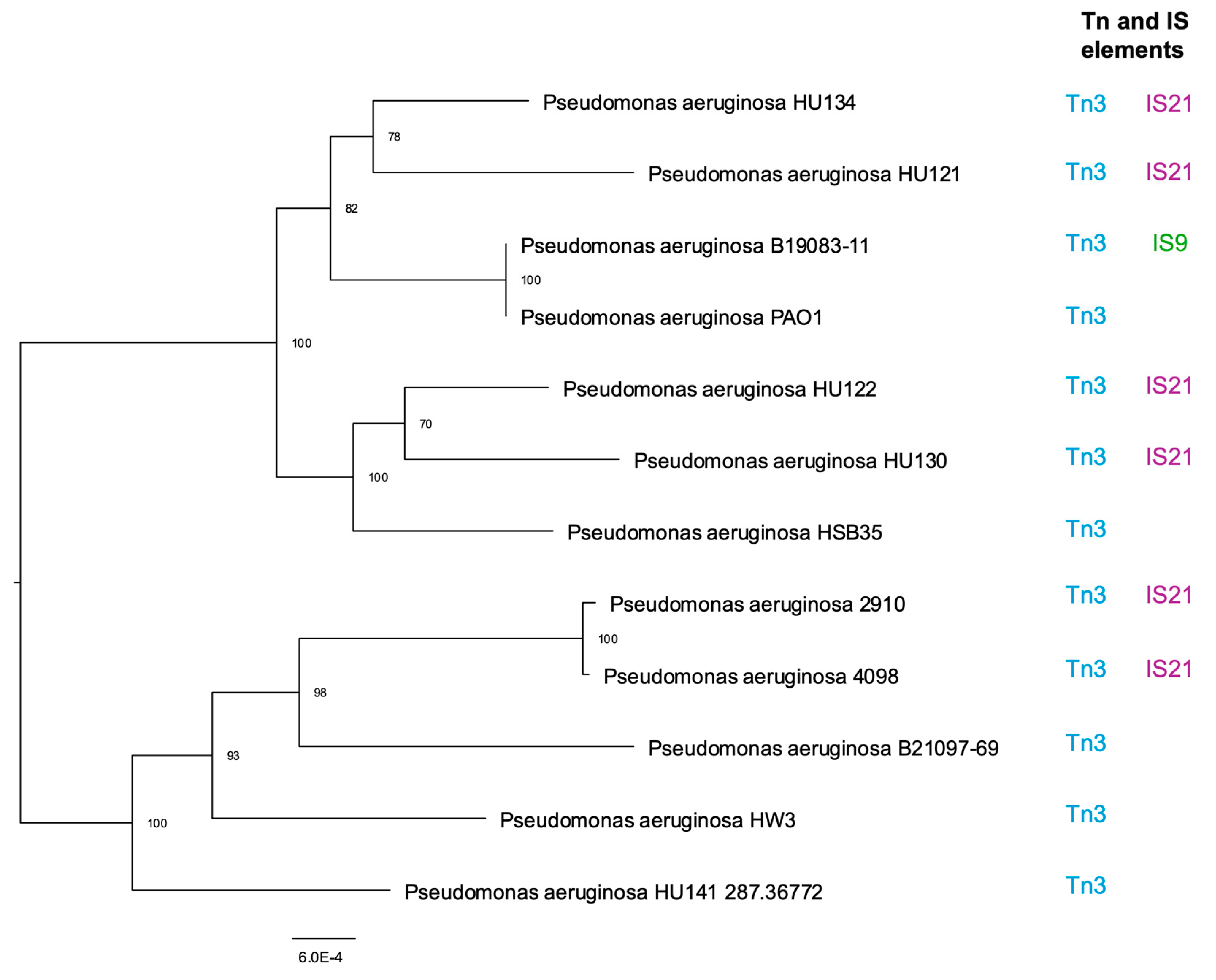
3.2. Tn and IS Elements

The presence of transposable elements including Tn3, IS6, IS3, IS5, and IS21 was identified in various bacterial isolates.
Tn3, a 4957-base pair transposon, was detected in all studied isolates with notably high prevalence. This transposon encodes three proteins: β-lactamase, which is responsible for resistance to β-lactam antibiotics; a transposase (encoded by the tnpA gene); and a resolvase (encoded by the tnpR gene).
In addition to Tn3, several IS elements were identified, including IS6. The isolate B19083-11 was the only one to exhibit IS29 (Supplementary Figure S1). In specific isolates, such as HU121, HU122, HU130, HU134, 2910, and 4098, IS21 was identified with high prevalence.
Mobile genetic elements IS21, IS6, and IS9 were detected in the genome of P. aeruginosa. They play a crucial role in genetic mobility by facilitating the insertion, deletion and rearrangement of genes, including those associated with antibiotic resistance and environmental adaptation.

3.3. Geno-Phenotype in Antibiotic Resistance

The comparative analysis of resistance to different antipseudomonal agents is shown in Table 3. It is noted that the examined isolates display resistance to a high number of antibiotic classes, so they are characterized by being extensively drug resistant (XDR).
The genomes of these isolates harbor several genes associated with resistance to β-lactams, aminoglycosides, quinolones, phenicols, sulfonamides, and colistin (Table 4).
The predominance of resistance genes to the β-lactam class is particularly high in all isolates, with class D (Oxacillinase β-lactamases) standing out; these have a serine-based structure. The blaOXA-396 gene is the most prevalent, found in four of the analyzed isolates, followed by blaOXA-494, detected in three isolates. Other genes such as blaOXA-846, blaOXA-488, blaOXA-848, blaOXA-395, and blaOXA-903 were detected in two of the examined isolates.
The β-lactamases of class A have a structure based on serine, similar to classes C and D. Most of the genes responsible for encoding these enzymes are located on plasmids, which implies the possibility of transfer between bacteria, although there are exceptions where such genes are found on the bacterial chromosome. Within this class belong the extended-spectrum β-lactamases (ESBL); as indicated by the name, they have a significantly broader spectrum of inhibition compared to penicillinases. They are capable of hydrolyzing all penicillins, including those associated with β-lactamase inhibitors, as well as almost all cephalosporins and Aztreonam. Strain B19083-11 is the only one to contain the blaSHV-12 gene.
Unlike other classes, class B has a metal-based structure, referred to as metallo-β-lactamases (MBL). The blaVIM-3 gene was detected in isolates 4098 and 2910. P. aeruginosa expressing MBL is resistant to all penicillins, all cephalosporins, and all carbapenems.
Class C β-lactamases, based on serine, are encoded by genes that are typically present in the bacterial chromosome. AmpC cephalosporinase is the main one, hydrolyzing penicillins and cephalosporins up to the third generation. AmpC includes different variants, referred to as PDCs (Pseudomonas-derived cephalosporinases). All examined isolates present different AmpC variants, with the blaPDC-3 variant detected in three isolates and blaPDC-16 in two of the isolates. The remaining variants were observed in only one strain.
The presence of the aminoglycoside class was confirmed in all isolates due to the presence of the aph(3′)-IIb gene. However, the gene aadA7 (also called ant(3″)-Ia) was exclusively detected in the HU141 strain. This gene confers resistance to gentamicin. The gene ant(2″)-Ia encodes an enzyme called nucleotidyltransferase, which modifies bacterial ribosomes, and it was detected in three of the isolates from Taiwan.
The genes aac(6′)-Ib3 and aac(6′)-Ib-cr (a variant of the aac(6′)-Ib gene) are responsible for encoding an enzyme called aminoglycoside acetyltransferase, which modifies antibiotics, hindering their effective binding to bacterial targets; they were detected in isolates B19083-11 and 2910. In strain B21097-69, the gene aac(6′)-IIa, another variant of the aac(6′)-Ib gene, was also identified. The aac(6′)-Ib-cr gene can also confer resistance to quinolones, such as ciprofloxacin.
For antibiotics belonging to the fosfomycin and amphenicols classes, a significant prevalence was observed among bacterial isolates. The fosA and fosX genes, as examples of single-nucleotide phosphotransferase, were identified as conferring resistance to fosfomycin and fosfomycin tromethamine antibiotics, which are commonly used in the treatment of bacterial infections caused by P. aeruginosa. fosA was present in all isolates and fosX was identified in one isolate. The genes catB2, catB7, and catB8 were detected among the bacterial isolates and correspond to different variants of the enzyme chloramphenicol acetyltransferase in P. aeruginosa. Remarkably, the catB7 variant was the most commonly detected in the isolates. The catB2 and catB8 variants, on the other hand, were identified in only one of the isolates, which is relatively uncommon considering that the catB8 gene is an additional variant of chloramphenicol acetyltransferase B found in P. aeruginosa.
The sul1 gene associated with bacterial resistance to sulfonamide antibiotics was detected in five isolates.
The tetG gene in P. aeruginosa encodes an efflux pump protein that actively expels tetracycline antibiotics from the bacterial cell, reducing their intracellular concentration and effectiveness. This gene was identified in only one isolate.

3.4. Virulome

Genomic approaches to the virulome have unveiled the presence of numerous genes associated with virulence factors in P. aeruginosa, spanning aspects such as adherence, motility, antimicrobial activity, antiphagocytosis, secretion systems, iron uptake, and quorum sensing, among others. Concerning adherence, genes linked to flagellum production (e.g., fliA-T, motA-D), the O-antigen of LPS, and type IV pili synthesis (e.g., pilA family genes, fimT, fimU, fimV) were identified, along with proteins related to type IV pili motility (e.g., chpA-E, pilG family genes).
Regarding antimicrobial activity, various virulence genes associated with phenazine biosynthesis, such as phzA, phzA2, phzB1, and phzB2, were detected.
In terms of antiphagocytosis, genes involved in alginate biosynthesis, such as alg44, alg8, algA, algC, algD, algE and others, were identified.
Biofilms play a crucial role in antimicrobial resistance, providing P. aeruginosa with structural and physiological protection that hinders the action of antibiotics and the immune system, making infections more persistent and difficult to eradicate. Several genes associated with biofilm formation were identified, including genes from the pel family (pelA, pelB and pelC), as well as the quorum sensing regulation system, mediated by lasR, lasI, rhlR and rhlI, responsible for biofilm maturation. Furthermore, different secretion systems were identified, notably the type VI secretion system (H-T6SS) and the type III secretion system (T3SS) of P. aeruginosa. Isolate 4098, belonging to ST4258, and isolate 2910, belonging to ST298, both contain the exoU gene. The exoU gene codes for the exoU toxin, a phospholipase-type enzyme that plays a crucial role in the pathogenicity of P. aeruginosa, facilitating cellular invasion and the destruction of host tissues.

3.5. Genomic Profiling

The comparative analysis of colistin-resistant P. aeruginosa genomes revealed differences between samples from Portugal and Taiwan compared to the reference strain PAO1 (Figure 2).
The Taiwanese isolates exhibited divergences from the reference strain, while intra-strain differences were less pronounced. Deletions were observed between reference isolates and colistin-resistant isolates in various gene clusters, including genes crucial for heme utilization and adhesion, belonging to the ShlA/HecA/FhaA family (Supplementary Tables S1 and S2).
Clusters of genes associated with membrane proteins and hypothetical proteins were also affected by deletions. However, genes related to Lipid A and LPS alterations, such as PmrA-PmrB, PhoP-PhoQ, ParR-ParS, and the ArnBCADTEF-PmrE operon, remained highly conserved, with a high percentage of similarity (>95%).
The isolates of P. aeruginosa from Portugal exhibited significant divergences compared to both the reference isolates and among themselves, when compared to the isolates from Taiwan. Remarkably, complete deletions were observed in gene clusters associated with Chloramphenicol O-acetyltransferase (CatB Family), accessory cholera enterotoxin, and mRNA interferase RelE in isolates HU121 and HUW3, while these genes were present in other isolates, albeit with differences in genetic similarity.
Additionally, the Type IV fimbrial biogenesis protein PilV gene showed significant modifications in the Portuguese isolates compared to the reference strain, with isolates HW3 and HU141 displaying low genetic similarity, approximately between 30 and 50%.
Like the Taiwan isolates, deletions were observed in gene clusters associated with T1SS RTX-secreted agglutinin in the Portuguese isolates. However, genes related to colistin resistance remained highly conserved.

3.6. Analysis of Mutations in Colistin-Resistant P. aeruginosa

The various genomes were compared to identify point mutations and variations in genetic content, aiming to assess potential within-host diversity. Several non-functional SNPs were detected, along with deleterious protein alterations, complex mutations involving deletions and insertions, and SNPs with a functional impact on genes related to colistin resistance.
The analysis of genetic mutations in various bacterial isolates revealed a wide array of alterations associated with colistin-resistance in P. aeruginosa (Table 5).
The identified modifications in the cprS gene, which is part of the two-component regulatory system CprR-CprS, play a pivotal role in this process by regulating bacterial resistance systems, including those related to colistin. These mutations have the potential to induce variations in the expression of genes involved in antibiotic resistance, thus affecting the bacterium’s ability to withstand antimicrobial effects. See Supplementary Figure S2 for the alignment using Clustal Omega of the HW3 strain against gb|AAG06466.1|+|cprS, which is the reference strain in the Comprehensive Antibiotic Resistance Database.
Similarly, the mutation in the pmrB gene, belonging to the two-component regulatory system PmrA-PmrB, which results in the substitution of tryptophan by glycine at position 25 of the protein, has been associated with colistin resistance in P. aeruginosa. We detected mutations in the phoQ gene, part of the phoP-phoQ two-component regulatory system, across different bacterial isolates.
Indeed, it is intriguing that, despite strain B21097-69 having more mutations in genes directly related to colistin, it does not exhibit the highest MIC. To determine whether there is any correlation between the mutations and MIC, the mutational analysis took into account not only genes directly linked to colistin resistance but also those potentially associated with bacterial resistance reported in the literature, as well as efflux pumps. The strain 4098 has more nonsynonymous mutations in resistance-related genes compared to the other isolates, particularly in the lepA genes.
Upon examining the mutations within the arnBCADTE-pmrE operon, numerous non-synonymous mutations linked to these genes are apparent. The protein changes evidenced in the inner membrane L-Ara4N transferase ArnT were identified in all colistin-resistant clinical isolates (Table 6). These changes include the following amino acid substitutions: Val266Ile, Leu11del, Ala282Ser, Arg502Gln, Ile509Val, Ala267Ser, Ala330Val, Leu439Ile, Val88Phe, Thr443Ile, Arg446His, Cys7Trp, His151Tyr, Thr166Ile, Ala265Val, Asn346Asp, Leu493fs, Gly156Arg, Asp232Asn, and 16Arg535Leu.
The complex mutation CysSer312SerGly appears to be present in all isolates associated with the ArnA protein. For the genes arnD, arnE, and arnF, a limited number of mutations were observed, suggesting a relatively stable genetic profile in these regions. In arnD protein, mutations included Phe58Leu, Glu25Asp, and Ser272Asn, with a complex mutation involving Ala118Val and Val123Ala. The ArnE protein exhibited the following mutations: Ala109Val, Thr13Ser, and Arg28His. The ArnF protein mutations comprised Val14Met, with complex mutations involving Ala118Val and Ala55Thr, along with Val123Ala and Val113Met.

4. Discussion

P. aeruginosa is a widely distributed bacterium that thrives in various environments, including soil, water, and even hospital settings. It is prevalent in both natural and human-made habitats. However, its significance extends beyond just being a common bacterium. P. aeruginosa is particularly concerning in medical contexts due to its remarkable adaptability and its tendency to cause persistent infections, especially in individuals with weakened immune systems [24].
The results presented in this study provide a comprehensive overview of the genetic diversity and distribution of certain P. aeruginosa subtypes, along with their clinical implications.
Our phylogenetic analysis provides valuable insights into the evolutionary relationships among different isolates of P. aeruginosa. The phylogenetic tree reveals significant genetic diversity, with distinct clades and branches suggesting evolutionary divergences over time. The presence of bootstrap values at each division of the tree provides a measure of statistical confidence in these evolutionary relationships. It is interesting to note that there is no clear distinction between isolates from Portugal and Taiwan, pointing to a global spread of these P. aeruginosa subtypes and highlighting the importance of global surveillance and international collaboration in controlling bacterial infections. The arrangement of isolates in close or distant groups can be explained by multiple factors that influence both the evolution and the adaptation of these microorganisms to specific environments. For example, even though the two bloodstream isolates from Taiwan originated from the same institute, the divergence in their clustering suggests that they may have acquired distinct genetic profiles from different infectious sources. In many cases, bacteremia can originate from UTIs or lung infections, resulting in different genomic profiles, since each niche imposes specific selective pressures and favors the acquisition of certain virulence and resistance determinants [8,25]. Additionally, the phylogenetic proximity observed between one of the Taiwanese isolates, P. aeruginosa PAO1, and the Portuguese UTI isolates may indicate the existence of a common ancestor or even the occurrence of horizontal gene exchange, which is an important mechanism for the dissemination of resistance and virulence genes [26,27]. This hypothesis is reinforced by studies that indicate that isolates associated with urinary tract infections often share similar genetic profiles, even in geographically distant contexts, due to adaptation to hospital environments with the intensive use of antibiotics. Thus, the presented phylogenetic analysis not only highlights the global heterogeneity of P. aeruginosa but also showcases the complexity of the evolutionary and epidemiological processes that contribute to the formation of clusters. Even isolates originating from the same hospital environment can follow different evolutionary trajectories, which reinforces the importance of conducting in-depth epidemiological and molecular studies for the development of more effective control and treatment strategies [28]. Another study conducted a comparative genomic analysis of clinical isolates of P. aeruginosa isolated from eye and cystic fibrosis patients. The analysis revealed significant variation in the size of the accessory genome among the 22 isolates studied, which correlated with the presence of genomic islands, insertion sequences, and prophages. The isolates exhibited diversity in sequence type and were dissimilar to globally epidemic P. aeruginosa clones. Notably, a majority of the eye isolates from India clustered within a single lineage. Indian eye isolates possessed genes linked to resistance against various antibiotics, which were absent in Australian isolates regardless of the infection source [29]. Another study investigated the evolutionary genomics of P. aeruginosa’s adaptation to the CF lung environment, where it transitions from a free-living environmental strain to one causing chronic infection. Whole-genome sequencing of 1000 P. aeruginosa isolates, including those isolated from CF patients, revealed that CF isolates were distributed across the phylogeny, indicating no genetic predisposition for chronic infection within specific clades. However, isolates from the CF niche experienced stronger positive selection on core genes compared to those from environmental or acute infection sources, indicating recent adaptation to the lung environment [30]. These findings contribute valuable insights into the genomic diversity of P. aeruginosa across different infection types and geographical locations.
The presence of transposable elements in bacteria is a widely observed characteristic that is of great importance in terms of their biology and adaptation. Among these elements, Tn3, IS6, IS3, IS5 and IS21 stand out; they were identified in several bacterial lineages. These transposable elements play a fundamental role in the genomic plasticity of bacteria, allowing for horizontal gene transfer, chromosomal reorganization, and the acquisition of new phenotypic characteristics, such as antibiotic resistance and adaptation to different environments [31,32]. In Tn3, several IS elements were identified, including IS6, whose importance in generating clusters of clinically relevant antibiotic resistance genes is becoming increasingly evident, and whose members may utilize an unusual transposition pathway [33]. Although not exclusive to P. aeruginosa, IS26 is widely distributed in various bacteria and plays a significant role in the dissemination of antibiotic resistance and genome rearrangement, representing an important factor to consider in studies of bacterial resistance and genomic adaptation [33]. IS3 and IS5 elements were also detected in all isolates; they are recognized for possessing associated transposases that facilitate DNA mobility within the bacterial genome. These transposable elements play crucial roles in bacterial evolution, allowing for gene transfer between organisms and contributing to adaptation to different environments and conditions [34]. IS21 and IS6 elements are pivotal in shaping P. aeruginosa’s genome, enabling rapid adaptation to antibiotics and environmental stresses. IS21 and IS6 elements drive genome rearrangements and insertions, promoting the spread of resistance genes such as carbapenemases (e.g., OXA-2). These IS families are linked to globally disseminated P. aeruginosa clones (e.g., ST463), which combine multidrug resistance with virulence factors such as ExoU [35,36]. In P. aeruginosa CMC-097, IS6100 (an IS6-like element) was identified adjacent to the In2020 integron. It carries tnpA2 and inverted repeats (IRs) that mediate recombination events. The IS6100 element (IAU57_09080) in CMC-097 facilitates the transfer of resistance genes within integrons, contributing to multidrug resistance [37,38]. In the clinical strain P. aeruginosa CMC-097, 13 copies of an IS21 family transposon were identified, contributing to genomic plasticity and resistance gene dissemination [39]. IS21 elements are frequently associated with carbapenem resistance integrons (e.g., In2020), which carry genes including blaOXA-2 (β-lactamase) and aacA27 (aminoglycoside acetyltransferase) [37,38,39,40].
The comparative analysis of resistance to various antipseudomonal agents reveals that the isolates exhibit resistance to multiple antibiotic classes, with a high prevalence of resistance to β-lactams, particularly class D enzymes. [40]. The blaOXA-396 gene is the most common, followed by blaOXA-494. Other genes such as blaOXA-846, blaOXA-488, blaOXA-848, blaOXA-395, and blaOXA-903 were detected in two of the examined isolates [41]. The class B carbapenemases genes, including blaVIM-3, were found in isolates 4098 and 2910, which contribute to resistance to a broad range of antibiotics. The detection of MBL, such as blaVIM-3, is particularly concerning due to its ability to confer resistance to a broad range of antibiotics, complicating the treatment of bacterial infections [42,43,44]. The isolate B19083-11 is the only one to contain the blaSHV-12 gene. Unlike other classes, class B has a metal-based structure, referred to as MBL.
The AmpC cephalosporinase, mainly represented by the blaPDC-3 and blaPDC-16 variants, was observed in all isolates. Increased expression of AmpC is associated with β-lactam resistance, particularly for combinations like ceftolozane/tazobactam and ceftazidime/avibactam [45,46,47,48]. Many different types of PDC have been documented, with certain variants associated with heightened resistance to ceftolozane/tazobactam and ceftazidime/avibactam [47,48,49,50]. Among isolates in the CC274 collection, a mutator strain named AUS601 displays significant resistance to ceftazidime, cefepime, and aztreonam, even without an increase in AmpC expression [51].
Additionally, various efflux pump genes, such as MexAB-OprM, MexCD-OprJ, and MexEF-OprN, were identified. These pumps contribute to multidrug resistance, complementing other mechanisms such as β-lactamase production and altered membrane permeability [52]. A mutational analysis of various genes revealed that the diverse isolates harbored mutations, primarily synonymous mutations in porins, multidrug efflux pumps, and membrane proteins, which are the primary drivers of prevalent carbapenem resistance [53,54,55]. During chronic CF respiratory infections, the MexAB-OprM efflux pump is subjected to significant mutational pressure, including the occurrence of inactivating mutations. This observation aligns with previous studies indicating that this efflux system is not essential and thus may be lost or deactivated in favor of overexpressing MexXY-OprM in certain subpopulations of CF P. aeruginosa [56].
The genes aac(6′)-Ib3, aac(6′)-Ib-cr, and aac(6′)-IIa, found in isolates B19083-11, 2910, and B21097-69, respectively, may confer resistance to aminoglycosides, while the gene aac(6′)-Ib-Hangzhou, identified in strain 2910 from Taiwan, is associated with Enterobacteria and also confers resistance to aminoglycosides [57,58,59] Additionally, the gene fosA, present in all isolates, and the gene fosX, identified in one isolate, confer resistance to antibiotics belonging to the classes of fosfomycin [60]. sul1 was detected in five isolates. Sul1 proteins confer resistance, which can be a significant public health concern [61,62].
The comparison between Portugal and Taiwan regarding antibiotic use and antibiotic stewardship strategies allows us to contextualize the resistance profiles observed in this study. In Portugal, antibiotic consumption has historically been high, especially in the community sector [63], although recent measures, such as the Prevention and Control of Antimicrobial Resistance Program (PPCRA), have been implemented to reduce inappropriate use [64]. Data from the European Centre for Disease Prevention and Control (ECDC) indicate a downward trend in total antibiotic consumption in recent years, in line with European guidelines for the rational use of antimicrobials [65].
On the other hand, Taiwan has a robust system for monitoring antibiotic use, with strict regulations established by the Taiwan National Antimicrobial Resistance Action Plan (TNARAP), which includes the strict control of prescriptions and the promotion of antibiotic stewardship programs in hospitals [66,67]. A study conducted by Lee et al. (2012) showed that, despite the high consumption of antibiotics in Taiwan, government initiatives have been effective in reducing unnecessary use, especially in hospital settings [66].
When comparing data from both countries, it is observed that, although Portugal and Taiwan face similar challenges in combating antimicrobial resistance, Taiwan implements a more centralized and rigorous control of prescriptions, while Portugal has focused on raising awareness and educating health professionals. The relationship between these different management styles and the resistance profiles identified in this study may provide important insights into the impact of national policies on the development of resistance in P. aeruginosa.
The comparative analysis of colistin-resistant P. aeruginosa genomes highlights divergences between isolates from Portugal and Taiwan when compared to the reference strain PAO1. Taiwanese isolates showed variations from the reference, with fewer intra-strain differences. Deletions were noted in various gene clusters, including those crucial for heme utilization and adhesion (ShlA/HecA/FhaA family). Particularly noteworthy is a large exoprotein whose function is presumably linked to heme utilization or adhesion, suggesting its importance for the virulence and persistence of P. aeruginosa, especially in nosocomial environments. Such deletions may lead to reduced virulence, colonization capacity, and altered genetic mobility, affecting bacterial evolution and antibiotic resistance acquisition [67]. P. aeruginosa biofilms represent a significant barrier to the efficacy of antibiotics, contributing to the chronicity and resistance of infections. Several genes are involved in biofilm formation and maintenance, including pelA, pelB and pelC, which encode to produce extracellular matrix polysaccharides, and pslA, pslB and pslC, which are essential for cell adhesion and biofilm structure [68]. Furthermore, the algD gene is directly related to the production of alginate, promoting resistance to the immune system and antibiotics [69]. The quorum-sensing regulatory system, mediated by lasR, lasI, rhlR and rhlI, is another key factor in biofilm maturation and the expression of virulence factors. The presence of biofilms not only protects bacteria against antimicrobial agents, but also favors the spread of resistance genes, worsening the challenges in treating infections caused by P. aeruginosa [70].
Clusters of genes related to T1SS RTX-secreted agglutinins underwent deletions, potentially reducing adhesion and toxin secretion, as well as altering the host immune response. Despite these deletions, colistin resistance genes remained conserved, indicating their importance [71,72].
Portuguese isolates exhibited significant genetic divergence, including complete deletions in some gene clusters and modifications in others, such as the PilV gene. These changes likely result from natural selection in response to environmental pressures, such as colistin exposure in hospitals. Molecular evolution also plays a role in the observed genetic variation, reflecting the bacteria’s adaptation to different environments.
The analysis of mutations in colistin-resistant P. aeruginosa identified various alterations, including non-functional SNPs, deleterious protein alterations, complex mutations involving deletions and insertions, and SNPs impacting genes related to colistin resistance. Mutations in regulatory networks, such as those in the cprS, pmrB, phoQ, parS, and parR genes, were identified, potentially affecting antibiotic resistance mechanisms. Alterations in these regulatory elements can modulate the expression of genes involved in colistin resistance pathways, thereby influencing the overall resistance phenotype of the bacterium [73]. Furthermore, the discrepancy between the mutation profile of the B21097-69 strain and its MIC underscores the complexity of the relationship between genotype and phenotype in antibiotic resistance. While this strain harbors more mutations directly related to colistin, its MIC does not necessarily reflect the highest resistance level observed among the isolates analyzed. This discrepancy suggests that factors beyond the presence of specific mutations may contribute to the overall resistance phenotype, such as the genetic background, epistatic interactions, or additional resistance mechanisms. Additionally, strain 4098 exhibited more nonsynonymous mutations in resistance-related genes, indicating potential implications for antibiotic resistance mechanisms. These mutations may alter the function or expression of key resistance determinants, leading to variations in resistance levels among bacterial isolates. Interpreting the mechanisms underlying colistin resistance presents challenges due to the intricate nature of bacterial response systems. Mutations in two-component regulators do not consistently correlate with clinical colistin resistance, indicating a potential interplay between various regulatory pathways. For instance, some studies suggest that individual two-component systems may not singularly dictate colistin resistance acquisition in P. aeruginosa, hinting at complex interactions between regulatory elements [74,75]. The kinase sensor PmrB activates the transcriptional response regulator PmrA either through a phosphotransfer relay or because of a mutation in pmrB. This two-component system drives bacterial responses to multiple stimuli and regulates modifications of the LPS [76,77]. In P. aeruginosa isolates B21097-69, 2910, and 4098, mutations in the pmrB gene have been identified. Specifically, strain 2910 exhibits two types of mutations (Trp25Gly and Gly469fs). The Ala247thr mutation detected in isolate B21097-69 involves lipid A and is generally associated with gain of function [78,79].
The detection of mutations in the phoQ gene across various bacterial isolates indicates that this gene may play a role in antibiotic resistance, including resistance to colistin. One specific mutation identified is both a frameshift and missense mutation, causing a shift in the reading frame and leading to a missense change at codon position 152. This alteration may have substantial implications for the structure and function of the PhoQ protein, which is essential to regulating bacterial resistance systems [80]. Yang et al. [75] showed that individual two-component systems may not be essential for the acquisition of colistin resistance in P. aeruginosa. However, it should be noted that the isolate with a premature stop codon in phoQ exhibited a high level of resistance [75]. The mutation in the phoQ gene described as a frameshift and missense (Val152fs) significantly impacts the function of the PhoQ protein. A frameshift mutation, such as Val152fs, causes a shift in the reading frame of the gene, leading to an altered and often truncated protein product. This specific mutation can result in a nonfunctional or partially functional PhoQ protein, disrupting its ability to respond to environmental signals and regulate target genes effectively. In the context of colistin resistance, mutations in phoQ can lead to either the increased or decreased expression of genes that modify LPS on the bacterial surface. These modifications, such as the addition of L-Ara4N or phosphoethanolamine to lipid A, reduce the negative charge of LPS and decrease the colistin binding affinity [76,77]. In P. aeruginosa, mutations in the phoQ gene, along with mutations in other regulatory genes such as pmrB, can enhance resistance to colistin by upregulating the arnBCADTEF operon, which encodes enzymes for LPS modification. The resulting changes in the outer membrane structure help the bacteria evade the antimicrobial activity of colistin, thus contributing to high levels of resistance [77].
Colistin resistance in P. aeruginosa can also be associated with alterations in LPS, particularly in lipid A, involving the operons pmrAB and arnBCADTEF-pmrE, which mediate the synthesis and transfer of pEtN and L-Ara4N, respectively [4]. The arnA gene mutation (arnA complex mutation; CysSerProGln312SerGlyProLys) introduces a complex amino acid change at position 312. ArnA is crucial in the biosynthesis of UDP- L-Ara4N, a key molecule in the lipid A modification of LPS [77,78,79,80]. These extensive changes can significantly alter the function of ArnA, impacting the overall resistance mechanism. The SNPs in the arnT gene cause multiple amino acid substitutions. ArnT is responsible for transferring L-Ara4N to lipid A. Each mutation can individually or collectively affect the enzyme’s efficiency, altering the bacterial membrane’s structure and enhancing resistance to colistin. The mutations in these genes collectively contribute to modifying the outer membrane of P. aeruginosa, reducing colistin’s ability to bind and exert its antimicrobial effect [78,79,80]. However, the phenotype of colistin resistance typically arises from a complex interplay of multiple mechanisms that enable the bacteria to evade antibiotic stress. In addition to modifications in LPS, P. aeruginosa can develop resistance through the overexpression of efflux pumps, which expel the antibiotic from the cell, and the production of enzymes that inactivate colistin before it can exert its bactericidal effect [53,80]. These mechanisms often work together, creating a robust defense system that makes treating P. aeruginosa infections particularly challenging.

5. Conclusions

P. aeruginosa is a significant pathogen that is responsible for serious hospital-acquired infections, particularly in immunocompromised patients and those with cystic fibrosis. It is one of the primary organisms responsible for nosocomial infections, including pneumonia and UTIs.
The genetic diversity and distribution of its specific subtypes show the pathogen’s adaptability to various environments, with UTIs being the most common source. High-risk clones such as ST235, associated with widespread dissemination and high antibiotic resistance, pose a significant public health threat. The phylogenetic analysis revealed no clear distinction between isolates from Portugal and Taiwan, highlighting its global spread. Mutations in genes such as phoQ and pmrB, and in the arnBCADTEF operon, contribute to colistin resistance by altering the LPS structure. This study aimed to provide genomic data from isolates from two countries, which are useful for identifying the determinants of antibiotic resistance, as well as prospecting data for potential new therapeutic targets.

Supplementary Materials

The following supporting information can be downloaded at: https://www.mdpi.com/article/10.3390/pathogens14040387/s1, Figure S1: Presence of IS26 in P. aeruginosa strain B19083-1; Figure S2: Alignment of Pseudomonas aeruginosa HW3 Strain with the Reference Strain cprS (gb|AAG06466.1|) using Clustal Omega; Table S1: Genomic annotating using BV-BRC tool for Portuguese isolates; Table S2:Genomic annotating using BV-BRC tool for Tawain isolates.

Author Contributions

T.D.S., H.-Y.W., T.-W.L., M.C., M.J.N.R., D.S., C.S., S.S., R.B. (Richard Bonnet) and R.B. (Racha Beyrouthy): methodology, data analyses, and writing—review and editing. M.H., G.I. and P.P.: concept, project supervision, writing—review and editing. All authors have read and agreed to the published version of the manuscript.

Funding

This research was funded by FCT (Fundação para a Ciência e a Tecnologia), related to a Ph.D. grant, with the reference 2020.05332.BD, and the projects UI/00772 and LA/P/0059/2020.

Institutional Review Board Statement

The study was conducted according to the guidelines of the Declaration of Helsinki, and approved by the Ethics Committee of University of Trás-os-Montes e Alto Douro (EC-UTAD) (protocol code BI087371847 and 2019-11-28).

Informed Consent Statement

Patient consent was waived due to the isolates were provided by the hospitals on culture plates, with no identifying information attached.

Data Availability Statement

Data are contained within the article and Supplementary Materials.

Acknowledgments

This work received support and help from FCT/MCTES (LA/P/0008/2020 DOI 10.54499/LA/P/0008/2020, UIDP/50006/2020 DOI 10.54499/UIDP/50006/2020 and UIDB/50006/2020 DOI 10.54499/UIDB/50006/2020), through national funds.

Conflicts of Interest

The authors declare no conflicts of interest.

References

  1. Laborda, P.; Sanz-García, F.; Hernando-Amado, S.; Martínez, J.L. Pseudomonas Aeruginosa: An Antibiotic Resilient Pathogen with Environmental Origin. Curr. Opin. Microbiol. 2021, 64, 125–132. [Google Scholar] [CrossRef] [PubMed]
  2. Sastre-Femenia, M.À.; Fernández-Muñoz, A.; Gomis-Font, M.A.; Taltavull, B.; López-Causapé, C.; Arca-Suárez, J.; Martínez-Martínez, L.; Cantón, R.; Larrosa, N.; Oteo-Iglesias, J.; et al. Pseudomonas aeruginosa Antibiotic Susceptibility Profiles, Genomic Epidemiology and Resistance Mechanisms: A Nation-Wide Five-Year Time Lapse Analysis. Lancet Reg. Health Eur. 2023, 34, 100736. [Google Scholar] [CrossRef] [PubMed]
  3. Avakh, A.; Grant, G.D.; Cheesman, M.J.; Kalkundri, T.; Hall, S. The Art of War with Pseudomonas aeruginosa: Targeting Mex Efflux Pumps Directly to Strategically Enhance Antipseudomonal Drug Efficacy. Antibiotics 2023, 12, 1304. [Google Scholar] [CrossRef] [PubMed]
  4. Jeannot, K.; Hagart, K.; Dortet, L.; Kostrzewa, M.; Filloux, A.; Plesiat, P.; Larrouy-Maumus, G. Detection of Colistin Resistance in Pseudomonas aeruginosa Using the MALDIxin Test on the Routine MALDI Biotyper Sirius Mass Spectrometer. Front. Microbiol. 2021, 12, 725383. [Google Scholar] [CrossRef]
  5. Biswas, S.; Brunel, J.M.; Dubus, J.C.; Reynaud-Gaubert, M.; Rolain, J.M. Colistin: An Update on the Antibiotic of the 21st Century. Expert Rev. Anti. Infect. Ther. 2012, 10, 917–934. [Google Scholar] [CrossRef]
  6. Fernández, L.; Álvarez-Ortega, C.; Wiegand, I.; Olivares, J.; Kocíncová, D.; Lam, J.S.; Martínez, J.L.; Hancock, R.E.W. Characterization of the Polymyxin B Resistome of Pseudomonas aeruginosa. Antimicrob. Agents Chemother. 2013, 57, 110–119. [Google Scholar] [CrossRef]
  7. Lee, J.Y.; Park, Y.K.; Chung, E.S.; Na, I.Y.; Ko, K.S. Evolved Resistance to Colistin and Its Loss Due to Genetic Reversion in Pseudomonas aeruginosa. Sci. Rep. 2016, 6, 25543. [Google Scholar] [CrossRef]
  8. Lee, J.Y.; Chung, E.S.; Na, I.Y.; Kim, H.; Shin, D.; Ko, K.S. Development of Colistin Resistance in PmrA-, PhoP-, ParR- and CprR-Inactivated Mutants of Pseudomonas aeruginosa. J. Antimicrob. Chemother. 2014, 69, 2966–2971. [Google Scholar] [CrossRef]
  9. McPhee, J.B.; Bains, M.; Winsor, G.; Lewenza, S.; Kwasnicka, A.; Brazas, M.D.; Brinkman, F.S.L.; Hancock, R.E.W. Contribution of the PhoP-PhoQ and PmrA-PmrB Two-Component Regulatory Systems to Mg2+-Induced Gene Regulation in Pseudomonas aeruginosa. J. Bacteriol. 2006, 188, 3995–4006. [Google Scholar] [CrossRef]
  10. Beceiro, A.; Llobet, E.; Aranda, J.; Bengoechea, J.A.; Doumith, M.; Hornsey, M.; Dhanji, H.; Chart, H.; Bou, G.; Livermore, D.M.; et al. Phosphoethanolamine Modification of Lipid A in Colistin-Resistant Variants of Acinetobacter baumannii Mediated by the PmrAB Two-Component Regulatory System. Antimicrob. Agents Chemother. 2011, 55, 3370–3379. [Google Scholar] [CrossRef]
  11. Moskowitz, S.M.; Ernst, R.K.; Miller, S.I. PmrAB, a Two-Component Regulatory System of Pseudomonas aeruginosa That Modulates Resistance to Cationic Antimicrobial Peptides and Addition of Aminoarabinose to Lipid A. J. Bacteriol. 2004, 186, 575–579. [Google Scholar] [CrossRef] [PubMed]
  12. Bolard, A.; Schniederjans, M.; Haüssler, S.; Triponney, P.; Valot, B.; Plésiat, P.; Jeannota, K. Production of Norspermidine Contributes to Aminoglycoside Resistance in Pmrab Mutants of Pseudomonas aeruginosa. Antimicrob. Agents Chemother. 2019, 63, 10. [Google Scholar] [CrossRef]
  13. Larsen, M.V.; Cosentino, S.; Rasmussen, S.; Friis, C.; Hasman, H.; Marvig, R.L.; Jelsbak, L.; Sicheritz-Pontén, T.; Ussery, D.W.; Aarestrup, F.M.; et al. Multilocus Sequence Typing of Total-Genome-Sequenced Bacteria. J. Clin. Microbiol. 2012, 50, 1355–1361. [Google Scholar] [CrossRef] [PubMed]
  14. Center for Genomic Epidemiology. Pseudomonas aeruginosa Serotyper. Available online: https://cge.food.dtu.dk/services/PAst/ (accessed on 25 May 2024).
  15. Tamura, K.; Stecher, G.; Kumar, S. MEGA11: Molecular Evolutionary Genetics Analysis Version 11. Mol. Biol. Evol. 2021, 38, 3022–3027. [Google Scholar] [CrossRef] [PubMed]
  16. Letunic, I.; Bork, P. Interactive Tree of Life (ITOL) v5: An Online Tool for Phylogenetic Tree Display and Annotation. Nucleic Acids Res. 2021, 49, W293–W296. [Google Scholar] [CrossRef]
  17. Bortolaia, V.; Kaas, R.S.; Ruppe, E.; Roberts, M.C.; Schwarz, S.; Cattoir, V.; Philippon, A.; Allesoe, R.L.; Rebelo, A.R.; Florensa, A.F.; et al. ResFinder 4.0 for Predictions of Phenotypes from Genotypes. J. Antimicrob. Chemother. 2020, 75, 3491–3500. [Google Scholar] [CrossRef]
  18. Alcock, B.P.; Huynh, W.; Chalil, R.; Smith, K.W.; Raphenya, A.R.; Wlodarski, M.A.; Edalatmand, A.; Petkau, A.; Syed, S.A.; Tsang, K.K.; et al. CARD 2023: Expanded Curation, Support for Machine Learning, and Resistome Prediction at the Comprehensive Antibiotic Resistance Database. Nucleic Acids Res. 2023, 51, D690–D699. [Google Scholar] [CrossRef]
  19. Olson, R.D.; Assaf, R.; Brettin, T.; Conrad, N.; Cucinell, C.; Davis, J.J.; Dempsey, D.M.; Dickerman, A.; Dietrich, E.M.; Kenyon, R.W.; et al. Introducing the Bacterial and Viral Bioinformatics Resource Center (BV-BRC): A Resource Combining PATRIC, IRD and ViPR. Nucleic Acids Res. 2023, 51, D678–D689. [Google Scholar] [CrossRef]
  20. Johansson, M.H.K.; Bortolaia, V.; Tansirichaiya, S.; Aarestrup, F.M.; Roberts, A.P.; Petersen, T.N. Detection of Mobile Genetic Elements Associated with Antibiotic Resistance in Salmonella enterica Using a Newly Developed Web Tool: MobileElementFinder. J. Antimicrob. Chemother. 2021, 76, 101–109. [Google Scholar] [CrossRef]
  21. Torsten Seemann Snippy. Available online: https://github.com/tseemann/snippy (accessed on 25 May 2024).
  22. Sievers, F.; Wilm, A.; Dineen, D.; Gibson, T.J.; Karplus, K.; Li, W.; Lopez, R.; McWilliam, H.; Remmert, M.; Söding, J.; et al. Fast, Scalable Generation of High-Quality Protein Multiple Sequence Alignments Using Clustal Omega. Mol. Syst. Biol. 2011, 7, 539. [Google Scholar] [CrossRef]
  23. De Sousa, T.; Hébraud, M.; Dapkevicius, M.L.N.E.; Maltez, L.; Pereira, J.E.; Capita, R.; Alonso-Calleja, C.; Igrejas, G.; Poeta, P. Molecular Sciences Genomic and Metabolic Characteristics of the Pathogenicity in Pseudomonas aeruginosa. Int. J. Mol. Sci. 2021, 22, 12892. [Google Scholar] [CrossRef] [PubMed]
  24. Fischer, S.; Dethlefsen, S.; Klockgether, J.; Tümmler, B. Phenotypic and Genomic Comparison of the Two Most Common ExoU-Positive Pseudomonas aeruginosa Clones, PA14 and ST235. mSystems 2020, 5, 10-1128. [Google Scholar] [CrossRef] [PubMed]
  25. Recio, R.; Sánchez-Diener, I.; Viedma, E.; Meléndez-Carmona, M.Á.; Villa, J.; Orellana, M.Á.; Mancheño, M.; Juan, C.; Zamorano, L.; Lora-Tamayo, J.; et al. Pathogenic Characteristics of Pseudomonas aeruginosa Bacteraemia Isolates in a High-Endemicity Setting for ST175 and ST235 High-Risk Clones. Eur. J. Clin. Microbiol. Infect. Dis. 2020, 39, 671–678. [Google Scholar] [CrossRef] [PubMed]
  26. del Barrio-Tofiño, E.; López-Causapé, C.; Oliver, A. Pseudomonas aeruginosa Epidemic High-Risk Clones and Their Association with Horizontally-Acquired β-Lactamases: 2020 Update. Int. J. Antimicrob. Agents 2020, 56, 106196. [Google Scholar] [CrossRef]
  27. Treepong, P.; Kos, V.N.; Guyeux, C.; Blanc, D.S.; Bertrand, X.; Valot, B.; Hocquet, D. Global Emergence of the Widespread Pseudomonas aeruginosa ST235 Clone. Clin. Microbiol. Infect. 2018, 24, 258–266. [Google Scholar] [CrossRef]
  28. Gómez-Zorrilla, S.; Juan, C.; Cabot, G.; Camoez, M.; Tubau, F.; Oliver, A.; Dominguez, M.A.; Ariza, J.; Peña, C. Impact of Multidrug Resistance on the Pathogenicity of Pseudomonas aeruginosa: In Vitro and in Vivo Studies. Int. J. Antimicrob. Agents 2016, 47, 368–374. [Google Scholar] [CrossRef]
  29. Sánchez-Diener, I.; Zamorano, L.; Peña, C.; Ocampo-Sosa, A.; Cabot, G.; Gómez-Zorrilla, S.; Almirante, B.; Aguilar, M.; Granados, A.; Calbo, E.; et al. Weighting the Impact of Virulence on the Outcome of Pseudomonas aeruginosa Bloodstream Infections. Clin. Microbiol. Infect. 2020, 26, 351–357. [Google Scholar] [CrossRef]
  30. Estepa, V.; Rojo-Bezares, B.; Torres, C.; Sáenz, Y. Genetic Lineages and Antimicrobial Resistance in Pseudomonas Spp. Isolates Recovered from Food Samples. Foodborne Pathog. Dis. 2015, 12, 486–491. [Google Scholar] [CrossRef]
  31. García-Castillo, M.; Del Campo, R.; Morosini, M.I.; Riera, E.; Cabot, G.; Willems, R.; Van Mansfeld, R.; Oliver, A.; Cantón, R. Wide Dispersion of ST175 Clone despite High Genetic Diversity of Carbapenem-Nonsusceptible Pseudomonas aeruginosa Clinical Strains in 16 Spanish Hospitals. J. Clin. Microbiol. 2011, 49, 2905–2910. [Google Scholar] [CrossRef]
  32. Zhao, Y.; Chen, D.; Ji, B.; Zhang, X.; Anbo, M.; Jelsbak, L. Whole-Genome Sequencing Reveals High-Risk Clones of Pseudomonas aeruginosa in Guangdong, China. Front. Microbiol. 2023, 14, 1134. [Google Scholar] [CrossRef]
  33. Subedi, D.; Vijay, A.K.; Kohli, G.S.; Rice, S.A.; Willcox, M. Comparative Genomics of Clinical Strains of Pseudomonas aeruginosa Strains Isolated from Different Geographic Sites. Sci. Rep. 2018, 8, 15668. [Google Scholar] [CrossRef]
  34. Dettman, J.R.; Kassen, R. Evolutionary Genomics of Niche-Specific Adaptation to the Cystic Fibrosis Lung in Pseudomonas aeruginosa. Mol. Biol. Evol. 2021, 38, 663–675. [Google Scholar] [CrossRef]
  35. Partridge, S.R.; Kwong, S.M.; Firth, N.; Jensen, S.O. Mobile Genetic Elements Associated with Antimicrobial Resistance. Clin. Microbiol. Rev. 2018, 31, 10-1128. [Google Scholar] [CrossRef]
  36. Benler, S.; Faure, G.; Altae-Tran, H.; Shmakov, S.; Zheng, F.; Koonin, E. Cargo Genes of Tn7-Like Transposons Comprise an Enormous Diversity of Defense Systems, Mobile Genetic Elements, and Antibiotic Resistance Genes. mBio 2021, 12, e02938-21. [Google Scholar] [CrossRef]
  37. Varani, A.; He, S.; Siguier, P.; Ross, K.; Chandler, M. The IS6 Family, a Clinically Important Group of Insertion Sequences Including IS26. Mob. DNA 2021, 12, 11. [Google Scholar] [CrossRef]
  38. Mahillon, J.; Léonard, C.; Chandler, M. IS Elements as Constituents of Bacterial Genomes. Res. Microbiol. 1999, 150, 675–687. [Google Scholar] [CrossRef]
  39. Mouton, J.W.; Den Hollander, J.G.; Horrevorts, A.M. Emergence of Antibiotic Resistance amongst Pseudomonas aeruginosa Isolates from Patients with Cystic Fibrosis. J. Antimicrob. Chemother. 1993, 31, 919–926. [Google Scholar] [CrossRef]
  40. Bai, Y.; Gong, Y.; Shen, F.; Li, H.; Cheng, Y.; Guo, J.; Liu, G.; Ji, A. fang Molecular Epidemiological Characteristics of Carbapenem-Resistant Pseudomonas aeruginosa Clinical Isolates in Southeast Shanxi, China. J. Glob. Antimicrob. Resist. 2024, 36, 301–306. [Google Scholar] [CrossRef]
  41. Pournaras, S.; Maniati, M.; Petinaki, E.; Tzouvelekis, L.S.; Tsakris, A.; Legakis, N.J.; Maniatis, A.N. Hospital Outbreak of Multiple Clones of Pseudomonas aeruginosa Carrying the Unrelated Metallo-β-Lactamase Gene Variants BlaVIM-2 and BlAVIM-4. J. Antimicrob. Chemother. 2003, 51, 1409–1414. [Google Scholar] [CrossRef]
  42. Tseng, S.P.; Hsueh, P.R.; Tsai, J.C.; Teng, L.J. Tn6001, a Transposon-like Element Containing the BlaVIM-3- Harboring Integron In450. Antimicrob. Agents Chemother. 2007, 51, 4187–4190. [Google Scholar] [CrossRef]
  43. Kazeminezhad, B.; Rad, A.B.; Gharib, A.; Zahedifard, S. BlaVIM and BlaIMP Genes Detection in Isolates of Carbapenem Resistant P. aeruginosa of Hospitalized Patients in Two Hospitals in Iran. Iran. J. Pathol. 2017, 12, 392. [Google Scholar] [CrossRef]
  44. Harris, P.N.A. Clinical Management of Infections Caused by Enterobacteriaceae That Express Extended-Spectrum β-Lactamase and AmpC Enzymes. Semin. Respir. Crit. Care Med. 2015, 36, 056–073. [Google Scholar] [CrossRef]
  45. Fraile-Ribot, P.A.; Cabot, G.; Mulet, X.; Periañez, L.; Luisa Martín-Pena, M.; Juan, C.; Pérez, J.L.; Oliver, A. Mechanisms Leading to in Vivo Ceftolozane/Tazobactam Resistance Development during the Treatment of Infections Caused by MDR Pseudomonas aeruginosa. J. Antimicrob. Chemother. 2018, 73, 658–663. [Google Scholar] [CrossRef]
  46. Cabot, G.; Ocampo-Sosa, A.A.; Tubau, F.; Macia, M.D.; Rodríguez, C.; Moya, B.; Zamorano, L.; Suárez, C.; Peña, C.; Martínez-Martínez, L.; et al. Overexpression of AmpC and Efflux Pumps in Pseudomonas aeruginosa Isolates from Bloodstream Infections: Prevalence and Impact on Resistance in a Spanish Multicenter Study. Antimicrob. Agents Chemother. 2011, 55, 1906–1911. [Google Scholar] [CrossRef]
  47. Cabot, G.; Bruchmann, S.; Mulet, X.; Zamorano, L.; Moyá, B.; Juan, C.; Haussler, S.; Olivera, A. Pseudomonas aeruginosa Ceftolozane-Tazobactam Resistance Development Requires Multiple Mutations Leading to Overexpression and Structural Modification of Ampc. Antimicrob. Agents Chemother. 2014, 58, 3091–3099. [Google Scholar] [CrossRef]
  48. García-Betancur, J.C.; De La Cadena, E.; Mojica, M.F.; Hernández-Gómez, C.; Correa, A.; Radice, M.A.; Castañeda-Méndez, P.; Jaime-Villalon, D.A.; Gales, A.C.; Munita, J.M.; et al. Comparative In Vitro Activity of Ceftolozane/Tazobactam against Clinical Isolates of Pseudomonas aeruginosa and Enterobacterales from Five Latin American Countries. Antibiotics 2022, 11, 1101. [Google Scholar] [CrossRef]
  49. Ruedas-López, A.; Alonso-García, I.; Lasarte-Monterrubio, C.; Guijarro-Sánchez, P.; Gato, E.; Vázquez-Ucha, J.C.; Vallejo, J.A.; Fraile-Ribot, P.A.; Fernández-Pérez, B.; Velasco, D.; et al. Selection of AmpC β-Lactamase Variants and Metalloβ- Lactamases Leading to Ceftolozane/Tazobactam and Ceftazidime/Avibactam Resistance during Treatment of MDR/XDR Pseudomonas aeruginosa Infections. Antimicrob. Agents Chemother. 2022, 66, e02067-21. [Google Scholar] [CrossRef]
  50. Berrazeg, M.; Jeannot, K.; Ntsogo Enguéné, V.Y.; Broutin, I.; Loeffert, S.; Fournier, D.; Plésiat, P. Mutations in β-Lactamase AmpC Increase Resistance of Pseudomonas aeruginosa Isolates to Antipseudomonal Cephalosporins. Antimicrob. Agents Chemother. 2015, 59, 6248–6255. [Google Scholar] [CrossRef]
  51. Ding, Y.; Hao, J.; Xiao, W.; Ye, C.; Xiao, X.; Jian, C.; Tang, M.; Li, G.; Liu, J.; Zeng, Z. Role of Efflux Pumps, Their Inhibitors, and Regulators in Colistin Resistance. Front. Microbiol. 2023, 14, 1207441. [Google Scholar] [CrossRef]
  52. Rojo-Bezares, B.; Estepa, V.; Cebollada, R.; de Toro, M.; Somalo, S.; Seral, C.; Castillo, F.J.; Torres, C.; Sáenz, Y. Carbapenem-Resistant Pseudomonas aeruginosa Strains from a Spanish Hospital: Characterization of Metallo-Beta-Lactamases, Porin OprD and Integrons. Int. J. Med. Microbiol. 2014, 304, 405–414. [Google Scholar] [CrossRef]
  53. Adewoye, L.; Sutherland, A.; Srikumar, R.; Poole, K. The MexR Repressor of the MexAB-OprM Multidrug Efflux Operon in Pseudomonas aeruginosa: Characterization of Mutations Compromising Activity. J. Bacteriol. 2002, 184, 4308–4312. [Google Scholar] [CrossRef] [PubMed]
  54. Suresh, M.; Nithya, N.; Jayasree, P.R.; Vimal, K.P.; Manish Kumar, P.R. Mutational Analyses of Regulatory Genes, MexR, NalC, NalD and MexZ of MexAB-OprM and MexXY Operons, in Efflux Pump Hyperexpressing Multidrug-Resistant Clinical Isolates of Pseudomonas aeruginosa. World J. Microbiol. Biotechnol. 2018, 34, 83. [Google Scholar] [CrossRef]
  55. Vettoretti, L.; Plésiat, P.; Muller, C.; El Garch, F.; Phan, G.; Attrée, I.; Ducruix, A.; Llanes, C. Efflux Unbalance in Pseudomonas aeruginosa Isolates from Cystic Fibrosis Patients. Antimicrob. Agents Chemother. 2009, 53, 1987–1997. [Google Scholar] [CrossRef] [PubMed]
  56. Galimand, M.; Lambert, T.; Gerbaud, G.; Courvalin, P. Characterization of the Aac(6’)-Ib Gene Encoding an Aminoglycoside 6’-N- Acetyltransferase in Pseudomonas aeruginosa BM2656. Antimicrob. Agents Chemother. 1993, 37, 1456–1462. [Google Scholar] [CrossRef] [PubMed]
  57. Gharbi, M.; Abbas, M.A.S.; Hamrouni, S.; Maaroufi, A. First Report of Aac(6′)-Ib and Aac(6′)-Ib-Cr Variant Genes Associated with Mutations in GyrA Encoded Fluoroquinolone Resistance in Avian Campylobacter coli Strains Collected in Tunisia. Int. J. Mol. Sci. 2023, 24, 16116. [Google Scholar] [CrossRef]
  58. Ramirez, M.S.; Nikolaidis, N.; Tolmasky, M.E. Rise and Dissemination of Aminoglycoside Resistance: The Aac(6′)-Ib Paradigm. Front. Microbiol 2013, 4, 121. [Google Scholar] [CrossRef]
  59. Van Hoek, A.H.A.M.; Mevius, D.; Guerra, B.; Mullany, P.; Roberts, A.P.; Aarts, H.J.M. Acquired Antibiotic Resistance Genes: An Overview. Front Microbiol 2011, 2, 203. [Google Scholar] [CrossRef]
  60. Zheng, D.; Bergen, P.J.; Landersdorfer, C.B.; Hirsch, E.B. Differences in Fosfomycin Resistance Mechanisms between Pseudomonas aeruginosa and Enterobacterales. Antimicrob. Agents Chemother. 2022, 66, e01446-21. [Google Scholar] [CrossRef]
  61. Ebrahim-Saraie, H.S.; Heidari, H.; Soltani, B.; Mardaneh, J.; Motamedifar, M. Prevalence of Antibiotic Resistance and Integrons, Sul and Smqnr Genes in Clinical Isolates of Stenotrophomonas maltophilia from a Tertiary Care Hospital in Southwest Iran. Iran J. Basic Med. Sci. 2019, 22, 872. [Google Scholar] [CrossRef]
  62. Venkatesan, M.; Fruci, M.; Verellen, L.A.; Skarina, T.; Mesa, N.; Flick, R.; Pham, C.; Mahadevan, R.; Stogios, P.J.; Savchenko, A. Molecular Mechanism of Plasmid-Borne Resistance to Sulfonamide Antibiotics. Nat. Commun. 2023, 14, 4031. [Google Scholar] [CrossRef]
  63. Pierson, L.S.; Pierson, E.A. Metabolism and Function of Phenazines in Bacteria: Impacts on the Behavior of Bacteria in the Environment and Biotechnological Processes. Appl. Microbiol. Biotechnol. 2010, 86, 1659–1670. [Google Scholar] [CrossRef] [PubMed]
  64. Qin, S.; Xiao, W.; Zhou, C.; Pu, Q.; Deng, X.; Lan, L.; Liang, H.; Song, X.; Wu, M. Pseudomonas aeruginosa: Pathogenesis, Virulence Factors, Antibiotic Resistance, Interaction with Host, Technology Advances and Emerging Therapeutics. Signal Transduct. Target Ther. 2022, 7, 199. [Google Scholar] [CrossRef]
  65. Trouillon, J.; Attrée, I.; Elsen, S. The Regulation of Bacterial Two-Partner Secretion Systems. Mol. Microbiol. 2023, 120, 159–177. [Google Scholar] [CrossRef]
  66. Linhartová, I.; Bumba, L.; Mašn, J.; Basler, M.; Osička, R.; Kamanová, J.; Procházková, K.; Adkins, I.; HejnováHolubová, J.; Sadílková, L.; et al. RTX Proteins: A Highly Diverse Family Secreted Bya Common Mechanism. FEMS Microbiol. Rev. 2010, 34, 1076–1112. [Google Scholar] [CrossRef]
  67. Elhosseiny, N.M.; El-Tayeb, O.M.; Yassin, A.S.; Lory, S.; Attia, A.S. The Secretome of Acinetobacter Baumannii ATCC 17978 Type II Secretion System Reveals a Novel Plasmid Encoded Phospholipase That Could Be Implicated in Lung Colonization. Int. J. Med. Microbiol. 2016, 306, 633–641. [Google Scholar] [CrossRef]
  68. Lo Sciuto, A.; Imperi, F. Aminoarabinosylation of Lipid a Is Critical for the Development of Colistin Resistance in Pseudomonas aeruginosa. Antimicrob. Agents Chemother. 2018, 62, 10-1128. [Google Scholar] [CrossRef]
  69. Lee, J.Y.; Ko, K.S. Mutations and Expression of PmrAB and PhoPQ Related with Colistin Resistance in Pseudomonas aeruginosa Clinical Isolates. Diagn. Microbiol. Infect. Dis. 2014, 78, 271–276. [Google Scholar] [CrossRef]
  70. Nirwan, P.K.; Chatterjee, N.; Panwar, R.; Dudeja, M.; Jaggi, N. Mutations in Two Component System (PhoPQ and PmrAB) in Colistin Resistant Klebsiella pneumoniae from North Indian Tertiary Care Hospital. J. Antibiot. 2021, 74, 450–457. [Google Scholar] [CrossRef]
  71. McPhee, J.B.; Lewenza, S.; Hancock, R.E.W. Cationic Antimicrobial Peptides Activate a Two-Component Regulatory System, PmrA-PmrB, That Regulates Resistance to Polymyxin B and Cationic Antimicrobial Peptides in Pseudomonas aeruginosa. Mol. Microbiol. 2003, 50, 205–217. [Google Scholar] [CrossRef]
  72. Chen, H.D.; Groisman, E.A. The Biology of the PmrA/PmrB Two-Component System: The Major Regulator of Lipopolysaccharide Modifications. Annu. Rev. Microbiol. 2013, 67, 83–112. [Google Scholar] [CrossRef]
  73. Barrow, K.; Kwon, D.H. Alterations in Two-Component Regulatory Systems of PhoPQ and PmrAB Are Associated with Polymyxin B Resistance in Clinical Isolates of Pseudomonas aeruginosa. Antimicrob. Agents Chemother. 2009, 53, 5150–5154. [Google Scholar] [CrossRef] [PubMed]
  74. Moskowitz, S.M.; Brannon, M.K.; Dasgupta, N.; Pier, M.; Sgambati, N.; Miller, A.K.; Selgrade, S.E.; Miller, S.I.; Denton, M.; Conway, S.P.; et al. PmrB Mutations Promote Polymyxin Resistance of Pseudomonas aeruginosa Isolated from Colistin-Treated Cystic Fibrosis Patients. Antimicrob. Agents Chemother. 2012, 56, 1019–1030. [Google Scholar] [CrossRef] [PubMed]
  75. Yang, B.; Liu, C.; Pan, X.; Fu, W.; Fan, Z.; Jin, Y.; Bai, F.; Cheng, Z.; Wu, W. Identification of Novel PhoP-PhoQ Regulated Genes That Contribute to Polymyxin B Tolerance in Pseudomonas aeruginosa. Microorganisms 2021, 9, 344. [Google Scholar] [CrossRef]
  76. Erdmann, M.B.; Gardner, P.P.; Lamont, I.L. The PitA Protein Contributes to Colistin Susceptibility in Pseudomonas aeruginosa. PLoS ONE 2023, 18, e0292818. [Google Scholar] [CrossRef] [PubMed]
  77. Miller, A.K.; Brannon, M.K.; Stevens, L.; Johansen, H.K.; Selgrade, S.E.; Miller, S.I.; Høiby, N.; Moskowitz, S.M. PhoQ Mutations Promote Lipid A Modification and Polymyxin Resistance of Pseudomonas aeruginosa Found in Colistin-Treated Cystic Fibrosis Patients. Antimicrob. Agents Chemother. 2011, 55, 5761–5769. [Google Scholar] [CrossRef]
  78. Gatzeva-Topalova, P.Z.; May, A.P.; Sousa, M.C. Structure and Mechanism of ArnA: Conformational Change Implies Ordered Dehydrogenase Mechanism in Key Enzyme for Polymyxin Resistance. Structure 2005, 13, 929–942. [Google Scholar] [CrossRef]
  79. Lee, J.Y.; Na, I.Y.; Park, Y.K.; Ko, K.S. Genomic Variations between Colistin-Susceptible and-Resistant Pseudomonas aeruginosa Clinical Isolates and Their Effects on Colistin Resistance. J. Antimicrob. Chemother. 2014, 69, 1248–1256. [Google Scholar] [CrossRef]
  80. López-Causapé, C.; Cabot, G.; del Barrio-Tofiño, E.; Oliver, A. The Versatile Mutational Resistome of Pseudomonas aeruginosa. Front. Microbiol. 2018, 9, 354671. [Google Scholar] [CrossRef]
Figure 1. Phylogenetic tree of various P. aeruginosa isolates. The tree illustrates the genetic relationships and divergence among the isolates, with bootstrap values indicating the level of statistical support for each branch. High bootstrap values reflect strong confidence in the specific branching points. Some of the Tn and IS elements were represented for each strain. SNPs were identified by mapping filtered reads against the reference genome P. aeruginosa PAO1 (NC_002516.2) using Snippy v4.3.6, with default parameters, including a minimum coverage of 10× and a minimum proportion of 0.9 to call a SNP. The Mobile Element Finder v1.0.3 and IS Finder detected mobile genetic elements associated with antimicrobial resistance, considering an element present when sequence coverage and identity exceeded 90%.
Pathogens 14 00387 g001
Figure 2. Graphical representation of the BLAST-based comparison of P. aeruginosa genomes using the BV-BRC tool. (A) Genome of the Portuguese isolates, where the outermost genome corresponds to P. aeruginosa PAO1, P. aeruginosa HW3, P. aeruginosa HSB35, P. aeruginosa HU141, P. aeruginosa HU134, P. aeruginosa HU130, P. aeruginosa HU122 and P. aeruginosa HU121. (B) Genome of the Taiwanese isolates, where the outermost genome corresponds to P. aeruginosa PAO1, P. aeruginosa B19083-11, P. aeruginosa B21097-69, P. aeruginosa 2910 and P. aeruginosa 4098.
Pathogens 14 00387 g002
Table 1. Sample of colistin-resistant P. aeruginosa isolates.
Isolate Reference No.Biosample AccessionHost
Disease
Isolation SourceYearGeolocation
HU121SAMN40216622Urinary tract infectionUrine2022Portugal
HU122SAMN40216623Urinary tract infectionUrine2022Portugal
HU130SAMN40216624Urinary tract infectionUrine2022Portugal
HU134SAMN40216625Urinary tract infectionUrine2022Portugal
HU141SAMN40216632Urinary tract infectionUrine2022Portugal
HW3SAMN40216626Wound infectionSkin wound2022Portugal
HSB35SAMN40216631Pulmonary infectionBronchial secretion2022Portugal
4098SAMN40216628Urinary tract infectionUrine2012Taiwan
2910SAMN40216627Urinary tract infectionUrine2010Taiwan
B19083-11SAMN40216629BacteremiaBlood2019Taiwan
B21097-69SAMN40216630BacteremiaBlood2021Taiwan
Table 2. Genomic information for the isolates (*), encompassing genome size (in base pairs), DNA GC percentage, contigs, protein-coding genes, rRNA genes, and RNA genes.
P. aeruginosa Reference No.Genome
Size (bp)
DNA GC (%)Protein Coding GenesrRNA GenestRNA GenesSTSerotype
HU1216,474,78266.3961335582438O6
HU1226,488,08766.366163558270O2
HU1306,707,78366.046450760348O12
HU1346,361,24966.4859976564258O6
HU1416,983,52365.856752463253O12
HW36,774,22865.9764867591601O11
HSB356,307,73066.4859663621053O2
40987,003,55265.95688012634258O11
29106,962,38966.0067081265298O11
B19083-116,713,74566.10647211633002O5
B21097-697,017,15166.0669771264235O11
(*) Acquired using the BV-BRC v3.32.31a tool; sequence types (ST) were obtained using the MLST v2.0.9 tool, while serotypes were identified using the PAst v1.0 tool.
Table 3. Antimicrobial resistance phenotypes of P. aeruginosa.
Class and⁄or AntimicrobialBreakpoints (mm; S≥/R<)MIC (mg/L; S≥/R<)Number of Resistant Isolates
Ampicillin--1
Amoxicillin/Clavulanate--2
Piperacillin50/180.001/169
Piperacillin/Tazobactam50/180.001/1611
Ceftazidime50/170.001/810
Cefepime50/210.001/811
Doripenem50/220.001/211
Ertapenem--2
Imipenem50/200.001/49
Meropenem20/1402/86
Aztreonam50/180.001/164
Ciprofloxacin50/260.001/0.511
Levofloxacin50/180.001/28
Amikacin15/1516/163
Tobramicyn18/1802/f21
Gentamicin15/15-5
Colistin *-2 *11
* The decision to utilize a breakpoint of 2 μg/mL for interpreting the colistin susceptibility results, despite the EUCAST recommendation of 4 μg/mL, is informed by recent discussions surrounding the efficacy and limitations of colistin. Concerns regarding inadequate drug exposure in patients with normal renal function, the inability to achieve bacterial stasis in pneumonia despite adequate exposure, and increased mortality rates associated with colistin monotherapy have prompted a reevaluation of breakpoints. The introduction of brackets in the proposed breakpoint tables (v12.0) aims to address these complexities, ensuring that communication reflects the nuances of colistin susceptibility and the need to combine therapy in systemic infections. Therefore, the decision to use a breakpoint of 2 μg/mL aligns with the evolving understanding of colistin’s role in therapy and the imperative to consider combination therapy in systemic infections, as outlined in the proposed EUCAST guidelines [23].
Table 4. Characteristics and distribution of the genotypes detected among the examined P. aeruginosa isolates.
IsolatesPhenotype ResistanceResistance Genes
HU121PIP/TAZ CAZ CIP FEP DOR IMP LVX PIP aph(3′)-Iib, catB7, blaPDC-3, blaOXA-486, blaOXA-903, fosA
HU122ERT PIP/TAZ CAZ CIP IMP LVX FEP DOR PIP aph(3′)-Iib, catB7, blaPDC-8, blaOXA-486, blaOXA-903, fosA
HU130PIP/TAZ CAZ CIP FEP DOR IMP MEM LVX PIP aph(3′)-Iib, catB8, blaPDC-5, blaOXA-396, blaOXA-494, fosA, sul1
HU134ERT PIP/TAZ CIP FEP DOR IMP PIP aph(3′)-Iib, catB7, blaPDC-3, blaOXA-396, blaOXA-494, fosA
HU141PIP/TAZ CAZ CIP FEP DOR GM TOB IMP LVX MEM PIP aph(3′)-Iib, aadA7, catB7, blaPDC-34, blaOXA-488, fosA, sul1
HW3AMP ATM CAZ CIP FEP DOR IMP LVX MEM PIP TZPaph(3′)-Iib, catB7, blaPDC-39, blaOXA-396, blaOXA-494, fosA,
HSB35ATM CAZ CIP FEP DOR LVX PIP TZPaph(3′)-Iib, catB7, blaOXA-396, blaOXA-847, blaOXA-494, blaPCD-1, fosA, crpP
4098AK AMC ATM CAZ CIP FEP DOR GM IPM MEM PIP TZPaph(3′)-Iib, catB7, blaVIM-3, blaPDC-16, blaOXA-395, blaOXA-848, fosA, sul1, qacE
2910AK ATM CAZ CIP FEP DOR GM IPM MEM PIP TZPaph(3′)-IIb, aac(6′)-Ib3, aac(6′)-Ib-cr, aac(6′)-Ib-Hangzhou, ant(2″)-Ia, catB7, blaVIM-3, blaPDC-16, blaOXA-395, blaOXA-848, fosA, fosX, sul1, crpP, qacE
B19083-11AK AMC CAZ CIP FEP DOR GM IPM LVX MEM TZPaph(3′)-IIb, aac(6′)-Ib3, aac(6′)-Ib-cr, ant(2″)-Ia, catB7, blaPDC-3, blaOXA-10, blaOXA-50, blaSHV-12, fosA, sul1, crpP
B21097-69CAZ CIP FEP DOR GM LVX TZPaph(3′)-IIb, aac(6′)-IIa, ant(2″)-Ia, catB2, catB7, blaPDC-35, blaOXA-488, blaOXA-17, fosA, crpP, tet(G), floR, qacE
pip (piperacillin); pip/taz (piperacillin/tazobactam); caz (ceftazidime); fep (cefepime); dor (doripenem); ipm (imipenem); mem (meropenem); ert (ertapenem); atm (aztreonam); amp (ampicillin); amc (amoxicillin/clavulanate); cip (ciprofloxacin); lvx (levofloxacin); gm (gentamicin); tob (tobramycin); ak (amikacin).
Table 5. Non-synonymous variations in regulatory networks and specific loci reported to have possible associations with colistin resistance in P. aeruginosa.
IsolatesRelated GenesType of MutationGene and Protein ChangeLocus_Tag
HU134cprSsnpmissense_variant c.347C > T p.Ala116ValPA3078
parSsnpmissense_variant c.1033G > A p.Ala345ThrPA1798
HU141parSsnpmissense_variant c.467C > T p.Pro156LeuPA1798
HW3cprSsnpmissense_variant c.322G > A p.Asp108AsnPA3078
missense_variant c.475G > A p.Val159Ile
missense_variant c.1158G > C p.Glu386Asp
2910parRsnpmissense_variant c.695G > A p.Gly232AspPA1799
pmrBsnpmissense_variant c.73T > G p.Trp25GlyPA4777
insframeshift_variant c.1403dupG p.Gly469fs
rsmAdelnon_coding_transcript_variantPA0905
4098parRsnpmissense_variant c.695G > A p.Gly232AspPA1799
pmrBsnpmissense_variant c.739G > A p.Ala247ThrPA4777
rsmAdelnon_coding_transcript_variantPA0905
B19083-11cprSsnpmissense_variant c.1282G > T p.Ala428SerPA3078
parRsnpmissense_variant c.259G > A p.Glu87LysPA1799
phoQcomplexframeshift_variant&missense_variant c p.Val152fsPA1180
rsmAdelnon_coding_transcript_variantPA0905
B21097-69cprSsnpmissense_variant c.47C > G p.Thr16SerPA3078
insframeshift_variant c.209_210insT p.Phe71fs
deldisruptive_inframe_deletion c.273_275delGCC p.Pro92del
snpmissense_variant c.1158G > C p.Glu386Asp
parSinsframeshift_variant c.248_249insA p.Gln84fsPA1798
parRsnpmissense_variant c.509G > A p.Ser170AsnPA1799
snpsynonymous_variant c.465C > A p.Ile155Ile
pmrBsnpmissense_variant c.739G > A p.Ala247ThrPA4777
phoQcomplexmissense_variant c.254_258delACGACinsTCGAT p.Tyr85PhePA1180
rsmAinsnon_coding_transcript_variantPA0905
delnon_coding_transcript_variant
snp—single-nucleotide polymorphism; ins—insertion; del—deletion; complex—more intricate variations or mutations in the DNA sequence.
Table 6. Non-synonymous variations of the arnBCADTE operon associated with colistin resistance in P. aeruginosa.
IsolatesRelated GenesType of MutationProtein ChangeLocus_Tag
HU121arnBsnpVal302AlaPA3552
arnAcomplexCysSer312SerGlyPA3554
arnTsnpVal266IlePA3556
HU122arnBsnpVal302AlaPA3552
arnCsnpArg198HisPA3553
arnAcomplexCysSer312SerGlyPA3554
arnTdelLeu11delPA3556
snpVal266Ile
snpAla282Ser
snpArg502Gln
snpIle509Val
HU130arnBsnpVal302AlaPA3552
arnCdelAla274_Phe277delPA3553
arnAcomplexCysSer312SerGlyPA3554
arnTsnpAla267SerPA3556
snpAla330Val
snpLeu439Ile
snpArg502Gln
snplle509Val
arnFsnpVal14MetPA3558
HU134arnBsnpLys286GluPA3552
snpVal302Ala
arnAcomplexCysSer312SerGlyPA3554
snpIle388Val
arnDsnpPhe58LeuPA3555
arnTsnpVal88PhePA3556
snpThr443Ile
snpArg446His
snpArg502Gln
snpIle509Val
arnFsnpVal14MetPA3558
HU141arnBsnpVal302AlaPA3552
snpGlu376Asp
arnCsnpAla327ValPA3553
arnAcomplexCysSer312SerGlyPA3554
snpIle388Val
arnDsnpGlu25AspPA3555
complexAla118Val
snpVal123Ala
arnTsnpCys7TrpPA3556
complexHis151Tyr
snpThr166Ile
snpAla265Val
snpIle509Val
arnEsnpAla109ValPA3557
arnFsnpVal14MetPA3558
complexAla55Thr
HW3arnBsnpVal302AlaPA3552
snpGlu376Asp
arnAcomplexPhe80TyrPA3554
snpThr297Ala
complexCysSer312SerGly
snpIle388Val
arnDsnpPhe58LeuPA3555
arnTsnpHis151TyrPA3556
snpAla267Ser
snpLeu337Gln
snpThr443Ala
snpIle509Val
arnFsnpVal113MetPA3558
HSB35arnBsnpLys286GluPA3552
snpVal302Ala
arnAcomplexCysSer312SerGlyPA3554
snpGln661Leu
arnDsnpSer272AsnPA3555
arnTsnpCys7TrpPA3556
snpAla267Ser
snpArg446His
snpVal511Met
2910arnBsnpThr143AlaPA3552
snpLys286Glu
snpVal302Ala
snpArg340Cys
snpGlu375Lys
snpGlu376Asp
arnCsnpAla327ValPA3553
arnAcomplexCysSer312SerGlyPA3554
snpIle388Val
arnDsnpGlu25AspPA3555
complexAla118Val
snpVal123Ala
arnTcomplexHis151TyrPA3556
snpThr166Ile
snpAla265Val
snpThr443Ala
snpIle509Val
arnEsnpThr13SerPA3557
4098arnBsnpThr143AlaPA3552
snpLys286Glu
snpVal302Ala
snpArg340Cys
snpGlu375Lys
snpGlu376Asp
arnCsnpAla327ValPA3553
arnAcomplexCysSer312SerGlyPA3554
snpIle388Val
arnDsnpGlu25AspPA3555
complexAla118Val
snpVal123Ala
arnTcomplexHis151TyrPA3556
snpThr166Ile
snpAla265Val
snpAsn346Asp
snpThr443Ala
complexLeu493fs
snpIle509Val
arnEsnpThr13SerPA3557
B19083-11arnBsnpVal302AlaPA3552
arnAcomplexCysSerProGln312SerGlyProLysPA3554
arnTsnpGly156ArgPA3556
snpAsp232Asn
snpAla267Ser
snpArg446His
snpIle509Val
B21097-69arnBsnpArg105SerPA3552
snpVal302Ala
snpGlu376Asp
arnCsnpAla327ValPA3553
arnAcomplexCysSer312SerGlyPA3554
snpIle388Val
insAla662fs
arnDsnpPhe58LeuPA3555
snpSer272Asn
arnTsnpCys7TrpPA3556
complexHis151Tyr
snpThr166Ile
snpAla265Val
snpThr443Ala
snpVal468Met
snpIle509Val
snp16Arg535Leu
arnEsnpArg28HisPA3557
snpAla109Val
arnFsnpVal14MetPA3558
Disclaimer/Publisher’s Note: The statements, opinions and data contained in all publications are solely those of the individual author(s) and contributor(s) and not of MDPI and/or the editor(s). MDPI and/or the editor(s) disclaim responsibility for any injury to people or property resulting from any ideas, methods, instructions or products referred to in the content.

