Article

Does the Hfq Protein Contribute to RNA Cargo Translocation into Bacterial Outer Membrane Vesicles?

by
Marisela Velez
1,* and
Véronique Arluison
2,3,*
1
Instituto de Catálisis y Petroleoquímica (CSIC), c/Marie Curie 2, Cantoblanco, 28049 Madrid, Spain
2
Laboratoire Léon Brillouin, UMR 12 CEA/CNRS, Site de Saclay, 91191 Gif-sur-Yvette, France
3
Université Paris Cité, UFR SDV, 35 Rue Hélène Brion, 75013 Paris, France
*
Authors to whom correspondence should be addressed.
Pathogens 2025, 14(4), 399; https://doi.org/10.3390/pathogens14040399
Submission received: 31 March 2025 / Revised: 17 April 2025 / Accepted: 18 April 2025 / Published: 21 April 2025
(This article belongs to the Section Bacterial Pathogens)

Abstract

:
Gram-negative bacteria release outer membrane vesicles (OMVs) that deliver various molecules, including virulence factors, to interact with their host. Recent studies have suggested that OMVs may also serve as carriers for RNAs, particularly small regulatory noncoding RNAs (sRNAs). For these RNAs to function effectively, they typically require a protein cofactor, Hfq, known as an RNA chaperone. In previous work, using molecular imaging, Circular Dichroism CD, and InfraRed FTIR spectroscopies, we demonstrated that Hfq interacts with the bacterial inner membrane and forms pores, suggesting a possible role in translocating RNA from the cytoplasm to periplasm and then to OMVs. In this study, we expand on our previous findings and provide evidence that RNA molecules bind to the Escherichia coli inner membrane in an Hfq-dependent manner. Moreover, we show that the lipid nature, in particular the presence of a cardiolipin-rich domain, is crucial for this interaction. These results reveal a new aspect of RNA translocation through the inner membrane, for further packaging in OMVs, and underscore the importance of Hfq in this mechanism.

1. Introduction

One important factor that enables bacteria to respond to external signals is the presence of small regulatory noncoding RNAs (sRNAs) [1,2]. Typically around 100 nucleotides long, these sRNAs regulate gene expression at the post-transcriptional level by binding imperfectly to their mRNA target(s) [3]. Nearly all prokaryotic organisms contain sRNAs [4], and extensive research on Gram-negative bacteria has revealed their crucial roles in various biological processes, such as virulence and nutrient acquisition [5,6,7]. For example, in the context of iron homeostasis, an sRNA called RyhB has been identified as a regulator of iron metabolism, helping bacteria to survive in iron-limited environments, for instance, within an eukaryotic host [8]. The action of sRNAs in general involves interactions with specific regions near the 5′ end of their target mRNAs, particularly within the translation initiation area (RBS, Ribosome-Binding Site, and AUG initiation codon). [2,3]. These interactions can either block or enhance the binding of the 30S ribosomal small subunit [3,9]. Because of their imperfect annealing to mRNA, sRNA action relies on additional protein factors, such as Hfq and ProQ, which act as chaperones, facilitating the sRNA-mRNA interaction [10]. Hfq, for instance, has multiple RNA-binding surfaces that allow it to bind both the sRNA and the mRNA simultaneously [11,12,13,14]. This increases the chance of base pairing and the kinetics of annealing. Hfq can also unwind or melt secondary structures in both sRNA and mRNA [15], exposing regions that are otherwise inaccessible for pairing. By holding the RNAs close and exposing their complementary regions, Hfq increases the kinetics of annealing—meaning the RNAs find and pair with each other more quickly [15]. The mechanisms of Hfq activities, however, are quite varied. Hfq does not only aid in sRNA:mRNA binding but also contributes to mRNA degradation [16,17]. Conversely, Hfq may also prevent sRNA degradation in some cases [18,19,20,21]. Moreover, Hfq shapes the bacterial chromosome [22,23] and influences various mechanisms related to DNA, such as replication, mutagenesis, or genetic recombination [24,25,26]. The effects of Hfq- on DNA-related processes may either be direct or indirect (i.e., using an sRNA-based regulation), as shown, for example, in its effect on the mutagenesis rate [26,27].
Structurally, Hfq is composed of two regions. The N-terminal region (NTR) of Escherichia coli Hfq forms a hexameric toroidal structure, referred to as an Sm-core [28,29], with six C-terminal regions (referred to as CTR in this manuscript), each composed of 38 amino acid residues, extending outward from the central core [11,30,31]. The hexameric structure and the RNA annealing function of Hfq mainly arise from the Sm-NTR [12,30,32]. Until recently, the role and structure of E. coli CTR were not well understood. However, recent studies have shown that the CTR adopts an amyloid-like structure [33,34]. The amyloid proteins are distinguished by their high β-sheet content, with a typical structure commonly referred to as the cross-β structure, and they typically exhibit a fibrillar morphology [35,36]. While amyloids are often linked to diseases such as Alzheimer’s in humans [37], they can also possess beneficial properties in some cases, and are thus referred to as functional amyloids [38,39]. This is particularly true for bacterial amyloids, which serve crucial and advantageous functions for the prokaryotic cell [40,41]. Functional bacterial amyloids, including Hfq-CTR, are usually characterized by a high content of alanine, asparagine, and threonine residues, and this is the case for Hfq-CTR (see Section 2.2). While the role of Hfq-CTR in RNA annealing remains somewhat uncertain and appears unnecessary for most sRNA-mediated regulations [32], its absence affects specific sRNA-based processes in vivo [42,43,44,45,46]. Furthermore, this domain has been shown to stabilize the subunit interface of the hexamer [31,43,47].
One crucial aspect of Hfq functions, precisely due to its amyloid CTR region, is its interaction with the bacterial membrane [48,49]. This interaction was first noted in vivo through imaging techniques [50,51], although the exact nature of the interaction remained unclear. It was estimated that about 50% of Hfq molecules are found in close proximity to the inner membrane [50]. Several hypotheses have been proposed regarding how Hfq interacts with the membrane, including binding to a membrane-bound nuclease or a post-translational modification involving an oxidized C:18 lipid [51,52,53]. However, using a synthetic peptide, it has been shown that the C-terminal region of Hfq can directly interact with the membrane without needing an intermediary protein or post-translational modification [48], utilizing the inherent ability of amyloids to bind to lipid bilayers [54,55]. Molecular microscopy studies have confirmed that Hfq’s interaction with the membrane results in membrane deformation and rupture [48]. Moreover, it has been shown that Hfq porates the inner membrane [49,56], and that discovery raised the possibility that Hfq might be exported in the periplasmic space. Transmission electron microscopy (TEM) studies have corroborated the proposal that Hfq is probably present in the periplasm [50], although the high resolution needed to definitively locate the protein between the inner and outer membranes of Gram-negative bacteria has prevented a clear conclusion (estimates of periplasm thickness vary from 10 to 50 nm [57]).
A significant element of bacterial communication involves outer membrane vesicles (OMVs) [58]. OMVs are tiny spherical structures (~100 nm) released by Gram-negative bacteria formed via the blebbing of the outer membrane [59,60]. They are essential for interkingdom cell-to-cell communication, in particular during the host infection. OMVs indeed mediate both bacterial virulence and immune modulation in a host [58,61,62,63,64]. Derived from the bacterial periplasm and outer membrane (OM), the composition of OMVs mirrors that of the periplasm and OM. OMVs contain a range of periplasmic and cytoplasmic components, collectively referred as cargo, including proteins, sugars (fragments of peptidoglycan), and toxins [60,65]. Beyond proteins, RNAs have also been identified within OMVs, gaining recognition as newly identified cargo [66,67]. These OMV-RNAs include small regulatory RNAs, tRNA, and mRNAs, although noncoding RNAs have been the more extensively studied [68,69]. The primary function of OMVs is to transport various biomolecules to host cells or other bacteria [62]. The cargo within OMVs can thus be internalized in the eukaryotic cell, transferring bacterial signaling cargos to alter the host cell’s behavior in ways that benefit bacterial proliferation. The presence of Hfq in the periplasm [50] raised the question of whether it might be encapsulated within OMVs. Western blot analysis has confirmed that Hfq is present in OMVs [70], though the exact location of the protein, in the membrane or within the lumen of OMVs, remains unclear.
In this work, we expanded on our earlier findings by showing that the E. coli Hfq-C-terminal region interacts with microdomains of the bacterial inner membrane, in agreement with our previous report [70]. Importantly, we demonstrate that inserted Hfq-CTR aids in the anchoring of RNA into these domains of the membrane. The RNA-IM interaction could thus be pivotal for RNA-driven regulatory processes involving OMVs [71], and our analysis may reveal mechanisms used by bacteria to export RNA in order to influence the genes expression profiles of their host [71].

2. Materials and Methods

2.1. Chemicals

All chemicals were purchased from Sigma-Aldrich (Saint Louis, MO, USA) or Thermofisher scientific (Waltham, MA, USA).

2.2. Preparation of Hfq-CTR

The sequence of the Hfq-CTR peptide corresponds to residues 64 to 102 in the full-length E. coli Hfq protein. The Hfq-CTR peptide was chemically synthetized (Proteogenix, Strasbourg, France).
Its sequence was: SRPVSHHSNAGGGTSSNYHHGSSAQNTSAQQDSEETE. The peptide was reconstituted in water at a concentration of 20 mg/mL (pH = 5.0). At this concentration, the peptide exhibits self-buffering properties and retains its activity [72].

2.3. DsrA sRNA Sequence and Preparation

For our analysis, we focused on a region of the DsrA small noncoding RNA (DsrA standing for “downstream of the replication initiator A” [73]). DsrA adopts a structure comprising three stem–loops (SL1, SL2, and SL3) with a lengthy linker region between SL1 and SL2 (Linker 1). SL3 functions as a Rho-independent transcription terminator. SL2 is involved in a dynamic conformational shift, enabling it to interact with various mRNAs [74]. In this paper, we focused our analysis on the SL1 and Linker 1 region that enhances the translation of rpoS mRNA, encoding the stress sigma factor σS [9,15]. The sequence of this region, referred to as DsrAcore in this manuscript, was AACACAUCAGAUUUCCUGGUGUAACGAAUUUUUUAAG. Before use, the DsrAcore oligonucleotide (Eurogentec, Seraing, Belgium) at 1mM (expressed in nucleotides) in water was melted at 80 °C for 1 min and then slowly cooled down at 20 °C to allow proper folding. This RNA was coupled to a biotin at the 5′ end.

2.4. Preparation of Small Unilamellar Vesicles (SUVs)

The protocol used for SUV preparation (2.4) and AFM imaging (2.5) was previously described in Turbant et al. [49]. Briefly, the E. coli Polar Extract (EPE) lipids from Avanti Polar Lipids (Alabaster, AL, USA) were prepared at a concentration of 10 mg/mL using a chilled chloroform:methanol 1:1 (v/v) solvent mixture. The EPE lipids formed a thin film by the gradual removal of solvent under a stream of N2(g), followed by an additional drying step for 30 min under nitrogen. The rehydration of the dried lipids was carried out in SUV buffer (10 mM Tris pH 7.5 containing 100 mM NaCl), after which vesicles were generated by gentle mixing at room temperature over 30 min. To obtain uniform SUVs, the lipid mixture was extruded approximately 30 times through a 0.2 µm polycarbonate filter using a Mini Extruder (Avanti Polar Lipids).

2.5. AFM Imaging in Solution

The suspension of SUVs was diluted to 0.4 g/L in SUV buffer supplemented with 2 mM CaCl2 and deposited onto freshly cleaved mica for an incubation at 37 °C for one hour. Afterward, the surface was rinsed thoroughly ten times with SUV buffer to remove non-adhered vesicles. The formation and integrity of the supported bilayer were verified by AFM imaging prior to the addition of the peptide. The Hfq-CTR peptide was applied to the bilayer at a concentration of 0.2 g/L and incubated at room temperature for 30 to 60 min inside a sealed chamber to prevent evaporation. The excess of peptides was removed by ten rinses with SUV buffer, and imaging continued in the same buffer. Imaging was conducted using rectangular silicon nitride cantilevers (PNP-DB from Nanoworld) on an Agilent Technologies 5500 AFM system (Santa Clara, CA, USA), operated in the tapping mode with a 15 kHz resonance frequency and a 0.48 N/m spring constant. Before initiating scans, the system was allowed to thermally equilibrate for approximately 30 min. Between five and ten locations were imaged at varying resolutions to ensure reproducibility. Images were analyzed using the WSxM 5.0 software (www.wsxmsolutions.com) and Gwyddion 2.62 free SPM analysis software (https://gwyddion.net/).
The RNA stock was diluted 3 times in water and incubated on top of the bilayer, previously incubated with Hfq-CTR for 40 min, after which the sample was extensively rinsed with SUV buffer before imaging.
The solution of colloidal particles modified with streptavidin (Alexa Fluor™ 488 Streptavidin, 5 nm colloidal gold conjugate, from Invitrogen/Thermofisher) was diluted 8 times in SUV buffer and incubated over the surface of the modified bilayer for 30 min. The sample was then again extensively rinsed with SUV buffer before imaging.

3. Results

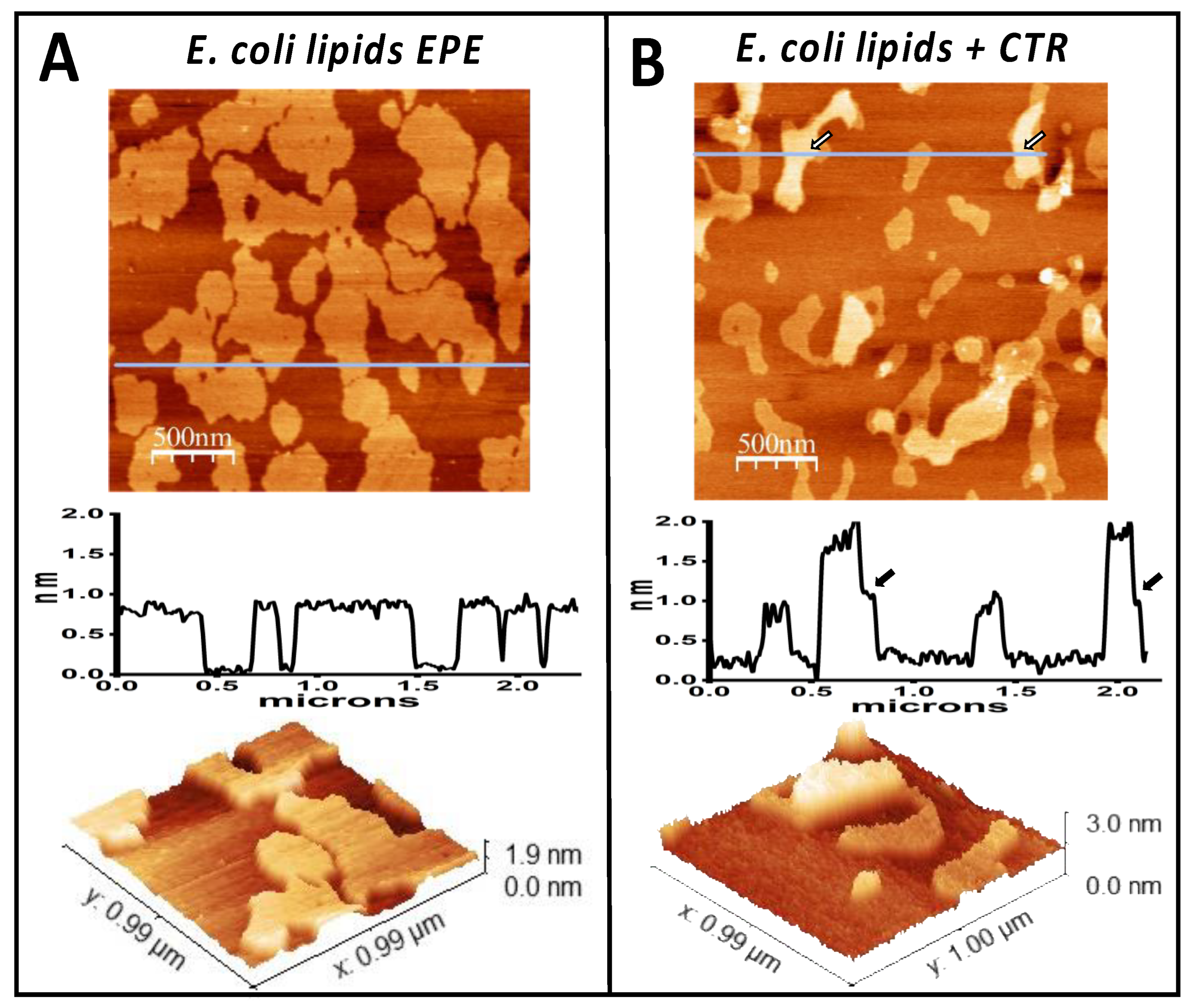
3.1. The Amyloid CTR Region of Hfq Binds to Membrane Microdomains

The E. coli inner membrane (IM) is mainly composed of four lipids: phosphatidylethanolamine (PE) (~75%), phosphatidylglycerol (PG) (~20%), cardiolipin (CL) (~5% during the exponential phase of growth, which can increase to 15–20% during the stationary phase [75,76]), and traces of phosphatidic acid (PA). To mimic the natural E. coli IM, we used EPE lipids extract containing 67% PE, 23% PG, and 10% CL. CL is a unique cone-shaped lipid and, due to its higher volume compared to the cylindrical shape of PG and PE and to its negative charge, it significantly influences membrane structure and dynamics [77]. While lipid rafts do not exist in bacteria, prokaryotes have equivalent membrane microdomains [78,79], and CL is the major component of these microdomains [80,81,82]. The conical shape of CL promotes its segregation, microdomain formation and facilitates negative membrane curvature [78]. Supported lipid bilayers of mixed composition including CL have been shown to present segregated domains that protrude around 1 nm from the rest. Figure 1A shows a supported lipid bilayer formed from the E. coli polar lipid extract. The domains observed are compatible with CL-rich microdomains [83]. Note that the lipids that interact the most with Hfq-CTR are phosphatidylglycerol (PG) and cardiolipin (CL—composed of two linked PG molecules [84]), whereas phosphatidylethanolamine (PE) and phosphatidic acid (PA) do not exhibit significant Hfq binding [56].
Figure 1B shows the membrane after exposure to Hfq-CTR for 45 min. A second layer of material was formed on top of some of the domains. These higher domains stabilize in about two hours into 1 nm high homogeneous and compact domains. Contrast in phase images obtained in tapping is associated with the mechanical properties of the sample. The different contrast observed between the higher and lower regions shown in Supplementary Figure S2 indicate that the presence of the protein could be rendering a certain stiffness to the bilayer. The fact that the phase images have a higher contrast in the higher regions indicates that these regions are mechanically more rigid than the bare lipids. Although this information is difficult to interpret at the molecular level, it does point to two possibilities: that the layer formed by the peptide is compact and, therefore, mechanically more rigid than the bilayer or that the peptide could be partially inserted into the membrane affecting its rigidity (as also described in [49]). It has been previously demonstrated that cardiolipin is the lipid that interacts most with Hfq-CTR [56], therefore reinforcing our hypothesis that the domains observed are enriched in cardiolipin. This hypothesis was strengthened by the preferred interaction of Hfq-CTR with these microdomains (Figure 1), as we previously demonstrated that cardiolipin is the lipid that interacts mostly with Hfq-CTR [56]. Note that a significant interaction between Hfq and PG also occurs, but in contrast to CL, PG is less prone to form membrane microdomains and is usually randomly distributed in the IM above 25 °C. We also observed that the domain size and shape may change over time after the interaction with Hfq-CTR, indicating that the lipid reordering might also by take place and that the composition of the domains might not be static.

3.2. The Amyloid CTR Region of Hfq Triggers RNA Insertion in the Memrbane

The supported lipid membranes were incubated with RNA after their exposure to the Hfq-CTR peptide. Figure 2A shows that, as expected, the surface, although somewhat rougher, probably due to the presence of RNA, did not show any significant structural difference with respect to the one only containing the peptide. The higher regions lying on top of the lipid domains remained. When this modified surface was incubated for 30 min with 5 nm colloidal gold beads conjugated with streptavidin, the surface underwent significant remodeling. Figure 2B shows how the 5 nm beads accumulate preferentially in certain regions of the surface, which is perfectly compatible with their selective accumulation, on the peptide-enriched higher regions. The profile clearly indicated their presence on the surface. Figure S1 shows the two controls that indicate that both peptide and RNA are needed for the colloidal particles to attach to the lipids. When a membrane was exposed to Hfq-CTR and colloidal beads, as shown in Figure S1A, there was no attachment of colloidal particles. The same was observed when the membrane was only exposed to RNA before colloidal particles (Figure S1B). We therefore confirm that there is no non-specific binding to the membrane of the RNA nor the streptavidin-modified beads.
Indeed, the interaction between the E. coli membrane and RNA is not inherently favorable. Cationic lipids are not naturally present in this membrane and, in the absence of proteins, the E. coli inner membrane displays negative charges, which originate from the anionic lipids present in this membrane (PE, PG, and CL [85,86]). This likely explains why the presence of Hfq-CTR is necessary for RNA to bind to IM, as shown in this paper.

4. Discussion

The presence of RNAs has been reported in OMVs from various Gram-negative bacteria [66,67,68,87]. Nevertheless, the mechanism by which these RNAs are packaged into OMVs remains unknown. Our previous studies suggested that these RNAs might be imported with the help of the Hfq chaperone, specifically using its CTR region, which physically interacts with the membrane and may allow the passive crossing of RNA through the IM to the periplasm [48,49]. In this paper, we show that Hfq-CTR is necessary for RNA to bind to the inner membrane, and this observation is compatible with RNA passing into the periplasm, where it can be encapsulated in OMVs (Figure 3). We have previously shown that Hfq is exported and present in the periplasm [50,70], and thus, believe that Hfq allows RNA periplasmic translocation for further export in OMVs. This mechanism indeed offers the advantage of protecting the RNAs from degradation both in the periplasm and within the OMV lumen, where RNases are abundant [19,88]. RNase I, for instance, mainly resides in the periplasm and could degrade unprotected periplasmic RNAs [89]. This association with Hfq could thus play an important role both in the translocation of the RNA through the inner membrane (using an unknown mechanisms), but also to prevent the premature degradation of the exported RNA immediately after its transit into the periplasm.
An important observation is the specific binding of Hfq or the Hfq:RNA complex to cardiolipin microdomains. CL is a unique, cone-shaped negative lipid, and its distinctive structure influences its propensity for phase transitions, with increasing membrane curvature, which affect vesicle budding [90,91,92]. This occurs mainly by CL sorting, which could be favored by Hfq binding. While cardiolipin is primarily found in the inner membrane, its presence in the E. coli outer membrane is also possible [93]. Thus, CL microdomains may attract Hfq and RNA to assist in their translocation within the periplasm, ultimately leading to embedding in OMVs (Figure 3). Another effect of CL, already reported for α-synuclein amyloid oligomers, is its ability to enhance pore formation in the inner mitochondrial membranes [94]. While in bacteria, CL has been proposed to prevent pore formation, this previous analysis did not use natural inner membranes and was conducted without a protein [95]. One can thus expect that, in addition to its role in OMV budding, CL attracts Hfq to promote the formation of pores in the inner membrane. Additionally, given Hfq’s role in RNA regulation, it is plausible that Hfq influences CL synthesis by modulating the expression or activity of enzymes involved in the CL biosynthetic pathway [96]. E. coli harbors three distinct enzymes that synthesize CL, ClsA, ClsB, and ClsC [97], and Hfq could influence their expression. Such a mechanism remains to be elucidated. Nevertheless, even if Hfq does not directly regulate cardiolipin synthases, it does regulate membrane stress responses and overall lipid homeostasis through the use of sRNAs. Hfq can thus potentially alter cardiolipin levels indirectly [96,98].

5. Conclusions

Although much is known about the passage of proteins across lipid bilayers, which often rely on precursors with a leader sequence for translocation through the membrane, data elucidating RNA translocation remain scarce. In this study, we present a model to better understand how Hfq might influence RNA translocation in the periplasm and its subsequent embedding into outer membrane vesicles. This work reveals potential mechanisms governing RNA transport and processing in bacterial systems, offering new insights into the interplay between RNA-binding proteins and membrane-associated pathways.

Supplementary Materials

The following supporting information can be downloaded at: https://www.mdpi.com/article/10.3390/pathogens14040399/s1. Figure S1. (A) E. coli lipid bilayer incubated in the presence of CTR and streptavidin colloidal gold beads. The height profile corresponds to the line shown on the upper image. The lower panel shows a three-dimensional representation of a small region. When the membrane is incubated only with Hfq-CTR, colloidal beads do not accumulate on the surface and only the cardiolipin-enriched domains with the Hfq-CTR are observed; (B) E. coli lipid bilayer incubated in the presence of RNA and streptavidin colloidal gold beads. When the membrane is not incubated with Hfq-CTR prior to the addition of RNA, colloidal particles do not accumulate on the surface and only the cardiolipin-enriched domains are observed. Figure S2. Topographic and phase images of the lipid bilayer incubated with the Hfq-CTR peptide. The regions with peptide appear darker in the phase image, indicating that their mechanical properties are different. The peptide regions appear stiffer than the pure lipid regions.

Author Contributions

Conceptualization, M.V. and V.A.; methodology, M.V.; software, M.V.; validation, M.V. and V.A.; formal analysis, M.V. and V.A.; investigation, M.V.; resources, M.V. and V.A.; writing—original draft preparation, M.V. and V.A.; writing—review and editing, M.V. and V.A.; project administration, V.A. All authors have read and agreed to the published version of the manuscript.

Funding

We acknowledge funding from International Emerging action IEA/PICS 08254 (V.A., M.V.). This study contributes to the IdEx Université de Paris ANR-18-IDEX-0001 (V.A.).

Institutional Review Board Statement

Not applicable.

Data Availability Statement

The data that support the findings of this study are available upon request from the corresponding authors.

Acknowledgments

We are grateful to Richard R. Sinden (SDMT, SD, USA) and G. Wegrzyn (Gdansk university, Poland) for the critical reading of the manuscript. We would like to thank Frank Wien (Synchrotron SOLEIL, France) and Florian Turbant (LLB and Synchrotron SOLEIL, France) for their support with the SRCD/OCD measurements conducted on the DISCO beamline at Synchrotron SOLEIL. These measurements (proposal 20230530; see References [49,56]) formed the foundation of this work. We also thank Wafa Achouak (CEA, Cadarache, France) for many fruitful discussions.

Conflicts of Interest

The authors declare no conflicts of interest.

Abbreviations

The following abbreviations are used in this manuscript:
Outer membrane vesicle: OMV; IM/OM: Inner/Outer membrane; CL: Cardiolipin; AFM: Atomic force microscopy; Hfq-NTR/CTR: Hfq N/C-terminal region; EPE: E. coli polar extract; sRNA: Small noncoding RNA; PG: Phosphatidylglycerol; SUV: Small unilamellar vesicle.

References

  1. Papenfort, K.; Melamed, S. Small RNAs, Large Networks: Posttranscriptional Regulons in Gram-Negative Bacteria. Annu. Rev. Microbiol. 2023, 77, 23–43. [Google Scholar] [CrossRef] [PubMed]
  2. Kavita, K.; de Mets, F.; Gottesman, S. New aspects of RNA-based regulation by Hfq and its partner sRNAs. Curr. Opin. Microbiol. 2018, 42, 53–61. [Google Scholar] [CrossRef]
  3. Gottesman, S. Trouble is coming: Signaling pathways that regulate general stress responses in bacteria. J. Biol. Chem. 2019, 294, 11685–11700. [Google Scholar] [CrossRef] [PubMed]
  4. Gelsinger, D.R.; DiRuggiero, J. The Non-Coding Regulatory RNA Revolution in Archaea. Genes 2018, 9, 141. [Google Scholar] [CrossRef]
  5. Svensson, S.L.; Sharma, C.M. Small RNAs in Bacterial Virulence and Communication. Microbiol. Spectr. 2016, 4, 169–212. [Google Scholar] [CrossRef]
  6. Pulvermacher, S.C.; Stauffer, L.T.; Stauffer, G.V. Role of the Escherichia coli Hfq protein in GcvB regulation of oppA and dppA mRNAs. Microbiology 2009, 155, 115–123. [Google Scholar] [CrossRef]
  7. Rice, J.B.; Vanderpool, C.K. The small RNA SgrS controls sugar-phosphate accumulation by regulating multiple PTS genes. Nucleic Acids Res. 2011, 39, 3806–3819. [Google Scholar] [CrossRef] [PubMed]
  8. Salvail, H.; Masse, E. Regulating iron storage and metabolism with RNA: An overview of posttranscriptional controls of intracellular iron homeostasis. Wiley Interdiscip. Rev. RNA 2012, 3, 26–36. [Google Scholar] [CrossRef]
  9. Majdalani, N.; Cunning, C.; Sledjeski, D.; Elliott, T.; Gottesman, S. DsrA RNA regulates translation of RpoS message by an anti-antisense mechanism, independent of its action as an antisilencer of transcription. Proc. Natl. Acad. Sci. USA 1998, 95, 12462–12467. [Google Scholar] [CrossRef]
  10. Melamed, S.; Adams, P.P.; Zhang, A.; Zhang, H.; Storz, G. RNA-RNA Interactomes of ProQ and Hfq Reveal Overlapping and Competing Roles. Mol. Cell 2020, 77, 411–425 e417. [Google Scholar] [CrossRef]
  11. Schumacher, M.A.; Pearson, R.F.; Moller, T.; Valentin-Hansen, P.; Brennan, R.G. Structures of the pleiotropic translational regulator Hfq and an Hfq- RNA complex: A bacterial Sm-like protein. Embo J. 2002, 21, 3546–3556. [Google Scholar] [CrossRef] [PubMed]
  12. Link, T.M.; Valentin-Hansen, P.; Brennan, R.G. Structure of Escherichia coli Hfq bound to polyriboadenylate RNA. Proc. Natl. Acad. Sci. USA 2009, 106, 19292–19297. [Google Scholar] [CrossRef]
  13. Sauer, E.; Schmidt, S.; Weichenrieder, O. Small RNA binding to the lateral surface of Hfq hexamers and structural rearrangements upon mRNA target recognition. Proc. Natl. Acad. Sci. USA 2012, 109, 9396–9401. [Google Scholar] [CrossRef]
  14. Dimastrogiovanni, D.; Frohlich, K.S.; Bandyra, K.J.; Bruce, H.A.; Hohensee, S.; Vogel, J.; Luisi, B.F. Recognition of the small regulatory RNA RydC by the bacterial Hfq protein. Elife 2014, 3, e05375. [Google Scholar] [CrossRef] [PubMed]
  15. Hwang, W.; Arluison, V.; Hohng, S. Dynamic competition of DsrA and rpoS fragments for the proximal binding site of Hfq as a means for efficient annealing. Nucleic Acids Res. 2011, 39, 5131–5139. [Google Scholar] [CrossRef] [PubMed]
  16. De Lay, N.; Schu, D.J.; Gottesman, S. Bacterial small RNA-based negative regulation: Hfq and its accomplices. J. Biol. Chem. 2013, 288, 7996–8003. [Google Scholar] [CrossRef]
  17. Vigoda, M.B.; Argaman, L.; Kournos, M.; Margalit, H. Unraveling the interplay between a small RNA and RNase E in bacteria. Nucleic Acids Res. 2024, 52, 8947–8966. [Google Scholar] [CrossRef]
  18. Folichon, M.; Arluison, V.; Pellegrini, O.; Huntzinger, E.; Regnier, P.; Hajnsdorf, E. The poly(A) binding protein Hfq protects RNA from RNase E and exoribonucleolytic degradation. Nucleic Acids Res. 2003, 31, 7302–7310. [Google Scholar] [CrossRef]
  19. Moll, I.; Afonyushkin, T.; Vytvytska, O.; Kaberdin, V.R.; Blasi, U. Coincident Hfq binding and RNase E cleavage sites on mRNA and small regulatory RNAs. RNA 2003, 9, 1308–1314. [Google Scholar] [CrossRef]
  20. Kornienko, I.V.; Aramova, O.Y.; Tishchenko, A.A.; Rudoy, D.V.; Chikindas, M.L. RNA Stability: A Review of the Role of Structural Features and Environmental Conditions. Molecules 2024, 29, 5978. [Google Scholar] [CrossRef]
  21. Sorger-Domenigg, T.; Sonnleitner, E.; Kaberdin, V.R.; Blasi, U. Distinct and overlapping binding sites of Pseudomonas aeruginosa Hfq and RsmA proteins on the non-coding RNA RsmY. Biochem. Biophys. Res. Commun. 2007, 352, 769–773. [Google Scholar] [CrossRef] [PubMed]
  22. Wien, F.; Gragera, M.; Matsuo, T.; Moroy, G.; Bueno-Carrasco, M.T.; Arranz, R.; Cossa, A.; Martel, A.; Bordallo, H.N.; Rudic, S.; et al. Amyloid-like DNA bridging: A new mode of DNA shaping. Nucleic Acids Res. 2025, 53, gkaf169. [Google Scholar] [CrossRef]
  23. Cossa, A.; Trepout, S.; Wien, F.; Groen, J.; Le Brun, E.; Turbant, F.; Besse, L.; Pereiro, E.; Arluison, V. Cryo soft X-ray tomography to explore Escherichia coli nucleoid remodeling by Hfq master regulator. J. Struct. Biol. 2022, 214, 107912. [Google Scholar] [CrossRef]
  24. Cech, G.M.; Pakula, B.; Kamrowska, D.; Wegrzyn, G.; Arluison, V.; Szalewska-Palasz, A. Hfq protein deficiency in Escherichia coli affects ColE1-like but not lambda plasmid DNA replication. Plasmid 2014, 73, 10–15. [Google Scholar] [CrossRef]
  25. Kubiak, K.; Wien, F.; Yadav, I.; Jones, N.C.; Vrønning Hoffmann, S.; Le Cam, E.; Cossa, A.; Geinguenaud, F.; van der Maarel, J.R.C.; Węgrzyn, G.; et al. Amyloid-like Hfq interaction with single-stranded DNA: Involvement in recombination and replication in Escherichia coli. QRB Discovery 2022, 3, e15. [Google Scholar] [CrossRef]
  26. Parekh, V.J.; Niccum, B.A.; Shah, R.; Rivera, M.A.; Novak, M.J.; Geinguenaud, F.; Wien, F.; Arluison, V.; Sinden, R.R. Role of Hfq in Genome Evolution: Instability of G-Quadruplex Sequences in E. coli. Microorganisms 2019, 8, 28. [Google Scholar] [CrossRef] [PubMed]
  27. Chen, J.; Gottesman, S. Hfq links translation repression to stress-induced mutagenesis in E. coli. Genes. Dev. 2017, 31, 1382–1395. [Google Scholar] [CrossRef] [PubMed]
  28. Wilusz, C.J.; Wilusz, J. Lsm proteins and Hfq: Life at the 3′ end. RNA Biol. 2013, 10, 592–601. [Google Scholar] [CrossRef]
  29. Mura, C.; Randolph, P.S.; Patterson, J.; Cozen, A.E. Archaeal and eukaryotic homologs of Hfq: A structural and evolutionary perspective on Sm function. RNA Biol. 2013, 10, 636–651. [Google Scholar] [CrossRef]
  30. Brennan, R.G.; Link, T.M. Hfq structure, function and ligand binding. Curr. Opin. Microbiol. 2007, 10, 125–133. [Google Scholar] [CrossRef]
  31. Arluison, V.; Folichon, M.; Marco, S.; Derreumaux, P.; Pellegrini, O.; Seguin, J.; Hajnsdorf, E.; Regnier, P. The C-terminal domain of Escherichia coli Hfq increases the stability of the hexamer. Eur. J. Biochem. 2004, 271, 1258–1265. [Google Scholar] [CrossRef] [PubMed]
  32. Olsen, A.S.; Moller-Jensen, J.; Brennan, R.G.; Valentin-Hansen, P. C-Terminally truncated derivatives of Escherichia coli Hfq are proficient in riboregulation. J. Mol. Biol. 2010, 404, 173–182. [Google Scholar] [CrossRef] [PubMed]
  33. Fortas, E.; Piccirilli, F.; Malabirade, A.; Militello, V.; Trepout, S.; Marco, S.; Taghbalout, A.; Arluison, V. New insight into the structure and function of Hfq C-terminus. Biosci. Rep. 2015, 35, e00190. [Google Scholar] [CrossRef]
  34. Berbon, M.; Martinez, D.; Morvan, E.; Grelard, A.; Kauffmann, B.; Waeytens, J.; Wien, F.; Arluison, V.; Habenstein, B. Hfq C-terminal region forms a beta-rich amyloid-like motif without perturbing the N-terminal Sm-like structure. Commun. Biol. 2023, 6, 1075. [Google Scholar] [CrossRef] [PubMed]
  35. Fitzpatrick, A.W.; Debelouchina, G.T.; Bayro, M.J.; Clare, D.K.; Caporini, M.A.; Bajaj, V.S.; Jaroniec, C.P.; Wang, L.; Ladizhansky, V.; Muller, S.A.; et al. Atomic structure and hierarchical assembly of a cross-beta amyloid fibril. Proc. Natl. Acad. Sci. USA 2013, 110, 5468–5473. [Google Scholar] [CrossRef]
  36. Gremer, L.; Scholzel, D.; Schenk, C.; Reinartz, E.; Labahn, J.; Ravelli, R.B.G.; Tusche, M.; Lopez-Iglesias, C.; Hoyer, W.; Heise, H.; et al. Fibril structure of amyloid-beta(1-42) by cryo-electron microscopy. Science 2017, 358, 116–119. [Google Scholar] [CrossRef]
  37. Ju, Y.; Tam, K.Y. Pathological mechanisms and therapeutic strategies for Alzheimer’s disease. Neural Regen. Res. 2022, 17, 543–549. [Google Scholar] [CrossRef]
  38. Maury, C.P. The emerging concept of functional amyloid. J. Intern. Med. 2009, 265, 329–334. [Google Scholar] [CrossRef]
  39. Otzen, D.; Riek, R. Functional Amyloids. Cold Spring Harb. Perspect. Biol. 2019, 11, a033860. [Google Scholar] [CrossRef]
  40. Giraldo, R. Defined DNA sequences promote the assembly of a bacterial protein into distinct amyloid nanostructures. Proc. Natl. Acad. Sci. USA 2007, 104, 17388–17393. [Google Scholar] [CrossRef]
  41. Khambhati, K.; Patel, J.; Saxena, V.; A, P.; Jain, N. Gene Regulation of Biofilm-Associated Functional Amyloids. Pathogens 2021, 10, 490. [Google Scholar] [CrossRef] [PubMed]
  42. Vecerek, B.; Rajkowitsch, L.; Sonnleitner, E.; Schroeder, R.; Blasi, U. The C-terminal domain of Escherichia coli Hfq is required for regulation. Nucleic Acids Res. 2008, 36, 133–143. [Google Scholar] [CrossRef] [PubMed]
  43. Vincent, H.A.; Henderson, C.A.; Stone, C.M.; Cary, P.D.; Gowers, D.M.; Sobott, F.; Taylor, J.E.; Callaghan, A.J. The low-resolution solution structure of Vibrio cholerae Hfq in complex with Qrr1 sRNA. Nucleic Acids Res. 2012, 40, 8698–8710. [Google Scholar] [CrossRef] [PubMed]
  44. Salim, N.N.; Faner, M.A.; Philip, J.A.; Feig, A.L. Requirement of upstream Hfq-binding (ARN)x elements in glmS and the Hfq C-terminal region for GlmS upregulation by sRNAs GlmZ and GlmY. Nucleic Acids Res. 2012, 40, 8021–8032. [Google Scholar] [CrossRef]
  45. Turbant, F.; Wu, P.; Wien, F.; Arluison, V. The Amyloid Region of Hfq Riboregulator Promotes DsrA:rpoS RNAs Annealing. Biology 2021, 10, 900. [Google Scholar] [CrossRef]
  46. Kavita, K.; Zhang, A.; Tai, C.H.; Majdalani, N.; Storz, G.; Gottesman, S. Multiple in vivo roles for the C-terminal domain of the RNA chaperone Hfq. Nucleic Acids Res. 2022, 50, 1718–1733. [Google Scholar] [CrossRef]
  47. Sarni, S.H.; Roca, J.; Du, C.; Jia, M.; Li, H.; Damjanovic, A.; Malecka, E.M.; Wysocki, V.H.; Woodson, S.A. Intrinsically disordered interaction network in an RNA chaperone revealed by native mass spectrometry. Proc. Natl. Acad. Sci. USA 2022, 119, e2208780119. [Google Scholar] [CrossRef]
  48. Malabirade, A.; Morgado-Brajones, J.; Trepout, S.; Wien, F.; Marquez, I.; Seguin, J.; Marco, S.; Velez, M.; Arluison, V. Membrane association of the bacterial riboregulator Hfq and functional perspectives. Sci. Rep. 2017, 7, 10724. [Google Scholar] [CrossRef]
  49. Turbant, F.; Waeytens, J.; Campidelli, C.; Bombled, M.; Martinez, D.; Grelard, A.; Habenstein, B.; Raussens, V.; Velez, M.; Wien, F.; et al. Unraveling Membrane Perturbations Caused by the Bacterial Riboregulator Hfq. Int. J. Mol. Sci. 2022, 23, 8739. [Google Scholar] [CrossRef]
  50. Diestra, E.; Cayrol, B.; Arluison, V.; Risco, C. Cellular electron microscopy imaging reveals the localization of the Hfq protein close to the bacterial membrane. PLoS ONE 2009, 4, e8301. [Google Scholar] [CrossRef]
  51. Taghbalout, A.; Yang, Q.; Arluison, V. The Escherichia coli RNA processing and degradation machinery is compartmentalized within an organized cellular network. Biochem. J. 2014, 458, 11–22. [Google Scholar] [CrossRef] [PubMed]
  52. Ikeda, Y.; Yagi, M.; Morita, T.; Aiba, H. Hfq binding at RhlB-recognition region of RNase E is crucial for the rapid degradation of target mRNAs mediated by sRNAs in Escherichia coli. Mol. Microbiol. 2011, 79, 419–432. [Google Scholar] [CrossRef]
  53. Obregon, K.A.; Hoch, C.T.; Sukhodolets, M.V. Sm-like protein Hfq: Composition of the native complex, modifications, and interactions. Biochim. Biophys. Acta 2015, 1854, 950–966. [Google Scholar] [CrossRef]
  54. Qiang, W.; Yau, W.M.; Schulte, J. Fibrillation of beta amyloid peptides in the presence of phospholipid bilayers and the consequent membrane disruption. Biochim. Biophys. Acta 2015, 1848, 266–276. [Google Scholar] [CrossRef] [PubMed]
  55. Nutini, A. Amyloid oligomers and their membrane toxicity—A perspective study. Prog. Biophys. Mol. Biol. 2024, 187, 9–20. [Google Scholar] [CrossRef]
  56. Turbant, F.; Machiels, Q.; Waeytens, J.; Wien, F.; Arluison, V. The Amyloid Assembly of the Bacterial Hfq Is Lipid-Driven and Lipid-Specific. Int. J. Mol. Sci. 2024, 25, 1434. [Google Scholar] [CrossRef]
  57. Graham, L.L.; Harris, R.; Villiger, W.; Beveridge, T.J. Freeze-substitution of gram-negative eubacteria: General cell morphology and envelope profiles. J. Bacteriol. 1991, 173, 1623–1633. [Google Scholar] [CrossRef] [PubMed]
  58. Sartorio, M.G.; Pardue, E.J.; Feldman, M.F.; Haurat, M.F. Bacterial Outer Membrane Vesicles: From Discovery to Applications. Annu. Rev. Microbiol. 2021, 75, 609–630. [Google Scholar] [CrossRef]
  59. Kulp, A.; Kuehn, M.J. Biological functions and biogenesis of secreted bacterial outer membrane vesicles. Annu. Rev. Microbiol. 2010, 64, 163–184. [Google Scholar] [CrossRef]
  60. Furuyama, N.; Sircili, M.P. Outer Membrane Vesicles (OMVs) Produced by Gram-Negative Bacteria: Structure, Functions, Biogenesis, and Vaccine Application. Biomed. Res. Int. 2021, 2021, 1490732. [Google Scholar] [CrossRef]
  61. Tsatsaronis, J.A.; Franch-Arroyo, S.; Resch, U.; Charpentier, E. Extracellular Vesicle RNA: A Universal Mediator of Microbial Communication? Trends Microbiol. 2018, 26, 401–410. [Google Scholar] [CrossRef] [PubMed]
  62. Schwechheimer, C.; Kuehn, M.J. Outer-membrane vesicles from Gram-negative bacteria: Biogenesis and functions. Nat. Rev. Microbiol. 2015, 13, 605–619. [Google Scholar] [CrossRef]
  63. Blenkiron, C.; Simonov, D.; Muthukaruppan, A.; Tsai, P.; Dauros, P.; Green, S.; Hong, J.; Print, C.G.; Swift, S.; Phillips, A.R. Uropathogenic Escherichia coli Releases Extracellular Vesicles That Are Associated with RNA. PLoS ONE 2016, 11, e0160440. [Google Scholar] [CrossRef]
  64. Lee, H.J. Microbe-Host Communication by Small RNAs in Extracellular Vesicles: Vehicles for Transkingdom RNA Transportation. Int. J. Mol. Sci. 2019, 20, 1487. [Google Scholar] [CrossRef]
  65. Dauros-Singorenko, P.; Blenkiron, C.; Phillips, A.; Swift, S. The functional RNA cargo of bacterial membrane vesicles. FEMS Microbiol. Lett. 2018, 365, fny023. [Google Scholar] [CrossRef]
  66. Ghosal, A.; Upadhyaya, B.B.; Fritz, J.V.; Heintz-Buschart, A.; Desai, M.S.; Yusuf, D.; Huang, D.; Baumuratov, A.; Wang, K.; Galas, D.; et al. The extracellular RNA complement of Escherichia coli. Microbiologyopen 2015, 4, 252–266. [Google Scholar] [CrossRef] [PubMed]
  67. Koeppen, K.; Hampton, T.H.; Jarek, M.; Scharfe, M.; Gerber, S.A.; Mielcarz, D.W.; Demers, E.G.; Dolben, E.L.; Hammond, J.H.; Hogan, D.A.; et al. A Novel Mechanism of Host-Pathogen Interaction through sRNA in Bacterial Outer Membrane Vesicles. PLoS Pathog. 2016, 12, e1005672. [Google Scholar] [CrossRef]
  68. Malabirade, A.; Habier, J.; Heintz-Buschart, A.; May, P.; Godet, J.; Halder, R.; Etheridge, A.; Galas, D.; Wilmes, P.; Fritz, J.V. The RNA Complement of Outer Membrane Vesicles from Salmonella enterica Serovar Typhimurium Under Distinct Culture Conditions. Front. Microbiol. 2018, 9, 2015. [Google Scholar] [CrossRef] [PubMed]
  69. Diallo, I.; Ho, J.; Lambert, M.; Benmoussa, A.; Husseini, Z.; Lalaouna, D.; Masse, E.; Provost, P. A tRNA-derived fragment present in E. coli OMVs regulates host cell gene expression and proliferation. PLoS Pathog. 2022, 18, e1010827. [Google Scholar] [CrossRef]
  70. Turbant, F.; Waeytens, J.; Blache, A.; Esnouf, E.; Raussens, V.; Wegrzyn, G.; Achouak, W.; Wien, F.; Arluison, V. Interactions and Insertion of Escherichia coli Hfq into Outer Membrane Vesicles as Revealed by Infrared and Orientated Circular Dichroism Spectroscopies. Int. J. Mol. Sci. 2023, 24, 11424. [Google Scholar] [CrossRef]
  71. Ajam-Hosseini, M.; Akhoondi, F.; Parvini, F.; Fahimi, H. Gram-negative bacterial sRNAs encapsulated in OMVs: An emerging class of therapeutic targets in diseases. Front. Cell Infect. Microbiol. 2023, 13, 1305510. [Google Scholar] [CrossRef]
  72. Turbant, F.; El Hamoui, O.; Partouche, D.; Sandt, C.; Busi, F.; Wien, F.; Arluison, V. Identification and characterization of the Hfq bacterial amyloid region DNA interactions. BBA Adv. 2021, 1, 100029. [Google Scholar] [CrossRef] [PubMed]
  73. Sledjeski, D.; Gottesman, S. A small RNA acts as an antisilencer of the H-NS-silenced rcsA gene of Escherichia coli. Proc. Natl. Acad. Sci. USA 1995, 92, 2003–2007. [Google Scholar] [CrossRef] [PubMed]
  74. Wu, P.; Liu, X.; Yang, L.; Sun, Y.; Gong, Q.; Wu, J.; Shi, Y. The important conformational plasticity of DsrA sRNA for adapting multiple target regulation. Nucleic Acids Res. 2017, 45, 9625–9639. [Google Scholar] [CrossRef]
  75. Oliver, P.M.; Crooks, J.A.; Leidl, M.; Yoon, E.J.; Saghatelian, A.; Weibel, D.B. Localization of anionic phospholipids in Escherichia coli cells. J. Bacteriol. 2014, 196, 3386–3398. [Google Scholar] [CrossRef]
  76. Cronan, J.E., Jr. Phospholipid alterations during growth of Escherichia coli. J. Bacteriol. 1968, 95, 2054–2061. [Google Scholar] [CrossRef] [PubMed]
  77. Kucerka, N.; Heberle, F.A.; Pan, J.; Katsaras, J. Structural Significance of Lipid Diversity as Studied by Small Angle Neutron and X-ray Scattering. Membranes 2015, 5, 454–472. [Google Scholar] [CrossRef]
  78. Mileykovskaya, E.; Dowhan, W. Cardiolipin membrane domains in prokaryotes and eukaryotes. Biochim. Biophys. Acta 2009, 1788, 2084–2091. [Google Scholar] [CrossRef]
  79. Mukhopadhyay, R.; Huang, K.C.; Wingreen, N.S. Lipid localization in bacterial cells through curvature-mediated microphase separation. Biophys. J. 2008, 95, 1034–1049. [Google Scholar] [CrossRef]
  80. Lopez, D.; Kolter, R. Functional microdomains in bacterial membranes. Genes. Dev. 2010, 24, 1893–1902. [Google Scholar] [CrossRef]
  81. Lopez, D. Molecular composition of functional microdomains in bacterial membranes. Chem. Phys. Lipids 2015, 192, 3–11. [Google Scholar] [CrossRef] [PubMed]
  82. Wessel, A.K.; Yoshii, Y.; Reder, A.; Boudjemaa, R.; Szczesna, M.; Betton, J.M.; Bernal-Bayard, J.; Beloin, C.; Lopez, D.; Volker, U.; et al. Escherichia coli SPFH Membrane Microdomain Proteins HflKC Contribute to Aminoglycoside and Oxidative Stress Tolerance. Microbiol. Spectr. 2023, 11, e0176723. [Google Scholar] [CrossRef] [PubMed]
  83. Domenech, O.; Sanz, F.; Montero, M.T.; Hernandez-Borrell, J. Thermodynamic and structural study of the main phospholipid components comprising the mitochondrial inner membrane. Biochim. Biophys. Acta 2006, 1758, 213–221. [Google Scholar] [CrossRef]
  84. Schlame, M. Cardiolipin synthesis for the assembly of bacterial and mitochondrial membranes. J. Lipid Res. 2008, 49, 1607–1620. [Google Scholar] [CrossRef] [PubMed]
  85. Card, G.L.; Trautman, J.K. Role of anionic lipid in bacterial membranes. Biochim. Biophys. Acta 1990, 1047, 77–82. [Google Scholar] [CrossRef]
  86. Pramanik, J.; Keasling, J.D. Stoichiometric model of Escherichia coli metabolism: Incorporation of growth-rate dependent biomass composition and mechanistic energy requirements. Biotechnol. Bioeng. 1997, 56, 398–421. [Google Scholar] [CrossRef]
  87. Ahmadi Badi, S.; Bruno, S.P.; Moshiri, A.; Tarashi, S.; Siadat, S.D.; Masotti, A. Small RNAs in Outer Membrane Vesicles and Their Function in Host-Microbe Interactions. Front. Microbiol. 2020, 11, 1209. [Google Scholar] [CrossRef] [PubMed]
  88. Hussein, M.; Jasim, R.; Gocol, H.; Baker, M.; Thombare, V.J.; Ziogas, J.; Purohit, A.; Rao, G.G.; Li, J.; Velkov, T. Comparative Proteomics of Outer Membrane Vesicles from Polymyxin-Susceptible and Extremely Drug-Resistant Klebsiella pneumoniae. mSphere 2023, 8, e0053722. [Google Scholar] [CrossRef]
  89. Zhu, L.Q.; Gangopadhyay, T.; Padmanabha, K.P.; Deutscher, M.P. Escherichia coli rna gene encoding RNase I: Cloning, overexpression, subcellular distribution of the enzyme, and use of an rna deletion to identify additional RNases. J. Bacteriol. 1990, 172, 3146–3151. [Google Scholar] [CrossRef]
  90. Beales, P.A.; Bergstrom, C.L.; Geerts, N.; Groves, J.T.; Vanderlick, T.K. Single vesicle observations of the cardiolipin-cytochrome C interaction: Induction of membrane morphology changes. Langmuir 2011, 27, 6107–6115. [Google Scholar] [CrossRef]
  91. Beltran-Heredia, E.; Tsai, F.C.; Salinas-Almaguer, S.; Cao, F.J.; Bassereau, P.; Monroy, F. Membrane curvature induces cardiolipin sorting. Commun. Biol. 2019, 2, 225. [Google Scholar] [CrossRef] [PubMed]
  92. Renner, L.D.; Weibel, D.B. Cardiolipin microdomains localize to negatively curved regions of Escherichia coli membranes. Proc. Natl. Acad. Sci. USA 2011, 108, 6264–6269. [Google Scholar] [CrossRef] [PubMed]
  93. Raetz, C.R.; Kantor, G.D.; Nishijima, M.; Newman, K.F. Cardiolipin accumulation in the inner and outer membranes of Escherichia coli mutants defective in phosphatidylserine synthetase. J. Bacteriol. 1979, 139, 544–551. [Google Scholar] [CrossRef] [PubMed]
  94. Ghio, S.; Camilleri, A.; Caruana, M.; Ruf, V.C.; Schmidt, F.; Leonov, A.; Ryazanov, S.; Griesinger, C.; Cauchi, R.J.; Kamp, F.; et al. Cardiolipin Promotes Pore-Forming Activity of Alpha-Synuclein Oligomers in Mitochondrial Membranes. ACS Chem. Neurosci. 2019, 10, 3815–3829. [Google Scholar] [CrossRef]
  95. Rocha-Roa, C.; Orjuela, J.D.; Leidy, C.; Cossio, P.; Aponte-Santamaria, C. Cardiolipin prevents pore formation in phosphatidylglycerol bacterial membrane models. FEBS Lett. 2021, 595, 2701–2714. [Google Scholar] [CrossRef]
  96. Klein, G.; Raina, S. Regulated Control of the Assembly and Diversity of LPS by Noncoding sRNAs. Biomed. Res. Int. 2015, 2015, 153561. [Google Scholar] [CrossRef]
  97. Tan, B.K.; Bogdanov, M.; Zhao, J.; Dowhan, W.; Raetz, C.R.; Guan, Z. Discovery of a cardiolipin synthase utilizing phosphatidylethanolamine and phosphatidylglycerol as substrates. Proc. Natl. Acad. Sci. USA 2012, 109, 16504–16509. [Google Scholar] [CrossRef]
  98. Guisbert, E.; Rhodius, V.A.; Ahuja, N.; Witkin, E.; Gross, C.A. Hfq modulates the sigmaE-mediated envelope stress response and the sigma32-mediated cytoplasmic stress response in Escherichia coli. J. Bacteriol. 2007, 189, 1963–1973. [Google Scholar] [CrossRef]
Figure 1. E. coli lipid bilayer incubated in the absence (A) or presence (B) of Hfq-CTR. Panel (A) shows the E. coli lipids bilayer. The height profile under the line shown on the upper image indicates that the domains are 0.8 nm higher than the rest of the membrane. The lower panel shows a three-dimensional representation of a small region. Panel (B) shows the E. coli lipid bilayer incubated in the presence of Hfq-CTR. The peptide accumulated on top of some of the domains, generating 1 nm high regions in some of them, as shown on the height profile. The arrows point the regions where the change in height occurs.
Pathogens 14 00399 g001
Figure 2. Effects of Hfq-CTR, RNA, and streptavidin gold beads on the E. coli lipid bilayer. (A) E. coli lipid bilayer (abbreviated as EPE) with HFq-CTR and RNA. The height profile under the line shown on the upper image indicates that some domain regions become higher, up to 1 nm above the domains, but no significant difference with respect to the bilayer prior to the incubation with RNA was detected. The arrows point the regions where the change in height occurs. The lower panel shows a three-dimensional representation of small region. (B) E. coli lipid bilayer incubated in the presence of Hfq-CTR, RNA, and streptavidin gold beads. The 5 nm diameter beads accumulate in some of the regions and the topographical profile detects reflects their presence.
Pathogens 14 00399 g002
Figure 3. Model of Hfq-CTR interaction with E. coli membrane microdomain and possible consequence on OMV formation. Hfq interacts with CL-rich microdomains (in orange). As shown previously, this may promote hole formation and passive export. In this paper, we show that Hfq, and precisely its amyloid CTR, and sRNA bound to Hfq-CTR interact specifically with membrane microdomains. Hfq can protect sRNA from RNAse degradation in the periplasm and/or OMV lumen. sRNA regulators controlling mRNAs are depicted as red open arrows and mRNAs as thick black lines; 5′ and 3′ of RNAs are depicted by a “ball and arrow head”, respectively; Hfq-NTR is depicted as a blue toroidal hexamer (Sm torus); Hfq-CTR as a blue amyloid β-strand; CL synthases as orange ellipses and RNAses as grey ellipses; positive and negative regulations by Hfq are indicated by arrows and T-shaped lines, and colored in green and red, respectively; the dotted line symbolizes the peptidoglycans between the outer and inner membranes (OMs/IMs).
Pathogens 14 00399 g003
Disclaimer/Publisher’s Note: The statements, opinions and data contained in all publications are solely those of the individual author(s) and contributor(s) and not of MDPI and/or the editor(s). MDPI and/or the editor(s) disclaim responsibility for any injury to people or property resulting from any ideas, methods, instructions or products referred to in the content.

