Article

Antiparasitic Activity of Chalepensin and Graveoline Isolated from Ruta chalepensis L.: In Vitro Evaluation Against Strongyloides venezuelensis

by
Nancy E. Rodríguez-Garza
1,2,
Miguel Marín
1,
Javier Sánchez-Montejo
1,
Joel H. Elizondo-Luévano
3,*,
Aldo F. Bazaldúa-Rodríguez
3,
Ramiro Quintanilla-Licea
3,
César I. Romo-Sáenz
2,4,
Rafael Peláez
1,5,
Antonio Muro
1 and
Julio López-Abán
1,*
1
Grupo de Enfermedades Infecciosas y Tropicales (e-INTRO), Instituto de Investigación Biomédica de Salamanca, Centro de Investigación de Enfermedades Tropicales de la Universidad de Salamanca (IBSAL-CIETUS), Facultad de Farmacia, Universidad de Salamanca, 37007 Salamanca, Spain
2
Departamento de Microbiología e Inmunología, Facultad de Ciencias Biológicas, Universidad Autónoma de Nuevo León, San Nicolás de los Garza 66455, Nuevo León, Mexico
3
Departamento de Química, Facultad de Ciencias Biológicas, Universidad Autónoma de Nuevo León, San Nicolás de los Garza 66455, Nuevo León, Mexico
4
Facultad de Medicina y Ciencias Biomédicas, Universidad Autónoma de Chihuahua, Chihuahua 31109, Chihuahua, Mexico
5
Laboratorio de Química Orgánica y Farmacéutica, Departamento de Ciencias Farmacéuticas, Facultad de Farmacia, Universidad de Salamanca, 37007 Salamanca, Spain
*
Authors to whom correspondence should be addressed.
Pathogens 2025, 14(5), 419; https://doi.org/10.3390/pathogens14050419 (registering DOI)
Submission received: 14 March 2025 / Revised: 18 April 2025 / Accepted: 23 April 2025 / Published: 25 April 2025

Abstract

:
Parasitic diseases constitute a significant challenge to global public health, with Strongyloides stercoralis ranking among the most prevalent and clinically significant parasites. The limitations of current nematocidal therapies highlight an urgent need for novel treatment strategies. In this study, the nematocidal activity of chalepensin and graveoline, two compounds isolated from Ruta chalepensis, was evaluated against larval and adult stages of Strongyloides venezuelensis (model for S. stercoralis). The in vitro efficacy of these compounds was assessed on third-stage infective larvae (L3) and adult parthenogenetic females at various time points, while cytotoxicity was determined using Vero cells to calculate selectivity indices (SI). Both compounds showed good antiparasitic activity, but chalepensin exhibited superior nematocidal activity compared to graveoline, with an LC50 of 3.9 µg/mL and an SI of 990 for L3, and an LC50 of 16.8 µg/mL and an SI of 200 for adult females at 72 h. Morphological analysis via scanning electron microscopy in adult females revealed that graveoline induced mostly cuticle detachment, while chalepensin caused protuberances across the parasite body. These findings suggest that both compounds possess promising antiparasitic potential, with chalepensin emerging as a particularly potent candidate for further exploration.

1. Introduction

Parasitic diseases pose a major global public health concern, particularly in tropical and subtropical regions where poor sanitary conditions facilitate their transmission [1]. Helminthic infections affect nearly a quarter of the world’s population, contributing significantly to disease burden and disability [2]. Among these infections, strongyloidiasis, caused by Strongyloides stercoralis, is one of the most neglected and challenging helminthiases to control and eradicate [3]. It is estimated to affect over 600 million people worldwide [4]. The infection begins when filariform larvae present in contaminated soil penetrate the host’s skin, subsequently migrating through the bloodstream to the lungs. From there, they ascend the respiratory tract, are swallowed, and reach the small intestine. There, they mature into adult females capable of reproducing via parthenogenesis [5]. Unlike most helminths, S. stercoralis has a unique autoinfective cycle that allows the parasite to persist within a host for decades, leading to chronic infections and posing significant therapeutic challenges [6]. Up to 70% of patients with chronic S. stercoralis infection may remain asymptomatic, with elevated blood eosinophil counts often serving as the only clinical indicator of strongyloidiasis [7]. In symptomatic cases, patients typically present with mild gastrointestinal and respiratory complaints, along with a characteristic skin rash known as larva currens [8]. Although chronic infections can be asymptomatic or exhibit mild symptoms, they carry the risk of progressing to hyperinfection syndrome, a severe, life-threatening disease with a reported fatality rate approaching 95% [9].
The World Health Organization (WHO) recommends ivermectin as the first-line treatment for strongyloidiasis due to its superior efficacy. In contrast, albendazole, which has comparatively lower efficacy, is considered a second-line therapeutic option [10]. However, high-quality evidence for the treatment of hyperinfection syndrome remains limited, and there have been reports of severe toxicities associated with high-dose ivermectin use [11]. For decades, ivermectin has been widely used in mass drug administration programs aimed at eliminating onchocerciasis and lymphatic filariasis. This extensive use has raised concerns about the potential emergence of drug resistance, an issue well-documented in veterinary medicine but not yet observed in human infections [10]. Given this potential threat, developing new therapeutic alternatives is needed.
The need for new antiparasitic therapeutic strategies has sparked growing interest in the use of natural products with antiparasitic activity. Several studies have demonstrated that plants exhibit promising nematocidal effects, opening new possibilities in the fight against helminths [12,13]. In a previous study, our research group evaluated the nematocidal activity of 12 ethnomedicinal Mexican plants against S. venezuelensis third-stage infective larvae (L3), with Ruta chalepensis standing out due to its potent activity [14]. R. chalepensis is a widely used plant in traditional medicine that has been studied for its attributed biological properties, including antimicrobial, antifungal, and antiparasitic activities [15]. The antiparasitic activity of this plant is reported against various protozoa, such as Entamoeba histolytica [16] and Plasmodium berghei [17], as well as helminths, including Meloidogyne incognita, M. javanica [18], Teladorsagia circumcincta, Trichostrongylus spp. [19], Haemonchus contortus [20] and Echinococcus granulosus [21].
Building on our previous findings, this study evaluated the in vitro nematocidal activity of two major compounds from R. chalepensis, graveoline and chalepensin, against Strongyloides venezuelensis (a widely used model for S. stercoralis) targeting both infective L3 and adult worms.

2. Materials and Methods

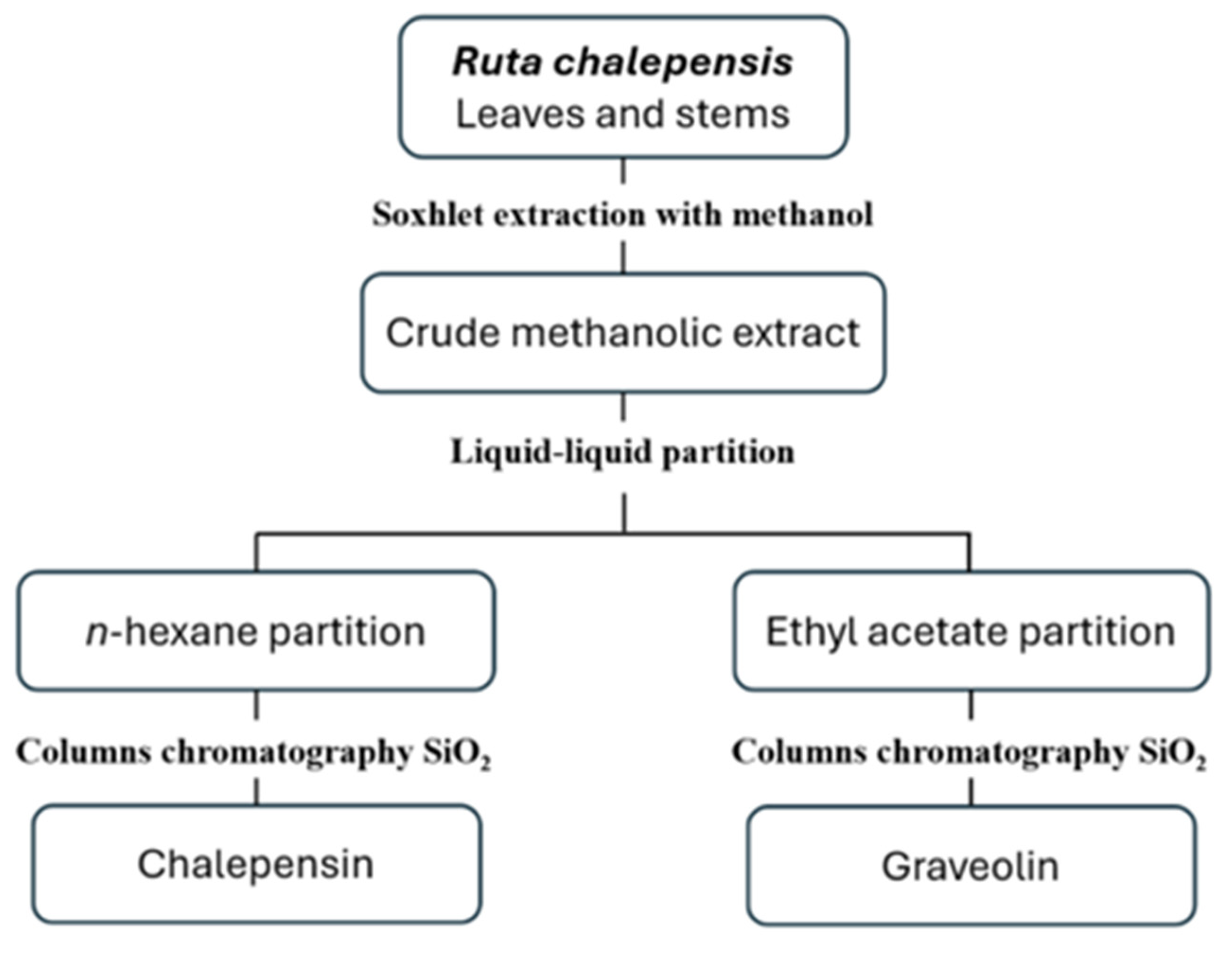
2.1. Plant Material and Isolation of Compounds

Our working group previously published the isolation and identification of the compounds chalepensin and graveoline obtained from the leaves and stems of the plant Ruta chalepensis L. (voucher FCB-UANL 30654) [22,23,24]. Chalepensin was obtained from the n-hexane partition of R. chalepensis through column chromatography using silica gel 60 G (Merck, Darmstadt, Germany), while graveoline was isolated from the ethyl acetate partition, also by columns chromatography with silica gel 60 G (Figure 1). The compounds were identified through spectroscopy and spectrometry using a Bruker Spectrometer (Model Advance DPX400, 9.4 Teslas; Bruker Corporation, Billerica, MA, USA) and subsequently compared with existing literature data. The spectroscopic analysis data for the compounds can be found in the Supplementary Materials (Supplementary Materials_Spectroscopic Data).

2.2. Ethical Statement

All animal procedures adhered to the ethical guidelines outlined in Spanish regulations (RD 53/2013) and the European Union Directive (2010/63/CE) regarding the care and use of laboratory animals. The experiments were conducted at the accredited Animal Experimentation Facilities of the University of Salamanca (Registration No.: PAE/SA/001). The study protocols received approval from the Ethics Committee of USAL (CBE 335 CEI 1080). Efforts were consistently made to minimize animal suffering. The animals were housed in standard polycarbonate cages under controlled conditions, including a 12 h light–dark cycle, temperatures maintained between 23 °C and 25 °C, and unrestricted access to food and water.

2.3. Nematocidal Activity of Compounds Against Strongyloides venezuelensis

The S. venezuelensis strain, provided by the Department of Parasitology at the Federal University of Minas Gerais (Brazil), was maintained through successive passages in 4-week-old male Wistar rats (150–200 g) to ensure the completion of its life cycle. The rats were subcutaneously inoculated with 12,000 infective L3 suspended in 500 μL of phosphate-buffered saline (PBS). Fecal samples from rats were collected between days 5 and 18 post-infection and cultured in 250 mL polyethylene containers with vermiculite and distilled water. The cultures were kept in a humid environment at 28 °C for 4 to 7 days (SANYO Electric Co., Ltd., Gunma, Japan). L3 were subsequently isolated using the Baermann method, washed three times with distilled water, and their viability was confirmed under a light microscope before starting the experiments [14].
We placed between 100 and 150 L3 into each well of flat-bottomed 96-well microplates (Corning Incorporated) with 100 µL of distilled water. The larvae were incubated at 28 °C for 30 min to allow them to adapt. Subsequently, they were exposed to the test compounds at concentrations ranging from 1 to 50 µM. As controls, 10 µM ivermectin (Sigma-Aldrich, St. Louis, MO, USA) served as the positive control, while distilled water, with or without 1% DMSO, was used as the negative control. The larvae were incubated at 28 °C for 72 h under humid conditions (SANYO Electric Co. Ltd.), and mortality was assessed after 24, 48, and 72 h. Mortality was defined as the absence of movement, which was verified under a light microscope at 40× magnification for at least 1 min, using visible light to stimulate movement [14].
The parasitic females of S. venezuelensis live in the small intestine mucosa of an infected host and produce eggs by parthenogenesis [25]. To obtain the adult females, seven days after infection, the rats were euthanized by cervical dislocation after unconsciousness induced by carbon dioxide, and their small intestines were excised, opened longitudinally, finely chopped, and placed into a sedimentation cup lined with gauze in PBS. The samples were incubated at 37 °C for 2 h. Females adults were recovered from the sediment, washed twice with 0.85% saline solution, and then subjected to two additional washes in Roswell Park Memorial Institute 1640 medium (RPMI-1640) (Gibco, Whaltman, MA, USA) supplemented with 10% heat-inactivated fetal bovine serum (FBS), 100 µg/mL streptomycin, and 100 U/mL penicillin (Gibco, Whaltman, MA, USA) [26].
Subsequently, 50 to 70 parthenogenetic females were placed in 96-well plates containing RPMI-1640 medium and incubated at 37 °C for 1 h to allow for adaptation. Following this period, the test compounds were applied at concentrations from 1 to 60 µM. The anti-parasitic activity was evaluated at 24, 48, and 72 h post-treatment by examining the parasites under a light microscope at 40× magnification for at least 1 min, using visible light to stimulate their movement. Based on their mobility, the parasites were classified into four categories: healthy, slightly affected (showing reduced movement), affected (exhibiting significantly reduced or abnormal movement), and dead (no observable movement) [26].

2.4. Scanning Electron Microscopy (SEM) on Parthenogenetic Females of Strongyloides venezuelensis

The effect of chalepensin and graveoline on adults of S. venezuelensis was observed by scanning electron microscopy (SEM). After 72 h of incubation, S. venezuelensis adults of each group of in vitro experiment were removed and fixed by immersing them immediately in 2.5% glutaraldehyde at room temperature for 3 h. The samples were then washed three times with 0.1 M phosphate buffer (pH 7.4) at room temperature for 30 min. After the final wash, they were stored at 4 °C. Subsequently, the samples were attached to Poly-L-Lysine in a humid chamber with 25% glutaraldehyde and incubated overnight. The next day, they were rinsed and fixed with 1% osmium tetroxide. After dehydration with alcohols, critical-point drying was performed, followed by metal coating. The process was conducted at the Electron Microscopy Facilities–NUCLEUS of the University of Salamanca, and visualization was carried out using a Jeol JSM-IT500 InTouchScope™ SEM (JEOL Ltd., Tokyo, Japan), SEM images were processed using the software Jeol v1250.

2.5. Cytotoxic Activity of Compounds in Vero Cells

African green monkey kidney epithelial cells (Vero, ATCC CCL-81) were used in this study. These cells were maintained in Dulbecco’s Modified Eagle Medium (DMEM; Gibco, Grand Island, NE, USA) supplemented with 10% FBS and 1% antibiotic and antimycotic solution (Life Technologies, Carlsbad, CA, USA). Cultures were incubated at 37 °C in a 5% CO2 atmosphere with 95% relative humidity.
Vero cells (1 × 10⁴ cells per well) were seeded into transparent 96-well flat-bottom polypropylene microplates (Corning Incorporated, Corning, NY, USA). After 24 h of incubation, the cells were treated with compounds at final concentrations ranging from 10 µM to 6 mM. Untreated culture medium with and with DMSO 1% was used as control. The cells were incubated for 48 h at 37 °C in a 5% CO2 atmosphere. Cell viability was determined using the colorimetric 3-[4,5-dimethylthiazol-2-yl]-2,5-diphenyltetrazolium bromide (MTT) assay (Affymetrix, Cleveland, OH, USA). Formazan crystals were dissolved in 100 µL of DMSO, and the optical density (OD) was measured at 570 nm using a microplate reader (MULTISKAN GO; Thermo Fisher Scientific, Waltham, MA, USA) [23]. The percentage of cell growth inhibition was calculated as follows (1):
%   C e l l   g r o w t h   i n h i b i t i o n = 100 D O 570   T r e a t e d   c e l l s D O 570   U n t r e a t e d   c e l l s × 100
In this study, Vero cells were employed as a mammalian cell model to assess the nonspecific cytotoxicity of the compounds. The selectivity indices (SIs) were calculated to know parasitic activity in relation to the toxicity in normal mammalian cells, where a high SI indicates strong parasitic activity with minimal toxicity to the cells [27]. The SI was determined by dividing the half-maximal inhibitory concentration (IC50) in Vero cells by the mean lethal concentration (LC50) for S. venezuelensis, using the following Formula (2):
S I = I C 50   V e r o   c e l l s L C 50   S .   v e n e z u e l e n s i s

2.6. Statistical Analysis

All statistical analyses were performed using the GraphPad Prism 8 software (GraphPad Software Inc., San Diego, CA, USA). The data presented here represent the means ± standard deviations (SDs) of triplicates obtained from at least three independent experiments, with a 95% confidence level. A non-regression analysis was used to calculate IC50 and LC50 values. Significant differences among different conditions were studied, using a one-way analysis of variance (1 way ANOVA) or t-test after homoscedasticity and normality studies. Post hoc Dunnett’s analyses were used to determine specific differences between groups and control ivermectin. Statistical differences were considered when p < 0.05.

3. Results

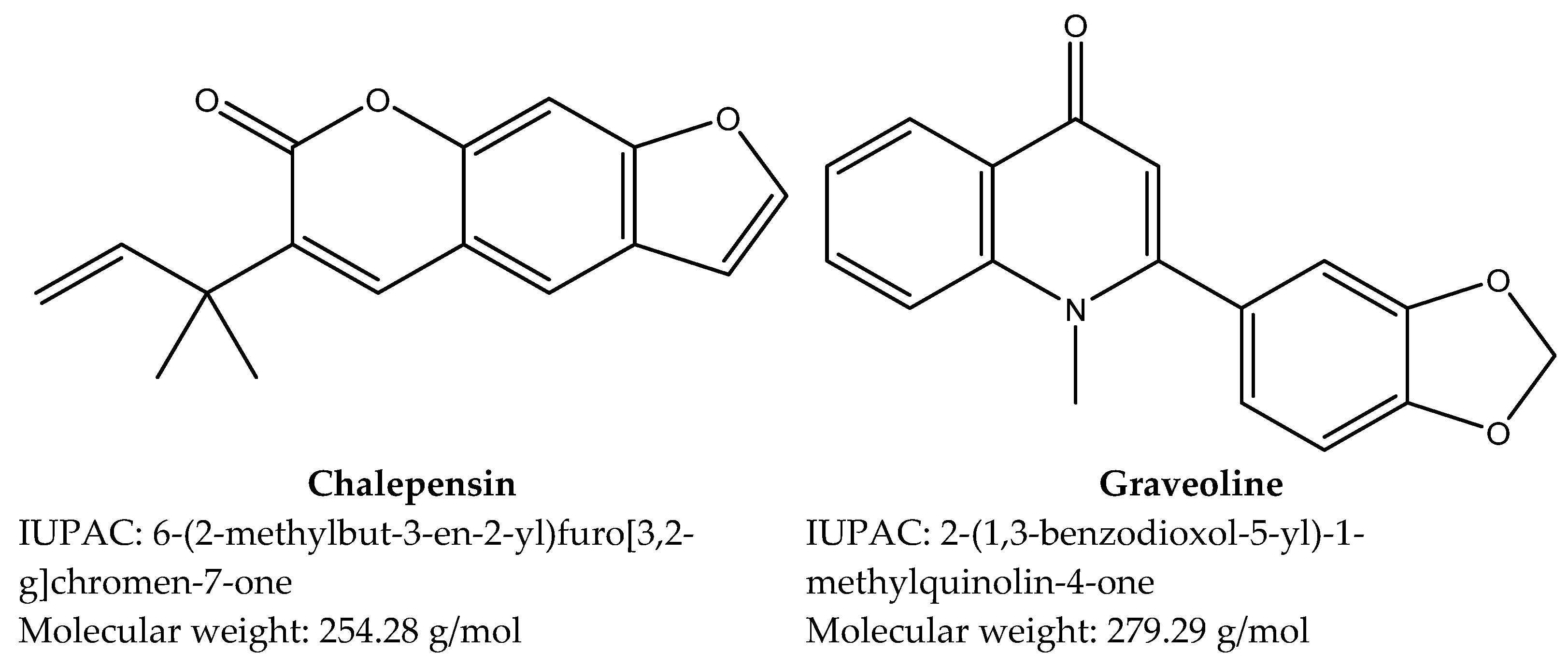
3.1. Compounds Isolated from Ruta chalepensis

Two compounds were successfully isolated from R. chalepensis, and their chemical structures were determined (Figure 2). The nematocidal activity of these compounds was evaluated against S. venezuelensis L3 and adults.

3.2. In Vitro Toxicity Against Vero Cells and Nematocidal Activity of Compounds Against Strongyloides venezuelensis L3

Nematocidal activity of the compounds against L3 of S. venezuelensis were tested at 24, 48, and 72 h and toxicity to Vero cells at 72 h (Table 1). The compounds were minimally toxic to Vero cells, and a slight time-dependent effect can be observed in both compounds against the infective L3. Both compounds showed good antiparasitic activity; however, the activity of chalepensin was 5 to 7 times lower than that of graveolin, depending on the incubation time, and was always similar than that of ivermectin (p > 0.05).

3.3. In Vitro Nematocidal Activity of Compounds in Strongyloides venezuelensis Adults

In Table 2, the results of the nematocidal activity of the compounds against female adults of S. venezuelensis at 24, 48, and 72 h are shown. Both compounds exhibited good antiparasitic activity with no time-dependent effect; at 24 h, they already show activity, which does not increase over time. In adults, as in larvae, chalepensin showed greater activity than graveoline. Furthermore, the antiparasitic effect observed against adults was lower than that obtained against larvae. In contrast, graveoline exhibited similar activity in both larvae and adults.
In Table 3, the status of the adults at different concentrations evaluated at 72 h is shown. With chalepensin, 100% mortality was observed at a dose of 30 µM, whereas with graveoline, a higher dose of 60 µM was required to achieve the same effect. In the control drug ivermectin at 10 µM, the parasites were slightly affected; however, with chalepensin at a lower dose of 3.75 µM, the same effect was observed.
The SEM images revealed distinct morphological alterations in adult parasites treated with compounds compared to the control at 72 h. In the control group, an intact cuticular structure was observed (Figure 3(A1,A2)), along with the characteristic folded morphology in the posterior region of the parasite (Figure 3(A3)). In contrast, chalepensin primarily induced the formation of numerous protuberances distributed along the entire surface of the parasite (Figure 3(C2)), accompanied by slight cuticle detachment (Figure 3(C1,C3)). Similarly, graveoline predominantly triggered cuticle detachment (Figure 3(D2)), suggesting a different mode of interaction with the parasite’s external structure. Notably, these specific alterations were absent in the ivermectin-treated group (Figure 3(B1–B3)), implying that chalepensin and graveoline may exert their antiparasitic effects through a mechanism distinct from that of ivermectin. Furthermore, despite their mechanistic differences, both compounds, along with ivermectin, induced observable morphological modifications, particularly in the posterior region of the parasite, reinforcing their potential impact on parasite viability and structural integrity.

4. Discussion

In this study, the antiparasitic potential of two major compounds from Ruta chalepensis was explored. Among the major compounds of R. chalepensis are chalepin, chalepensin, rutamarin [28], psoralen, bergapten [29], and graveoline [23]. In this study, the compounds evaluated were chalepensin and graveoline.
Chalepensin has been isolated from various plants, particularly those belonging to the Rutaceae family. It has shown in vitro activity against E. histolytica [16]; however, this is the first report of its antiparasitic activity in more complex organisms, such as helminths. Against E. histolytica trophozoites, chalepensin exhibits an LC50 of 180.77 µM; in comparison, our results indicate a 53-fold and 11-fold lower LC50 for larvae and adults of S. venezuelensis, respectively, highlighting its antinematicidal potential. This compound has demonstrated not only antiprotozoal activity but also antimicrobial properties, cytotoxic effects against several cancer cell lines, calcium antagonism, inhibition of platelet aggregation, and a mechanism-based inhibition of the cytochrome P450 (CYP) 2A6 enzyme [30]. Similarly, graveoline is also found in plants of the Rutaceae family. This compound exhibits a wide range of pharmacological effects, including antibacterial activity, spasmolytic properties, and antitumor potential [31], but no antiparasitic activity has been reported to date.
There are already some reports on the activity of plant-isolated compounds against S. venezuelensis larvae. For example, a study evaluated the activity of various compounds isolated from Macleaya cordata and Chelidonium majus, where the most active compound from M. cordata was protopine, with an LC50 of 33 μM, while from C. majus it was dehydrocorydaline, with an LC50 of 12 μM. However, upon determining the SIs using HL60 cells, these compounds showed very low SIs, being >0.3 and 1, respectively. Nevertheless, other compounds with higher IC50 values also demonstrated significantly higher SI. This was the case for oxysanguinarine and L-tetrahydrocolumbamine, both with SIs > 500 [32]. In contrast, in this study, the evaluated compounds yielded better results, with chalepensin being far superior, presenting an LC50 of 3.4 μM and an SI of 990 using Vero cells, and graveoline an LC50 of 24.4 μM and an SI of 26.2.
This study also evaluated the activity against S. venezuelensis adults. Few studies report activity at this stage of the parasite; most focus only on larvae. However, evaluating adults is highly relevant since they are responsible for making the parasitic infection persistent by continuously producing eggs [25]. With graveoline, similar results were obtained between larvae and adults, whereas with chalepensin, the IC50 increased almost fivefold in adults. It has already been reported that adults are more resistant than larvae; even ivermectin is not as effective in adults as it is in larvae [26]. In this study, chalepensin showed better results than ivermectin in adults. At 10 µM of ivermectin, the parasites were only slightly affected; however, with chalepensin at a lower dose of 3.75 µM, the same effect was observed. This highlights the antiparasitic potential of this compound against S. venezuelensis.
The morphology of adults exposed to lethal concentrations of the compounds was also analyzed, revealing several marked differences compared to healthy controls. In a study evaluating the effect of Mentha × villosa essential oil on Schistosoma mansoni adults, it was found to cause bubble lesions over the entire body of the worms, as well as damaged tegument and exposed musculature in some worms, along with tegument erosion [33]. Similarly, in the same parasite, it has been shown that allicin causes vesicle formation [34], while miltefosine induces tegument peeling [35]; which aligns with the observations made with graveoline, where tegument erosion was mainly noted, and chalepensin, where protuberances were observed along the body. With ivermectin, no significant morphological differences were observed compared to the control. This could be due to its mechanism of action, as ivermectin is a positive allosteric modulator that selectively opens glutamate-gated chloride channels in nematodes. This action leads to the inhibition of feeding by affecting pharyngeal muscles, paralysis by impacting motor nerves, blockage of egg release, and loss of host immunosuppression by preventing the opening of the excretory/secretory pore [36]. In contrast, the mechanism of action of chalepensin and graveoline, as they induce cuticular disruptions, may be more similar to that of praziquantel, which acts as a calcium agonist, increasing calcium concentration inside parasite cells, which leads to tegument disruption. Additionally, it exhibits synergy with the host immune system by exposing parasite antigens, allowing antibodies to recognize the parasite and activate the immune response [37].
The results of this study highlight the nematocidal activity of the compounds chalepensin and graveoline. However, it is essential to evaluate them in an in vivo murine model to confirm their antiparasitic activity and rule out any potential toxicity, as well as to conduct further assays to elucidate its mechanism of action.

5. Conclusions

This study demonstrated the antiparasitic effect of chalepensin and graveoline, isolated from Ruta chalepensis, against Strongyloides venezuelensis third-stage larvae and adults. Both compounds exhibited nematocidal activity and selective toxicity against parasites compared to mammalian cells, highlighting their potential as alternative treatments. Additionally, they induced morphological changes in the parasites, particularly in their cuticle, suggesting a possible mechanism of action. These findings indicate that chalepensin and graveoline possess promising antiparasitic potential, with chalepensin emerging as a particularly potent candidate for further investigation.

Supplementary Materials

The following supporting information can be downloaded at https://www.mdpi.com/article/10.3390/pathogens14050419/s1, Figures S1.1–S1.12: Spectroscopic analysis of chalepensin; Figures S2.1–S2.22: Spectroscopic analysis of graveoline.

Author Contributions

Conceptualization: N.E.R.-G.; methodology: M.M., J.S.-M., R.Q.-L. and A.F.B.-R.; validation: J.L.-A., A.F.B.-R. and C.I.R.-S.; formal analysis: N.E.R.-G. and M.M.; investigation: N.E.R.-G., J.L.-A. and M.M.; resources: J.L.-A., R.P. and R.Q.-L.; data curation: N.E.R.-G. and M.M.; writing—original draft preparation: N.E.R.-G.; writing—review and editing: J.L.-A., M.M., J.S.-M. and J.H.E.-L.; visualization: N.E.R.-G.; supervision: J.L.-A. and R.Q.-L.; project administration: J.L.-A. and R.Q.-L.; funding acquisition: J.H.E.-L., R.Q.-L., R.P., A.M. and C.I.R.-S. All authors have read and agreed to the published version of the manuscript.

Funding

This research was funded by the Consejo Nacional de Humanidades, Ciencia y Tecnología (CONHACYT) of Mexico. N.E.R.-G. received a doctoral grant numbered 808132 (CVU: 1006989); J.H.E.-L. received a postdoctoral grant numbered I1200/331/2023 (CVU: 418935); and C.I.R.-S. received a postdoctoral grant numbered I1200/331/2023 (CVU: 445572). The research was also funded by MCIN/AEI/doi 10.13039/501100011033, and co-founded by European Union grant PID2021-127471OB-I00 (J.L.-A. and R.P. principal researcher).

Institutional Review Board Statement

The animal study protocol received approval (18 December 2023) from the Ethics Committee of the University of Salamanca (CBE 335 CEI 1080) for research involving animals.

Informed Consent Statement

Not applicable.

Data Availability Statement

The datasets generated or analyzed during the present study are available from the corresponding authors.

Acknowledgments

The authors thank the CONHACYT, SNII, the Asociación Universitaria Iberoamericana de Postgrado (AUIP), the Fundación Universidad de Salamanca (USAL)—Banco Santander, and the Electron Microscopy Facilities–NUCLEUS of the University of Salamanca. The Doctoral Fellowship Program of “Junta de Castilla y León” is co-funded by “Fondo Social Europeo” (M.M. and J.S.-M. ORDEN EDU/601/2020).

Conflicts of Interest

The authors declare no conflicts of interest.

References

  1. Nag, V.L.; Kalita, J.M. Epidemiology of Parasitic Infections. In Textbook of Parasitic Zoonoses; Springer: Singapore, 2022; pp. 51–58. [Google Scholar]
  2. Eslahi, A.V.; Hashemipour, S.; Olfatifar, M.; Houshmand, E.; Hajialilo, E.; Mahmoudi, R.; Badri, M.; Ketzis, J.K. Global Prevalence and Epidemiology of Strongyloides stercoralis in Dogs: A Systematic Review and Meta-Analysis. Parasit. Vectors 2022, 15, 21. [Google Scholar] [CrossRef] [PubMed]
  3. Eslahi, A.V.; Badri, M.; Nahavandi, K.H.; Houshmand, E.; Dalvand, S.; Riahi, S.M.; Johkool, M.G.; Asadi, N.; Hoseini Ahangari, S.A.; Taghipour, A.; et al. Prevalence of Strongyloidiasis in the General Population of the World: A Systematic Review and Meta-Analysis. Pathog. Glob. Health 2021, 115, 7–20. [Google Scholar] [CrossRef] [PubMed]
  4. Buonfrate, D.; Bisanzio, D.; Giorli, G.; Odermatt, P.; Fürst, T.; Greenaway, C.; French, M.; Reithinger, R.; Gobbi, F.; Montresor, A.; et al. The Global Prevalence of Strongyloides stercoralis Infection. Pathogens 2020, 9, 468. [Google Scholar] [CrossRef]
  5. Luvira, V.; Siripoon, T.; Phiboonbanakit, D.; Somsri, K.; Watthanakulpanich, D.; Dekumyoy, P. Strongyloides stercoralis: A Neglected but Fatal Parasite. Trop. Med. Infect. Dis. 2022, 7, 310. [Google Scholar] [CrossRef]
  6. Karanam, L.S.K.; Basavraj, G.K.; Papireddy, C.K.R. Strongyloides stercoralis Hyper Infection Syndrome. Indian J. Surg. 2021, 83, 582–586. [Google Scholar] [CrossRef]
  7. Yeh, M.Y.; Aggarwal, S.; Carrig, M.; Azeem, A.; Nguyen, A.; Devries, S.; Destache, C.; Nguyen, T.; Velagapudi, M. Strongyloides stercoralis Infection in Humans: A Narrative Review of the Most Neglected Parasitic Disease. Cureus 2023, 15, e46908. [Google Scholar] [CrossRef] [PubMed]
  8. Gordon, C.A.; Utzinger, J.; Muhi, S.; Becker, S.L.; Keiser, J.; Khieu, V.; Gray, D.J. Strongyloidiasis. Nat. Rev. Dis. Primers 2024, 10, 6. [Google Scholar] [CrossRef]
  9. Alam, A.M.; Ozdemir, C.; Reza, N. Strongyloides stercoralis Infection in the UK: A Systematic Review and Meta-Analysis of Published Cases. Clin. Med. 2024, 24, 100227. [Google Scholar] [CrossRef]
  10. Henriquez-Camacho, C.; Pérez-Molina, J.A.; Buonfrate, D.; Rodari, P.; Gotuzzo, E.; Luengo, B.; Plana, M.N. Ivermectin vs Moxidectin for Treating Strongyloides stercoralis Infection: A Systematic Review. Parasitology 2025, 151, 1–7. [Google Scholar] [CrossRef]
  11. Czeresnia, J.M.; Weiss, L.M. Strongyloides stercoralis. Lung 2022, 200, 141–148. [Google Scholar] [CrossRef]
  12. Khamesipour, F.; Kheyri, P.; Shojaat, S.; Chelgerdi Dehkordi, B.; Basirpour, B.; Afzal, S.S.; Hejazi, S.H. A Review of the Effect of Medicinal Plant on Helminthic Infections. Infect. Dis. Herb. Med. 2021, 2, 127. [Google Scholar] [CrossRef]
  13. Ranasinghe, S.; Armson, A.; Lymbery, A.J.; Zahedi, A.; Ash, A. Medicinal Plants as a Source of Antiparasitics: An Overview of Experimental Studies. Pathog. Glob. Health 2023, 117, 535–553. [Google Scholar] [CrossRef] [PubMed]
  14. Rodríguez-Garza, N.E.; Gomez-Flores, R.; Quintanilla-Licea, R.; Elizondo-Luévano, J.H.; Romo-Sáenz, C.I.; Marín, M.; Sánchez-Montejo, J.; Muro, A.; Peláez, R.; López-Abán, J. In Vitro Anthelmintic Effect of Mexican Plant Extracts and Partitions Against Trichinella spiralis and Strongyloides venezuelensis. Plants 2024, 13, 3484. [Google Scholar] [CrossRef]
  15. Coimbra, A.T.; Ferreira, S.; Duarte, A.P. Genus Ruta: A Natural Source of High Value Products with Biological and Pharmacological Properties. J. Ethnopharmacol. 2020, 260, 113076. [Google Scholar] [CrossRef] [PubMed]
  16. Quintanilla-Licea, R.; Mata-Cárdenas, B.; Vargas-Villarreal, J.; Bazaldúa-Rodríguez, A.; Kavimngeles-Hernández, I.; Garza-González, J.; Hernández-García, M. Antiprotozoal Activity against Entamoeba histolytica of Plants Used in Northeast Mexican Traditional Medicine. Bioactive Compounds from Lippia graveolens and Ruta chalepensis. Molecules 2014, 19, 21044–21065. [Google Scholar] [CrossRef]
  17. Dessalegn Asmelashe, G.; Abdelwuhab, M.; Asrie, A.B. In Vivo Antiplasmodial Activity Evaluation of the Leaves of Ruta chalepensis L. (Rutaceae) against Plasmodium berghei. Discov. Phytomedicine 2017, 4, 1. [Google Scholar] [CrossRef]
  18. Ntalli, N.G.; Manconi, F.; Leonti, M.; Maxia, A.; Caboni, P. Aliphatic Ketones from Ruta chalepensis (Rutaceae) Induce Paralysis on Root Knot Nematodes. J. Agric. Food Chem. 2011, 59, 7098–7103. [Google Scholar] [CrossRef]
  19. Ortu, E.; Sanna, G.; Scala, A.; Pulina, G.; Caboni, P.; Battacone, G. In Vitro Anthelmintic Activity of Active Compounds of the Fringed Rue Ruta chalepensis against Dairy Ewe Gastrointestinal Nematodes. J. Helminthol. 2017, 91, 447–453. [Google Scholar] [CrossRef]
  20. Jasso-Díaz, G.; Torres-Hernández, G.; Zamilpa, A.; Becerril-Pérez, C.M.; Ramírez-Bribiesca, J.E.; Hernández-Mendo, O.; Sánchez-Arroyo, H.; Olmedo-Juárez, A.; González-Cortazar, M.; Mendoza-De Gives, P. Ruta chalepensis Full Extract and Organic Phases Exhibit Nematocidal Activity against Haemonchus contortus Eggs and Infective Larvae (L3). Helminthologia 2022, 59, 46–54. [Google Scholar] [CrossRef]
  21. Jalil, P.J.; Shnawa, B.H.; Hamad, S.M.; Hamad, B.S.; Ahmed, M.H. The Efficiency of Fabricated Ag/ZnO Nanocomposite Using Ruta chalepensis L. Leaf Extract as a Potent Protoscolicidal and Anti-Hydatid Cysts Agent. J. Biomater. Appl. 2023, 38, 629–645. [Google Scholar] [CrossRef]
  22. Bazaldúa-Rodríguez, A.F.; Quintanilla-Licea, R.; Verde-Star, M.J.; Hernández-García, M.E.; Vargas-Villarreal, J.; Garza-González, J.N. Furanocoumarins from Ruta chalepensis with Amebicide Activity. Molecules 2021, 26, 3684. [Google Scholar] [CrossRef] [PubMed]
  23. Elizondo-Luévano, J.H.; Rodríguez-Garza, N.E.; Bazaldúa-Rodríguez, A.F.; Romo-Sáenz, C.I.; Tamez-Guerra, P.; Verde-Star, M.J.; Gomez-Flores, R.; Quintanilla-Licea, R. Cytotoxic, Anti-Hemolytic, and Antioxidant Activities of Ruta chalepensis L. (Rutaceae) Extract, Fractions, and Isolated Compounds. Plants 2023, 12, 2203. [Google Scholar] [CrossRef] [PubMed]
  24. Quintanilla-Licea, R.; Mata-Cárdenas, B.; Vargas-Villarreal, J.; Bazaldúa-Rodríguez, A.; Verde-Star, M. Antiprotozoal Activity against Entamoeba histolytica of Furocoumarins Isolated from Ruta chalepensis. Planta Med. 2016, 81, S1–S381. [Google Scholar] [CrossRef]
  25. Maeda, Y.; Palomares-Rius, J.E.; Hino, A.; Afrin, T.; Mondal, S.I.; Nakatake, A.; Maruyama, H.; Kikuchi, T. Secretome Analysis of Strongyloides venezuelensis Parasitic Stages Reveals That Soluble and Insoluble Proteins Are Involved in Its Parasitism. Parasit. Vectors 2019, 12, 21. [Google Scholar] [CrossRef]
  26. Marín, M.; Sánchez-Montejo, J.; Ramos, S.; Muro, A.; López-Abán, J.; Peláez, R. Deciphering Chemical Rules for Drug Penetration into Strongyloides. Pharmaceutics 2024, 16, 1224. [Google Scholar] [CrossRef]
  27. Mansour, K.A.; El-Neketi, M.; Lahloub, M.-F.; Elbermawi, A. Nanoemulsions of Jasminum humile L. and Jasminum grandiflorum L. Essential Oils: An Approach to Enhance Their Cytotoxic and Antiviral Effects. Molecules 2022, 27, 3639. [Google Scholar] [CrossRef]
  28. Al-Majmaie, S.; Nahar, L.; Rahman, M.M.; Nath, S.; Saha, P.; Talukdar, A.D.; Sharples, G.P.; Sarker, S.D. Anti-MRSA Constituents from Ruta chalepensis (Rutaceae) Grown in Iraq, and In Silico Studies on Two of Most Active Compounds, Chalepensin and 6-Hydroxy-Rutin 3′,7-Dimethyl Ether. Molecules 2021, 26, 1114. [Google Scholar] [CrossRef] [PubMed]
  29. Juneja, K.; Beuerle, T.; Sircar, D. Enhanced Accumulation of Biologically Active Coumarin and Furanocoumarins in Callus Culture and Field-grown Plants of Ruta chalepensis Through LED Light-treatment. Photochem. Photobiol. 2022, 98, 1100–1109. [Google Scholar] [CrossRef]
  30. Nahar, L.; Al-Majmaie, S.; Al-Groshi, A.; Rasul, A.; Sarker, S.D. Chalepin and Chalepensin: Occurrence, Biosynthesis and Therapeutic Potential. Molecules 2021, 26, 1609. [Google Scholar] [CrossRef]
  31. Li, Z.; Mu, C.; Wang, B.; Jin, J. Graveoline Analogs Exhibiting Selective Acetylcholinesterase Inhibitory Activity as Potential Lead Compounds for the Treatment of Alzheimer’s Disease. Molecules 2016, 21, 132. [Google Scholar] [CrossRef]
  32. Satou, T.; Koga, M.; Matsuhashi, R.; Koike, K.; Tada, I.; Nikaido, T. Assay of Nematocidal Activity of Isoquinoline Alkaloids Using Third-Stage Larvae of Strongyloides ratti and S. venezuelensis. Vet. Parasitol. 2002, 104, 131–138. [Google Scholar] [CrossRef]
  33. Matos-Rocha, T.J.; Cavalcanti, M.G.D.S.; Veras, D.L.; Feitosa, A.P.S.; Gonçalves, G.G.A.; Portela-Junior, N.C.; Lúcio, A.S.S.C.; Silva, A.L.D.; Padilha, R.J.R.; Marques, M.O.M.; et al. Ultrastructural Changes in Schistosoma mansoni Male Worms After In Vitro Incubation with the Essential Oil of Mentha x villosa Huds. Rev. Inst. Med. Trop. 2016, 58, 4. [Google Scholar] [CrossRef] [PubMed]
  34. Lima, C.M.B.L.; Freitas, F.I.d.S.; Morais, L.C.S.L.d.; Cavalcanti, M.G.d.S.; Silva, L.F.d.; Padilha, R.J.R.; Barbosa, C.G.S.; Santos, F.A.B.d.; Alves, L.C.; Diniz, M.d.F.F.M. Ultrastructural Study on the Morphological Changes to Male Worms of Schistosoma mansoni after in Vitro Exposure to Allicin. Rev. Soc. Bras. Med. Trop. 2011, 44, 327–330. [Google Scholar] [CrossRef] [PubMed]
  35. Bertão, H.G.; da Silva, R.A.R.; Padilha, R.J.R.; de Azevedo Albuquerque, M.C.P.; Rádis-Baptista, G. Ultrastructural Analysis of Miltefosine-Induced Surface Membrane Damage in Adult Schistosoma mansoni BH Strain Worms. Parasitol. Res. 2012, 110, 2465–2473. [Google Scholar] [CrossRef] [PubMed]
  36. Martin, R.J.; Robertson, A.P.; Choudhary, S. Ivermectin: An Anthelmintic, an Insecticide, and Much More. Trends Parasitol. 2021, 37, 48–64. [Google Scholar] [CrossRef]
  37. Thomas, C.M.; Timson, D.J. The Mechanism of Action of Praziquantel: Can New Drugs Exploit Similar Mechanisms? Curr. Med. Chem. 2020, 27, 676–696. [Google Scholar] [CrossRef]
Figure 1. General diagram of isolation of chalepensin and graveoline from Ruta chalepensis leaves and stems.
Pathogens 14 00419 g001
Figure 2. Structure of isolated compounds chalepensin and graveoline from Ruta chalepensis determined by spectrometric analysis.
Pathogens 14 00419 g002
Figure 3. SEM images at 1000× magnification (scale = 10 µm) of S. venezuelensis parthenogenetic adult females with different treatments at 72 h. (A) Control; (B) ivermectin at 10 µM; (C) chalepensin at 30 µM; (D) graveoline at 60 µM. In each picture cephalic (1), body (2) and caudal (3) parts are depicted in parasites in the different treatment conditions.
Pathogens 14 00419 g003
Table 1. Mean and standard deviation of in vitro Vero toxicity at 72 h IC50, nematocidal activity of compounds LC50 in Strongyloides venezuelensis infective third-stage larvae (L3) at 24, 48, and 72 h, and selectivity index (SI) at 72 h.
CompoundVero Cells IC50 (µM)S. venezuelensis L3 LC50 (µM)SI
24 h48 h72 h
Chalepensin3365.9 ± 58.35.7 ± 1.2 a,*3.9 ± 1.3 a,*3.4 ± 0.9 a,*990.0
Graveoline640.3 ± 26.428.3 ± 6.7 b25.9 ± 5.9 b24.4 ± 5.8 b26.2
Ivermectin ND2.1± 0.11.6 ± 0.31.5 ± 0.3ND
ANOVA-F(2,23) = 36.59
p = 0.0004
F(2,23) = 45.46
p = 0.0002
F(2,23) = 42.08
p = 0.0003
-
Dunnett’s comparison to ivermectin-a p = 0.4878
b p = 0.0004
a p = 0.6464
b p = 0.0003
a p = 0.7294
b p = 0.0003
-
* No significant differences in comparison with ivermectin (p > 0.05). a Comparison between chalepensin and ivermectin. b Comparison between graveoline and ivermectin. ND: Not determined.
Table 2. Mean and standard deviations of in vitro nematocidal activity LC50 of chalepensin and graveoline against S. venezuelensis parasitic female adults and selectivity index (SI) at 72 h.
CompoundLC50 in µMSI
24 h48 h72 h
Chalepensin17.3 ± 3.117.1 ± 2.916.8 ± 2.4200.4
Graveoline27.8 ± 4.826.9 ± 5.126.5 ± 3.224.2
t-test p = 0.0334p = 0.0444p = 0.0137-
The t-test differences between compounds were considered when p < 0.05.
Table 3. Status categories of S. venezuelensis female adults at different concentrations of the compounds.
CompoundConcentration µMCategory
Chalepensin30.0All dead
15.0Most affected and some dead
7.5Slightly affected
3.8Slightly affected
1.9Healthy
Graveoline60.0All dead
30.0Most dead and some affected
15.0Slightly affected
7.5Healthy
3.8Healthy
Ivermectin10.0Slightly affected
Disclaimer/Publisher’s Note: The statements, opinions and data contained in all publications are solely those of the individual author(s) and contributor(s) and not of MDPI and/or the editor(s). MDPI and/or the editor(s) disclaim responsibility for any injury to people or property resulting from any ideas, methods, instructions or products referred to in the content.

