Communication

Effect of Wearing Running Shoes on Lower Limb Kinematics by Using OpenSim Simulation Software

by
Gunarajulu Renganathan
1,*,
Hamidreza Barnamehei
2,3,
Swagata Das
1 and
Yuichi Kurita
1
1
Department of System Cybernetics, Hiroshima University, Hiroshima 739-8522, Japan
2
Gene and Linda Voiland School of Chemical and Bioengineering, Washington State University, Pullman, WA 99164, USA
3
Department of Integrative Physiology and Neuroscience, Washington State University, Pullman, WA 99164, USA
*
Author to whom correspondence should be addressed.
Actuators 2022, 11(6), 152; https://doi.org/10.3390/act11060152
Submission received: 25 April 2022 / Revised: 1 June 2022 / Accepted: 4 June 2022 / Published: 6 June 2022
(This article belongs to the Special Issue Soft Exoskeleton and Supernumerary Limbs for Human Augmentation)

Abstract

:
(1) Flatfoot is a common malformation in both children and adults, in which a proper arch fails to develop. This study aimed to see how over-the-counter running shoes improved the gait patterns of flatfoot patients. (2) Methods: Three healthy flatfoot subjects were included in the study. Flatfoot was diagnosed by a lateral talometatarsal angle of more than 4 degrees and a talocalcaneal angle of more than 30 degrees. All the patient data were captured using Vicon motion caption cameras. The subjects were allowed to walk at self-selected speeds with and without running shoes. (3) Results: Significant differences in lower limb kinematics were observed between barefoot and running shoe gait. In addition, by wearing the running shoes, the center of mass and lower limb kinematics changed. (4) Conclusion: The improvement in balance and control was clearly indicated, and the change in gait on the entire lower limb influenced normalizing the stresses of the foot with running shoes. These valuable results can be used for rehabilitation programs.

1. Introduction

Foot postures are generally classified into three categories: neutral, cavus, and flatfoot (FF), with normal, high, and low medial arch height, respectively. Flat feet are a kind of lever arm disease that can affect gait kinematics and balance control patterns. Treatment options include surgery, exercise therapy, physiotherapy, and the prescription of shoes and other rehabilitation equipment. The foot of a person with a flatfoot deformity is subjected to increased mechanical overloading. This is linked to musculoskeletal diseases in the lower limbs, such as plantar fasciitis, Achilles tendinitis, and patella–femoral joint discomfort. Foot insoles have been demonstrated to be an effective therapy for reducing the symptoms of flatfoot patients [1].

1.1. Characteristics of Flatfoot

  • Flatfoot promotes excessive pronation and reduced stress absorption;
  • When a typical foot comes into contact with the ground, it is subjected to a pressure of 1.5-times the body weight, but those with flat feet experience higher tiredness due to a lack of shock absorption;
  • Obesity can also cause irregular foot mobility by increasing the stress on the feet during the stance phase.

1.2. Current State-of-the-Art

Flatfoot subjects may not have adequately activated supportas a consequence of overuse when performing activities that put recurring loads on the feet. Excessive pronation of the subtalar joints may cause damage to the medial side of the knees [2]. Based on previous literature [3], it has been found that the medio-lateral kinematic evaluations are essential to evaluate the foot. Furthermore, most of the flatfoot studies reported on treatment after surgery, flatfoot dynamics, and EMG characteristics of the lower extremities. Few studies reported on the effects of foot insoles/shoes [4,5,6]. However, the relationship between balance control, hip kinematics, and wearing running shoes is not clear. Therefore, the goals of the current study were (1) to examine the flatfoot gait kinematics with and without running shoes and (2) to compare the balance control relationship between the with-shoe and without-shoe groups for flatfoot subjects in different directions.

2. Materials and Methods

2.1. Participants

Three flatfoot subjects (67.3 ± 6.8 kg, 172 ± 4.4 cm, 31.5 ± 8.5 years) participated in the study. All subjects read carefully and then signed an ethical form of consent provided by the Science and Research University.

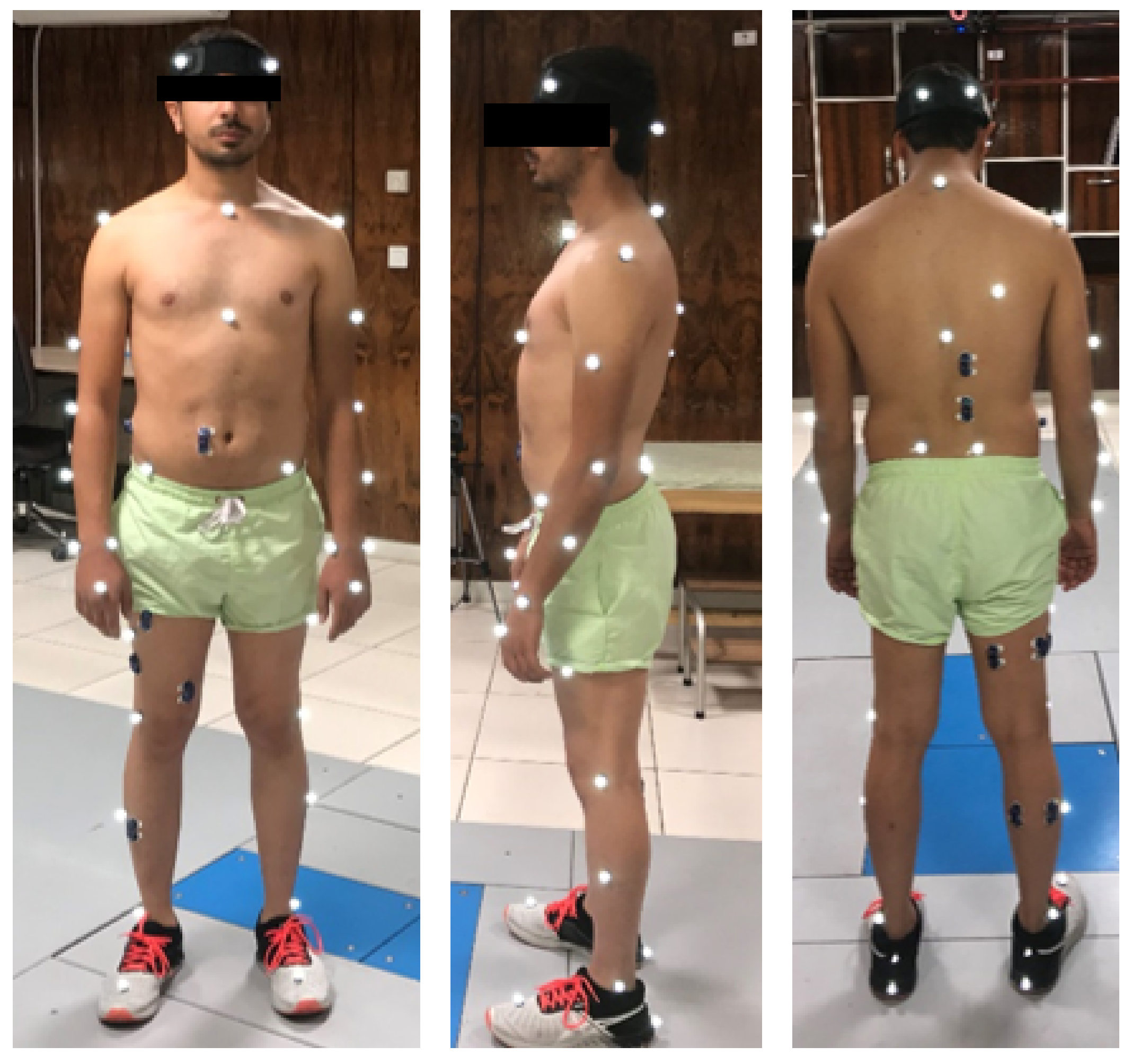
2.2. Instrumentation

Following the Navicular drop test and resting calcaneal position test, a 3D gait analysis was performed. Subjects were asked to perform a bilateral stance posture assessment for model creation and processing prior to gait acquisition. As a result, all subjects were instructed to walk barefoot at a self-selected and comfortable pace across an 8-meter walkway, replicating their daily gait. A starting point was established to standardize gait initiation. Twelve Vicon motion-capturing cameras (Vicon MX, Oxford, UK, 200 Hz sampling frequency) were used to record the kinematics data. For motion capture, 35 reflective markers were affixed over the anatomical landmarks, as shown in Figure 1. Each participant was asked to walk with and without shoes (running shoes made of TPU), and the data were obtained for 5 successive trials. The trial was discarded if the subjects failed to produce their daily gait, and a new trial was conducted for the study. Trials that were clear with all the marker data were selected for further processing.

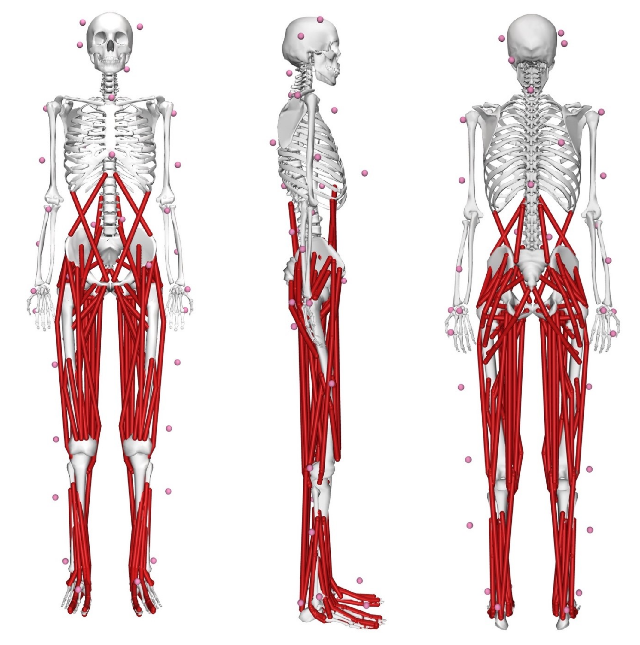
2.3. Musculoskeletal Modeling

We used the Rajagopal (2016) model of OpenSim (Stanford University, Stanford, CA, USA, 4.3 version)m which consists of 37 degrees of freedom (20 DOFs in the lower body and 17 in the torso and upper body), 80 muscle-tendon actuators to actuate the lower limbs, and 17 torque actuators to simulate the gait [7], as shown in Figure 2. The generic model was scaled by mass, height, and marker data in the static position. The scaling procedure was performed by the scaling tool of the OpenSim software. The inverse kinematics (IK) was used to quantify the hip, knee, and ankle kinematics [8,9,10]. The IK estimates a weighted least-squares equation to minimize the length between model marker data ( x i ) and surface reflective marker data ( x ¯ i ) in each time interval [11]:
m i n q ( i = 1 N w i x ¯ i x i ( q ) 2 )
where q presents the vector of the generalized coordinates of the model and w i the weight of the ith marker.

2.4. Data Analysis

The Motion Kinematics and Kinetics Analyser (Mokka) software was used to distinguish the on-set and off-set from the C3D file and convert it into a TRC file [12,13]. Mokka can also be used for extracting the ground reaction force (GRF) and EMG data associated with the subject. The duration time of one cycle gait was normalized to a percentage to facilitate the timing comparison between groups and subjects [14].

2.5. Statistics

The data were statistically processed with the Matlab 2022 software (MathWorks, Natick, MA, USA). The statistics analysis was performed for the lower limb kinematics for gait with and without running shoes. The results are presented as mean values and standard deviations (SDs) with a confidence level of 95%.

3. Results

3.1. Kinematic Analysis

The ankle, hip, and knee angles of each lower limb (Left/Right) were analyzed for one complete gait cycle and are presented in Figure 3, 5 and 7.
The hip kinematics of the flatfoot subjects with its maximum joint angle characteristics are visualized in Figure 3 and Figure 4. From the above Figure 3, there was no significant difference in hip kinematics with and without shoes. From Figure 4, it is indicated that the flatfoot subjects exhibited higher FE compared to other ranges of motion. Compared to left and right flatfoot hip kinematics, Flexion/Extension was higher in both the with-shoe and without-shoe cases. The absolute values of the hip AA were observed to be higher in the right compared to the left. Hip rotation showed higher differences in joint angles with a decrease of −187.4% and an increase of −4.9% in the right and left flat foot during gait phase [15].
The knee angle of the flatfoot subjects and its maximum joint angle with and without shoes are depicted in Figure 5 and Figure 6.
From the above Figure 5 and Figure 6, there was no significant difference between with- and without-shoe conditions in flatfoot subjects. The knee kinematics of flatfoot subjects with and without shoes were similar [15]. The percent of change between with- and without-shoe groups of the knee was found to have a decrease of 12.4% and an increase of 0.4% in the right and left knee.
The ankle angle of the flatfoot subjects and its maximum joint angle with and without shoes are depicted in Figure 7 and Figure 8.
From the above Figure 7 and Figure 8, it is clearly shown that the ankle kinematics of flatfoot subjects with and without shoes were distinctly separable. A smaller ankle dorsiflexion angle was observed while using shoes for one complete gait cycle. The percent of change between the with- and without-shoe group was found to be a decrease of 41.96% and 35.2% in the right and left ankle angle. It is clearly evident that while wearing shoes, the DOF and ankle kinematics were reduced, indicating less pressure was required to initiate the movement than normal walking. Hence, the ankle plays a major role in flatfoot kinematics, and an orthosis will help balance control in flatfoot subjects, as well as help in reducing ankle pronation deformity.

3.2. COM Displacement and Velocity Results

COM variables (displacement and velocity) were obtained through body kinematics and are tabulated in Table 1 and Table 2.
According to Figure 9, it is clear that the flatfoot group with shoes had a larger variability in the COM compared to the without-shoes (barefoot) group [15]. The resultant medio-lateral motion of the COM during locomotion can be used as a functional indicator to identify a person who is at greater risk of falling [16]. The flatfoot subjects with shoes showed an increase in medio-lateral motion, which indicates compensatory adjustments are established to counter the balance disturbance in the frontal plane.

4. Discussion

Flatfoot is a condition that causes several injuries, such as pain due to the alternation in the gait patterns, speed, balance, and control. This will consequently result in the risk of falling and decreasing the mobility functions. Hence, the presented study compared the kinematics of gait with and without running shoes and the medio-lateral relationship for balance in flatfoot subjects. The kinematics results indicated that flatfoot disorder alters the lower body kinematics, balance, and control. Flatfoot subjects have lesser ankle dorsiflexion and a lesser knee extension peak, leading to a lack of mobility. It was also indicated that flatfoot groups have a different range of motion (ROM), similar to previous literature [4,15]. It was shown that the above kinematic variables can be customized and compensate for normal gait by wearing shoes. Based on this study, the pronation deformity will improve when wearing shoes, and this can reduce the ankle angle [4,15,16]. The hip and knee flexion need to be absorbed at the foot level usually during dynamic impact. Running shoes act as a cushion and balance support for flatfoot subjects. This can be compensated by providing counterparts within the shoe to absorb the abnormal changes in the gait. Additionally, the medio-lateral COM and its role in balance control were detailed in this study [16]. The counter balance in the frontal plane is essential for flatfoot subjects to reduce the risk of falling. The above needs were addressed by wearing shoes. Further, The presented data can be cross-validated with normal subjects to differentiate the lower limb kinematics. Plantar pressure characteristics can also be recorded to understand the pressure distribution variations in the foot region. Furthermore, a balance compensation strategy for flatfoot subjects needs to be developed. In this way, the presented data will be helpful in the development of foot orthoses for flatfoot subjects suitable for dynamic activities.

5. Conclusions

The main goal of the current study was to quantify the kinematic variables and COM relationship in flatfoot subjects with and without running shoes. The shoe and barefoot groups were significantly different in the kinematics of lower limb joints. Running shoes can alter the kinematic variables of the lower limbs in flatfoot subjects. Hence, in order to protect, restore, and reduce the side effects of the foot and posture, wearing suitable shoes can be a fine option for flatfoot disorder. Furthermore, the presented study described the parameters essential for balance and control. Further attempts are needed to evaluate the specific modification strategy of subjects with flatfoot, which could use different running shoes with visual input. In addition, the correlation between kinematics and kinematicvariables in the evaluation of running shoes will be helpful to uncover the pathological aspects.

Author Contributions

Conceptualization, G.R., S.D. and H.B.; methodology, G.R. and H.B.; software, G.R.; validation, G.R., S.D. and H.B.; formal analysis, G.R.; writing—original draft preparation, G.R., S.D., and H.B.; writing—review and editing, G.R., S.D., Y.K. and H.B.; visualization, G.R.; supervision, Y.K.; project administration, S.D. All authors have read and agreed to the published version of the manuscript.

Funding

This research received no external funding.

Institutional Review Board Statement

The study was conducted in accordance with the Declaration of Helsinki, and the protocol was approved by the Ethics Committee of 9908-0508.

Informed Consent Statement

Informed consent was obtained from all subjects involved in the study.

Data Availability Statement

The data presented in this study are available on request from the corresponding author.

Conflicts of Interest

The authors declare no conflict of interest.

Abbreviations

The following abbreviations are used in this manuscript:
EMGElectromyography
COMCenter of mass
ROMRange of motion
DOFDegree of freedom
IKInverse kinematics

References

  1. Delp, S.L.; Anderson, F.C.; Arnold, A.S.; Loan, P.; Habib, A.; John, C.T.; Guendelman, E.; Thelen, D.G. OpenSim: Open-source software to create and analyze dynamic simulations of movement. IEEE Trans. Biomed. Eng. 2007, 54, 1940–1950. [Google Scholar] [CrossRef] [PubMed] [Green Version]
  2. Lee, C.R.; Kim, M.K. The effects on muscle activation of flatfoot during gait according to the velocity on an ascending slope. J. Phys. Ther. Sci. 2014, 26, 675–677. [Google Scholar] [CrossRef] [PubMed] [Green Version]
  3. Prachgosin, T.; Chong, D.Y.; Leelasamran, W.; Smithmaitrie, P.; Chatpun, S. Medial longitudinal arch biomechanics evaluation during gait in subjects with flexible flatfoot. Acta Bioeng. Biomech. 2015, 17, 121–130. [Google Scholar] [PubMed]
  4. Marouvo, J.; Sousa, F.; Fernandes, O.; Castro, M.A.; Paszkiel, S. Gait Kinematics Analysis of Flatfoot Adults. Appl. Sci. 2021, 11, 7077. [Google Scholar] [CrossRef]
  5. Chen, Y.; Su, F.; Huang, C. Effects of the shoes and insoles on gait patterns in the flatfoot. J. Biomech. 2007, 40, S472. [Google Scholar] [CrossRef]
  6. Kim, M.K.; Lee, Y.S. Kinematic analysis of the lower extremities of subjects with flat feet at different gait speeds. J. Phys. Ther. Sci. 2013, 25, 531–533. [Google Scholar] [CrossRef] [PubMed] [Green Version]
  7. Rajagopal, A.; Dembia, C.L.; DeMers, M.S.; Delp, D.D.; Hicks, J.L.; Delp, S.L. Full-body musculoskeletal model for muscle-driven simulation of human gait. IEEE Trans. Biomed. Eng. 2016, 63, 2068–2079. [Google Scholar] [CrossRef] [PubMed]
  8. Barnamehei, H.; Zhou, Y.; Zhang, X.; Vasavada, A. Cervical spine intervertebral kinematics estimated from inverse kinematics and compared to dynamic X-ray data. Gait Posture 2020, 81, 36–37. [Google Scholar] [CrossRef]
  9. Barnamehei, H.; Ghomsheh, F.T.; Cherati, A.S.; Pouladian, M. Kinematic models evaluation of shoulder complex during the badminton overhead forehand smash task in various speed. Inform. Med. Unlocked 2021, 26, 100697. [Google Scholar] [CrossRef]
  10. Barnamehei, H.; Ghomsheh, F.T.; Cherati, A.S.; Pouladian, M.; Aminishahsavarani, A.; Golfeshan, N. Shoulder Kinematics and Kinetics Comparison Between Amateur and Professional Athletics During High-Speed Overhead Tasks: Computer Simulation Study. In International Conference on NeuroRehabilitation; Springer: Vigo, Spain, 2020; pp. 71–75. [Google Scholar]
  11. Lu, T.W.; O’connor, J. Bone position estimation from skin marker co-ordinates using global optimisation with joint constraints. J. Biomech. 1999, 32, 129–134. [Google Scholar] [CrossRef]
  12. Barnamehei, H.; Kharazi, M.; Barnamehei, M. Heel Height Affects Lower Extremity Joint Moments during Walking with OpenSim Software. 2017. Available online: https://www.researchgate.net/profile/Hamidreza-Barnamehei/publication/318592221_P83_Heel_height_affects_lower_extremity_joint_moments_during_walking_with_OpenSim_software/links/5b4ba88b45851519b4c01f3b/P83-Heel-height-affects-lower-extremity-joint-moments-during-walking-with-OpenSim-software.pdf (accessed on 15 March 2020).
  13. Barnamehei, H.; Ghomsheh, F.T.; Cherati, A.S.; Pouladian, M. Muscle and joint force dependence of scaling and skill level of athletes in high-speed overhead task: Musculoskeletal simulation study. Inform. Med. Unlocked 2020, 20, 100415. [Google Scholar] [CrossRef]
  14. Bagheri, F.; Barnamehei, H.; Kharazi, M.; Panahi, A.; Golfeshan, N.; Razaghi, M.; Rostami, M.; Barnamehei, M.; Naghavi, N.; Jafarloo, S.A. Comparison of biarticular muscles effects on balance variation between bare-foot and wearing high-heel shoe conditions: Experimental and simulation by OpenSim. Gait Posture 2020, 81, 20–21. [Google Scholar] [CrossRef]
  15. Barnamehei, H.; Karimidastjerdi, M.; Torabigoudarzi, M.; Kharazi, M.; Barnamehei, M.; Golfeshan, N.; Naghavi, N.; Jafarloo, S.A.; Ghorbani, M. Simulation of flatfoot gait with and without running shoes by evaluation of hip kinematics and balance control variations via OpenSim. Gait Posture 2020, 81, 32–33. [Google Scholar] [CrossRef]
  16. Chou, L.S.; Kaufman, K.R.; Hahn, M.E.; Brey, R.H. Medio-lateral motion of the center of mass during obstacle crossing distinguishes elderly individuals with imbalance. Gait Posture 2003, 18, 125–133. [Google Scholar] [CrossRef]
Figure 1. Experimental setup used for GAIT measurements. The motion analysis lab for kinematics and kinetics measurements in Movafaghian (Sharif University of Technology, Tehran, Iran) consists of high-speed motion captures and force plates.
Actuators 11 00152 g001
Figure 2. The anterior view, lateral view, and posterior view sides of the Rajagopal (2016) musculoskeletal model of OpenSim (Stanford University, Stanford, CA, USA, 4.3 version) consisting of surface markers, 37 degrees of freedom (20 DOFs in the lower body and 17 in the torso and upper body), 80 muscle–tendon actuators to actuate the lower limbs, and 17 torque actuators to simulate the gait.
Actuators 11 00152 g002
Figure 3. Hip kinematics of flatfoot subject with and without running shoes. r-right; l-left; FE-flexion/extension; AA-abduction/adduction; RR-internal/external rotation.
Actuators 11 00152 g003
Figure 4. Maximum hip joint angles of flatfoot subjects with and without shoes. r-right; l-left.
Actuators 11 00152 g004
Figure 5. Knee kinematics of flatfoot subjects with and without shoes. Red line indicates mean knee joint angle with shoe group and Blue line indicated mean knee joint angle without shoe group.
Actuators 11 00152 g005
Figure 6. Maximum knee joint angles of flatfoot subjects with and without shoes. r-right; l-left.
Actuators 11 00152 g006
Figure 7. Ankle kinematics of flatfoot subjects with and without shoes. Red line indicates mean ankle joint angle with shoe group and Blue line indicated mean ankle joint angle without shoe group.
Actuators 11 00152 g007
Figure 8. Maximum ankle (dorsiflexion) joint angles of flatfoot subjects with and without shoes. r-right; l-left.
Actuators 11 00152 g008
Figure 9. COM variables (displacement and velocity) of flatfoot subjects with and without shoes. Red line indicates mean ankle joint angle with shoe group and Blue line indicated mean ankle joint angle without shoe group.
Actuators 11 00152 g009
Table 1. Mean and standard deviation of COM variables (displacement).
Without Shoe
COM
With Shoe
COM
COMxCOMyCOMzCOMxCOMyCOMz
Mean0.0117942−0.00234−0.009240.0051975−0.002879−0.009582
Stddev *−0.005516−0.000559−5.33 × 10 5 −0.005516−0.000559−5.33 × 10 5
* Standard deviation.
Table 2. Mean and standard deviation of COM variables (velocity).
Without Shoe
COM
With Shoe
COM
COMxCOMyCOMzCOMxCOMyCOMz
Mean0.81813750.02023290.00306890.8754778−0.0045990.0037519
Stddev *0.09335340.09783070.01575230.09335340.09783070.0157523
* Standard deviation.
Publisher’s Note: MDPI stays neutral with regard to jurisdictional claims in published maps and institutional affiliations.

