Review

Biointerface Materials for Cellular Adhesion: Recent Progress and Future Prospects

by
John V. L. Nguyen
1,2 and
Ebrahim Ghafar-Zadeh
1,2,3,*
1
Biologically Inspired Sensors and Actuators (BioSA), Lassonde School of Engineering, York University, Toronto, ON M3J 1P3, Canada
2
Department of Biology, Faculty of Science, York University, Toronto, ON M3J 1P3, Canada
3
Department of Electrical Engineering and Computer Science, Lassonde School of Engineering, York University, Toronto, ON M3J 1P3, Canada
*
Author to whom correspondence should be addressed.
Actuators 2020, 9(4), 137; https://doi.org/10.3390/act9040137
Submission received: 3 November 2020 / Revised: 7 December 2020 / Accepted: 10 December 2020 / Published: 11 December 2020
(This article belongs to the Section Actuators for Medical Instruments)

Abstract

:
While many natural instances of adhesion between cells and biological macromolecules have been elucidated, understanding how to mimic these adhesion events remains to be a challenge. Discovering new biointerface materials that can provide an appropriate environment, and in some cases, also providing function similar to the body’s own extracellular matrix, would be highly beneficial to multiple existing applications in biomedical and biological engineering, and provide the necessary insight for the advancement of new technology. Such examples of current applications that would benefit include biosensors, high-throughput screening and tissue engineering. From a mechanical perspective, these biointerfaces would function as bioactuators that apply focal adhesion points onto cells, allowing them to move and migrate along a surface, making biointerfaces a very relevant application in the field of actuators. While it is evident that great strides in progress have been made in the area of synthetic biointerfaces, we must also acknowledge their current limitations as described in the literature, leading to an inability to completely function and dynamically respond like natural biointerfaces. In this review, we discuss the methods, materials and, possible applications of biointerface materials used in the current literature, and the trends for future research in this area.

1. Introduction

Attachment is a fundamental process observed across nature, from the macroscopic burrs of burdock plants hooking onto passing animals, down to the proteins which mediate cell–cell attachment. Evident across the multitude of scales in biology, many biological processes rely on the state of attachment onto another surface in order to proceed with the actions of normal physiology. In particular, processes including, but not limited to, cell differentiation, cell cycle control, cell migration, and cell survival are all tightly regulated by the current state of attachment of the cell [1], supporting the belief that cellular fate mechanisms are due, at least in part, to the properties of the cell’s surroundings, which may be other organic materials such as cells, inorganic materials such as chemicals, or even physical properties such as the underlying substrate texture. In the native context of the cellular environment, the extracellular matrix (ECM) is the natural scaffolding of the cells, providing a suitable substrate for cellular adhesion, structural support for the three-dimensional shape for the collective cells in forming the tissue, and even providing biochemical support by being a sink for various signaling proteins and hormones [2]. These properties of the ECM provide key considerations to us when selecting or designing biointerfaces that can enhance cellular adhesion, and function as effective mediators of biological actuation.
While adhesion and actuation are different physical features, their relation to each other is not to be overlooked, and situations where both are apparent are especially significant in the context of biological cells. Cell-substrate interactions are critical for cellular adhesion onto surfaces and the mechanics of materials that interface the cells have to be considered in order to comprehensively select or design new biointerfaces or ECM mimetics. The beating heart is a noteworthy example where the mechanics the cellular substrates are not just simply involved in organ-level structure but are also essential for the proper functioning of the organ [3]. At the cellular scale, cardiac muscle contractions can be thought of as cellular adhesion and actuation atop an underlying substrate. Mechanotransduction, the conversion of mechanical signals into electrochemical signals, is another biological process that is dependent on cellular actuation. In instances of mechanotransduction, mechanical forces applied to the cell through focal adhesion points can modulate cellular behavior [4]. Actuation of the joints between bones are a biological instance where mechanotransduction can be observed. The articular cartilage found in joints is subject to a variety of mechanical forces such as shear, compression and tension and these mechanical forces are absorbed by the ECM surrounding the cartilage. The ECM is then able to dissipate and transmit the mechanical forces to ECM-adhering chondrocytes, which then convert the mechanical forces into various biochemical signals, including those that then direct either the formation or degradation of the cartilage matrix [5,6]. With these concepts in mind, it should be clear that biointerfaces with adhesion-enhancing features are a key contributor to the mechanical properties of the cell, including cellular motion and actuation, and their proper functioning.
In cell–cell or cell–ECM binding, adhesion is typically facilitated by an interaction between the cell adhesion molecules (CAMs) on the surface of the cell and the partner cell or ECM. However, in biomaterials, these CAMs may not be natively present. Integration of adhesion-facilitating proteins into biomaterial surfaces has been used, but this requires careful consideration of the chemical properties. For example, one important chemical aspect is the hydrophilicity of the biointerface material. Biomaterials with a surface that is very hydrophobic will have more protein adhesion onto the surface, but also more protein denaturation, and biomaterials with a surface that is very hydrophilic will have poor adhesion of proteins onto the surface [7,8]. Indeed, having robust protein adhesion and absorption into the surface of biomaterial and the maintenance of protein conformation requires a careful balance between making the surface either too hydrophobic or too hydrophilic. Considering physical properties, substrate stiffness is a property that has been thoroughly investigated and known to alter the adhesive ability of cells. Prior works have observed that cells on soft substrates show high levels of adhesion, greater spreading and the establishment of focal adhesion points between the cell and the substrates, and that cells on rigid substrates show lower levels of adhesion and less spreading [9,10,11]. These are examples some of the classical methods that biointerface materials have used to modulate cell adhesion. Further examples in this paper will address more modern approaches.
Recent advances in areas of bioengineering, such as biosensors, high-throughput screening (HTS), and tissue engineering have benefited from the current selection of biointerface materials and technologies; however, many of these materials are still imperfect, and do not provide all of the benefits that are imparted by a natural ECM. Indeed, it must be recognized that progress in the areas of biomaterials and biointerfaces are dependent on, and perhaps even limited by, the current knowledge available in related fields, and the materials available at our disposal. As an example, the discovery of the Arg–Gly–Asp tripeptide, or RGD motif, within the amino acid sequence of the ECM-bound protein, fibronectin, in 1984 had provided sparkling insight into a key mediator for cellular attachment [12,13,14,15], and while the potential for incorporation of the RGD motif into biomaterials had initially been predicted by as early as 1990 [16], it took more than a decade from the initial report of the RGD motif for successes in incorporating the key tripeptide into common biomaterials to be reported [17,18,19,20]. Therefore, while it is important to identify what features of biological systems we would like to have when selecting or designing new biomaterials and biointerfaces, a prerequisite step is that we must first identify the aspects of the natural environment of the biological system which gives rise to these desirable features. Consequently, by depending on the progress of discovery in other fields, such as cell biology and materials science, in order to identify and incorporate these key environmental aspects into our synthetic materials, it will appear as though developments in biomaterials and biointerface design has kept out of pace with current innovation; however, accounting for the interdisciplinary aspect of this field, we must consider this delay in the translation of knowledge to be an inevitable part of the process.
While it is still an evolving area of study, great strides in progress have taken place in the development of biointerfaces, as evident by the recent literature. Novel materials show promise in many areas of application, especially in applications pertaining to medical therapeutics and diagnostics, collectively known as theranostics [21,22,23]. Focusing on the ability of these biointerfaces to provide appropriate cell-substrate interactions or biomolecule–substrate interactions would be a wise first step when developing tools for use either inside or outside of the laboratory. This is especially evident when considering both the current trends in bioengineering aiming to miniaturize standard biomedical instrumentation tools down into so called labs-on-a-chip (LOCs) or micro total analysis systems (µTASs), and the push to have these tools integrated into consumer electronics, such as smartphones or smartwatches [24]. Thinking about how the biointerfaces of these tools should act to immobilize and detect biological elements and their respective signals is a current issue of great interest. Therefore, our review shall focus on the biointerfaces used in the current literature, with an explicit focus on biointerfaces which enhance cellular adhesion, and the future direction of biointerface design and its applications.

2. Biointerfaces for Non-Specific Cell Type Adhesion

The most traditional use of biointerfaces for non-specific cell adhesion is in cell culturing applications. While glass was the initial substrate for cell cultures, being used in landmark experiments on culturing cells in vitro [25,26], in the modern lab, most instances of cell culturing utilize polystyrene plates. Polystyrene plates have numerous benefits, such as its optical clarity, ease of molding and ability to withstand sterilization by irradiation, and additionally, its disposability and low cost make it attractive in applications where concerns of contaminant residues from reused glassware could interfere with results, and so a fresh dish would avoid this possibility. However, the surface chemistry of untreated polystyrene is hydrophobic, making it an unsuitable surface for cellular adhesion and consequently, also unsuitable for applications requiring cell culture of adherent cells [27]. To rectify this, several different methods have previously attempted to improve tissue culturing the ability of polystyrene, including oxidation of the plate surface by sulfuric acid, etching by gamma irradiation, treatment by gas plasma under vacuum and treatment by corona discharge under atmospheric conditions [28,29,30,31], with the latter two methods forming highly energetic oxygen ions which bind to the surface, forming polar, hydrophilic groups on the plate surface. While these methods were able to improve the ability of the cells to adhere to the polystyrene-based plates, they still fell short of natural cellular substrates, such as the ECM, and do not permit cell cultures that function like cells in vivo. To overcome this, a variety of biointerface materials have been developed and evaluated, incorporating natural and synthetic materials to make hybrid biointerface materials.
In our survey of the current literature, many different biointerface materials are evident, with each presenting their own advantages and disadvantages. Some may be used alone or blended with another substrate material as a composite mixture. A summary of base substrate materials, with examples from the literature, is presented in Table 1. We note that even though specific cell types are listed within the table, there is no indication in the referenced literature that these biointerface materials are tailored exclusively for one cell type. Given that biological tissue is composed of multiple cell types which interact with each other, the inclusion of non-specific biointerfaces in new technologies should be able to more accurately model tissue composition in vivo, as opposed to biointerfaces tailored for cell type-specific binding, permitting applications where multiple cell types are used, such as in co-cultures. There is a wide range in the variety of biointerface materials, and one of the simplest ways to categorize these materials is by whether they are naturally derived or synthetic. Naturally derived biointerface materials (e.g., alginate, collagen and corn starch) are noted to be inherently biocompatible, but with any natural product, there may be batch-to-batch variations, undefined matrix compositions (including possible natural impurities) and restricted possibilities to modify these materials, all of which could hinder its ability to function in many applications [32].

2.1. Current Trends in Biointerface Materials for Non-Specific Cell Type Adhesion

From the current literature, three common trends are evident when evaluating novel biointerface materials, (1) multiple materials and processes that were previously used alone are now being used in conjunction to create new, hybrid materials and processes, (2) there is extensive use of the ECM and ECM-derived components, such as proteins and proteoglycans and (3) the recent growth in the pace of published papers describing three-dimensional biointerface materials and three-dimensional applications for these materials.

2.1.1. Utilization of Multiple Materials and Processes for Novel Biointerface Material Properties

In many instances, a variety of different materials and processes can be blended to create hybrid materials that carry features of multiple materials and processes. Several such papers have already been discussed in this review that combines multiple materials to create hybrid materials that have new properties that are suitable for biological applications, typically biological elements incorporated into a hydrogel; however, recent work has also demonstrated that there is a benefit to the incorporation of inorganic materials into biointerface materials as well. Here, we present work by Ahadian and colleagues which incorporated vertically aligned carbon nanotubes (CNTs) into a methacrylated gelatin (GelMA) hydrogel (Figure 1) to create a biological scaffold with tunable electrical and mechanical properties [50]. They observed numerous physical advantages with their hybrid hydrogel system, including anisotropic electrical conductivity and superior mechanical strength as compared to GelMA hydrogels with either no CNTs, or with randomly distributed CNTs. Additionally, they tested their hybrid GelMA-CNT hydrogel’s ability to culture skeletal muscle cells and observed an increase in the number of functional myofibers yielded as compared to skeletal muscle cells cultured on GelMA hydrogels with either no CNTs, or with randomly distributed CNTs, and were able to increase myogenic gene and protein expression by applying electrical stimulation (ES) along the direction of the aligned CNTs. This work demonstrates the incorporation of ES into these novel biointerface materials to alter its properties, and along with other recent works, also demonstrates the possibility of creating dynamic biointerface materials that are manipulatable by external factors, such as magnetic field [51], mechanical force [52], temperature [53], pH, and light [54]. The development of useful, dynamic and manipulatable biointerface surfaces using a variety of different stimuli has been demonstrated, as evident from the surveyed literature, and many of these have shown incredible abilities that can be altered even while in use.

2.1.2. Biointerface Materials with Integrated ECM and ECM-Related Elements

The ECM is the natural scaffolding of cells, and so, being able to have cells grow on an ECM would be ideal for mimicking the in vivo environment. While it is possible to produce an ECM by tissue decellularization [55], it is not a simple process. Instead, as an alternative, select components from the ECM can be incorporated into biomaterials, improving the biocompatibility and ECM-like properties [56,57], while not going through the challenges of decellularizing and adapting an ECM for a specific use.

2.1.3. Biointerface Materials for Three-Dimensional Applications

Research in the areas of tissue engineering and regenerative medicine have all grown in popularity in the last few decades, as their success in the lab would imply a range of new possibilities in the area of medical treatments [58,59]. The common factor between these areas is that they all involve the increasing utilization of three-dimensional cell scaffolds [60]. Three-dimensional cell scaffolds have been established as a means by which cells can adhere to in order to be structurally supported in a three-dimensional configuration while also maintaining regular function. Three-dimensional scaffolds have even been developed on the microscale, where their versatility in the study of basic cell biology has been noted [61]. Several methods are currently in use to produce three-dimensional cell scaffolds in the laboratory and a small sample of these are summarized, with examples from the literature, and presented in Table 2. We highlight two significant works that take different approaches in order to achieve production of three-dimensional scaffolds. Work by Rajzer and colleagues developed a three-dimensional scaffold using two different three-dimensional fabrication techniques, 3D printing and electrospinning, with the goal of developing a scaffold to reconstruct nasal cartilage and subchondral bone [62]. In 3D printing (using the Fused Deposition Modelling, or FDM, method), solidified filaments of the material are extruded from a heated nozzle onto a platform where it cools and hardens. The nozzle moves around the platform, forming the shape of the printed object, layer-by-layer. In the electrospinning method, the nozzle is connected to a high voltage source which charges the material, and by electrostatic repulsion, the material stretches out into a thin stream, with a diameter on the nanometer scale. The material then dries and solidifies while in flight, collecting on a grounded plate. Rajzer and colleagues had 3D printed a poly (l-lactic acid) scaffold with large pores and electrospun a gelatin scaffold on top of the poly (l-lactic acid) scaffold. They noted that the large pores allowed cells to easily grow into the scaffold, while the gelatin provided binding sites for integrin adhesion, while osteogenon added to the gelatin improved mineralization of the scaffold at the cartilage/bone interface. Using both of these techniques in conjunction allows imparts an improved ability for the scaffold to integrate into the surrounding tissue and also remain structurally supported.
While most methods of producing a three-dimensional scaffold discussed in this review require making a synthetic surface for the cells to adhere to, tissue decellularization involves isolation of a 3D ECM by breaking the cell membranes and washing away the cells on a piece of tissue. The resulting product is a decellularized ECM that can be reseeded with new cells. The clear advantage with tissue decellularization is that the complex structure of the tissue pre-decellularization can be maintained, eliminating the need to direct cell growth into a specific shape to form tissue or even entire organs. Perhaps the most notable potential application for decellularized tissue is in its potential for organ transplantation, where a donor organ can be decellularized and reseeded with cells from the recipient [63], greatly reducing or even eliminating the risk of organ rejection and the need for immunosuppressant drugs. The disadvantages to this method are that it still requires a donor organ and that the recipient must have healthy cells that can be used to reseed the donor ECM, although studies have demonstrated that it is possible to use the ECM from other animals, and especially porcine ECM, for recellularization with human cells allowing for the possibility of xenografts [64,65], and that the collected cells used for reseeding of the ECM do not have to be of the same type of cell as the original cells on the decellularized ECM, as other cell types can first be transformed into induced pluripotent stem cells then into the desired cell type, allowing for de novo organ genesis even if the recipient had no cells of the desired type available [66,67]. Recent works have focused on combining both ECM from other animals and human induced pluripotent stem cells [67,68,69,70]. Research in this area has strong implications for both artificial organ creation and in laboratory applications involving three-dimensional cells, such as on organs-on-a-chip. Although with this growth in described three-dimensional materials, we also have to acknowledge that the rapid progress in two-dimensional materials has not slowed down. The recent advances in two-dimensional interface models have made significant contributions to understanding the mechanism of cellular adhesion [71], and cellular impact of extracellular stimulation, such as substrate roughness and stiffness [72,73,74]. These advances in two-dimensional biointerface materials are likely to be able to trickle into three-dimensional biointerface materials and be further studied in three-dimensional models.
Table 2. Examples of three-dimensional cell culturing methods from recent literature.
MethodBase Scaffold MaterialScaffold ShapeCell TypeDescribed 1 Potential ApplicationsRef.
MoldedHydrogel (cellulose)CustomMouse fibroblastDrug delivery and tissue engineering[75]
3D printedHydrogel (polyHIPEs)CustomHuman MSCSynthetic bone graft and tissue engineering[76]
ElectrospunHydrogel (GelMA)ThreadHuman fibroblastSynthetic skin graft, tissue engineering and wound dressing[77]
Hanging dropletHydrogel (alginate)SphericalHuman colon cells and human liver cellsDrug screening[78]
Decellularized tissueECMOriginating tissueHuman liver cellsArtificial organ, disease modelling, drug screening and tissue engineering[79]
Direct laser writingProtein-functionalized photoresistCustomHuman lung adenocarcenoma and mouse embryo fibroblastTissue engineering and regenerative medicine[80]
1 “Described” indicates the use or possible use of the material, as stated by the authors in the referenced literature. GelMA = gelatin methacryloyl, MSC = mesenchymal stem cell, polyHIPEs = polymerized high internal phase emulsions.

3. Biointerfaces for Cell Type-Specific Adhesion

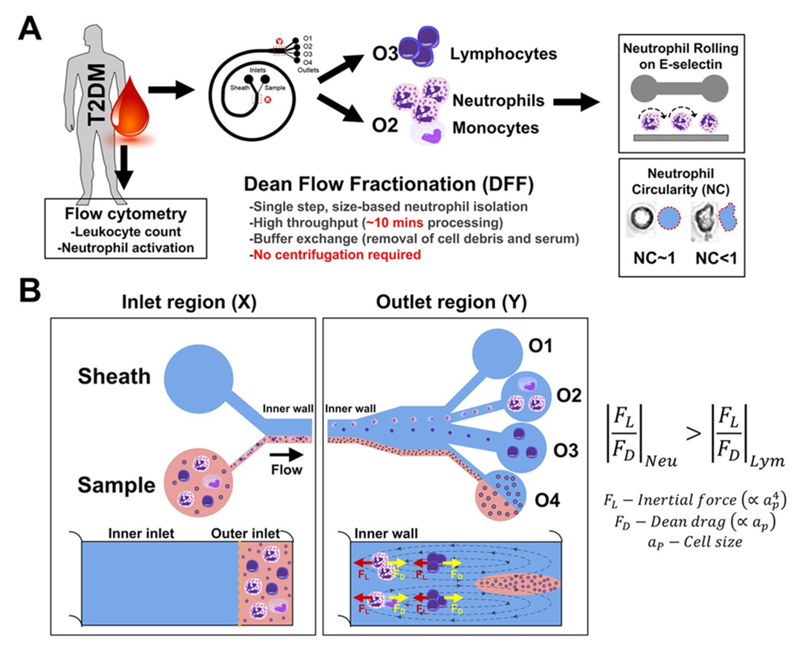
Cell type-specific methods are one of the most crucial methods used in the laboratory. Considering instances of cell-specific events in nature, it is known that in order to selectively identify or isolate cells of a specific type, we must exploit cellular characteristics that are possessed by that cell type, and not by others. While cell identification has classically been performed manually by microscopy. Automation of these processes has led to machinery that can both quickly and accurately detect cellular characteristics and even sort cells. Flow cytometry and flow-assisted cell sorting (FACS) are the gold standard for automated cell identification and sorting; however, the equipment required for this takes up space, the cost of the machinery may be prohibitive to research groups and it requires training on the use and interpretation of results. Given these constraints, Hou and colleagues had developed a microfluidic system that can isolate neutrophils from blood, using a method called Dean Flow Fractionation [81]. In their method, the unpurified blood sample flows through a spiral microchannel, experiencing centrifugal acceleration directed radially outwards, and at the end of the microchannel are four outlets. With careful tuning of the centrifugal acceleration experienced by the cells, the neutrophils can be directed to one of the specific outlets (Figure 2). Evidently, this method exploits the differences in the sizes of the cells, and therefore, would be challenging to use in applications where the differences in size between the desired cell type group and the undesired cell type group are minimal, requiring precise tuning of the system. Similarly, other methods also exist which do not require identifying the cell type of each individual cell prior to isolation, such as density gradient centrifugation or magnetic-activated cell sorting (MACS); however, with these methods, there is still a requirement to know what cells are present in the population, or rather, the densities of the cells and what antigens may be present on the cell surface, respectively.
It may perhaps be more practical to eliminate the necessity of having to first isolate the desired cell type before it is detected, as doing so would eliminate a step that could otherwise consume additional time or materials, while also balancing the need of keeping such a process true to its intended purpose. A solution to this challenge would be to employ a method that selectively interacts with only the desired cell type, even in the presence of undesired cell types or other unwanted particulates. This can be achieved by using biointerfaces that selectively promote cellular adhesion for only the certain cells of interest. A selection of current interfaces used in the literature is presented in Table 3.
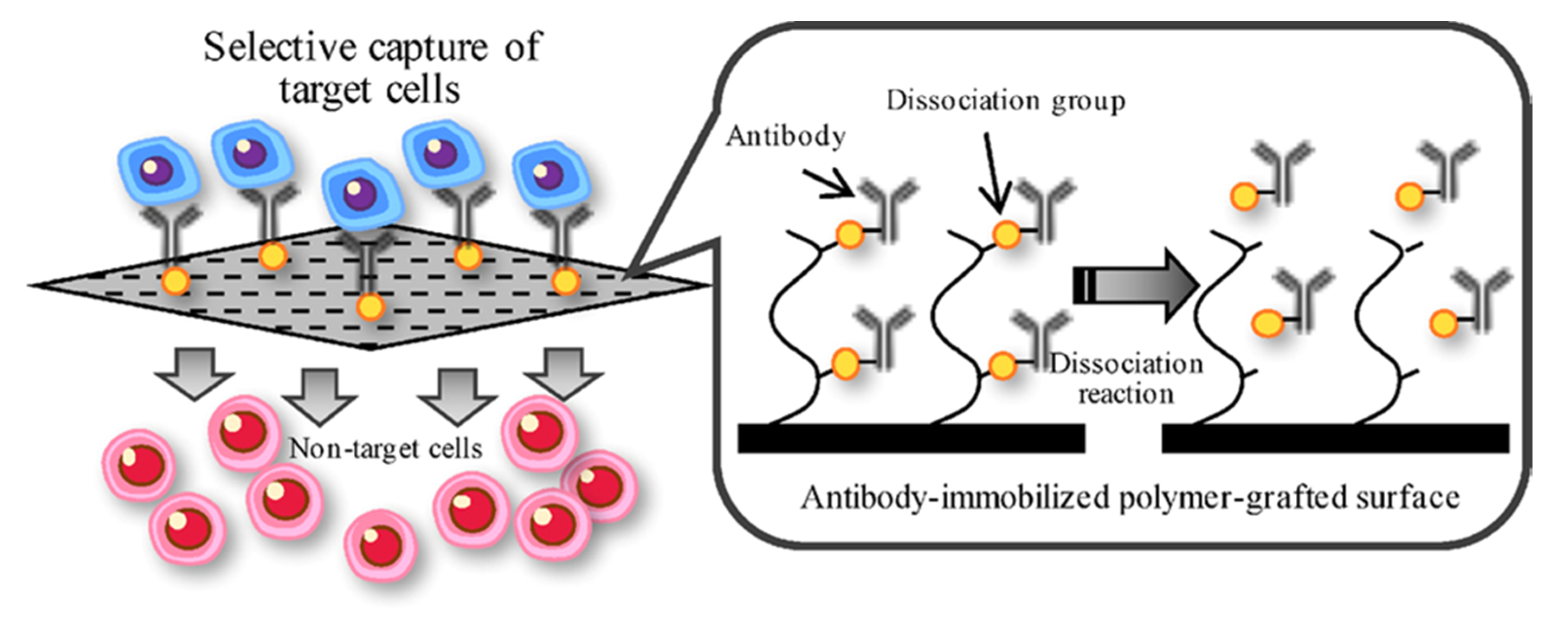
Previously, the prominent method of facilitating cell type-specific adhesion had focused on the use of peptide fragments, such as Arg–Gly–Asp (RGD), Arg–Gly–Asp–Ser (RGDS), Leu–Asp–Val (LDV) and others [14,15,92,93]. Currently, the most used methods of cell type-specific adhesion onto a surface rely primarily on the immobilization of antibodies onto a substrate, binding to cells that possess the complementary antigen molecule on the cell surface. For example, Kimura and colleagues had described two different ways to immobilize antibodies to capture specific cell types [94]. In one method, the antibodies were immobilized onto a surface by desthiobiotin–avidin interaction, and once the target cells were captured, the antibody with the bound cell could be released from the surface by the addition of biotin-modified polymer which exchanges places with desthiobiotin. In the other method, the antibodies were immobilized by double-stranded DNA, and when the target cells were captured, the antibody with the bound cell could be released from the surface by the addition of DNase. A general representation of this process is shown in Figure 3.

3.1. Current Trends in Biointerface Materials for Cell Type-Specific Adhesion

From our survey of the literature, it appears as though the rate of innovation in producing novel cell type-specific biointerfaces that promote adhesion is lower than that of non-specific cell type biointerfaces. The likely rationale for this is that there already exists a variety of methods for the isolation of specific cell types, using machines or other laboratory techniques, such as FACS, density gradient centrifugation or MACS, as mentioned previously, and in many cases, it is feasible to use these techniques when specific cell types are required. Additionally, the need for material surfaces that promote adhesion for only a specific cell type had not been previously recognized. However, personalized medicine has increasingly become both a popular research focus and a cultural phenomenon, inspiring the commercialization of many consumer devices designed to monitor biomedical signals while integrated into our personal electronic devices, such as our smartphones and smartwatches. We see two avenues where research in cell type-specific biointerfaces will go: (1) precise and portable medical diagnostics and (2) biocompatible and implantable materials.

3.1.1. Precise and Portable Medical Diagnostics

The creation of cell type-specific biointerfaces permits the possibility of developing precise and portable medical diagnostic tools. Our bodily fluids contain an abundant number of cells and other biomolecules that are indicative of our overall health and can provide us with the information necessary for the early detection of disease. Regrettably, the accessibility to this information seems to be prohibitive to those who cannot afford the time commitment or cost to see a physician or lack the appropriate testing facilities in their area. This issue is even worse in under-developed nations, where infectious diseases are even more prevalent, and so, there is an urgent need for solutions where it is feasible to test patient samples at the point-of-care environment. Laksanasopin and colleagues had written on the development and use of a microfluidic smartphone dongle that can diagnose HIV and syphilis and display the results on the screen of the device in under 15 min [95]. Their solution was based on the enzyme-linked immunosorbent assay (ELISA) and used a finger prick in order to obtain the 2 μL of blood required for the test. Striebel and colleagues sought to capture the macrophages in blood, using patterned antifouling polymer brushes for controlled surface adhesion [96]. Knowlton and colleagues described their 3D-printed smartphone-based point-of-care tool which can sort cells, and it does so by combining magnetic focusing and fluorescence imaging of fluorescently stained cells [97]. Similarly, Tran and colleagues described their smartphone-based device which also used magnetics and fluorescent imaging; however, their method used magnetic iron oxide nanoparticles surrounded by luminescent quantum dots [98]. Clearly, these reports demonstrate that laboratory methods are emerging as techniques that can be taken out of the lab and become more integrated with our everyday electronics.
While one front of precise and portable medical diagnostics involves the increased accessibility of bioanalysis tools into our lives, another aspect is that better biointerfaces will allow for the detection of disease markers which were previously difficult to detect, most markedly mentioned in the recent literature is the circulating tumor cell (CTC). CTCs occur in very low concentrations in the bloodstream, and have been referred to as a problem that is analogous to looking for a needle in a haystack [99]; however, given that CTCs are present in patients suffering from all of the major carcinomas [100], they are a very attractive target for early detection of cancer. Currently, the FDA has only approved one method for CTC isolation from blood, CellSearch®, which utilizes magnetic beads coated with anti-epithelial cell adhesion molecule (EpCAM) antibodies [100]; however, the cells isolated by this method cannot maintain viability, impacting the ability to study these cells and tailor treatment in a personalized medicine fashion. Myung and colleagues demonstrated the use of poly(amidoamine) (PAMAM) dendrimers on a glass surface to immobilize antihuman epidermal growth factor receptor (EGFR) antibodies (Figure 4), aiming to capture non-small-cell lung carcinoma CTCs which express EGFR in a majority of patients [85]. Mitchell and colleagues used sodium dodecanoate to immobilize E-selectin onto halloysite nanotubes, demonstrating the ability to capture CTCs without antibodies [91]. Yan and colleagues immobilized biotinylated antibodies onto gold-plated polydimethylsiloxane (PDMS) by using thiol-DNA-biotin-avidin as intermediary coupling molecules and had this system on a microchip, demonstrating an ability to capture CTCs using common PDMS-based microfluidics [89]. Brinkmann and colleagues and Liu and colleagues had demonstrated the compatibility of ELISA in a PDMS-based microfluidic platform in order to capture CTCs [101,102]. These works represent the future direction of biomaterials as tools to interact with and capture specific biomolecules and cells for medical applications, such as disease diagnosis or early disease detection.

3.1.2. Biocompatible and Implantable Materials

While the collective trend of non-specific cell type biointerfaces was to produce 3D materials that could eventually replace tissue and organs, we observed that the trend in cell type-specific biointerfaces was towards improving the biocompatibility and healing properties of implantable materials. In the surveyed literature, there was a focus on the utilization of cell type-specific biointerfaces to accelerate the endothelization process of stents. Stents are used in a variety of applications, and the insertion of stents are a common treatment for both coronary artery blockages and aneurysms. In both cases, it would be beneficial to restore the structural integrity of the endothelium, and so being able to accelerate the process of forming the endothelial layer would mean a quicker healing process for the patient and possibly better clinical outcomes. Stainless steel is a common material used for the manufacture of stents. Benvenuto and colleagues had written about their method of capturing endothelial progenitor cells (EPC) onto stainless steel stents by using immobilized antibodies directed against an EPC surface receptor [83]. They were able to immobilize the antibodies by using S-(11-Trichlorosilylundecanyl)-benzenethiosulfonate as an intermediary between the stainless steel and the antibodies. Qi and colleagues had also sought to develop a method of capturing EPCs onto stainless steel, although instead of antibodies they had used DNA aptamers, which are noted for their ability to have even a higher affinity to their targets than antibodies [90,103]. Qi and colleagues had deposited a plasma polymerized allylamine film onto the stainless steel in order to immobilize the antibodies. Ultimately, the endothelization of vascular devices remains to be a topic of great importance [104]. The modification or functionalization of the surface of vascular devices and other implantable materials is a direction of application for cell type-specific biointerfaces in the future.

4. Considerations for Selecting Biointerface Materials for Cell Adhesion

Clearly, the selection of the biointerface requires consideration of both material properties and the desired usage. We identify four considerations that must be made when selecting a biointerface material, (1) the required dimensions that cells must be able to occupy (i.e., two dimensions or three dimensions), (2) the target cell(s) of interest, (3) the application that this is to be used for and (4) the method used to detect the cells.

4.1. Dimensions of Cell Occupation

Although cell culturing had traditionally been performed using two-dimensional methods, multiple studies have indicated that two-dimensional cell cultures do not mimic normal cell function in vivo, and because of this, have different genomic and proteomic profiles, respond to drugs differently, and that the three-dimensional cellular environment more closely represents normal cell physiology [105,106,107]; however, the clear disadvantage is the increased difficulty in forming these three-dimensional cell culture systems, and perhaps in many cases, two-dimensions is sufficient for investigating the questions at hand. Upon careful consideration, it must then be asked how we can definitively determine whether a question can be accurately answered using the traditional two-dimensional systems, or whether the use of the three-dimensional system is warranted.
Breslin and O’Driscoll evaluated the response of three breast cancer cell lines (BT474, HCC1954 and EFM192A) when cultured in two-dimensional versus three-dimensional systems, with the three-dimensional system using a poly(2-hydroxyethyl methacrylate)-based hydrogel [108]. They observed significant differences between the two-dimensional and three-dimensional systems in all three cell lines, including differences in cell morphology, viability, the efficacy of anticancer drugs (neratinib and docetaxel), the gene expression profiles for genes involved in cell survival pathways, drug targets and a drug efflux pump, and the activity of a drug-metabolizing enzyme. With this perspective on the many differences between two-dimensional and three-dimensional culture of cancer cells, it may seem beneficial that applications involving cancer use three-dimensional systems for cell culture as it best represents the typical formation of this disease in vivo. These findings have serious implications for the numerous bioinstrumentation devices proposed for cancer modelling, screening for cancer, or the screening of anticancer drugs.

4.2. Target Cell(s) of Interest

Incompatibilities may also exist between the cell culturing biointerface material and the cells. Polylysine is commonly used as a material which increases the adhesion of cultured cells onto the cell culture plates, and it can exist as one out of two possible enantiomers. The poly-L-lysine amino acid precursor occurs naturally, and in some cell lines, proteases that can break down poly-L-lysine are produced [109], therefore preventing the cells from adhering to poly-L-lysine-coated plates. However, poly-D-lysine is made using a synthetic precursor that cannot be broken down by those same proteases, and therefore, would be preferred over poly-L-lysine when culturing those cell lines as the adhesion enhancing properties can be maintained.

4.3. Application-Specific Challenges

The desire for appropriate cell-substrate interactions must also be balanced by consideration of the challenges of the proposed application. Poldervaart and colleagues had developed hydrogel scaffolds containing epithelial progenitor cells for implantation into nude mice, with the goal of creating vascular networks in vivo [110]. The hydrogels were blended Matrigel–alginate mixtures that showed increasing structural rigidity as the proportion of alginate in the blend increased; however, with an increasing proportion of alginate (and a decreasing proportion of Matrigel), the vascular networks that emerged showed less branching, indicating an inverse correlation between the ability to form vascular networks in vivo and the structural strength of the chosen scaffold material. In contrast, Cavo and colleagues also used a Matrigel–alginate hydrogel mixture, with the exception that they had the goal of modelling breast cancer in vitro [111]. They noted the success in accurately modelling human breast cancer processes, including the malignant morphology, spread and invasion abilities. Although both groups used a Matrigel–alginate mixture, their differing applications for this one composite material demonstrated the impact of matching suitable biointerface materials to their proposed application.

4.4. Cell Detection Methods

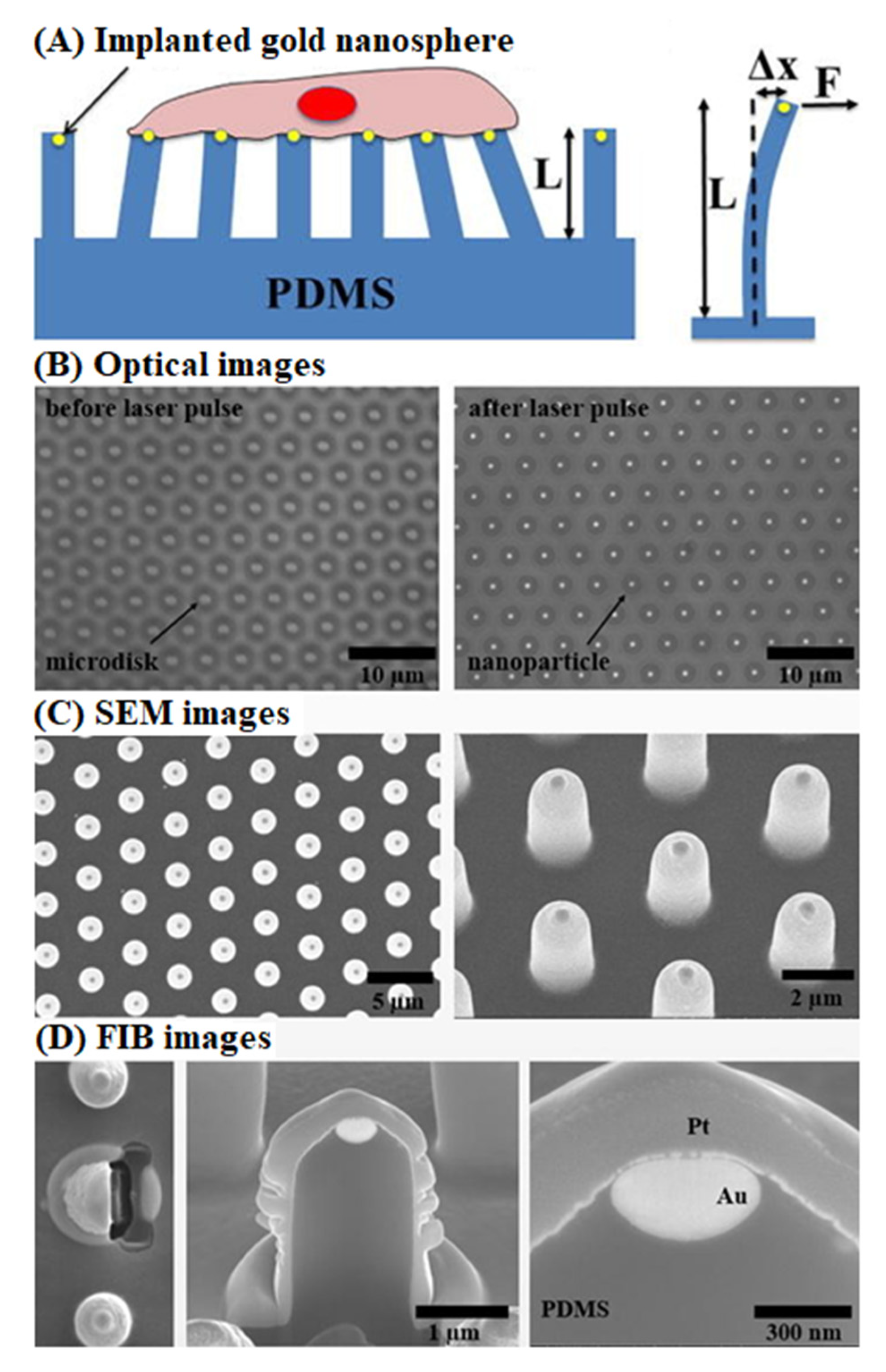
A majority of surveyed literature has detected cells by using optically based methods, such as fluorescence microscopy, making it a necessity that the biointerface materials used for cell culture provide adequate optical transparency for visualization of the cells. It should be noted that other methods have recently gained traction as suitable cellular detection methods, such as complementary metal–oxide–semiconductor (CMOS) capacitive sensors [112], and micropillar structures [113]. Capacitive sensors rely on cells coming into contact with electrodes and completing the electrical circuit in order to detect the presence of the cells at the location of the electrodes. Micropillars rely on cells pushing down on tiny vertical structures labeled with a fluorescent marker or light scattering particle (Figure 5). As the cells push down and deflect the micropillars, a camera captures the movement of the fluorescent or light scattering particle and computes the force applied by the cell required to deflect the pillars to the extent that the fluorescent/scattered light moved while also accounting for the stiffness of the micropillar, allowing this system to detect the presence of the cells at the location of the pillars and to determine the amount of downward force being applied by the cell.
Both capacitive sensors and micropillar structures rely on cell contact for detection and additionally, also do not require any fluorescent labeling of the cell itself, providing two different means for label-free cell detection. As the capacitive sensor does not rely on any form of light detection, the optical transparency of the cell culturing material is not of concern, allowing for a wider range of possible biointerface materials, including ones that are opaque. However, this requires that the selected cell culture material must not impede the detection of the cells, either by electrically insulating the cells from the contact electrodes or by acting to continually bridge the gap between the electrodes, even in the absence of a cell. These methods have been demonstrated for numerous lab-on-a-chip applications [114,115], and it should be expected that these gain a greater level of adoption into bioinstrumentation devices in the future.

5. Dynamic and Smart Materials for Biological Applications

While this review highlighted the biological and chemical aspects of biointerfaces for cellular adhesion and their use as bioactuators, there are other considerations that may also be made when trying to promote the adhesion of cells onto a surface. Ghafar-Zadeh and colleagues had reviewed the reported literature pertaining to cardiac regeneration from the standpoint of the engineered stem cell microenvironment and noted the role of chemical, structural, microfluidic and mechanical cues on the adhesion ability of the cells [116]. Although these cues are a factor in promoting cellular adhesion, they ultimately remain in a static state in what we consider to be traditional biointerfaces. As we consider the dynamic nature of biological systems and the ultimate goal of producing synthetic materials that can mimic the cellular environment, the natural progression for biointerfaces would be to create dynamic biointerfaces that can change in response to external stimuli.
Gomes and colleagues reviewed the literature in the area of dynamic and functional biointerfaces and noted the recent literature on biointerface materials that can respond to pH, temperature, mechanical forces, light, magnetic fields and electrical potential and electrical fields [117]. The creation of biointerface materials that respond to a variety of stimuli represent functions that are performed by a variety of different tissue types in living systems. These responsive biointerface materials appear to be well-suited for applications in artificial organs, biological sensing, HTS, medical diagnostics, cell separation and sorting and even the creation of lifelike robotics. While these dynamic biointerfaces have been proven in large 2D scaffolds, Hippler and colleagues have also demonstrated a dynamic ability in 3D microstructures, with actuating surfaces controllable by temperature and light [118].
Though given the attractive potential in using these very same biointerface materials in biosensing, Guo and colleagues have reviewed the literature for uses describing smart materials for biosensing applications [119]. Similar to those described for cell adhesion, modern materials for biosensing applications utilize both nanomaterials and hydrogels, and unlike any of the reviewed literature focusing on cell adhesion, there also appears to be the use of photonic crystals for the reflection of light, although works in the previous section did describe the use gold nanospheres in a similar function, as a light source and for positional tracking of the cell. Materials that can dynamically react to stimuli, like an ECM, can be highly efficient substrates for both cell culturing applications and biosensing. In particular, we note areas highlighted by Guo and colleagues that could serve in high-impact biosensing applications, glucose sensing and protein sensing. One highlighted study performed by Zhu and colleagues developed a hydrogel wound dressing for optical monitoring of pH and glucose [120]. This wound dressing protects the wound environment to promote healing but also integrates smart sensing functions that result in color changes of the hydrogel can be analyzed into quantitative information using a smartphone. Another highlighted study by Duan and colleagues created an immunochromatographic strip that can be used for the detection of diseases and tested their system with the Ebola virus [121]. While the creation and use of a lateral flow assay have already been seen for point-of-care diagnosis of pregnancy state, having a rapid assay for virus detection would have strong applications for clinical diagnosis of many diseases. Further development into both dynamic and smart materials provide great benefit for practical applications involving cells or biomolecules.

6. Summary and Conclusions

This review highlights the exciting state of biointerfaces tailored for cellular adhesion, and the use of these biointerfaces as bioactuators in systems in which the mechanical movement of cells would be considered. While there are many different methods reported for cellular adhesion, we see the field as roughly reducible to two categories: biointerfaces for (1) non-specific cell type adhesion and (2) cell type-specific adhesion, though both are not mutually exclusive. While cellular adhesion may be thought of as a biological phenomenon, there are numerous engineered approaches that also contribute to the promotion of cellular adhesion. Ultimately, these biointerface materials are well suited to becoming a basis for a variety of tools within and outside of the lab, and further study is required to fully realize their potential.

7. Future Perspectives

Throughout the review, we have sought to mention the major trends in both non-specific cell type adhesion and cell type-specific adhesion and have made numerous predictions. We reiterate here that we predict non-specific cell type biointerfaces to utilize multiple different materials and processes, integrating controllable and autonomic components to dynamically change their adhesive properties. Parlak and colleagues have created a dynamic bioelectrocatalytic biointerface that is responsive to both light and temperature [122]. In their system, light and temperature function as input signals to control the catalytic ability of their biointerface using Boolean logic, mimicking the output of AND or OR gates. This is a prime example of the interaction between biology and engineering that we expect to see more of in future works. Non-specific cell type biointerfaces are also predicted to contain either more ECM components or have a more refined composition with the goal of developing synthetic cellular environments that can mimic the ECM and even other aspects of the natural cellular environment. While 2D techniques have traditionally been used in the lab when studying cells, we predict that 3D methods will dominate future studies of modelling and screening and that given the diverse composition of cell types in the body, non-specific cell type biointerfaces will be a prime consideration when refining these studies.
With regards to cell type-specific biointerfaces, we see two directions where research will strongly proceed. Precise and portable medical diagnostics are becoming increasingly popular in consumer electronics, and there is consumer interest in having our health information easily accessible and attainable from outside of the physician’s office. Smartphone applications that facilitate the testing and interpretation of diseases have even been developed and studied [123], demonstrating the ability to use our personal communication devices as tools for public health. Additionally, cell type-specific biointerfaces are one of the top contenders for use in the diagnosis of new diseases. With the elucidation of cellular biomarkers from our bodily fluids, cell type-specific biointerfaces could be used as ultra-sensitive tools for disease diagnosis and early disease detection. Biocompatible and implantable materials is also a strong area of application for cell type-specific biointerfaces, where it can be applied to accelerate the process of healing.
Lastly, we wish to highlight the current challenge of culturing hematopoietic stem cells (HSC) ex vivo. HSCs are essential for the establishment and maintenance of our circulatory system, being differentiated into specialized cells such as red blood cells, white blood cells and platelets and replenishing those cells as needed. The ability to take HSCs out of their environment would allow us the ability to better study these cells and possibly use them for medical treatments, such as transplants. However, the viability of HSCs in the body strongly depends on the cellular environment of these cells, called the HSC niche and therefore, extraction of HSCs from the body removes them from this environment which is integral for their continued viability. Although a variety of engineered methods have been proposed for the establishment of the HSC niche ex vivo, there is still no method that can achieve this, and so currently, expansion of HSCs cannot be performed ex vivo [124]. This is a challenge that we believe could be tackled from the standpoint of using appropriate biointerfaces as bioactuators that can mimic the pertinent biological, chemical and physical aspects of the natural environment, and so should be of great interest to researchers in this area.

Author Contributions

Conceptualization, J.V.L.N. and E.G.-Z.; formal analysis, J.V.L.N.; investigation, J.V.L.N.; resources, J.V.L.N. and E.G.-Z.; data curation, J.V.L.N.; writing—original draft preparation, J.V.L.N.; writing—review and editing, J.V.L.N. and E.G.-Z.; visualization, J.V.L.N.; supervision, E.G.-Z.; project administration, E.G.-Z.; funding acquisition, J.V.L.N. and E.G.-Z. All authors have read and agreed to the published version of the manuscript.

Funding

This research was funded by the Natural Sciences and Engineering Research Council of Canada (NSERC), Alexander Graham Bell Canada Graduate Scholarship—Master’s (CGS M) to J.V.L.N.

Conflicts of Interest

The authors declare no conflict of interest. The funders had no role in the design of the study; in the collection, analyses, or interpretation of data; in the writing of the manuscript, or in the decision to publish the results.

References

  1. Huang, S.; Ingber, D.E. The structural and mechanical complexity of cell-growth control. Nat. Cell Biol. 1999, 1, E131–E138. [Google Scholar] [CrossRef]
  2. Lu, P.; Takai, K.; Weaver, V.M.; Werb, Z. Extracellular Matrix degradation and remodeling in development and disease. Cold Spring Harbor Perspect. Biol. 2011, 3, a005058. [Google Scholar] [CrossRef]
  3. Nitsan, I.; Drori, S.; Lewis, Y.E.; Cohen, S.; Tzlil, S. Mechanical communication in cardiac cell synchronized beating. Nat. Phys. 2016, 12, 472–477. [Google Scholar] [CrossRef]
  4. Wang, N. Review of cellular mechanotransduction. J. Phys. D Appl. Phys. 2017, 50, 233002. [Google Scholar] [CrossRef]
  5. Fitzgerald, J.B.; Jin, M.; Dean, D.; Wood, D.J.; Zheng, M.H.; Grodzinsky, A.J. Mechanical Compression of Cartilage Explants Induces Multiple Time-dependent Gene Expression Patterns and Involves Intracellular Calcium and Cyclic AMP. J. Biol. Chem. 2004, 279, 19502–19511. [Google Scholar] [CrossRef] [PubMed] [Green Version]
  6. Fitzgerald, J.B.; Jin, M.; Grodzinsky, A.J. Shear and compression differentially regulate clusters of functionally related temporal transcription patterns in cartilage tissue. J. Biol. Chem. 2006, 281, 24095–24103. [Google Scholar] [CrossRef] [PubMed] [Green Version]
  7. Ikada, Y. Surface modification of polymers for medical applications. Biomaterials 1994, 15, 725–736. [Google Scholar] [CrossRef]
  8. Bacakova, L.; Filova, E.; Parizek, M.; Ruml, T.; Svorcik, V. Modulation of cell adhesion, proliferation and differentiation on materials designed for body implants. Biotechnol. Adv. 2011, 29, 739–767. [Google Scholar] [CrossRef]
  9. Pelham, R.J.; Wang, Y.L. Cell locomotion and focal adhesions are regulated by substrate flexibility. Proc. Natl. Acad. Sci. USA 1997, 94, 13661–13665. [Google Scholar] [CrossRef] [Green Version]
  10. Wong, J.Y.; Velasco, A.; Rajagopalan, P.; Pham, Q. Directed movement of vascular smooth muscle cells on gradient-compliant hydrogels. Langmuir 2003, 19, 1908–1913. [Google Scholar] [CrossRef]
  11. Engler, A.; Bacakova, L.; Newman, C.; Hategan, A.; Griffin, M.; Discher, D. Substrate Compliance versus Ligand Density in Cell on Gel Responses. Biophys. J. 2004, 86, 617–628. [Google Scholar] [CrossRef] [Green Version]
  12. Hynes, R.O. Integrins: A family of cell surface receptors. Cell 1987, 48, 549–554. [Google Scholar] [CrossRef]
  13. Gehlsen, K.R.; Dillner, L.; Engvall, E.; Ruoslahti, E. The human laminin receptor is a member of the integrin family of cell adhesion receptors. Science 1988, 241, 1228–1229. [Google Scholar] [CrossRef] [PubMed]
  14. Ruoslahti, E.; Pierschbacher, M.D. New perspectives in cell adhesion: RGD and integrins. Science 1987, 238, 491–497. [Google Scholar] [CrossRef]
  15. Pierschbacher, M.D.; Ruoslahti, E. Cell attachment activity of fibronectin can be duplicated by small synthetic fragments of the molecule. Nature 1984, 309, 30–33. [Google Scholar] [CrossRef]
  16. Langer, R.; Cima, L.G.; Tamada, J.A.; Wintermantel, E. Future directions in biomaterials. Biomaterials 1990, 11, 738–745. [Google Scholar] [CrossRef]
  17. Rowley, J.A.; Madlambayan, G.; Mooney, D.J. Alginate hydrogels as synthetic extracellular matrix materials. Biomaterials 1999, 20, 45–53. [Google Scholar] [CrossRef]
  18. Lévesque, S.G.; Shoichet, M.S. Synthesis of cell-adhesive dextran hydrogels and macroporous scaffolds. Biomaterials 2006, 27, 5277–5285. [Google Scholar] [CrossRef]
  19. Ouyang, L.; Highley, C.B.; Rodell, C.B.; Sun, W.; Burdick, J.A. 3D Printing of Shear-Thinning Hyaluronic Acid Hydrogels with Secondary Cross-Linking. ACS Biomater. Sci. Eng. 2016, 2, 1743–1751. [Google Scholar] [CrossRef]
  20. Qayyum, A.S.; Jain, E.; Kolar, G.; Kim, Y.; Sell, S.A.; Zustiak, S.P. Design of electrohydrodynamic sprayed polyethylene glycol hydrogel microspheres for cell encapsulation. Biofabrication 2017, 9. [Google Scholar] [CrossRef]
  21. Huang, H.; Lovell, J.F. Advanced Functional Nanomaterials for Theranostics. Adv. Funct. Mater. 2017, 27. [Google Scholar] [CrossRef] [PubMed] [Green Version]
  22. Peng, H.; Liu, X.; Wang, G.; Li, M.; Bratlie, K.M.; Cochran, E.; Wang, Q. Polymeric multifunctional nanomaterials for theranostics. J. Mater. Chem. B 2015, 3, 6856–6870. [Google Scholar] [CrossRef] [PubMed] [Green Version]
  23. Lim, E.K.; Kim, T.; Paik, S.; Haam, S.; Huh, Y.M.; Lee, K. Nanomaterials for theranostics: Recent advances and future challenges. Chem. Rev. 2015, 115, 327–394. [Google Scholar] [CrossRef] [PubMed]
  24. Zhang, D.; Liu, Q. Biosensors and bioelectronics on smartphone for portable biochemical detection. Biosens. Bioelectron. 2016, 75, 273–284. [Google Scholar] [CrossRef]
  25. Harrison, R.G.; Greenman, M.J.; Mall, F.P.; Jackson, C.M. Observations of the living developing nerve fiber. Anat. Rec. 1907, 1, 116–128. [Google Scholar] [CrossRef] [Green Version]
  26. Harrison, R.G. On the stereotropism of embryonic cells. Science 1911, 34, 279–281. [Google Scholar] [CrossRef] [Green Version]
  27. Curtis, A.S.G.; Forrester, J.V.; McInnes, C.; Lawrie, F. Adhesion of cells to polystyrene surfaces. J. Cell Biol. 1983, 97, 1500–1506. [Google Scholar] [CrossRef] [Green Version]
  28. Martin, G.R.; Rubin, H. Effects of cell adhesion to the substratum on the growth of chick embryo fibroblasts. Exp. Cell Res. 1974, 85, 319–333. [Google Scholar] [CrossRef]
  29. Bentley, K.L.; Klebe, R.J. Fibronectin binding properties of bacteriologic petri plates and tissue culture dishes. J. Biomed. Mater. Res. 1985, 19, 757–769. [Google Scholar] [CrossRef]
  30. Ertel, S.I.; Chilkoti, A.; Horbett, T.A.; Ratner, B.D. Endothelial cell growth on oxygen-containing films deposited by radio-frequency plasmas: The role of surface carbonyl groups. J. Biomater. Sci., Polym. Ed. 1992, 3, 163–183. [Google Scholar] [CrossRef]
  31. Amstein, C.F.; Hartman, P.A. Adaptation of plastic surfaces for tissue culture by glow discharge. J. Clin. Microbiol. 1975, 2, 46–54. [Google Scholar] [PubMed]
  32. Liu, Z.Q.; Wei, Z.; Zhu, X.L.; Huang, G.Y.; Xu, F.; Yang, J.H.; Osada, Y.; Zrínyi, M.; Li, J.H.; Chen, Y.M. Dextran-based hydrogel formed by thiol-Michael addition reaction for 3D cell encapsulation. Colloids Surf. B 2015, 128, 140–148. [Google Scholar] [CrossRef] [PubMed]
  33. Utech, S.; Prodanovic, R.; Mao, A.S.; Ostafe, R.; Mooney, D.J.; Weitz, D.A. Microfluidic Generation of Monodisperse, Structurally Homogeneous Alginate Microgels for Cell Encapsulation and 3D Cell Culture. Adv. Healthc. Mater. 2015, 4, 1628–1633. [Google Scholar] [CrossRef] [PubMed] [Green Version]
  34. Chen, F.; Yu, S.; Liu, B.; Ni, Y.; Yu, C.; Su, Y.; Zhu, X.; Yu, X.; Zhou, Y.; Yan, D. An Injectable Enzymatically Crosslinked Carboxymethylated Pullulan/Chondroitin Sulfate Hydrogel for Cartilage Tissue Engineering. Sci. Rep. 2016, 6, 20014. [Google Scholar] [CrossRef] [PubMed]
  35. Fitzgerald, K.A.; Guo, J.; Tierney, E.G.; Curtin, C.M.; Malhotra, M.; Darcy, R.; O’Brien, F.J.; O’Driscoll, C.M. The use of collagen-based scaffolds to simulate prostate cancer bone metastases with potential for evaluating delivery of nanoparticulate gene therapeutics. Biomaterials 2015, 66, 53–66. [Google Scholar] [CrossRef] [PubMed]
  36. Ceseracciu, L.; Heredia-Guerrero, J.A.; Dante, S.; Athanassiou, A.; Bayer, I.S. Robust and biodegradable elastomers based on corn starch and polydimethylsiloxane (PDMS). ACS Appl. Mater. Interfaces 2015, 7, 3742–3753. [Google Scholar] [CrossRef] [PubMed]
  37. Giobbe, G.G.; Crowley, C.; Luni, C.; Campinoti, S.; Khedr, M.; Kretzschmar, K.; de Santis, M.M.; Zambaiti, E.; Michielin, F.; Meran, L.; et al. Extracellular matrix hydrogel derived from decellularized tissues enables endodermal organoid culture. Nat. Commun. 2019, 10, 5658. [Google Scholar] [CrossRef] [Green Version]
  38. Nguyen, T.-U.; Bashur, C.A.; Kishore, V. Impact of elastin incorporation into electrochemically aligned collagen fibers on mechanical properties and smooth muscle cell phenotype. Biomed. Mater. 2016, 11, 025008. [Google Scholar] [CrossRef] [Green Version]
  39. Sireesha, M.; Jagadeesh Babu, V.; Ramakrishna, S. Biocompatible and biodegradable elastomer/fibrinogen composite electrospun scaffolds for cardiac tissue regeneration. RSC Adv. 2015, 5, 103308–103314. [Google Scholar] [CrossRef]
  40. Hsiao, C.-T.; Cheng, H.W.; Huang, C.M.; Li, H.R.; Ou, M.H.; Huang, J.R.; Khoo, K.H.; Yu, H.W.; Chen, Y.Q.; Wang, Y.K.; et al. Fibronectin in cell adhesion and migration via N-glycosylation. Oncotarget 2017, 8, 70653–70668. [Google Scholar] [CrossRef] [Green Version]
  41. Li, L.; Lu, C.; Wang, L.; Chen, M.; White, J.; Hao, X.; McLean, K.M.; Chen, H.; Hughes, T.C. Gelatin-Based Photocurable Hydrogels for Corneal Wound Repair. ACS Appl. Mater. Interfaces 2018, 10, 13283–13292. [Google Scholar] [CrossRef] [PubMed]
  42. Hyysalo, A.; Ristola, M.; Mäkinen, M.E.L.; Häyrynen, S.; Nykter, M.; Narkilahti, S. Laminin α5 substrates promote survival, network formation and functional development of human pluripotent stem cell-derived neurons in vitro. Stem Cell Res. 2017, 24, 118–127. [Google Scholar] [CrossRef] [PubMed]
  43. Dolega, M.E.; Abeille, F.; Picollet-D’hahan, N.; Gidrol, X. Controlled 3D culture in Matrigel microbeads to analyze clonal acinar development. Biomaterials 2015, 52, 347–357. [Google Scholar] [CrossRef]
  44. Bu, X.; Li, J.; Yang, S.; Sun, J.; Deng, Y.; Yang, Y.; Wang, G.; Peng, Z.; He, P.; Wang, X.; et al. Surface Modification of C3N4 through Oxygen-Plasma Treatment: A Simple Way toward Excellent Hydrophilicity. ACS Appl. Mater. Interfaces 2016, 8, 31419–31425. [Google Scholar] [CrossRef] [PubMed]
  45. Kamande, J.W.; Nagendran, T.; Harris, J.; Taylor, A.M. Multi-compartment Microfluidic Device Geometry and Covalently Bound Poly-D-Lysine Influence Neuronal Maturation. Front. Bioeng. Biotechnol. 2019, 7, 84. [Google Scholar] [CrossRef] [PubMed] [Green Version]
  46. Lam, J.; Clark, E.C.; Fong, E.L.S.; Lee, E.J.; Lu, S.; Tabata, Y.; Mikos, A.G. Evaluation of cell-laden polyelectrolyte hydrogels incorporating poly(L-Lysine) for applications in cartilage tissue engineering. Biomaterials 2016, 83, 332–346. [Google Scholar] [CrossRef] [PubMed] [Green Version]
  47. Macková, H.; Plichta, Z.; Hlídková, H.; Sedláček, O.; Konefal, R.; Sadakbayeva, Z.; Dušková-Smrčková, M.; Horák, D.; Kubinová, Š. Reductively Degradable Poly(2-hydroxyethyl methacrylate) Hydrogels with Oriented Porosity for Tissue Engineering Applications. ACS Appl. Mater. Interfaces 2017, 9, 10544–10553. [Google Scholar] [CrossRef]
  48. Sanzari, I.; Buratti, E.; Huang, R.; Tusan, C.G.; Dinelli, F.; Evans, N.D.; Prodromakis, T.; Bertoldo, M. Poly(N-isopropylacrylamide) based thin microgel films for use in cell culture applications. Sci. Rep. 2020, 10, 6162. [Google Scholar] [CrossRef]
  49. Enayati, M.S.; Behzad, T.; Sajkiewicz, P.; Rafienia, M.; Bagheri, R.; Ghasemi-Mobarakeh, L.; Kolbuk, D.; Pahlevanneshan, Z.; Bonakdar, S.H. Development of electrospun poly (vinyl alcohol)-based bionanocomposite scaffolds for bone tissue engineering. J. Biomed. Mater. Res. Part A 2018, 106, 1111–1120. [Google Scholar] [CrossRef]
  50. Ahadian, S.; Ramón-Azcón, J.; Estili, M.; Liang, X.; Ostrovidov, S.; Shiku, H.; Ramalingam, M.; Nakajima, K.; Sakka, Y.; Bae, H.; et al. Hybrid hydrogels containing vertically aligned carbon nanotubes with anisotropic electrical conductivity for muscle myofiber fabrication. Sci. Rep. 2014, 4, 4271. [Google Scholar] [CrossRef] [Green Version]
  51. Zhao, W.; Odelius, K.; Edlund, U.; Zhao, C.; Albertsson, A.C. In Situ Synthesis of Magnetic Field-Responsive Hemicellulose Hydrogels for Drug Delivery. Biomacromolecules 2015, 16, 2522–2528. [Google Scholar] [CrossRef] [PubMed]
  52. Epstein, A.K.; Hong, D.; Kim, P.; Aizenberg, J. Biofilm attachment reduction on bioinspired, dynamic, micro-wrinkling surfaces. New J. Phys. 2013, 15. [Google Scholar] [CrossRef] [Green Version]
  53. Han, D.; Lu, Z.; Chester, S.A.; Lee, H. Micro 3D Printing of a Temperature-Responsive Hydrogel Using Projection Micro-Stereolithography. Sci. Rep. 2018, 8, 1963. [Google Scholar] [CrossRef] [PubMed]
  54. GhavamiNejad, A.; Samarikhalaj, M.; Aguilar, L.E.; Park, C.H.; Kim, C.S. pH/NIR light-controlled multidrug release via a mussel-inspired Nanocomposite Hydrogel for Chemo-Photothermal cancer therapy. Sci. Rep. 2016, 6, 33594. [Google Scholar] [CrossRef] [PubMed] [Green Version]
  55. Gilbert, T.W.; Sellaro, T.L.; Badylak, S.F. Decellularization of tissues and organs. Biomaterials 2006, 27, 3675–3683. [Google Scholar] [CrossRef] [PubMed]
  56. Wade, R.J.; Burdick, J.A. Engineering ECM signals into biomaterials. Mater. Today 2012, 15, 454–459. [Google Scholar] [CrossRef]
  57. Hinderer, S.; Layland, S.L.; Schenke-Layland, K. ECM and ECM-like materials—Biomaterials for applications in regenerative medicine and cancer therapy. Adv. Drug Delivery Rev. 2016, 97, 260–269. [Google Scholar] [CrossRef]
  58. Mao, A.S.; Mooney, D.J. Regenerative medicine: Current therapies and future directions. Proc. Natl. Acad. Sci. USA 2015, 112, 14452–14459. [Google Scholar] [CrossRef] [Green Version]
  59. Youngblood, R.L.; Truong, N.F.; Segura, T.; Shea, L.D. It’s All in the Delivery: Designing Hydrogels for Cell and Non-viral Gene Therapies. Mol. Ther. 2018, 26, 2087–2106. [Google Scholar] [CrossRef] [Green Version]
  60. Bishop, E.S.; Mostafa, S.; Pakvasa, M.; Luu, H.H.; Lee, M.J.; Wolf, J.M.; Ameer, G.A.; He, T.C.; Reid, R.R. 3-D bioprinting technologies in tissue engineering and regenerative medicine: Current and future trends. Genes Dis. 2017, 4, 185–195. [Google Scholar] [CrossRef]
  61. Hippler, M.; Lemma, E.D.; Bertels, S.; Blasco, E.; Barner-Kowollik, C.; Wegener, M.; Bastmeyer, M. 3D Scaffolds to Study Basic Cell Biology. Adv. Mater. 2019, 31, 1808110. [Google Scholar] [CrossRef] [PubMed] [Green Version]
  62. Rajzer, I.; Kurowska, A.; Jabłoński, A.; Jatteau, S.; Śliwka, M.; Ziąbka, M.; Menaszek, E. Layered gelatin/PLLA scaffolds fabricated by electrospinning and 3D printing- for nasal cartilages and subchondral bone reconstruction. Mater. Des. 2018, 155, 297–306. [Google Scholar] [CrossRef]
  63. Badylak, S.F.; Taylor, D.; Uygun, K. Whole-Organ Tissue Engineering: Decellularization and Recellularization of Three-Dimensional Matrix Scaffolds. Annu. Rev. Biomed. Eng. 2011, 13, 27–53. [Google Scholar] [CrossRef] [PubMed]
  64. Yoeruek, E.; Bayyoud, T.; Maurus, C.; Hofmann, J.; Spitzer, M.S.; Bartz-Schmidt, K.-U.; Szurman, P. Decellularization of porcine corneas and repopulation with human corneal cells for tissue-engineered xenografts. Acta Ophthalmol. 2012, 90, e125–e131. [Google Scholar] [CrossRef]
  65. Barakat, O.; Abbasi, S.; Rodriguez, G.; Rios, J.; Wood, R.P.; Ozaki, C.; Holley, L.S.; Gauthier, P.K. Use of decellularized porcine liver for engineering humanized liver organ. J. Surg. Res. 2012, 173, e11–e25. [Google Scholar] [CrossRef]
  66. Abraham, S.; Sheridan, S.D.; Miller, B.; Rao, R.R. Stable propagation of human embryonic and induced pluripotent stem cells on decellularized human substrates. Biotechnol. Progr. 2010, 26, 1126–1134. [Google Scholar] [CrossRef]
  67. Gilpin, S.E.; Ren, X.; Okamoto, T.; Guyette, J.P.; Mou, H.; Rajagopal, J.; Mathisen, D.J.; Vacanti, J.P.; Ott, H.C. Enhanced lung epithelial specification of human induced pluripotent stem cells on decellularized lung matrix. Ann. Thorac. Surg. 2014, 98, 1721–1729. [Google Scholar] [CrossRef] [Green Version]
  68. Minami, T.; Ishii, T.; Yasuchika, K.; Fukumitsu, K.; Ogiso, S.; Miyauchi, Y.; Kojima, H.; Kawai, T.; Yamaoka, R.; Oshima, Y.; et al. Novel hybrid three-dimensional artificial liver using human induced pluripotent stem cells and a rat decellularized liver scaffold. Regen. Ther. 2019, 10, 127–133. [Google Scholar] [CrossRef]
  69. Kitano, K.; Schwartz, D.M.; Zhou, H.; Gilpin, S.E.; Wojtkiewicz, G.R.; Ren, X.; Sommer, C.A.; Capilla, A.V.; Mathisen, D.J.; Goldstein, A.M.; et al. Bioengineering of functional human induced pluripotent stem cell-derived intestinal grafts. Nat. Commun. 2017, 8, 765. [Google Scholar] [CrossRef] [Green Version]
  70. Lu, T.Y.; Lin, B.; Kim, J.; Sullivan, M.; Tobita, K.; Salama, G.; Yang, L. Repopulation of decellularized mouse heart with human induced pluripotent stem cell-derived cardiovascular progenitor cells. Nat. Commun. 2013, 4, 2307. [Google Scholar] [CrossRef]
  71. Yu, L.; Hou, Y.; Xie, W.; Camacho, J.L.C.; Cheng, C.; Holle, A.; Young, J.; Trappmann, B.; Zhao, W.; Melzig, M.F.; et al. Ligand Diffusion Enables Force-Independent Cell Adhesion via Activating α5β1 Integrin and Initiating Rac and RhoA Signaling. Adv. Mater. 2020, 32, 2002566. [Google Scholar] [CrossRef] [PubMed]
  72. Deng, J.; Zhao, C.; Spatz, J.P.; Wei, Q. Nanopatterned Adhesive, Stretchable Hydrogel to Control Ligand Spacing and Regulate Cell Spreading and Migration. ACS Nano 2017, 11, 8282–8291. [Google Scholar] [CrossRef] [PubMed]
  73. Hou, Y.; Yu, L.; Xie, W.; Camacho, L.C.; Zhang, M.; Chu, Z.; Wei, Q.; Haag, R. Surface Roughness and Substrate Stiffness Synergize to Drive Cellular Mechanoresponse. Nano Lett. 2020, 20, 748–757. [Google Scholar] [CrossRef] [PubMed]
  74. Hou, Y.; Xie, W.; Yu, L.; Camacho, L.C.; Nie, C.; Zhang, M.; Haag, R.; Wei, Q. Surface Roughness Gradients Reveal Topography-Specific Mechanosensitive Responses in Human Mesenchymal Stem Cells. Small 2020, 16, 1905422. [Google Scholar] [CrossRef] [Green Version]
  75. Yang, X.; Liu, G.; Peng, L.; Guo, J.; Tao, L.; Yuan, J.; Chang, C.; Wei, Y.; Zhang, L. Highly Efficient Self-Healable and Dual Responsive Cellulose-Based Hydrogels for Controlled Release and 3D Cell Culture. Adv. Funct. Mater. 2017, 27. [Google Scholar] [CrossRef]
  76. Whitely, M.; Cereceres, S.; Dhavalikar, P.; Salhadar, K.; Wilems, T.; Smith, B.; Mikos, A.; Cosgriff-Hernandez, E. Improved in situ seeding of 3D printed scaffolds using cell-releasing hydrogels. Biomaterials 2018, 185, 194–204. [Google Scholar] [CrossRef]
  77. Zhao, X.; Sun, X.; Yildirimer, L.; Lang, Q.; Lin, Z.Y.; Zheng, R.; Zhang, Y.; Cui, W.; Annabi, N.; Khademhosseini, A. Cell infiltrative hydrogel fibrous scaffolds for accelerated wound healing. Acta Biomater. 2017, 49, 66–77. [Google Scholar] [CrossRef] [Green Version]
  78. Aeby, E.A.; Misun, P.M.; Hierlemann, A.; Frey, O. Microfluidic Hydrogel Hanging-Drop Network for Long-Term Culturing of 3D Microtissues and Simultaneous High-Resolution Imaging. Adv. Biosyst. 2018, 2, 1800054. [Google Scholar] [CrossRef]
  79. Mazza, G.; Rombouts, K.; Rennie Hall, A.; Urbani, L.; Vinh Luong, T.; Al-Akkad, W.; Longato, L.; Brown, D.; Maghsoudlou, P.; Dhillon, A.P.; et al. Decellularized human liver as a natural 3D-scaffold for liver bioengineering and transplantation. Sci. Rep. 2015, 5, 13079. [Google Scholar] [CrossRef]
  80. Richter, B.; Hahn, V.; Bertels, S.; Claus, T.K.; Wegener, M.; Delaittre, G.; Barner-Kowollik, C.; Bastmeyer, M. Guiding Cell Attachment in 3D Microscaffolds Selectively Functionalized with Two Distinct Adhesion Proteins. Adv. Mater. 2017, 29. [Google Scholar] [CrossRef]
  81. Hou, H.W.; Petchakup, C.; Tay, H.M.; Tam, Z.Y.; Dalan, R.; Chew, D.E.K.; Li, H.; Boehm, B.O. Rapid and label-free microfluidic neutrophil purification and phenotyping in diabetes mellitus. Sci. Rep. 2016, 6. [Google Scholar] [CrossRef] [PubMed]
  82. Černochová, P.; Blahová, L.; Medalová, J.; Nečas, D.; Michlíček, M.; Kaushik, P.; Přibyl, J.; Bartošíková, J.; Manakhov, A.; Bačáková, L.; et al. Cell type specific adhesion to surfaces functionalised by amine plasma polymers. Sci. Rep. 2020, 10, 9357. [Google Scholar] [CrossRef] [PubMed]
  83. Benvenuto, P.; Neves, M.A.D.; Blaszykowski, C.; Romaschin, A.; Chung, T.; Kim, S.R.; Thompson, M. Adlayer-mediated antibody immobilization to stainless steel for potential application to endothelial progenitor cell capture. Langmuir 2015, 31, 5423–5431. [Google Scholar] [CrossRef]
  84. Lin, Q.K.; Hou, Y.; Xu, X.; Tang, J.; Han, Y.; Chen, H.; Ji, J. Anti-CD34 antibody functionalized swollen polymeric coating for endothelial cell rapid selectively capture. Int. J. Polym. Mater. Polym. Biomater. 2015, 64, 99–103. [Google Scholar] [CrossRef]
  85. Myung, J.H.; Roengvoraphoj, M.; Tam, K.A.; Ma, T.; Memoli, V.A.; Dmitrovsky, E.; Freemantle, S.J.; Hong, S. Effective Capture of Circulating Tumor Cells from a Transgenic Mouse Lung Cancer Model Using Dendrimer Surfaces Immobilized with Anti-EGFR. Anal. Chem. 2015, 87, 10096–10102. [Google Scholar] [CrossRef] [Green Version]
  86. Kimura, T.; Nakamura, N.; Sasaki, N.; Hashimoto, Y.; Sakaguchi, S.; Kimura, S.; Kishida, A. Capture and release of target cells using a surface that immobilizes an antibody via desthiobiotin-avidin interaction. Sens. Mater. 2016, 28, 1255–1263. [Google Scholar] [CrossRef] [Green Version]
  87. Kimura, T.; Nakamura, N.; Umeda, K.; Hashimoto, Y.; Kishida, A. Capture and release of cells using a temperature-responsive surface that immobilizes an antibody through DNA duplex formation. J. Biomater. Sci., Polym. Ed. 2017, 28, 1172–1182. [Google Scholar] [CrossRef] [PubMed]
  88. Schneider, A.-K.; Nikolov, P.M.; Giselbrecht, S.; Niemeyer, C.M. DNA-SMART: Biopatterned Polymer Film Microchannels for Selective Immobilization of Proteins and Cells. Small 2017, 13, 1603923. [Google Scholar] [CrossRef]
  89. Yan, S.; Chen, P.; Zeng, X.; Zhang, X.; Li, Y.; Xia, Y.; Wang, J.; Dai, X.; Feng, X.; Du, W.; et al. Integrated Multifunctional Electrochemistry Microchip for Highly Efficient Capture, Release, Lysis, and Analysis of Circulating Tumor Cells. Anal. Chem. 2017, 89, 12039–12044. [Google Scholar] [CrossRef]
  90. Qi, P.; Yan, W.; Yang, Y.; Li, Y.; Fan, Y.; Chen, J.; Yang, Z.; Tu, Q.; Huang, N. Immobilization of DNA aptamers via plasma polymerized allylamine film to construct an endothelial progenitor cell-capture surface. Colloids Surf. B 2015, 126, 70–79. [Google Scholar] [CrossRef]
  91. Mitchell, M.J.; Castellanos, C.A.; King, M.R. Immobilized surfactant-nanotube complexes support selectin-mediated capture of viable circulating tumor cells in the absence of capture antibodies. J. Biomed. Mater. Res. Part A 2015, 103, 3407–3418. [Google Scholar] [CrossRef] [PubMed] [Green Version]
  92. Komoriya, A.; Green, L.J.; Mervic, M.; Yamada, S.S.; Yamada, K.M.; Humphries, M.J. The minimal essential sequence for a major cell type-specific adhesion site (CS1) within the alternatively spliced type III connecting segment domain of fibronectin is leucine-aspartic acid-valine. J. Biol. Chem. 1991, 266, 15075–15079. [Google Scholar] [PubMed]
  93. Humphries, M.J.; Akiyama, S.K.; Komoriya, A.; Olden, K.; Yamada, K.M. Identification of an alternatively spliced site in human plasma fibronectin that mediates cell type-specific adhesion. J. Cell Biol. 1986, 103, 2637–2647. [Google Scholar] [CrossRef] [Green Version]
  94. Kimura, T.; Nakamura, N.; Hashimoto, Y.; Sakaguchi, S.; Kimura, S.; Kishida, A. Selective cell capture and release using antibody-immobilized polymer-grafted surface. Kobunshi Ronbunshu 2018, 75, 155–163. [Google Scholar] [CrossRef] [Green Version]
  95. Laksanasopin, T.; Guo, T.W.; Nayak, S.; Sridhara, A.A.; Xie, S.; Olowookere, O.O.; Cadinu, P.; Meng, F.; Chee, N.H.; Kim, J.; et al. A smartphone dongle for diagnosis of infectious diseases at the point of care. Sci. Transl. Med. 2015, 7, 273re1. [Google Scholar] [CrossRef] [PubMed] [Green Version]
  96. Striebel, J.; Vorobii, M.; Kumar, R.; Liu, H.-Y.; Yang, B.; Weishaupt, C.; Rodriguez-Emmenegger, C.; Fuchs, H.; Hirtz, M.; Riehemann, K. Controlled Surface Adhesion of Macrophages via Patterned Antifouling Polymer Brushes. Adv. NanoBiomed Res. 2020. [Google Scholar] [CrossRef]
  97. Knowlton, S.; Joshi, A.; Syrrist, P.; Coskun, A.F.; Tasoglu, S. 3D-printed smartphone-based point of care tool for fluorescence- and magnetophoresis-based cytometry. Lab Chip 2017, 17, 2839–2851. [Google Scholar] [CrossRef]
  98. Tran, M.V.; Susumu, K.; Medintz, I.L.; Algar, W.R. Supraparticle Assemblies of Magnetic Nanoparticles and Quantum Dots for Selective Cell Isolation and Counting on a Smartphone-Based Imaging Platform. Anal. Chem. 2019, 91, 11963–11971. [Google Scholar] [CrossRef]
  99. Van der Toom, E.E.; Verdone, J.E.; Gorin, M.A.; Pienta, K.J. Technical challenges in the isolation and analysis of circulating tumor cells. Oncotarget 2016, 7, 62754–62766. [Google Scholar] [CrossRef] [Green Version]
  100. Allard, W.J.; Matera, J.; Miller, M.C.; Repollet, M.; Connelly, M.C.; Rao, C.; Tibbe, A.G.J.; Uhr, J.W.; Terstappen, L.W.M.M. Tumor cells circulate in the peripheral blood of all major carcinomas but not in healthy subjects or patients with nonmalignant diseases. Clin. Cancer Res. 2004, 10, 6897–6904. [Google Scholar] [CrossRef] [Green Version]
  101. Brinkmann, F.; Hirtz, M.; Haller, A.; Gorges, T.M.; Vellekoop, M.J.; Riethdorf, S.; Müller, V.; Pantel, K.; Fuchs, H. A versatile microarray platform for capturing rare cells. Sci. Rep. 2015, 5, 15342. [Google Scholar] [CrossRef] [PubMed] [Green Version]
  102. Liu, H.; Koch, C.; Haller, A.; Joosse, S.A.; Kumar, R.; Vellekoop, M.J.; Horst, L.J.; Keller, L.; Babayan, A.; Failla, A.V.; et al. Evaluation of Microfluidic Ceiling Designs for the Capture of Circulating Tumor Cells on a Microarray Platform. Adv. Biosyst. 2020, 4, 1900162. [Google Scholar] [CrossRef] [PubMed] [Green Version]
  103. Tan, W.; Donovan, M.J.; Jiang, J. Aptamers from cell-based selection for bioanalytical applications. Chem. Rev. 2013, 113, 2842–2862. [Google Scholar] [CrossRef] [PubMed] [Green Version]
  104. Jana, S. Endothelialization of cardiovascular devices. Acta Biomater. 2019, 99, 53–71. [Google Scholar] [CrossRef] [PubMed]
  105. Jensen, C.; Teng, Y. Is It Time to Start Transitioning From 2D to 3D Cell Culture? Front. Mol. Biosci. 2020, 7, 33. [Google Scholar] [CrossRef] [PubMed] [Green Version]
  106. Duval, K.; Grover, H.; Han, L.H.; Mou, Y.; Pegoraro, A.F.; Fredberg, J.; Chen, Z. Modeling physiological events in 2D vs. 3D cell culture. Physiology 2017, 32, 266–277. [Google Scholar] [CrossRef] [PubMed]
  107. Kapałczyńska, M.; Kolenda, T.; Przybyła, W.; Zajączkowska, M.; Teresiak, A.; Filas, V.; Ibbs, M.; Bliźniak, R.; Łuczewski, Ł.; Lamperska, K. 2D and 3D cell cultures—A comparison of different types of cancer cell cultures. Arch. Med. Sci. 2018, 14, 910–919. [Google Scholar] [CrossRef]
  108. Breslin, S.; O’Driscoll, L. The relevance of using 3D cell cultures, in addition to 2D monolayer cultures, when evaluating breast cancer drug sensitivity and resistance. Oncotarget 2016, 7, 45745–45756. [Google Scholar] [CrossRef] [Green Version]
  109. Tsuyuki, E.; Tsuyuki, H.; Stahmann, M.A. The synthesis and enzymatic hydrolysis of poly-D-lysine. J. Biol. Chem. 1956, 222, 479–485. [Google Scholar]
  110. Poldervaart, M.T.; Gremmels, H.; van Deventer, K.; Fledderus, J.O.; Öner, F.C.; Verhaar, M.C.; Dhert, W.J.A.; Alblas, J. Prolonged presence of VEGF promotes vascularization in 3D bioprinted scaffolds with defined architecture. J. Controlled Release 2014, 184, 58–66. [Google Scholar] [CrossRef]
  111. Cavo, M.; Caria, M.; Pulsoni, I.; Beltrame, F.; Fato, M.; Scaglione, S. A new cell-laden 3D Alginate-Matrigel hydrogel resembles human breast cancer cell malignant morphology, spread and invasion capability observed “in vivo”. Sci. Rep. 2018, 8, 5333. [Google Scholar] [CrossRef] [PubMed] [Green Version]
  112. Nabovati, G.; Ghafar-Zadeh, E.; Letourneau, A.; Sawan, M. Smart Cell Culture Monitoring and Drug Test Platform Using CMOS Capacitive Sensor Array. IEEE Trans. Biomed. Eng. 2019, 66, 1094–1104. [Google Scholar] [CrossRef] [PubMed]
  113. Xiao, F.; Wen, X.; Tan, X.H.M.; Chiou, P.Y. Plasmonic micropillars for precision cell force measurement across a large field-of-view. Appl. Phys. Lett. 2018, 112, 033701. [Google Scholar] [CrossRef] [PubMed]
  114. Li, H.Y.; Dauriac, V.; Thibert, V.; Senechal, H.; Peltre, G.; Zhang, X.X.; Descroix, S. Micropillar array chips toward new immunodiagnosis. Lab Chip 2010, 10, 2597–2604. [Google Scholar] [CrossRef] [PubMed]
  115. Ghafar-Zadeh, E.; Sawan, M. CMOS Capacitive Sensors for Lab-on-Chip Applications; Springer: Dordrecht, The Netherlands, 2010. [Google Scholar] [CrossRef]
  116. Ghafar-Zadeh, E.; Waldeisen, J.R.; Lee, L.P. Engineered approaches to the stem cell microenvironment for cardiac tissue regeneration. Lab Chip 2011, 11, 3031–3048. [Google Scholar] [CrossRef] [PubMed] [Green Version]
  117. Gomes, B.S.; Simões, B.; Mendes, P.M. The increasing dynamic, functional complexity of bio-interface materials. Nat. Rev. Chem. 2018, 2. [Google Scholar] [CrossRef]
  118. Hippler, M.; Blasco, E.; Qu, J.; Tanaka, M.; Barner-Kowollik, C.; Wegener, M.; Bastmeyer, M. Controlling the shape of 3D microstructures by temperature and light. Nat. Commun. 2019. [Google Scholar] [CrossRef] [Green Version]
  119. Guo, Z.; Liu, H.; Dai, W.; Lei, Y. Responsive principles and applications of smart materials in biosensing. Smart Mater. Med. 2020, 1, 54–65. [Google Scholar] [CrossRef]
  120. Zhu, Y.; Zhang, J.; Song, J.; Yang, J.; Du, Z.; Zhao, W.; Guo, H.; Wen, C.; Li, Q.; Sui, X.; et al. A Multifunctional Pro-Healing Zwitterionic Hydrogel for Simultaneous Optical Monitoring of pH and Glucose in Diabetic Wound Treatment. Adv. Funct. Mater. 2020, 30, 1905493. [Google Scholar] [CrossRef]
  121. Duan, D.; Fan, K.; Zhang, D.; Tan, S.; Liang, M.; Liu, Y.; Zhang, J.; Zhang, P.; Liu, W.; Qiu, X.; et al. Nanozyme-strip for rapid local diagnosis of Ebola. Biosens. Bioelectron. 2015, 74, 134–141. [Google Scholar] [CrossRef]
  122. Parlak, O.; Beyazit, S.; Tse-Sum-Bui, B.; Haupt, K.; Turner, A.P.F.; Tiwari, A. Programmable bioelectronics in a stimuli-encoded 3D graphene interface. Nanoscale 2016, 8, 9976–9981. [Google Scholar] [CrossRef] [PubMed] [Green Version]
  123. Balán, I.C.; Lopez-Rios, J.; Nayak, S.; Lentz, C.; Arumugam, S.; Kutner, B.; Dolezal, C.; Macar, O.U.; Pabari, T.; Wang Ying, A.; et al. SMARTtest: A Smartphone App to Facilitate HIV and Syphilis Self- and Partner-Testing, Interpretation of Results, and Linkage to Care. AIDS Behav. 2020, 24, 1560–1573. [Google Scholar] [CrossRef] [PubMed]
  124. Kumar, S.; Geiger, H. HSC Niche Biology and HSC Expansion Ex Vivo. Trends Mol. Med. 2017, 23, 799–819. [Google Scholar] [CrossRef] [PubMed]
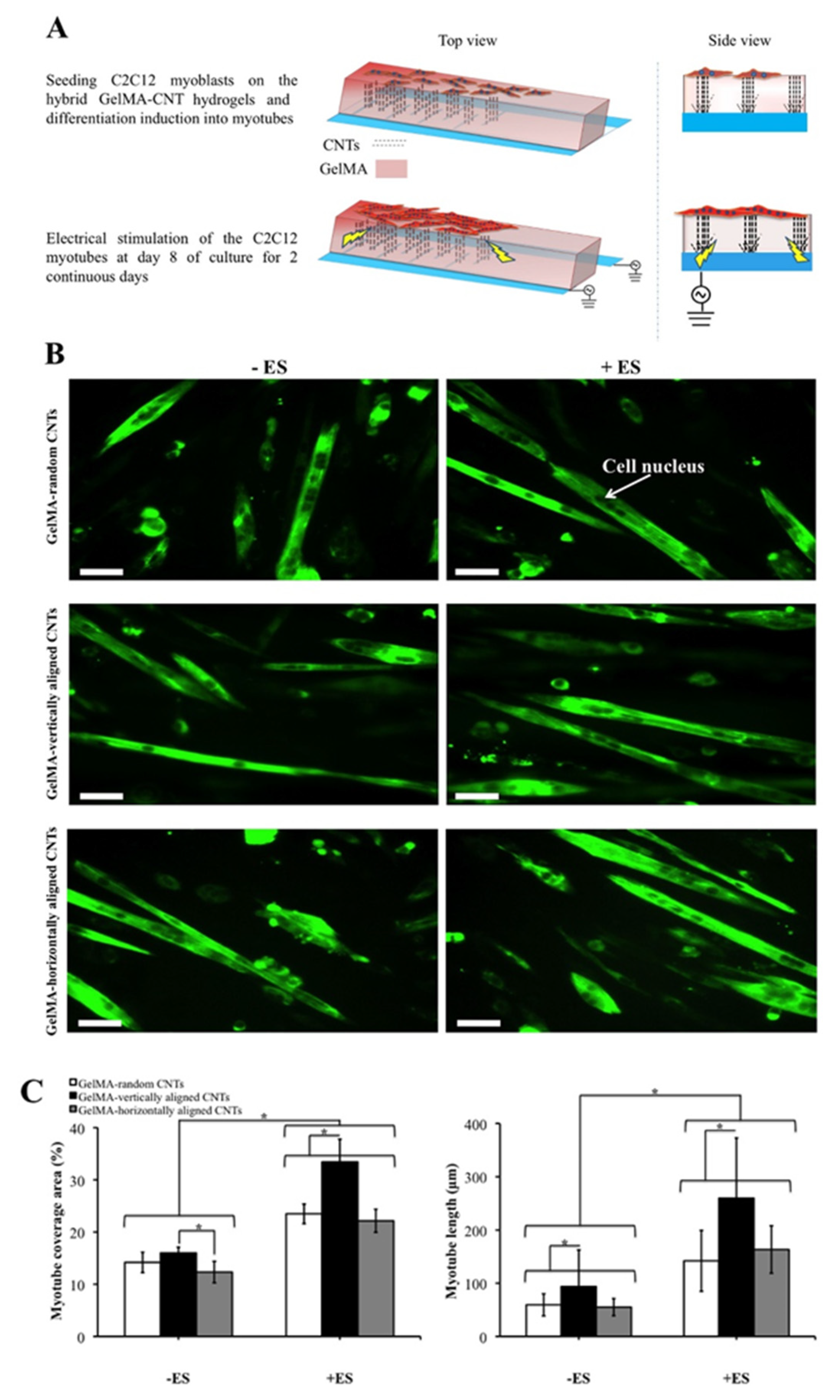
Figure 1. Differentiation of C2C12 myoblasts on GelMA−0.3 mg/mL carbon nanotube (CNT) hydrogels and characterization of the C2C12 myotubes obtained under electrical stimulation (ES). (A) Schematic representation of the procedure used to produce and electrically stimulate C2C12 myotubes. (B) Immunostaining of the fast skeletal myosin heavy chain in the C2C12 myotubes fabricated on hybrid methacrylated gelatin (GelMA)-random CNT, GelMA-vertically aligned CNT and GelMA-horizontally aligned CNT hydrogels with and without ES application (indicated as +ES and −ES, respectively) on day 10 of culture. Cell nuclei within the C2C12 myotubes were obvious after the staining procedure. The ES parameters were as follows: a voltage of 8 V, a frequency of 1 Hz and a duration of 10 ms. The scale bars represent 50 μm. (C) Quantification of the myotube coverage area and myotube length of the C2C12 myotubes fabricated on hybrid GelMA-random CNT, GelMA-vertically aligned CNT and GelMA-horizontally aligned CNT hydrogels with and without ES on day 10 of culture. Data in part (C) are presented as mean ± standard deviation obtained from at least 40 myotubes of 2 independent experiments. Asterisks indicate significant differences between samples (* p < 0.05). Reproduced from [50].
Actuators 09 00137 g001
Figure 2. Single step and label-free neutrophil sorting using Dean Flow Fractionation (DFF) microfluidic technology. (A) Experimental workflow for neutrophil isolation and phenotyping in type 2 diabetes mellitus (T2DM) patients. Blood samples are lysed and processed using the 2-inlet, 4-outlet spiral microdevice for efficient size-based neutrophil sorting. The purified neutrophils are used for in vitro cell rolling assay in a microchannel functionalized with E-selectin, as well as shape measurement (neutrophil circularity). (B) Schematic illustration of DFF separation principle. Under the influence of Dean vortices, small cellular constituents (platelets and lysed red blood cells (RBCs)) and free haemoglobin migrate laterally towards inner wall and back to outer wall due to Dean drag forces (FD (yellow arrows)). Larger leukocytes experience additional strong inertial lift forces (FL (red arrows)) and due to the strong dependence of FL and FD on cell size, larger neutrophils/monocytes (10–12 μm) focus closer to the inner wall and are sorted into outlet 2 while smaller lymphocytes (~7–8 μm) are collected at outlet 3. Outlet 4 is used for removal of platelets, lysed RBCs, and free haemoglobin. Reproduced from [81].
Actuators 09 00137 g002
Figure 3. Representation of cell type-specific capture onto a surface using antibodies. Blue cells represent target cells, red cells represent non-target cells, yellow circle represents the dissociation group. Reproduced from [94].
Actuators 09 00137 g003
Figure 4. Development of surfactant-nanotube complexes to fabricate nanostructured biomaterial surfaces for flow-based tumor cell capture assays. (A) Mixing and adsorption of sodium dodecanoate (NaL) surfactant to halloysite nanotubes (HNT) to create surfactant-nanotube complexes (NaL-HNT). (B) Stability of NaL-HNT and HNT dispersions (1.1 wt %) 24-h postmixing and adsorption. (C) Fabrication of nanostructured biomaterial surfaces. Polyurethane (PU) flow device surfaces coated with poly-l-lysine (PLL) to immobilize NaL-HNT and HNT. E-selectin (ES) is then adsorbed to HNT-coated surfaces, and tumor cells are perfused over surfaces at physiologically relevant flow rates to enable tumor cell capture. CTC: circulating tumor cell. Reproduced from [91].
Actuators 09 00137 g004
Figure 5. (A) Schematics of a plasmonic micropillar platform for the cell force measurement. Each polymer micropillar tip is embedded with a single gold nanosphere that serves as a strong and point-source-like light source for precision position tracking. (B) Microscopy optical images which show that gold microdisks transformed into gold nanospheres after pulse laser annealing. (C) Scanning electron microscopy (SEM) images show micropillars with transferred gold nanospheres. (D) Focused ion beam (FIB) images show that these transferred gold nanospheres are physically anchored inside the pillar and have an oblate spheroid shape with a long axis of 473 nm and a short axis of 268 nm. Platinum (Pt) coating shown in the images is for FIB imaging and not a part of the original pillar structure. Modified from [113].
Actuators 09 00137 g005
Table 1. Examples of non-specific cell type biointerfaces that promote adhesion.
Base Adhesion LayerDescribed 1 Dimensions of GrowthTarget CellsCellular Detection Method 2Described 1 Potential ApplicationsRef.
Alginate3DMouse MSCFluorescent imagingCell encapsulation, pharmaceutical research, tissue engineering and regenerative medicine[33]
Chondroitin sulfate3DPorcine ACDNA quantification and fluorescent imagingCartilage tissue engineering and regenerative medicine[34]
Collagen3DHuman PCCFluorescence imagingDisease modelling and drug screening[35]
Cornstarch3DHuman osteosarcomaFluorescence imagingUnspecified[36]
Dextran3DRat BMSC and mouse EFFluorescence imagingCell encapsulation[32]
Extracellular matrix3DHuman FH, human FSIC, human GS, human LDC, human SIC, and mouse SICBright field and fluorescence imagingDisease modelling, tissue regeneration and tissue repair[37]
Elastin3DRat ASMCFluorescence imagingArtificial vascular graft[38]
Fibrinogen3DHuman cardiomyocytesFluorescence imaging and scanning electron microscopyTissue engineering and tissue regeneration[39]
Fibronectin3DHuman BOSC, human CEC, and human FFFluorescence and phase contrast imagingWound dressing[40]
Gelatin3DMouse SCTFFluorescence imagingDrug delivery and tissue engineering[41]
Hyaluronic acid3DMouse EFFluorescence imagingRegenerative medicine and tissue engineering[19]
Laminin2DHuman PSC-derived neuronsFluorescence imagingRegenerative medicine[42]
Matrigel3DHuman PCFluorescence and phase contrast imagingDisease modelling[43]
Oxygen plasma 32DRat ADSCFluorescence imagingTissue engineering[44]
Poly-D-lysine2DHuman ESCFluorescence imagingDisease modelling[45]
Poly-L-lysine3DRabbit marrow-derived MSCBright field imagingTissue engineering and tissue regeneration[46]
Poly(ethylene glycol)3DHuman BGC, human DF, rat AGPC, and rat PICFluorescence imagingCell delivery and tissue engineering[20]
Poly(2-hydroxyethyl methacrylate)3DHuman MSCFluorescence imagingDrug delivery and tissue engineering[47]
Poly(N-isopropylacrylamide)2DMouse myoblastBright field and fluorescence imagingElectronics for cell culture[48]
Poly(vinyl alcohol)3DHuman BOSCFluorescence imagingTissue engineering[49]
1 “Described” indicates the use or possible use of the material, as stated by the authors in the referenced literature. 2 The Cellular Detection Method column only includes methods that were performed solely to confirm cell viability, presence or distribution of the cells in/on the base adhesion layer. 3 “Oxygen plasma” is a treatment method applied to the substrate material; cells do not bind directly onto the oxygen plasma. 2D = two-dimensional, 3D = three-dimensional, AC = auricular chondrocyte, ADSC = adipose-tissue-derived stromal cells, AGPC = adrenal gland pheochromocytoma cell, ASMC = aortic smooth muscle cell, BMSC = bone marrow mesenchymal stem cell, BOSC = bone osteosarcoma cell, BGC = brain glioblastoma cell, CEC = cervical epithelial cell, DF = dermal fibroblast, EF = embryo fibroblast, ESC = embryonic stem cell, FF = foreskin fibroblast, FH = fetal hepatocyte, FSIC = fetal small intestinal cell, GS = gastric cell, KF = kidney fibroblast, LDC = liver duct cell, MSC = mesenchymal stem cell, PCC = prostate cancer cell, PC = prostate cell, PIC = pancreas insulinoma cell, PSC = pluripotent stem cell, SCTF = subcutaneous connective tissue fibroblast, SIC = small intestinal cell.
Table 3. Examples of cell type-specific biointerfaces used for cell capture.
Recognition Element TypeTarget Cell(s)Described 1 Potential Applications(s)Coupling Method/Linker 2SubstrateCellular Detection Method 3Ref.
Amine plasma polymerBovine EC, human keratinocyte, human SF, mouse myoblasts and rat VSMCRegenerative medicineN/AN/AFluorescence imaging[82]
AntibodyEPC 4Biomedical devicesS-(11-Trichlorosilylundecanyl)-benzenethiosulfonate316L stainless steelN/A[83]
AntibodyHuman EC and human VSMCImplantable materialsPolyethylenimie + heparin + chitosanPoly(ethylene terephalate)Fluorescence imaging[84]
AntibodyMouse LCMedical diagnostic and prognosticPolyethylene glycol + poly(amidoamine) dendrimersEpoxy-functionalized glassFluorescence imaging[85]
AntibodyMouse BM and mouse spleenCell therapy, immune therapy and regenerative medicineBiotin + avidin + desthiobiotinPolyethylene filmFluorescence imaging[86]
AntibodyMouse BMCell therapySingle stranded DNA + single stranded DNAPolyethylene filmFluorescence imaging[87]
AntibodyHuman EKMedical diagnostics and implantable materialsSingle stranded DNA + single stranded DNA + streptavidin + biotinCyclic olefin polymer filmFluorescence imaging[88]
AntibodyHuman EKMedical diagnostics and implantable materialsSingle stranded DNA + single stranded DNA + streptavidin + biotinPolycarbonate filmFluorescence imaging[88]
AntibodyHuman BCC, human CCC, human HCC, human NSCLCC and human PCCMedical diagnostics and monitoringThiol + DNA + biotin + avidin + biotinGold-plated PDMSFluorescence imaging[89]
DNA (aptamer)Human SMC, human UVEC and rat MSC-derived EPCImplantable materialsPlasma polymerized allylamine316L stainless steelFluorescence imaging and QCM-D[90]
Protein (E-selectin)Human BA, human BC, human CA and human LAMedical diagnosticsSodium dodecanoateHalloysite nanotubesFluorescence and non-fluorescence imaging[91]
1 “Described” indicates the use or possible use of the material, as stated by the authors in the referenced literature. 2 “Coupling Method/Linker” represents either the intermediary molecule between the substrate and recognition element or the reagents required to produce the intermediary molecule. 3 The Cellular Detection Method column only includes methods that were performed solely to confirm cell viability, presence or distribution of the cells in/on the base adhesion layer. 4 The authors state that their method would be suitable for the capture of EPC, but do not use any cell types in their paper to demonstrate this. BCC = breast cancer cell, CCC = cervical carcinoma cell, BM = bone marrow, EC = endothelial cell, EK = embryonal kidney EPC = endothelial progenitor cell, HCC = hepatocellular carcinoma cells, LC = lung cancer, MSC = mesenchymal stem cell, NSCLCC = non-small-cell lung cancer cells, PCC = prostate cancer cell, PDMS = polydimethylsiloxane, QCM-D = quartz crystal microbalance with dissipation, SF = skin fibroblast, SMC = smooth muscle cell, UVEC = umbilical vein endothelial cell, VSMC = vascular smooth muscle cell.
Publisher’s Note: MDPI stays neutral with regard to jurisdictional claims in published maps and institutional affiliations.