Simple Summary
This study was performed to increase the understanding of the capacity of horse skin to detect pain when directly compared to human skin. The study focused on gluteal skin where horses are most often struck with whips during racing. The study was designed to inform the debate surrounding the use of whip strikes in horse racing where there is increasing pressure on the global racing industry to justify whip use. At the core of the debate is the question—do horses experience pain when being whipped? The study used microscopic studies of skin from 10 deceased humans and 20 euthanased horses to explore any differences between the species in their skin structure and nerve supply. The results revealed no significant difference between humans and horses in either the concentration of nerve endings in the outer pain-detecting layer of skin (epidermis) or in the thickness of this layer. In horses, this layer was deeper on the right than on the left. The collagen layer (dermis) of skin which is not involved in pain detection was significantly thinner in humans than in horses. These findings show that, although horse skin is thicker overall than human skin, the part of the skin that is thicker does not insulate them from pain that is generated during a whip strike, and that humans and horses have the equivalent basic anatomic structures to detect pain in the skin.
Abstract
The current project aims to build on knowledge of the nociceptive capability of equine skin to detect superficial acute pain, particularly in comparison to human skin. Post-mortem samples of gluteal skin were taken from men (n = 5) and women (n = 5), thoroughbreds and thoroughbred types (mares, n = 11; geldings, n = 9). Only sections that contained epidermis and dermis through to the hypodermis were analysed. Epidermal depth, dermal depth and epidermal nerve counts were conducted by a veterinary pathologist. The results revealed no significant difference between the epidermal nerve counts of humans and horses (t = 0.051, p = 0.960). There were no significant differences between epidermal thickness of humans (26.8 µm) and horses (31.6 µm) for reference (left side) samples (t = 0.117, p = 0.908). The human dermis was significantly thinner than the horse dermis (t = −2.946, p = 0.007). Epidermal samples were thicker on the right than on the left, but only significantly so for horses (t = 2.291, p = 0.023), not for humans (t = 0.694, p = 0.489). The thicker collagenous dermis of horse skin may afford some resilience versus external mechanical trauma, though as this is below the pain-detecting nerve endings, it is not considered protective from external cutaneous pain. The superficial pain-sensitive epidermal layer of horse skin is as richly innervated and is of equivalent thickness as human skin, demonstrating that humans and horses have the equivalent basic anatomic structures to detect cutaneous pain. This finding challenges assumptions about the physical capacity of horses to feel pain particularly in comparison to humans, and presents physical evidence to inform the discussion and debate regarding the ethics of whipping horses.
1. Introduction
The use of whips in horse racing is increasingly being questioned on ethical, welfare [1,2,3,4,5,6,7,8,9,10,11], social sustainability [10], and legal grounds [8]. Racing industry proponents argue that “the whip is used for safety (of both rider and horse) or to encourage the horse to perform to its best when in contention”, and that horses in many jurisdictions are protected from pain by whip padding and rules that govern whip use [12]. Some industry commentators have asserted that racing horses are immune to any pain from whip strikes chiefly because they are adrenalinised [13]. Others point out that horses may be physically brutal with one another in social interactions or conflict [14] and have therefore evolved to be robust and stoic. At the core of the debate is a fundamental question—do horses experience pain when whipped?
It is broadly accepted in science and society that most animals experience pain when struck. This is reflected by current societal norms, ethics, and laws that prohibit the striking or hitting animals. However, this protection is not extended to certain animal species—these being almost exclusively large domestic species—including the horse. It is reasonable to question why the assumption exists, and crucially, what scientific evidence exists to demonstrate that horses do or do not feel pain when being whipped.
Assumptions that horses are insensitive to pain may be influenced by historic factors and colloquialisms. The term “thick-skinned”, to denote insensitivity, has been in the English lexicon for over 400 years [15]. In parallel, the scientific community previously taxonomically defined and grouped certain species by having thick skin. The term “pachyderm” (from the Greek “pachys”, meaning “thick” and “derma”, meaning “skin”) long grouped elephants, hippopotamus, horses, pigs, and other large species together [16].
In recent decades, limited scientific data have become available that document the full skin thickness of horses, with it reportedly ranging between 1.2 to 7 mm. [17,18]. This is indeed considerably thicker than full thickness human skin, reported at 1.2 mm thick on average in multiple studies [19,20,21], with a range of 0.52 to 2.0 mm [21]. However, interpreting the skin thickness as a whole as an indicator of skin sensitivity fails to account for the complexity of the skin organ and, crucially, that detection of pain by mammalian skin overwhelmingly occurs in the very superficial layer of the skin (the epidermis), where the source of the painful stimulus (e.g., a strike or a heat source) comes into contact with these cells. Pain receptors of the skin are, in the vast majority, found in the epidermis, terminating in the mid to superficial epidermis [22,23]. These are fine, non-myelinated fibrils that detect damage to epidermal cells [22]. There may also be some dermal structures that have potential to act as nociceptors under some circumstances, such as thermal noxious stimulation [24] and following experimental selective ablation of epidermal nociceptors [25].
Reported data on horse epidermal thickness are sparse, though a mean epidermal thickness of 53 μm has been reported [26]. A recent study reported depth of equine epidermal from the thorax as 29 μm and from the limbs as 46 μm [27]. In comparison, four studies of human epidermal thickness found a mean epidermal depth of 51.2, 52.2, 53.0, and 91.8 μm, respectively [19,20,21,28]. What these data suggested is that where it counts, in terms of pain sensitivity, horse skin may be no more meaningfully thick than human skin. However, because each prior study used variable methods to measure skin, the existing data across the two species could not be reliably compared.
Skin thickness aside, the capacity of skin to detect pain must also depend on the presence and density of the microscopic neural structures of the epidermis. Pain is a complex and primordial physiological function key to survival because it provides a powerful incentive to avoid tissue damage. The basic and absolute need for pain detection is reflected in the remarkable evolutionary conservation of nociception from metazoa to mammals [29]. There is no evidence of significant differences in the neural structures involved in nociception between mammalian species [30]. However, there are gaps in our detailed knowledge concerning presence and density of cutaneous nociceptive fibres in many animal species, including horses.
The capacity of the skin to detect pain normally is recognized to have a relationship with the number or density of nerve fibres in the epidermis, as demonstrated by rare diseases in humans that cause insensitivity to pain [31,32], and the use of intra-epidermal nerve counts to diagnose peripheral sensory neuropathies [23,33]. However, despite the long-standing debate surrounding capacity of horses to experience pain, there are no known reports in the scientific literature that describe and quantify intra-epidermal nerves in the horse.
This study aims to address these knowledge gaps to move us closer to an informed understanding of the capacity of the horse to experience cutaneous pain. This work has been conducted in part to inform the debate surrounding the use of whip strikes in the global horse racing industry, a debate which may become integral to the sustainability of the industry as it moves into the future.
The current project aimed to build on our knowledge of the nociceptive structures of equine skin, particularly in comparison to human skin. The project was designed to test three hypotheses derived from anecdotal approaches to the ability of horses to experience acute superficial pain. First, that horses have thicker epidermal skin layers than humans. Second, that horses have thicker dermal skin layers than humans. Third, that horses have fewer cutaneous nociceptive neurological structures than humans. The current study focused on Thoroughbred horses and horses of Thoroughbred type because of the relevance of the Thoroughbred to racing.
2. Materials and Methods
2.1. Ethics Approval
This work was approved by Macquarie University Human Anatomy Governance Committee (project number 2019-4). Animal Ethics Committee approval was not required for the use of tissues retrieved post-mortem.
2.2. Horse Skin Acquisition
A team of three personnel (one team leader, O.J., and two graduate students) attended an export abattoir to collect the equine tissues for this project. They identified Thoroughbreds and Thoroughbred types (n = 20) that were to be processed in a batch. No history was available for the horses. Targeted horses were sexed (mares, n = 11; geldings, n = 9) and marked with adhesive stickers. After euthanasia, their age was estimated from their dentition and recorded (see Table 1). Skin samples were removed using a scalpel from both the left and right gluteal region and added to container of paraformaldehyde phosphate buffer solution, labelled with the identify code of the horse. They were stored for a minimum of 30 days at room temperature prior to trimming.

Table 1.
The distribution age ranges of men (n = 5), women (n = 5), mares (n = 11), and geldings (n = 9), from which six sections were taken.
2.3. Human Skin Acquisition
Human skin was sourced from the Surgical Skills and Anatomy Centre (SSAC), Faculty of Medicine Health and Health Sciences, Macquarie University, NSW, Australia. Samples were acquired from ten fresh frozen human cadavers (men, n = 5; women, n = 5) and the age of the cadavers recorded (see Table 1). Skin samples were removed from both the left and right gluteal region, and added to a container of paraformaldehyde phosphate buffer solution, labelled with the identify code of the cadaver. They were stored for a minimum of 30 days at room temperature prior to trimming.
While every attempt was made to obtain three samples from each side of each subject, in some cases this was not possible. Missing data were as follows: nerve count data from horse sample #126, both sides (age quartile 1); nerve count, epidermal and dermal data from human sample #44,7 right side (age quartile 3); and nerve count data from human sample #453, both sides (age quartile 3). The problem of missing data was addressed statistically by use of a mixed model with a random effect structure, rather than the solely fixed effect model that had been planned. Mixed models are a method considered appropriate for unbalanced designs and for repeated measures such as was encountered in this data-set.
2.4. Histology Processing and Staining
All samples were submitted to a National Association of Testing Authorities (NATA) accredited histology laboratory for sample trimming and slide preparation. Skin from both horses and humans was trimmed into full thickness sections (epidermis to hypodermis inclusive) and placed in cassettes. Samples were embedded in paraffin wax, sectioned at 16 µm and stained with hematoxylin and eosin (HE) using established histological techniques. Slides were then cover-slipped using the adhesive dibutylphthalate polystyrene xylene.
2.5. Photographic Images
Photographic images of the horse skin sections stained with hematoxylin and eosin were taken on a BX-51 microscope, using a Jenoptik Gryphax Kapella camera (Jenoptik Group, Jena, Germany). A full thickness (epidermis to hypodermis) image was taken in the centre of each skin section (three on each slide). Images were taken using the panoramic function in the Jenoptik software (Jenoptik Group, Jena, Germany). Each image was 1100 pixels in width, and each pixel represented 1.05887 μm in real distance. Photographic images of the human skin sections stained with hematoxylin and eosin were taken on a CX43 microscope (Olympus, Shinjuku-ku, Tokyo, Japan), using an Olympus EP50 camera (Olympus, Shinjuku-ku, Tokyo, Japan). A full thickness (epidermis to hypodermis) single image or two sequential images were taken in the centre of each skin section (three on each slide), using the EPview software (Olympus, Shinjuku-ku, Tokyo, Japan). Each image was 1100 pixels in width, and each pixel represented 1.18181 μm in real distance. All images were saved as JPEG files, and in total 6 photographs were taken of skin from each horse or human; three from each side of the body. An image of the epidermis and superficial dermis was taken in three sequential regions of the skin section, excluding areas of stain precipitation or tissue folds. Each image was 2080 pixels in width and each pixel was 0.58140 μm in real distance. All images were saved as JPEG files and, in total, six photographs were taken of skin from horse or human; three from each side of the body.
2.6. Analysis of H&E Sections
All images were analysed by a specialist veterinary pathologist using Motic Images Plus software (Motic, Kowloon, Hong Kong). Only sections that contained epidermis and dermis through to the hypodermis were analysed. One human sample that did not contain epidermis despite rotation and re-embedding of the tissue and was excluded from analysis. The width of each photographic image was confirmed as 1100 pixels using PixelZoomer software (Matthias Schütz; www.pixelzoomer.com) and each pixel represented 2.9 μm in the photographic software. The image was orientated with the epidermis parallel to the short axis of the image and the area of the epidermis and dermis was measured using the Motic Images Plus freehand tool. In skin samples with invaginations of hair follicles (horse skin), the natural contour of the epidermis was followed across the ostia. In samples with anastomosing rete ridges (human skin), the natural contour of the epidermis was followed. The average depth of the epidermis and dermis was then calculated in microns by dividing the area by the width of the image and corrected for the number of microns per pixel.
2.7. Immunohistochemistry Protocol
Paraffin embedded blocks were submitted to a diagnostic histology laboratory for PGP9.5 immunohistochemistry. Three micrometre sections were cut from each block, and three sections were placed on each slide. Immunohistochemical staining PGP9.5 was performed using the Leica Bond III automated staining platform (Leica Biosystems, Melbourne, VIC, Australia). Heat-mediated antigen retrieval (100 °C) was used at pH 9 for 30 min. The primary antibody (Anti-PGP9.5, Polyclonal Rabbit) was incubated for 30 min at ambient temperature. Bond Polymer Refine Red Detection (DS9390) was used with the standard Leica protocol.
2.8. Analysis of IHC Sections
The project used standardised and recognized methodology for quantifying intra-epidermal and dermal innervation, as used by the European Federation of Neurological Societies (EFNS) in the assessment of peripheral neuropathies [33]. Morphometric methods were used to quantify the density of neural structures in the epidermis as a count of intra-epidermal nerve fibres per mm of epidermis. The EFNS guidelines [33] were followed to count each nerve fibre crossing the basement membrane at the dermal–epidermal junction as a single unit. Nerve fibres branching in the epidermis were counted as one unit, nerves that split below the basement membrane were counted as two units, nerve fibres that failed to cross the basement membrane or were present in the epidermis but do not cross the basement membrane were not counted. Each fibre to be counted was indicated with a marker, with a total count being recorded for each examined section of skin.
2.9. Statistical Analysis
Epidermal depth (µm), dermal depth (µm) and nerve count (epidermal BM/mm), were modelled using a linear mixed effect model, with log transformation of epidermal depth and nerve count because of improved diagnostic plotting following such transformation. The model included the interaction between Species (Horse or Human) and Sex (Female or Male), as well as the age of the subject (the age was split into species quartiles due to the mean and variance differences between species) and the side (Right or Left) from which the sample was taken, both nested within species. Additional to these fixed effects, a fixed section number effect (First, Second and Third) was included to correct for any biases caused by the viewing order of samples and a random individual effect to account for individual variation). Models were fit using R Statistical software [34], making use of the “lme4” package [35]. Statistical significance was accepted at p < 0.05.
3. Results
3.1. Overview
Histological examination of horse and human skin revealed similar anatomical architecture with clear demarcation between the epidermis, dermis and hypodermis and shared fundamental histological features. The epidermis of stratified squamous epithelium contained the stratum basale embedded with variable numbers of pigmented cells on a basement membrane, stratum granulosum, stratum spinosum and stratum corneum. The superficial papillary dermis extended from the epidermal basement membrane and consisted of loosely separated collagen bundles that surrounded primary hair follicles and associated adnexal glandular tissue. The deeper reticular dermis extended from the follicular–adnexal units and consisted of more tightly packed collagen bundles. The hypodermis contained a predominance of adipose tissue with loose strands of collagen. Observation differences between the horse and human skin included greater density of follicular-adnexal units in the horse. While both equine and human epidermis form distinct rete ridges at the dermo-epidermal junction, these tended to be thinner and deeper with occasional anastomosis noted in human skin (see Figure 1).
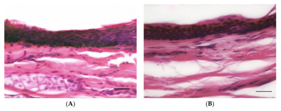
Figure 1.
Representative histologic images of horse (A) and human (B) epidermis and superficial dermis of gluteal skin illustrating comparable epidermal thickness (Haematoxylin and Eosin, 400×, 16 μm sections. Bar = 20 μm).
3.2. Epidermal Thickness
Plots of the data showed some signs of a right skew, so introduction of a logarithmic transformation was decided upon. The logarithmic boxplots are presented for reference purposes (see Figure 2).
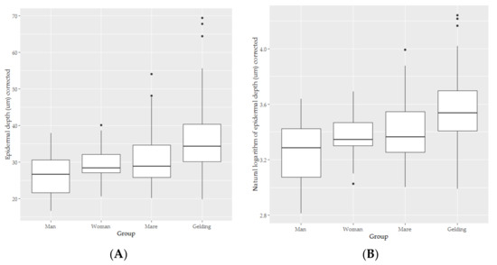
Figure 2.
The box-and-whisker plots for the epidermal depth (A) and natural logarithm of epidermal depth (B) of skin from men (n = 5), women (n = 5), mares (n = 11) and geldings (n = 9).
The marginal R2 and conditional R2 for the epidermal thickness model were 0.216 and 0.585, respectively, indicating that both fixed effects (marginal R2) and fixed and random effects considered together (conditional R2) account for substantial proportions of the variance in epidermal depth. The individual random effect for the epidermis model accounted for 47% of the variance. The fixed effect coefficients calculated for this model appear in Table 2.
Table 2.
The fixed effect coefficients calculated for a linear mixed model exploring the epidermis of men (n = 5), women (n = 5), mares (n = 11) and geldings (n = 9). Significant findings appear in bold.
No significant differences were found between humans and horses for left sides samples (t = 0.117, p = 0.908, see Figure 3). Humans had skin 0.185 units thinner on the log scale which corresponds to approximately 16.9% thinner.
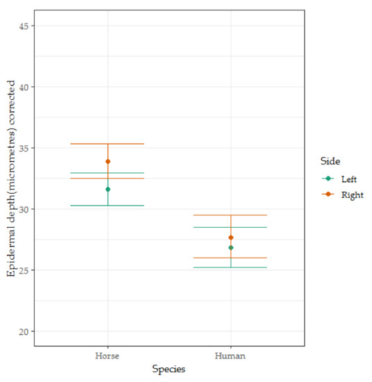
Figure 3.
The box-and-whisker plots for the corrected epidermal depth of skin from men (n = 5), women (n = 5), mares (n = 11) and geldings (n = 9). Coefficients and error bars are shown on the original measurement scale for ease of interpretation. This figure illustrates the significant interaction effect found between the side from which the sample was taken and the species from which the sample was taken.
3.3. Dermal Thickness
There was some suggestion of negative skew (see Figure 4), but the diagnostic residual plots were adequate.
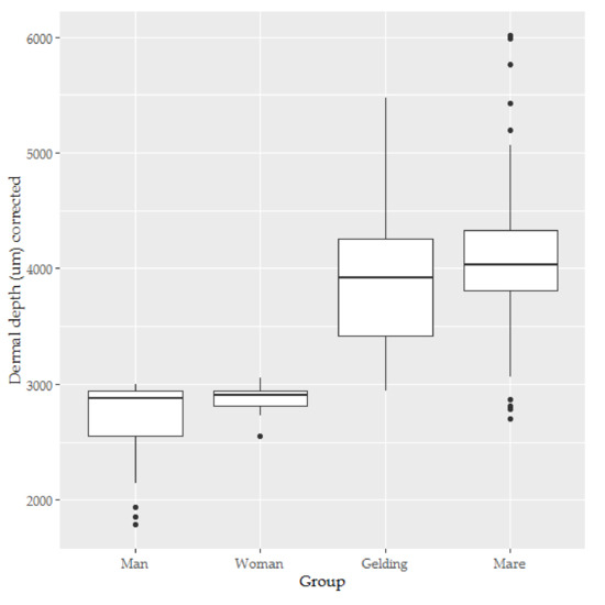
Figure 4.
The box-and-whisker plots for the dermal depth of skin from men (n = 5), women (n = 5), mares (n = 11) and geldings (n = 9).
The marginal R2 and conditional R2 for the dermal thickness model were 0.548 and 0.867, respectively, indicating that both fixed effects (marginal R2) and fixed and random effects considered together (conditional R2) account for substantial proportions of the variance in dermal depth. The individual random effect for the dermis model accounted for 71% of the variance. The fixed effect coefficients calculated for this model appear in Table 3.
Table 3.
The coefficients calculated for a linear mixed model exploring the dermis of men (n = 5), women (n = 5), mares (n = 11) and geldings (n = 9). Significant findings appear in bold.
The human dermis was significantly thinner than the horse dermis (t = −2.946, p = 0.007, see Figure 5).
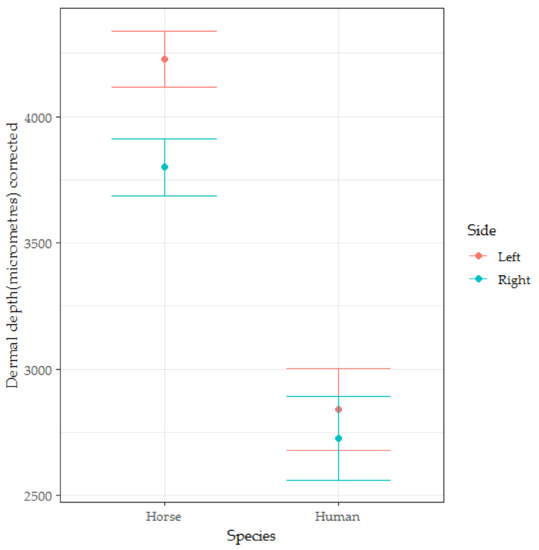
Figure 5.
The box-and-whisker plots for the dermal depth of skin from men (n = 5), women (n = 5), mares (n = 11) and geldings (n = 9). Coefficients and error bars are shown on the original measurement scale for ease of interpretation. This figure illustrates the significant interaction effect found between the side from which the sample was taken and the species from which the sample was taken.
3.4. Epidermal Nerve Count
Typical examples of the results of the immunohistochemistry studies are shown in Figure 6. The distributions of nerve counts scores appear in Figure 7.
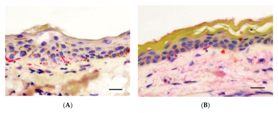
Figure 6.
Representative skin sections stained with immunohistochemistry using anti-PGP9.5 antibodies from Horse (A) and Human (B) skin. Images include epidermis and superficial dermis. Nerve endings are selectively stained with a red dye (*). Sections illustrate the finding that epidermal nerve endings are found in equivalent density across horse and human skin (anti-PGP9.5 immunohistochemistry, 400× magnification. Bar = 20 μm).
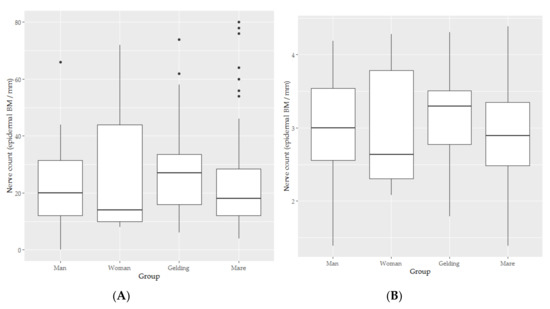
Figure 7.
The box-and-whisker plots for the epidermal nerve count (A) and natural logarithm of epidermal depth (B) of skin from men (n = 5), women (n = 5), mares (n = 11) and geldings (n = 9).
Some concerning shapes on the QQ and Scale location plots were found for in the residuals of the linear mixed model of the untransformed data, but were ameliorated with log transformation, apparently due to the presence of true zeros and positive skew in the original data.
The marginal R2 and conditional R2 for the model (see Table 4) of the transformed nerve count data (see Figure 7, panel B) were 0.042 and 0.716 respectively, indicating that both fixed effects (marginal R2) were relatively unimportant for this model but random individual effect still accounted for substantial proportions of the variance in epidermal nerve count, in this case, 70% of the variance. The relative unimportance of the fixed effects, which include Species compared to the thickness of skin layers is interesting to note. The coefficients calculated for this model appear in Table 4.
Table 4.
The coefficients calculated for a linear mixed model exploring the epidermal nerve count of men (n = 5), women (n = 5), mares (n = 11) and geldings (n = 9). Significant findings appear in bold.
There was no significant difference between human and horse nerve counts (t = 0.051, p = 0.960, see Figure 8).
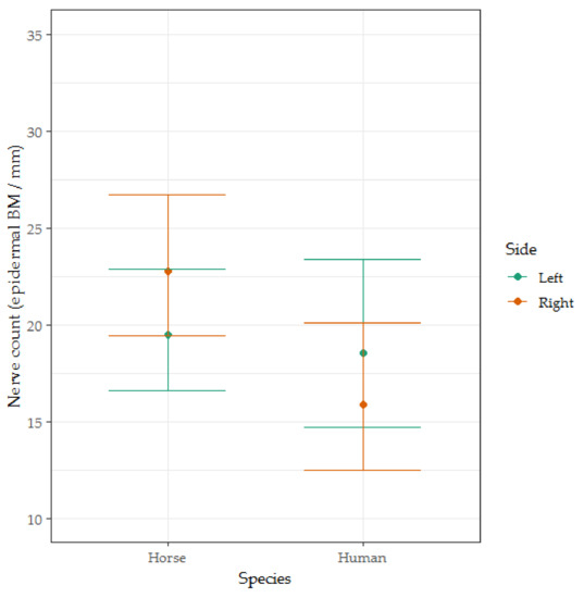
Figure 8.
The box-and-whisker plots for the epidermal nerve count of men (n = 5), women (n = 5), mares (n = 11) and geldings (n = 9). Coefficients and error bars are shown on the original measurement scale for ease of interpretation. This figure illustrates the significant interaction effect found between the side from which the sample was taken and the species from which the sample was taken.
4. Discussion
The results revealed no significant differences between epidermal thickness of humans and horses for left-sided (reference) samples. This refutes our first hypothesis, that horses have thicker epidermal skin layers than humans. There was mild asymmetry of the thickness of the horse epidermis between left and right sides, and the right horse epidermis was therefore marginally thicker than right human epidermis. This is an unusual finding and is discussed below. As was expected, the horse dermis was significantly thicker than the human dermis, supporting our second hypothesis. The structural integrity of the relatively thick equine dermis may withstand forceful impacts better than human dermis. However, the facts of cutaneous innervation with respect to nociception demonstrate that dermal thickness cannot be a significant factor in skin sensitivity. There were no significant differences between the epidermal nerve counts of humans and horses, refuting the third hypothesis that horses have fewer dermal nociceptive neurological structures than humans.
Together, these findings indicate that horse skin is virtually indistinguishable from human skin with respect to the basic anatomical structures relevant to cutaneous pain detection. These observations must refresh old assumptions made about the capacity of the “thick-skinned” horse to experience pain in comparison to humans.
It is recognized that pain is an enormously complex process, with huge amount of modulation of nociceptive signalling between noxious stimulation at the skin and the perception of pain in the higher centres of the central nervous. The current study does not attempt to assume, based on these findings, that the end-experience of pain in the horse is the same as in humans, as such a comparison is currently outside the capability of science, even when comparing the experience of pain between two humans. However, this does mean that broad similarity between species permits valid comparisons about pain perception, even in the absence of absolute equivalence of skin structures.
An observational study of horses racing in Australia [4] found 83% of whip strikes caused indentations of the skin of the horses whipped, and comparative studies in mice and humans showed such deformation is likely to be detected by cutaneous nociceptors [36], as did a recent study in horses [37]. Fundamentally, if an anti-nociceptive/analgesic state arises in all racing horses, it begs the question that if the whip has lost its salience, then why use it at all? Recent evidence indicates that certain distinct stress-related, affective states are characterised by enhanced, not reduced pain perception [38,39,40,41].
Repeated whipping is common in racing even though the IFHA forbids “using the whip with excessive frequency” [42]. The accumulated damage from successive strikes is likely to create more pain and inflammation. This assumption is based on the results of a feline study [43] in that repeated application reduced the thresholds for nociceptor activation and enhanced the duration of their responses. In Australia, racing thoroughbreds may be struck no more than five times prior to the final 100 m but after this point they can be struck at the jockey’s discretion [44]. This can equate to a strike per stride and effectively this means that 18 strikes per race are within the rules [13].
Forceful impact to equine skin may occur outside racing and other forms of equestrian activity. It is accepted that horses can cause severe injuries to one another, for example in disputes over focal resources such as food [14]. However, an important distinction to make in this context is that they are not being exposed to harm or injury by commission of an act by a human that is of no benefit to the horse.
In the current study, the horse epidermis was deeper on the right than on the left- and right-sided horse epidermis was thicker than human epidermis (which showed a non-significant trend towards similar left–right asymmetry). Bilateral asymmetry of epidermal thickness is a highly unexpected finding. The authors are not aware of reported asymmetry for measurements of normal skin thickness (epidermis or dermis) in any human or mammalian study. Localised or regional thickening of the epidermis may occur in pathologic states, and is known as epidermal hyperplasia or acanthosis. Epidermal thickening occurs in response to chronic insult or injury to the epidermis [45]. Thinning of the epidermis can occur in pathological states. However, these are generally restricted to cutaneous endocrinopathies and the epidermal thinning in these conditions is bilaterally symmetrical. Other pathological or physiological causes cannot be entirely excluded. There is evidence that horses tend to use their left eye when approaching potential threats [46], which could arguably eventuate in a bias for left-sided injury. So, one might have predicted that the skin on the left side of horses needs to be stronger than that on the right side. An investigation into epidermal asymmetry in horse skin would be a worthwhile future study.
The role of pain in horse sports is part of the growing debate about what constitutes ethical equitation [7,47]. In a flight animal, such as the horse, being unable to resolve aversive cutaneous stimulation causes distress. Horses have evolved to run away from such stimuli since the most likely natural cause of such stimulation is contact from a predator. Repeated strikes of the whip in horses that are fatigued as they end a race [3] are likely to be distressing and cause suffering. The horses’ loss of agency is described in the literature as helplessness and repeated, inescapable, treatment of this sort is thought to lead to learned helplessness [48].
The concept of ethical equitation [1] advocates a three-step process for stakeholders who seek to retain the social license to operate [13]. It demands that we identify the causes of distress in the horses we ride, that we mitigate these stressors as much as possible and we justify the retention of those that cannot be mitigated. The current findings suggest the ethical future for racing is one in which whips are carried but not used for so-called encouragement. Fortunately, this model is already well established in the UK where apprentice jockeys compete in hands-and-heels series [49].
A minor limitation of the current study is that human cadavers were fresh frozen, while the equine tissues were processed immediately after euthanasia. It is possible, but highly unlikely, that this contributed to the similarity of the tissues. The imbalance between the numbers of human and equine samples was caused by the 2020 national lockdown in Australia that interrupted access to specimens from human anatomy classes. A further minor limitation is that we were unable to gather data on any potential structural difference in the hairs of horses and humans. It appears unlikely that hairs afford meaningful protection from the impacts of whips, given that whips cause cavitation waves through the skin and underlying tissues [50]. Studies of anaesthetized horses may assist in this domain and might also be helpful in revealing the thermographic responses of horse skin to whipping. Future adjunctive research should include histopathological examination of bilateral skin and subcutaneous tissues from horses which have died within seven days of a racing event. This could reveal any evidence of inflammation and repair at the site of recent whip strikes.
5. Conclusions
This primary aim of this project was to improve our understanding of the neuroanatomy of horse skin with respect to cutaneous pain sensation. The project achieved this aim through quantitative methods, using research material that can be openly accessed by interested parties. The results revealed no significant difference between the epidermal nerve counts of humans and horses. There were no significant differences between epidermal thickness of humans and horses for left side samples. The human dermis was significantly thinner than the horse dermis. The horse epidermis was deeper on the right than the left. These findings indicate that the superficial pain-sensitive epidermal layer of horse skin is as richly innervated and is of equivalent thickness as human skin, demonstrating that humans and horses have the equivalent key anatomical structures to detect cutaneous pain.
Author Contributions
Dermal-epidermal Conceptualization, L.T.; methodology, L.T.; M.S.; and P.M.; formal analysis, M.S. and B.W.; investigation, M.S.; resources, R.A., I.J., and O.J.; data curation, M.S.; B.W.; writing—Original draft preparation, L.T. and P.M.; writing—Review and editing, L.T., M.S., P.M., R.A., I.J., C.J., O.J., and B.W.; visualization, B.W.; supervision, M.S. and P.M; project administration, P.M.; funding acquisition, P.M. All authors have read and agreed to the published version of the manuscript.
Funding
This research was funded by RSPCA Australia. Open access publication was supported by the Sydney School of Veterinary Science and RSPCA Australia.
Acknowledgments
The first approach to this research question appeared on ABC TV’s Catalyst program. We are grateful to the abattoir management and staff for their help and cooperation during the collection of horse skin. Janos Tomka is warmly thanked for his help in obtaining the human tissues. We appreciate the input and contribution of the staff at Royal Prince Alfred Hospital for the histology and immunostaining work. We are also grateful to the individuals whose donated cadavers were the source of the human skin samples. Kevin Keay, Daniel Boorman and Michelle Gerke-Duncan are thanked for their advice and help in preparing and storing the horse samples.
Conflicts of Interest
The authors declare no conflict of interest.
References
- Jones, B.; McGreevy, P.D. Ethical equitation: Applying a cost-benefit approach. J. Vet. Behav. 2010, 5, 196–202. [Google Scholar] [CrossRef]
- McGreevy, P.D.; Oddie, C. Holding the whip hand-A note on the distribution of jockeys’ whip hand preferences in Australian Thoroughbred racing. J. Vet. Behav. 2011, 6, 287–289. [Google Scholar] [CrossRef]
- Evans, D.; McGreevy, P.D. An investigation of racing performance and whip use by jockeys in Thoroughbred races. PLoS ONE 2011, 6, e15622. [Google Scholar] [CrossRef]
- McGreevy, P.D.; Corken, R.A.; Salvin, H.; Black, C.M. Whip use by jockeys in a sample of Australian Thoroughbred races-An observational study. PLoS ONE 2012, 7, e33398. [Google Scholar] [CrossRef] [PubMed]
- Graham, R.; McManus, P. Changing human-animal relationships in sport: An analysis of the UK and Australian horse racing whips debates. Animals 2016, 6, 32. [Google Scholar] [CrossRef] [PubMed]
- Ban on Whip Use Would Be Positive for Racing, Suggests John Francome. Available online: http://horsetalk.co.nz/2015/11/12/ban-whip-use-positive-racing-francome/#axzz40UAtJE5b (accessed on 23 September 2020).
- McLean, A.N.; McGreevy, P.D. Ethical equitation: Capping the price horses pay for human glory. J. Vet. Behav. 2010, 5, 203–209. [Google Scholar] [CrossRef]
- Jones, B.; Goodfellow, J.; Yeates, J.; McGreevy, P.D. A Critical analysis of the British horseracing authority’s review of the use of the whip in horseracing. Animals 2015, 5, 138–150. [Google Scholar] [CrossRef] [PubMed]
- McGreevy, P.D.; Hawson, L.A.; Salvin, H.; McLean, A. A note on the force of whip impacts delivered by jockeys using forehand and backhand strikes. J. Vet. Behav. 2013, 8, 395–399. [Google Scholar] [CrossRef]
- Bergmann, I. Sustainability, thoroughbred racing and the need for change. Pferdeheilkunde Equine Med. 2015, 31, 490–498. [Google Scholar] [CrossRef]
- Heleski, C.R.; Anthony, R. Science alone is not always enough: The importance of ethical assessment for a more comprehensive view of equine welfare. J. Vet. Behav. 2012, 7, 169–178. [Google Scholar] [CrossRef]
- British Horseracing Authority. Responsible Regulation: A Review of the Use of the Whip in Horseracing. 2011. Available online: http://www.britishhorseracing.com/wp-content/uploads/2014/03/WhipReview.pdf (accessed on 24 September 2020).
- McGreevy, P.D.; McManus, P. Why Horse-Racing in Australia Needs a Social Licence to Operate. The Conversation, 3 November 2017. Available online: https://theconversation.com/why-horse-racing-in-australia-needs-a-social-licence-to-operate-79492 (accessed on 10 November 2020).
- McGreevy, P.D. Equine Behavior-A Guide for Veterinarians and Equine Scientists; W.B Saunders: London, UK, 2012. [Google Scholar]
- Porter, N. Thick-skinned. In Webster’s Revised Unabridged Dictionary of the English Language; C. & G. Merriam Co.: Springfield, MA, USA, 1913. [Google Scholar]
- Cuvier, G.; Vallentin, R.; Latreille, P.A.; MacMurtrie, H. The Animal Kingdom, Arranged According to Its Organization: Serving as a Foundation for the Natural History of Animals, and an Introduction to Contemporary Anatomy; G. Henderson: London, UK, 1834. [Google Scholar]
- Wakuri, H.; Mutoh, K.; Ichikawa, H.; Liu, B. Microscopic anatomy of the equine skin with special reference to the dermis. Okajimas Folia Anat. Jpn. 1995, 72, 177–183. [Google Scholar] [CrossRef] [PubMed]
- Volkering, M.E. Variation of Skin Thickness over the Equine Body and the Correlation between Skin Fold Measurement and Actual Skin Thickness. Doctoral Thesis, Utrecht University, Utrecht, The Netherlands, 2009. [Google Scholar]
- Southwood, W.F.W. The thickness of the skin. Plast Reconstr Surg 1995, 15, 423–429. [Google Scholar] [CrossRef] [PubMed]
- Artz, C.P.; Moncrief, J.A.; Pruitt, B.A. Burns: A team Approach; W. B. Saunders Company: Philadelphia, PA, USA, 1979; pp. 24–44. [Google Scholar]
- Lee, Y.; Hwang, K. Skin thickness of Korean adults. Surg. Radiol. Anat. 2002, 24, 183–189. [Google Scholar] [CrossRef] [PubMed]
- Schummer, A.; Wilkens, H.; Vollmerhaus, B.; Habermehl, K.H. Skin and cutaneous organs of the horse. In The Circulatory System, the Skin, and the Cutaneous Organs of Domestic Mammals; Schummer, A., Wilkens, H., Vollmerhaus, B., Habermehl, K.H., Eds.; Springer: New York, NY, USA, 1981; p. 537. [Google Scholar]
- Ebenezer, G.J.; Hauer, P.; Gibbons, C.; McArthur, J.C.; Polydefkis, M. Assessment of epidermal nerve fibers. J. Neuropathol. Exp. Neurol. 2007, 66, 1059–1073. [Google Scholar] [CrossRef]
- Rosenberger, D.C.; Binzen, U.; Treede, R.-D.; Greffrath, W. The capsaicin receptor TRPV1 is the first line defense protecting from acute non damaging heat: A translational approach. J. Transl. Med. 2020, 18, 28. [Google Scholar] [CrossRef]
- Mense, S. Anatomy of nociceptors. In The Senses: A Comprehensive Reference; Bushnell, M.C., Smith, D.V., Beauchamp, G.K., Firestei, S.J., Eds.; Academic Press: New York, NY, USA, 2008; pp. 11–41. [Google Scholar]
- Talukdar, A.H.; Calhoun, M.L.; Stinson, A.W. Microscopic anatomy of the skin of the horse. Am. J. Vet. Res. 1972, 33, 2365–2390. [Google Scholar]
- Jørgensen, E.; Lazzarini, G.; Pirone, A.; Jacobsen, S.; Miragliotta, V. Normal microscopic anatomy of equine body and limb skin: A morphological and immunohistochemical study. Ann. Anat. 2018, 218, 205–212. [Google Scholar] [CrossRef]
- Sato, K.; Sugibayashi, K.; Morimoto, Y. Species differences in percutaneous absorption of nicorandil. J. Pharm. Sci. 1991, 80, 104–107. [Google Scholar] [CrossRef]
- Smith, E.S.J.; Lewin, G.R. Nociceptors: A phylogenetic view. J. Comp. Physiol. A 2009, 195, 1089–1106. [Google Scholar] [CrossRef]
- National Research Council (US) Committee on Recognition and Alleviation of Pain in Laboratory Animals. Pain in Research Animals: General Principles and Considerations; National Academies Press: Washington, DC, USA, 2009. [Google Scholar]
- Dyck, P.J.; Mellinger, J.F.; Reagan, T.J.; Horowitz, S.J.; McDonald, J.W.; Litchy, W.J.; Daube, J.R.; Fealey, R.D.; Go, V.L.; Kao, P.C.; et al. Not ‘indifference to pain’ but varieties of hereditary sensory and autonomic neuropathy. Brain 1983, 106, 373–390. [Google Scholar] [CrossRef]
- Nolano, M.; Crisci, C.; Santoro, L.; Barbieri, F.; Casale, R.; Kennedy, W.; Wendelschafer-Crabb, G.; Provitera, V.; Di Lorenzo, N.; Caruso, G. Absent innervation of skin and sweat glands in congenital insensitivity to pain with anhidrosis. Clin. Neurophysiol. 2000, 111, 1596–1601. [Google Scholar] [CrossRef]
- Lauria, G.; Cornblath, D.R.; Johansson, O.; McArthur, J.C.; Mellgren, S.I.; Nolano, M.; Rosenberg, N.; Sommer, C. EFNS guidelines on the use of skin biopsy in the diagnosis of peripheral neuropathy. Eur. J. Neurol. 2005, 12, 747–758. [Google Scholar] [CrossRef] [PubMed]
- RC Team. R: A Language and Environment for Statistical Computing; R Foundation for Statistical Computing: Vienna, Austria, 2019; Available online: http://www.R-project.org/ (accessed on 10 November 2020).
- Bates, D.; Mächler, M.; Bolker, B.; Walker, S. Fitting linear mixed-effects models usinglme4. J. Stat. Softw. 2015, 67, 1–48. [Google Scholar] [CrossRef]
- Lewin, G.R.; Moshourab, R. Mechanosensation and pain. J. Neurobiol. 2004, 61, 30–44. [Google Scholar] [CrossRef]
- Taylor, P.; Crosignani, N.; Lopes, C.; Rosa, A.C.; Luna, S.P.L.; Filho, J.N.P. Mechanical nociceptive thresholds using four probe configurations in horses. Vet. Anaesth. Analg. 2016, 43, 99–108. [Google Scholar] [CrossRef]
- Sluka, K.A.; Rasmussen, L.A. Fatiguing exercise enhances hyperalgesia to muscle inflammation. Pain 2010, 148, 188–197. [Google Scholar] [CrossRef] [PubMed]
- Chen, X.; Green, P.G.; Levine, J.D. Stress enhances muscle nociceptor activity in the rat. Neuroscience 2011, 185, 166–173. [Google Scholar] [CrossRef] [PubMed]
- Dinal, O.A.; Levinel, J.D.; Greenl, P.G. Enhanced cytokine-induced mechanical hyperalgesia in skeletal muscle produced by a novel mechanism in rats exposed to unpredictable sound stress. Eur. J. Pain 2011, 15, 796–800. [Google Scholar] [CrossRef]
- Rivat, C.; Becker, C.; Blugeot, A.; Zeau, B.; Mauborgne, A.; Pohl, M.; Benoliel, J.-J. Chronic stress induces transient spinal neuroinflammation, triggering sensory hypersensitivity and long-lasting anxiety-induced hyperalgesia. Pain 2010, 150, 358–368. [Google Scholar] [CrossRef]
- International Federation of Horseracing Authorities. International Agreement on Breeding, Racing and Wagering. Available online: http://www.horseracingintfed.com/resources/2018Agreement.pdf (accessed on 14 April 2018).
- Bessou, P.; Perl, E.R. Response of cutaneous sensory units with unmyelinated fibers to noxious stimuli. J. Neurophysiol. 1969, 32, 1025–1043. [Google Scholar] [CrossRef]
- Racing Australia. Australian Rules of Racing. 2020. Available online: https://www.racingaustralia.horse/FreeServices/Australian_Rules_Of_Racing.aspx (accessed on 20 September 2020).
- Hargis, A.M.; Myers, S. The Integument. In Pathologic Basis of Veterinary Disease, 6th ed.; Zachary, J.F., Ed.; Elsevier: St. Louis, MO, USA, 2018; p. 1022. [Google Scholar]
- LaRose, C.; Richard-Yris, M.-A.; Hausberger, M.; Rogers, L.J. Laterality of horses associated with emotionality in novel situations. Laterality 2006, 11, 355–367. [Google Scholar] [CrossRef] [PubMed]
- McLean, A.N.; McGreevy, P.D. Horse-training techniques that may defy the principles of learning theory. J. Vet. Behav. 2010, 5, 187–195. [Google Scholar] [CrossRef]
- Hall, C.; Goodwin, D.; Heleski, C.; Randle, H.; Waran, N. Is there evidence of learned helplessness in horses? J. Appl. Anim. Welf Sci. 2008, 11, 249–266. [Google Scholar] [CrossRef] [PubMed]
- Thompson, K.; McManus, P.; Stansall, D.; Wilson, B.J.; McGreevy, P.D. Is whip use important to thoroughbred racing integrity? What stewards’ reports reveal about fairness to punters, jockeys and horses. Animals 2020, 10, 1985. [Google Scholar] [CrossRef] [PubMed]
- Barington, K.; Jensen, H.E. The impact of force on the timing of bruises evaluated in a porcine model. J. Forensic Leg. Med. 2016, 40, 61–66. [Google Scholar] [CrossRef]


Publisher’s Note: MDPI stays neutral with regard to jurisdictional claims in published maps and institutional affiliations. |
© 2020 by the authors. Licensee MDPI, Basel, Switzerland. This article is an open access article distributed under the terms and conditions of the Creative Commons Attribution (CC BY) license (http://creativecommons.org/licenses/by/4.0/).