Communication

Molecular Detection of Arthropod-Borne Pathogens in Eurasian Badgers (Meles meles) from the United Kingdom

by
Lisa Guardone
1,
Valentina Virginia Ebani
1,*,
Ranieri Verin
2,
Simona Nardoni
1,
Antonio Consolazione
1,
Malcolm Bennett
3 and
Francesca Mancianti
1
1
Department of Veterinary Sciences, University of Pisa, Viale delle Piagge 2, 56124 Pisa, Italy
2
Department of Comparative Biomedicine and Food Science, University of Padova, Viale dell’Università 16, 35020 Legnaro, Padova, Italy
3
School of Veterinary Medicine and Science, University of Nottingham, Nottingham LE12 5RD, UK
*
Author to whom correspondence should be addressed.
Animals 2020, 10(3), 446; https://doi.org/10.3390/ani10030446
Submission received: 30 January 2020 / Revised: 24 February 2020 / Accepted: 3 March 2020 / Published: 6 March 2020

Abstract

:

Simple Summary

Blood-sucking arthropods can cause infections in domestic and wild animals, as well as in humans by transmitting pathogens while blood feeding. During the first part of the twenty-first century, diseases caused by arthropods, including ticks, have increased significantly in Europe. In this study, the presence of different types of pathogens in badgers from Great Britain was evaluated by means of molecular techniques. While no bacteria were found, a blood parasite (Babesia sp.) was identified in half of the investigated animals. The same parasite has been reported in badgers from Spain, Scotland, China and Hungary, and recently also in a wild cat in Bosnia Herzegovina, a wolf in Italy and hunting dogs in Hungary, showing its widespread occurrence and potential presence in different host species. The impact on the health of these hosts and also of other wild carnivores needs to be further investigated.

Abstract

Arthropod-borne diseases (ABD) are of increasing interest in veterinary and public health. Eurasian badgers (Meles meles) are known to harbor a wide range of pathogens, but information on their role as ABD reservoirs and their potential epidemiological relevance is limited. This study aimed to investigate the occurrence of arthropod-borne pathogens, specifically piroplasmids and the bacteria Anaplasma phagocytophilum, Ehrlichia canis, Coxiella burnetii, Francisella tularensis and Bartonella spp., in badgers from Great Britain (GB). Blood and heart samples from 18 badgers were examined using PCR and sequencing. A neighbour-joining (NJ) phylogram was also produced. Nine animals tested positive for Babesia sp., while none of the samples was positive for the investigated bacteria. The sequences obtained clustered with other sequences of Babesia sp. from badgers from GB and elsewhere, including China, Hungary, Spain and Italy, showing a widespread distribution of this parasite in badgers. Badger-associated Babesia DNA was also found recently in a wild cat in Bosnia Herzegovina, in a wolf in Italy and in dogs in Hungary. Further investigations are needed to understand the epidemiology of this putative pathogen and its impact on the health of wild and domestic carnivores.

1. Introduction

The incidence and distribution of arthropod-borne diseases have increased in Europe in recent decades as a result of increased awareness, global travel and trade and climatic and other environmental changes [1]. The main arthropod vectors threatening public health across Europe are mosquitoes, sandflies and ticks [2]. As an example, the UK reported the non-native mosquito Aedes albopictus, a known vector for dengue and chikungunya virus, in 2016. Culex modestus, the mosquito vector for the West Nile virus, has been detected since 2010. For tick-borne diseases, the changing distribution of Ixodes ricinus, the Lyme borreliosis vector, has been reported in parallel with an increase in the human reports of Lyme disease [3]. Among arthropod-borne diseases, tick-borne diseases (TBD) are particularly important to both human and veterinary medicine [4], and possibly to conservation biology. Several TBD have been reported in wildlife [5]. The adaptability of some species of wild carnivores to urban and peri-urban environments [6], and their role in the life cycle of pathogens of public health and veterinary interest is becoming increasingly evident [7].
The Eurasian (European) badger (Meles meles, Mustelidae family, Carnivora order) is widespread throughout Europe, and while it prefers mixed pasture and broadleaf woodlands, its generalist, highly adaptable nature enables it to exploit a wide variety of habitats [8,9]. Although relatively abundant in European countries, lower densities have been reported in some areas, sometime associated with hunting and “baiting”, and strong population fluctuations have been observed. This has prompted the implementation of conservation regulations and protected status since the 1970s and 1980s in several countries, such as the United Kingdom, Ireland, Spain, Portugal, Italy, Belgium, the Netherlands, Albania, Greece, Estonia, Luxemburg and Hungary [8]. The badger is Britain’s largest indigenous carnivore species and it has its own protection legislation, which is additional to general wildlife conservation legislation. Combined figures from England, Wales and Scotland suggest an estimated population of approximately 300,000 badgers [10].
Badgers may have an epidemiological role in infections of veterinary and public health importance, including the rabies virus, and most notably, bovine tuberculosis (Mycobacterium bovis), [11,12] as well as a range of other viruses, bacteria and endo- and ectoparasites [13]. Transmission, and therefore the veterinary and medical significance, may be enhanced in the case of relatively high population densities [9]. In the past decades, similarly to red foxes [14], populations of badgers have increased and adapted to peri-urban and urban environments across Europe [15], thus representing a possible source of pathogens for domestic animals and humans [16].
Among relevant arthropod-borne pathogens are Babesia, Cytauxzoon and Theileria species, all related hemoprotozoans collectively known as piroplasmids or piroplasms. Their life cycle is indirect; asexual reproduction occurs inside erythrocytes in the vertebrate hosts while the sexual phase takes place in ixodid ticks, which ingest and subsequently transmit the parasites while taking a blood meal [17]. An especially wide vertebrate host range is known for Babesia species, such that all vertebrate species may be susceptible to infection as long as they are also a host for Babesia-vector ticks. Various ticks have been reported as piroplasmid vectors. For Babesia and Theileria the most important genera are Rhipicephalus, Dermacentor, Haemaphysalis, Boophilus, Ixodes and Hyalomma, depending on the host species and the geographical area [17]. Cytauxzoon is mainly transmitted by Dermacentor and Amblyomma in North America, while its vector remains unknown in Europe [18].
Other tick-borne infections reported in Europe include Ehrlichia canis, which infects dogs, cats and wild canids and is usually transmitted by the brown dog-tick Rhipicephalus sanguineus, although other ticks may be involved in the transmission [19]. Anaplasma phagocytophilum is responsible for disease mainly in humans, horses and dogs, but it may also infect several wild mammal species and it is largely transmitted by ixodid ticks [20]. Coxiella burnetii may be transmitted by various tick species, although humans and animals usually acquire the infection through ingestion of raw milk and dairy products or inhalation of contaminated aerosols [21]. Francisella tularensis is able to infect several species of mammals, mainly lagomorphs, and is transmitted through hematophagous arthropods, but also through contact with infected animals as well as by drinking contaminated water [22], although it has never been reported in Great Britain. Finally, some Bartonella species may also be transmitted by ticks, as well as by other hematophagous arthropods [23], although tick competence is debated [24]. Bartonellae may infect domestic animals, mainly dogs and cats, even though wild mammals are frequently involved in the epidemiology of these agents [25].
This study investigated the occurrence of piroplasmids and arthropod-borne bacteria Anaplasma phagocytophilum, Ehrlichia canis, Coxiella burnetii, Francisella tularensis and Bartonella spp. in Eurasian badgers from Northwest England (GB).

2. Materials and Methods

2.1. Sampling and Post Mortem Examination

Eighteen carcasses of Eurasian badgers (8 males and 10 females), of different ages, were collected between 2016 and 2017 from Cheshire (Northwest England), United Kingdom. In all cases, the cause of death was attributed to road traffic collision. Carcass collection and sampling was carried out as part of a broader national project, focused on the prevalence of bovine tuberculosis in badgers.
The study had formal ethical approval from both the University of Liverpool and the University of Nottingham.
Ticks were not collected, as soon after the death of the host the majority of the ectoparasites drop off the hosts, making ticks generally not evaluable at post mortem examination.
A full post mortem examination was carried out on each subject within 24 h of receiving the carcasses. In the meantime, these were stored at 4 °C. Sex and age (juveniles or adults) of each carcass was recorded and based on body mass and dental wear as previously described [12]. Both blood and heart samples were collected from eight of the badgers, while for 10 animals only the heart was available (Table 1).

2.2. Molecular Analysis

Total DNA was extracted from blood (200 µL) and heart (10 mg) samples using the DNeasy Blood and Tissue Kit (QIAGEN), following the manufacturer’s blood and tissue protocols, respectively.
The total DNA obtained was submitted to different PCR protocols, targeting different gene fragments and using different primer pairs to screen for DNA of piroplasmids, A. phagocytophilum, E. canis, C. burnetii, F. tularensis and Bartonella spp. (Supplementary Materials, Table S1). For some pathogens, such as A. phagocytophilum and E. canis, a nested PCR protocol was used. PCR amplifications were performed using Wonder Taq (Euroclone, Milano, Italy) and an automated thermal cycler (Gene-Amp PCR System 2700, Perkin Elmer, Norwalk, CT, USA). Sterile distilled water instead of DNA was used as negative control. DNA extracted from slides for indirect immunofluorescent assay coated with A. phagocytophilum, E. canis, C. burnetii, F. tularensis and Bartonella henselae (Fuller Laboratories, Fullerton, CA, USA), and a known positive sample of B. annae previously sequenced [26] were included as positive controls.
PCR products were analysed by electrophoresis on 2% agarose gel stained with GelRed® Nucleic Acid Gel Stain (Biotium). SharpMass™ 100 Plus Ladder (Euroclone, Milano, Italy) was used as a DNA marker. PCR products of the expected length and with a sufficient concentration were forward and reverse Sanger sequenced by an external company. Nucleotide sequences were analysed using Bioedit version 7.0.9 [27]. Adjustments were made after visual checking and consensus sequences were compared against those deposited in GenBank by using the National Center for Biotechnology Information (NCBI) Basic Local Alignment Search Tool (BLAST). A neighbour-joining (NJ) phylogram was constructed using Bioedit and MEGA-X software.

3. Results

At post mortem examination all the subjects showed good to fair body condition with adequate adipose tissue deposits (Table 1). No gross lesions, including lesions suggestive of bovine tuberculosis, other than road traffic related injuries, were observed.
DNA of arthropod-borne bacteria was not detected in any sample. DNA of piroplasmids was amplified in seven out of the eight blood samples examined, as well as in two additional heart samples (Table 1). Thus, overall, piroplasmid DNA was successfully amplified in 50% of the sampled badgers. Seven amplicons from blood samples were sequenced (the positive heart samples could not be sequenced due to their low concentration). All positive samples were identified as Babesia sp. In fact, all the sequences produced in the present study showed 100% identity values with sequences of Babesia sp. “type A” isolated from a badger in China and in Spain [16], as well as with Babesia sp. isolated from Felis silvetris in Bosnia and Herzegovina [28]. Slightly lower identity values (99.81–99.79%) were observed with sequences of Babesia sp. “type A” isolated from badgers in the UK (Scotland) [10]. Details of the BLAST results, including the accession number of the sequences with the highest identity values are reported in Supplementary Materials, Table S2.
The NJ phylogram was constructed by aligning and trimming (to a final length of 455 bp) the seven sequences of the small subunit (18S) ribosomal RNA gene obtained in the present study, 19 sequences of badger-associated Babesia sp. and 44 other piroplasmids sequences already included in the phylogenetic analysis in recent works on Babesia in badgers [9,10]. All the sequences obtained in the present study clustered together, along with other Babesia sp. isolated from badgers from the UK (n = 1, deposited as “type A”), Italy (n = 3), Hungary (n = 2), Spain (n = 2, one of which deposited as “type A”) and China (n = 2, deposited as “type A”). Moreover, badger-associated Babesia sp. isolated from a wolf in Italy, from a wild cat in Bosnia Herzegovina and from a dog in Hungary also clustered in this group. All these sequences clustered separately from another cluster including four sequences from badgers (one from Spain, two from the UK and one from China) described as “type B” (Figure 1).

4. Discussion

None of the samples tested contained PCR-detectable A. phagocytophilum, E. canis, F. tularensis, C. burnetii, or Bartonella spp. As the number of samples tested was very small, any conclusion drawn from these results should be tentative. However, it is not surprising that E. canis and F. tularensis infection were not found as E. canis in British dogs has so far been largely (although not completely) limited to those with a history of travel to mainland Europe [29], and F. tularensis has never been reported in Great Britain, Ireland or Iceland [30]. A novel Ehrlichia sp. was found in a badger in Northern Spain [31] and isolates recently obtained from Italian badgers showed 99% similarity with it [32]. C. burnetii infection is endemic in British livestock, and seropositivity is common in people working with them. Infection often occurs without any obvious signs of disease in both animals and humans [33,34,35]. The role of wildlife in the epidemiology/ecology of Q-fever is unclear globally [36] and unknown in Great Britain, but the fact that the badgers in this study came from one of the densest dairy farming areas of Great Britain and none were infected suggests that they probably do not play a role in the ecology of the infection. A. phagocytophilum and Bartonella spp. infections are both found in domestic animals and wildlife in Great Britain and A. phagocytophilum was detected in 0.74% of ixodid ticks collected from British dogs [37]. However, as in this study, A. phagocytophilum was not detected in badgers in The Netherlands, Czech Republic, and Spain and only a low prevalence was observed among badgers in another study conducted in Belgium and The Netherlands, as well as in Italy, suggesting badgers do not play a significant epidemiological role in this infection ([32] and references therein). A previous survey found what appeared to be a novel Bartonella sp., most closely related to B. clarridgeiae, in 12% of badgers in northern Spain [38]. B. clarridgeiae infection is largely associated with cats and dogs, and more recently humans [39], so further investigation of a larger number of badgers would be interesting.
Tick-borne hemoparasites of the genus Babesia are reported to be the second most commonly found parasites in the blood of mammals after trypanosomes. Infection has been reported globally in a great variety of wild mammal species and some birds [17,40,41], as well as in badgers and other wildlife [5,16,26,42,43]. In recent reports, Babesia parasites have been molecularly characterised and the species’ descriptions in older studies have been questioned. Babesia from wildlife hosts may be distinguished as two types: parasites largely specific to a particular host, and babesias typically found in closely related domestic animals, e.g., B. bigemina in gazelles or B. canis in wolves [17]. According to the most recent molecular studies, Babesia spp. of carnivores can be divided into three groups: (i) the B. microti group infecting felids, canids, mustelids and procyonids; (ii) the prototheilerid group infecting felids, canids, herpestids and hyaenids; and (iii) the Babesia sensu stricto group infecting canids, procyonids, and ursids [44].
Babesia epidemiology in badgers has recently attracted the interest of research groups in various countries, as shown by the increasing number of publications in which variable prevalence values have been reported (Table 2). Although only a small number of badgers were tested in this study, the prevalence found is in broad agreement with the 70% prevalence found in 64 badgers sampled from East/Central Scotland. Prior to the work of Bartley et al. [10], which describes the first molecular identification of Babesia DNA from badgers in Britain, piroplasmosis were reported in this host in southwest England [45]. However, only low parasitemia of intraerythrocytic organisms resembling piroplasms were reported and these were not specifically identified. The vectors are likely Ixodes canisuga and I. hexagonus, badger ticks that are common in the area [46]. A subsequent study tested a total of 718 blood samples from 263 individual badgers over a three-year period, and found 77.2% individuals positive for B. missirolii (based on morphology) at least once over 3 years [47].
There are few reports of babesiosis in badgers from other countries, and they often describe only small-scale surveys (Table 2). As mentioned, the interest seems to have increased in the last decade following the report of a new piroplasmid in the Eurasian badger in Spain (accession number FJ225390) [42]. The species was shown to be distantly related to both Theileria/Babesia annae and a piroplasmid from Lontra canadensis and is considered as belonging to the group of the Babesia microti-like organisms found in the dog and small carnivores. Subsequent case reports and epidemiological studies reported the same type (named Babesia sp. “type A” by some authors) as well as different sequence types (including the genotype called Babesia sp. “type B” in some studies) occurring in other countries (Table 2). In particular, studies performed in Spain confirmed the presence of Babesia in badgers in that country [16]. Lately, Babesia sp. was identified in badgers in Hungary [9,44], Korea (although in this case without molecular investigation) [48] and very recently also in Italy [32,49]. In addition, a few Babesia sequences not referred to in any publication are also deposited in GenBank. These are derived from a badger from Xinjiang, China (Babesia sp. badger isolate Badger-1; MG799847.1) and from I. canisuga ticks collected from a red fox in Germany (Babesia sp. 4 NAN2012; JX679177.1).
Badger-associated Babesia sp. DNA has also been amplified from the blood of one European wild cat (Felis silvestris) in Bosnia and Herzegovina. This record, together with the finding of an Hepatozoon sp. type typical of the badger in F. silvestris, has been attributed to the sharing of burrows between badgers and other animals, which favours transmission of the potential vectors, especially nidicolous arthropods [28]. Additional epidemiological information may be obtained by investigating ticks rather than just vertebrate hosts. In Spain, a recent study found B. annae/T. annae in a pool of nymphs of I. canisuga from a badger, despite the badger not being detectably infected [7].
The high similarity of “type A” sequences obtained from Europe and China suggests this is a badger-adapted species/genotype [49]. Moreover, the finding of badger-associated Babesia sp. “type A” in a wild cat in Bosnia and Herzegovina [28], and recently in a grey wolf in Italy [49] and in six dogs in Hungary, one with relevant clinical signs [44], demonstrates the possibility of infection of a broad range of carnivore hosts and highlights the circulation of these species within the order Caniformia [32]. Walking in woodlands and hunting activities were identified as risk factors for dogs as these are associated with infestation with ticks [44].

5. Conclusions

This study confirms the widespread occurrence of badger-associated Babesia sp. (type A). As this and previous studies have mainly investigated a fragment of the 18 rRNA gene, further analysis of additional gene targets would be informative. Moreover, recent reports of the same badger-associated Babesia species/genotype in a wild cat, wolf and in clinically affected domestic dogs underlines the need for larger scale molecular surveys to investigate the potential risk posed by this putative pathogen for other wild carnivores as well as for domestic animals.
Even though the obtained results suggest that M. meles is not pivotal in the epidemiology of the other investigated arthropod-borne pathogens in the examined area, further investigation of a larger number of badgers could be useful to better understand their role in these infections.

Supplementary Materials

The following are available online at https://www.mdpi.com/2076-2615/10/3/446/s1, Table S1: Gene targets, amplicon length, primer pairs and PCR conditions used for the different pathogens, Table S2: Details of the results of the BLAST analysis (https://blast.ncbi.nlm.nih.gov/Blast.cgi) for the samples PCR positive for piroplasmids.

Author Contributions

Conceptualization, F.M. and V.V.E.; methodology, V.V.E. and L.G.; validation, R.V., M.B. and F.M.; formal analysis, L.G.; investigation, L.G., V.V.E., S.N. and A.C.; resources, F.M., R.V., M.B.; data curation, L.G.; writing—original draft preparation, L.G. and V.V.E.; writing—review and editing, R.V., M.B.; supervision, F.M. All authors have read and agreed to the published version of the manuscript.

Funding

This research received no external funding.

Conflicts of Interest

The authors declare no conflict of interest.

References

  1. Medlock, J.M.; Leach, S.A. Effect of climate change on vector-borne disease risk in the UK. Lancet Infect. Dis. 2015, 15, 721–730. [Google Scholar] [CrossRef]
  2. Baylis, M. Potential impact of climate change on emerging vector-borne and other infections in the UK. Environ. Health 2017, 16, 112. [Google Scholar] [CrossRef] [Green Version]
  3. Medlock, J.M.; Hansford, K.M.; Vaux, A.G.; Cull, B.; Gillingham, E.; Leach, S. Assessment of the public health threats posed by vector-borne disease in the United Kingdom (UK). Int. J. Environ. Res. Public Health 2018, 15, 2145. [Google Scholar] [CrossRef] [Green Version]
  4. Dantas-Torres, F.; Chomel, B.B.; Otranto, D. Ticks and tick-borne diseases: A One Health perspective. Trend Parasitol. 2012, 28, 437–446. [Google Scholar] [CrossRef]
  5. Alvarado-Rybak, M.; Solano-Gallego, L.; Millán, J. A review of piroplasmid infections in wild carnivores worldwide: Importance for domestic animal health and wildlife conservation. Parasites Vectors 2016, 9, 538. [Google Scholar] [CrossRef] [PubMed] [Green Version]
  6. Gehrt, S.D.; Riley, S.P.D.; Cypher, B.L. Urban Carnivores, Ecology, Conflict, and Conservation; John Hopkins University Press: Baltimore, MD, USA, 2010. [Google Scholar]
  7. Millán, J.; Proboste, T.; De Mera, I.G.F.; Chirife, A.D.; De La Fuente, J.; Altet, L. Molecular detection of vector-borne pathogens in wild and domestic carnivores and their ticks at the human–wildlife interface. Ticks Tick Borne Dis. 2016, 7, 284–290. [Google Scholar] [CrossRef] [PubMed]
  8. Piza Roca, C.; La Haye, M.J.J.; Jongejans, E. Environmental drivers of the distribution and density of the European badger (Meles meles): A review. Lutra 2014, 57, 87–109. [Google Scholar]
  9. Hornok, S.; Trauttwein, K.; Takács, N.; Hodžić, A.; Duscher, G.G.; Kontschán, J. Molecular analysis of Ixodes rugicollis, Candidatus Neoehrlichia sp.(FU98) and a novel Babesia genotype from a European badger (Meles meles). Ticks Tick Borne Dis. 2017, 8, 41–44. [Google Scholar] [CrossRef] [PubMed]
  10. Bartley, P.M.; Wilson, C.; Innes, E.A.; Katzer, F. Detection of Babesia DNA in blood and spleen samples from Eurasian badgers (Meles meles) in Scotland. Parasitology 2017, 144, 1203–1210. [Google Scholar] [CrossRef]
  11. Smith, G.C.; Wilkinson, D. Modelling disease spread in a novel host: Rabies in the European badger Meles meles. J. Appl. Ecol. 2002, 39, 865–874. [Google Scholar] [CrossRef]
  12. Sandoval Barron, E.; Swift, B.; Chantrey, J.; Christley, R.; Gardner, R.; Jewell, C.; McGrath, I.; Mitchell, A.; O’Cathail, C.; Prosser, A.; et al. A study of tuberculosis in road traffic-killed badgers on the edge of the British bovine TB epidemic area. Sci. Rep. 2018, 8, 17206. [Google Scholar] [CrossRef] [PubMed] [Green Version]
  13. Hancox, M. Parasites and infectious diseases of the Eurasian badger (Meles meles L.): A review. Mammal Rev. 1980, 10, 151–162. [Google Scholar] [CrossRef]
  14. Gloor, S.; Bontadina, F.; Hegglin, D.; Deplazes, P.; Breitenmoser, U. The rise of urban fox populations in Switzerland. Mamm. Biol. 2001, 66, 155–164. [Google Scholar]
  15. Judge, J.; Wilson, G.J.; Macarthur, R.; Delahay, R.J.; McDonald, R.A. Density and abundance of badger social groups in England and Wales in 2011–2013. Sci. Rep. 2014, 4, 3809. [Google Scholar] [CrossRef] [PubMed] [Green Version]
  16. Barandika, J.F.; Espí, A.; Oporto, B.; Del Cerro, A.; Barral, M.; Povedano, I.; García-Pérez, A.L.; Hurtado, A. Occurrence and genetic diversity of piroplasms and other apicomplexa in wild carnivores. Parasitol. Open 2016, 2, e6. [Google Scholar] [CrossRef] [Green Version]
  17. Schnittger, L.; Rodriguez, A.E.; Florin-Christensen, M.; Morrison, D.A. Babesia: A world emerging. Infect Genet Evol 2012, 12, 1788–1809. [Google Scholar] [CrossRef]
  18. Veronesi, F.; Ravagnan, S.; Cerquetella, M.; Carli, E.; Olivieri, E.; Santoro, A.; Pesaro, S.; Berardi, S.; Rossi, G.; Ragni, B.; et al. First detection of Cytauxzoon spp. infection in European wildcats (Felis silvestris silvestris) of Italy. Ticks Tick Borne Dis. 2016, 7, 853–858. [Google Scholar] [CrossRef]
  19. Sainz, Á.; Roura, X.; Miró, G.; Estrada-Peña, A.; Kohn, B.; Harrus, S.; Solano-Gallego, L. Guideline for veterinary practitioners on canine ehrlichiosis and anaplasmosis in Europe. Parasites Vectors 2015, 8, 75. [Google Scholar] [CrossRef] [Green Version]
  20. Stuen, S.; Granquist, E.G.; Silaghi, C. Anaplasma phagocytophilum—A widespread multi-host pathogen with highly adaptive strategies. Front. Cell. Infect. Microbiol. 2013, 3, 31. [Google Scholar] [CrossRef] [Green Version]
  21. Berri, M.; Rekiki, A.; Boumedine, K.S.; Rodolakis, A. Simultaneous differential detection of Chlamydophila abortus, Chlamydophila pecorum and Coxiella burnetii from aborted ruminant’s clinical samples using multiplex PCR. BMC Microbiol. 2009, 9, 130. [Google Scholar] [CrossRef] [Green Version]
  22. Ellis, J.; Oyston, P.C.; Green, M.; Titball, R.W. Tularemia. Clin. Microbiol. Rev. 2002, 15, 631–646. [Google Scholar] [CrossRef] [PubMed] [Green Version]
  23. Reis, C.; Cote, M.; Le Rhun, D.; Lecuelle, B.; Levin, M.L.; Vayssier-Taussat, M.; Bonnet, S.I. Vector competence of the tick Ixodes ricinus for transmission of Bartonella birtlesii. PLoS Negl. Trop. Dis. 2011, 5. [Google Scholar] [CrossRef] [PubMed] [Green Version]
  24. Telford, S.R., III; Wormser, G.P. Bartonella spp. transmission by ticks not established. Emerg. Infect. Dis. 2010, 16, 379. [Google Scholar] [CrossRef]
  25. De Sousa, K.C.M.; Do Amaral, R.B.; Herrera, H.M.; Santos, F.M.; Macedo, G.C.; De Andrade Pinto, P.C.E.; Barros-Battesti, D.M.; Machado, R.Z.; André, M.R. Genetic Diversity of Bartonella spp. in wild mammals and ectoparasites in Brazilian Pantanal. Microb. Ecol. 2018, 76, 544–554. [Google Scholar] [CrossRef] [PubMed] [Green Version]
  26. Ebani, V.V.; Rocchigiani, G.; Nardoni, S.; Bertelloni, F.; Vasta, V.; Papini, R.A.; Verin, R.; Poli, A.; Mancianti, F. Molecular detection of tick-borne pathogens in wild red foxes (Vulpes vulpes) from Central Italy. Acta Trop. 2017, 172, 197–200. [Google Scholar] [CrossRef] [PubMed]
  27. Hall, T.A. BioEdit: A User-Friendly Biological Sequence Alignment Editor and Analysis Program for Windows 95/98/NT; Nucleic acids symposium series; Oxford Academic: London, UK, 1999; Volume 41, No. 41; pp. 95–98. [Google Scholar]
  28. Hodžić, A.; Alić, A.; Duscher, G.G. High diversity of blood-associated parasites and bacteria in European wild cats in Bosnia and Herzegovina: A molecular study. Ticks Tick Borne Dis. 2018, 9, 589–593. [Google Scholar] [CrossRef] [PubMed]
  29. Wilson, H.E.; Mugford, A.R.; Humm, K.R.; Kellett-Gregory, L.M. Ehrlichia canis infection in a dog with no history of travel outside the United Kingdom. J. Small Anim. Pract. 2013, 54, 425–427. [Google Scholar] [CrossRef]
  30. Gürcan, Ş. Epidemiology of tularemia. Balkan Med. J. 2014, 31, 3–10. [Google Scholar] [CrossRef] [PubMed]
  31. Garcı́a-Pérez, A.L.; Oporto, B.; Espí, A.; Del Cerro, A.; Barral, M.; Povedano, I.; Barandika, J.; Hurtado, A. Anaplasmataceae in wild ungulates and carnivores in northern Spain. Ticks Tick Borne Dis. 2016, 7, 264–269. [Google Scholar] [CrossRef]
  32. Battisti, E.; Zanet, S.; Khalili, S.; Trisciuoglio, A.; Hertel, B.; Ferroglio, E. Molecular survey on vector-borne pathogens in alpine wild carnivorans. Front. Vet. Sci. 2020, 7, 1. [Google Scholar] [CrossRef] [Green Version]
  33. Halsby, K.D.; Kirkbride, H.; Walsh, A.L.; Okereke, E.; Brooks, T.; Donati, M.; Morgan, D. The epidemiology of Q fever in England and Wales 2000–2015. Vet. Sci. 2017, 4, 28. [Google Scholar] [CrossRef] [Green Version]
  34. Thomas, D.R.; Treweek, L.; Salmon, R.L.; Kench, S.M.; Coleman, T.J.; Meadows, D.; Morgan-Capner, P.; Caul, E.O. The risk of acquiring Q fever on farms: A seroepidemiological study. Occup. Environ. Med. 1995, 52, 644–647. [Google Scholar] [CrossRef] [PubMed] [Green Version]
  35. Valergakis, G.E.; Russell, C.; Grogono-Thomas, R.; Eisler, M.C.; Bradley, A.J. Coxiella burnetii in bulk tank milk of dairy cattle in south-west England. Vet. Rec. 2012, 171, 156. [Google Scholar] [CrossRef] [PubMed]
  36. Meerburg, B.G.; Reusken, C.B. The role of wild rodents in spread and transmission of Coxiella burnetii needs further elucidation. Wildl. Res. 2011, 38, 617–625. [Google Scholar] [CrossRef]
  37. Smith, F.D.; Wall, L.E.R. Prevalence of Babesia and Anaplasma in ticks infesting dogs in Great Britain. Vet. Parasitol. 2013, 198, 18–23. [Google Scholar] [CrossRef]
  38. Gerrikagoitia, X.; Gil, H.; García-Esteban, C.; Anda, P.; Juste, R.A.; Barral, M. Presence of Bartonella species in wild carnivores of northern Spain. Appl. Environ. Microbiol. 2012, 78, 885–888. [Google Scholar] [CrossRef] [Green Version]
  39. Vieira-Damiani, G.; De Paiva Diniz, P.P.V.; Pitassi, L.H.U.; Sowy, S.; Scorpio, D.G.; Lania, B.G.; Drummond, M.R.; Soares, T.C.B.; Barjas-Castro, M.D.L.; Breitschwerdt, E.B.; et al. Bartonella clarridgeiae bacteremia detected in an asymptomatic blood donor. J. Clin. Microbiol. 2015, 53, 352–356. [Google Scholar] [CrossRef] [Green Version]
  40. Soares, H.S.; Marcili, A.; Barbieri, A.R.; Minervino, A.H.; Moreira, T.R.; Gennari, S.M.; Labruna, M.B. Novel piroplasmid and Hepatozoon organisms infecting the wildlife of two regions of the Brazilian Amazon. Int. J. Parasitol. Parasites Wildl. 2017, 6, 115–121. [Google Scholar] [CrossRef]
  41. Young, K.M.; Corrin, T.; Wilhelm, B.; Uhland, C.; Greig, J.; Mascarenhas, M.; Waddell, L.A. Zoonotic Babesia: A scoping review of the global evidence. PLoS ONE 2019, 14. [Google Scholar] [CrossRef] [Green Version]
  42. Bartley, P.M.; Hamilton, C.; Wilson, C.; Innes, E.A.; Katzer, F. Detection of Babesia annae DNA in lung exudate samples from red foxes (Vulpes vulpes) in Great Britain. Parasites Vectors 2016, 9, 84. [Google Scholar] [CrossRef] [Green Version]
  43. Gimenez, C.; Casado, N.; Criado-Fornelio, Á.; De Miguel, F.Á.; Dominguez-Peñafiel, G. A molecular survey of Piroplasmida and Hepatozoon isolated from domestic and wild animals in Burgos (northern Spain). Vet. Parasitol. 2009, 162, 147–150. [Google Scholar] [CrossRef] [PubMed]
  44. Hornok, S.; Horváth, G.; Takács, N.; Kontschán, J.; Sz˝oke, K.; Farkas, R. Molecular identification of badger-associated Babesia sp. DNA in dogs: Updated phylogeny of piroplasms infecting Caniformia. Parasit Vectors 2018, 11, 235. [Google Scholar] [CrossRef]
  45. Peirce, M.A.; Neal, C. Piroplasmosis in British badgers (Meles meles). Vet. Rec. 1974, 94, 493–494. [Google Scholar] [CrossRef]
  46. Gallagher, J.; Nelson, J. Cause of ill health and natural death in badgers in Gloucestershire. Tuberculosis 1979, 10, 14–16. [Google Scholar]
  47. Macdonald, D.W.; Anwar, M.; Newman, C.; Woodroffe, R.; Johnson, P.J. Inter-annual differences in the age-related prevalences of Babesia and Trypanosoma parasites of European badgers (Meles meles). J. Zool. 1999, 247, 65–70. [Google Scholar] [CrossRef]
  48. Hong, S.H.; Kim, H.J.; Jeong, Y.I.; Cho, S.H.; Lee, W.J.; Kim, J.T.; Lee, S.E. Serological and molecular detection of Toxoplasma gondii and Babesia microti in the blood of rescued wild animals in Gangwon-do (Province), Korea. Korean J. Parasitol. 2017, 55, 207. [Google Scholar] [CrossRef] [Green Version]
  49. Santoro, M.; Auriemma, C.; Lucibelli, M.G.; Borriello, G.; D’Alessio, N.; Sgroi, G.; Veneziano, V.; Galliero, G.; Fusco, G. Molecular Detection of Babesia spp. (Apicomplexa: Piroplasma) in Free-Ranging Canids andMustelids from Southern Italy. Front. Vet. Sci. 2019, 6, 269. [Google Scholar] [CrossRef] [Green Version]
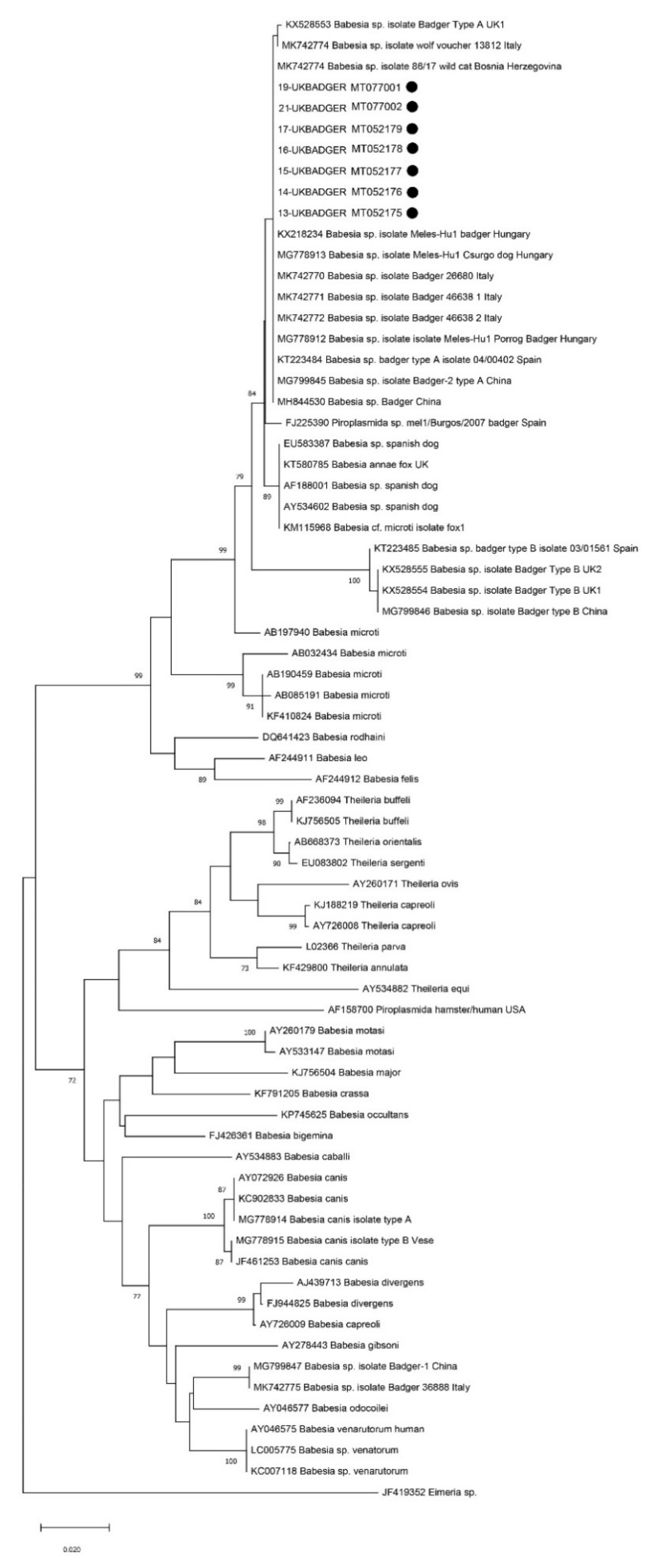
Figure 1. Neighbour-joining phylogram created with 7 sequences of the small subunit (18S) ribosomal RNA gene obtained in the present survey, 19 sequences of badger-associated Babesia sp. and 44 other piroplasmids sequences included in the phylogenetic analysis in recent works on Babesia sp. in badgers [9,10].
Animals 10 00446 g001
Table 1. Details of the sampled Eurasian badgers: sex, age class, weight, body condition score (BCS), status of the carcass at the time of sampling, availability of different tissues and results of the PCR for amplification of piroplasmid DNA. n.a.: not available.
NSexAge ClassWeight (kg)BCSCarcass StatusPCR—Piroplasmid Blood SamplesPCR—Piroplasmid Heart Samples
1FYoung7.64/5Goodn. a.Neg
2FAdult11.64/5Goodn. a.Neg
3FAdult12.83/5Goodn. a.Pos
4MAdult123/5Mildly autolyticn. a.Pos
5FJuvenile6.74/5Moderately autolyticn. a.Neg
6FAdult12.84/5Goodn. a.Neg
7FAdult14.14/5Goodn. a.Neg
8MAdult15.83/5Autolyticn. a.Neg
9MAdult10.14/5Goodn. a.Neg
10FAdult14.33/5Autolyticn. a.Neg
11MAdult10.34/5GoodPosPos
12MJuvenile10.53/5Very goodPosPos
13MJuvenile9.83/5GoodPosPos
14MAdult11.43/5GoodPosPos
15FJuvenile9.43/5GoodPosPos
16FAdult9.44/5GoodNegNeg
17FAdult11.63/5GoodPosPos
18MJuvenile8.32/5AutolyticPosPos
Table 2. Literature studies (since 2009) that report badger-associated Babesia sp. in Eurasian badgers (Meles meles) or in other carnivore species or ticks.
ReferencesGeographical AreaSpecies NPositive/Examined (Prevalence %)Babesia sp. Found
Battisti et al. 2020 [32]ItalyBadger (M. meles)4541/45 (91.1%)Badger-associated Babesia spp. type A and B; B. capreoli, Babesia sp. DO23163
Santoro et al. 2019 [49]ItalyBadger (M. meles)137/13 (53.8%)Two sequence types: badger-associated Babesia spp. from MK742770 to MK742773 and MK742775
Wolf (Canis lupus)131/13 (7.7%)Badger-associated Babesia spp. (MK742774)
Hodžić et al. 2018 [28]Bosnia and HerzegovinaEuropean wild cat (Felis silvestris silvestris)185.5%Badger-associated Babesia sp. (MF614153)
Bartley et al. 2017 [10]UK (Scotland)Badger (M. meles)64overall: 70.2%; Type A: 12·8% spleen and 38.3% blood; Type B: 19.1% spleen and 46.8% blood. Both type A and B sequences 10.6% spleen and 31.9% bloodBabesia Badger type A UK1 (KX528553), Badger type B UK1 (KX528554) and Badger type B UK2 (KX528555)
additional sequences registered under accession nr. From KY250472 to KY250477 *
Hornok et al. 2017 [9]HungaryBadger (M. meles)1case report (n = 1) Babesia sp. Meles-Hu1 (KX218234)
Ixodes rugicollis31/3 (33.3%)Babesia sp. Meles-Hu1
Hornok et al. 2018 [44]HungaryBadger (M. meles)55/5 (100%); Babesia sp. Meles-Hu1 (MG778912)
Dogs906/90 (6.7%);Babesia sp. Meles-Hu1 (MG778913)
Ixodes canisuga2718/27 (66.7%)Babesia sp. Meles-Hu1
Hong et al. 2017 [48]KoreaBadgers32/3 (66.7%)Babesia microti
Barandika et al. 2016 [16]SpainBadger (M. meles)12264/122 (52.2%)Babesia sp. badger type A (KT223484) and B (KT223485)
Millán et al. 2016 [7]SpainBadger (M. meles)30-
Gimenez et al. 2009 [43]SpainBadger (M. meles)51/5 (20%)Piroplasmida sp. mel1/Burgos/2007 (FJ225390)
* These sequences were not included in the NJ dendrogram as they only overlap the sequences obtained in the present study for approximately 220 bp.