Article

Postpartum Uterine Involution in Martina Franca Jennies

1
Faculty of Veterinary Medicine, Università degli Studi di Teramo, Località Piano D’Accio, 64100 Teramo, Italy
2
Department of Veterinary Medicine, Università degli Studi di Milano, Via dell’Università, 6, 26900 Lodi, Italy
3
Department of Veterinary Medicine, Università degli Studi di Sassari, Via Vienna, 2, 07100 Sassari, Italy
4
Department of Neuroscience, Imaging and Clinical Sciences, Università "d’Annunzio" di Chieti-Pescara, Via dei Vestini, 31, 66100 Chieti Scalo, Italy
*
Author to whom correspondence should be addressed.
Animals 2021, 11(10), 2762; https://doi.org/10.3390/ani11102762
Submission received: 27 August 2021 / Revised: 14 September 2021 / Accepted: 15 September 2021 / Published: 22 September 2021
(This article belongs to the Section Animal Reproduction)

Abstract

:

Simple Summary

The assessment of a normal postpartum course is pivotal to maximize the likelihood to achieve a new pregnancy, especially in equids. However, postpartum (PP) uterine involution was studied in the horse but, to date, very little is known about the donkey, and many aspects of this fine-tuning reproductive phase remain to be clarified. The present study aimed to investigate the PP uterine involution in normal foaling Martina Franca jennies by ultrasonography, histology, and cytology. The results showed that ultrasonographic differences between the post-pregnant and non-post-pregnant uterine horn disappeared by PP Day 7. Endometrial glands area and perimeter showed a significant constant trend of decrease from PP Day 1 to PP Day 28. Epithelial thickness significantly increased from PP Days 1–3 to PP Day 7, and again, concurrently with the foal heat, to PP Day 14. Endometrial cytology showed a marked neutrophil reduction at PP Day 7, and an eosinophil increase from the first 3 days after parturition to the PP Days 7–14. Based on the combined ultrasonographic, histological, and cytological findings, the results of the present study suggest that, in Martina Franca jennies with normal foaling, the PP uterine involution can be considered complete at PP Day 14.

Abstract

This study investigated the postpartum (PP) uterine involution in nine multiparous Martina Franca jennies with at term, normal, and singleton foaling. Transrectal ultrasonography performed at Days 1, 3, 7, 14, 21, and 28 showed that the uterine tip diameters did not differ between the post-pregnant (PPH) and non-post-pregnant uterine horns (NPPH), whereas the diameter of the middle PPH was larger than the NPPH until Day 7 (p < 0.05). The diameter of the corpora-cornual junction resulted larger in the PPH than NPPH at Day 7 (p < 0.05). At Day 3, the microcaruncolae were not detected. Endometrial glands (GL) number increased, with the highest value on Day 28. Endometrial GL area and perimeter decreased (p < 0.001) from Day 1 to Day 28. Epithelial thickness increased from Days 1–3 to Day 7 (p < 0.001), and concurrently with the foal heat to Day 14 (p < 0.001), with a decrease at Days 21–28. A marked neutrophils reduction on Day 7 and eosinophil increase from the first three days to Days 7–14 was observed. The results suggest that, in Martina Franca jennies with normal foaling, the PP uterine involution can be considered complete on Day 14.

1. Introduction

After parturition, the involution of the uterus and the restoration of normal genital system conditions are essential to allow a new pregnancy in all species, but even more in the horse and the donkey, in which the first postpartum estrus, known as “foal heat”, occurs shortly after parturition. This event provides a possibility for a new pregnancy, when postpartum proceeds normally, with timed uterine involution and first heat ovulation. Therefore, the assessment of a normal postpartum course is pivotal to maximize the likelihood to achieve a new pregnancy.
In the horse, macroscopic postpartum changes can be monitored by transrectal palpation [1] and ultrasonography [2], whilst the microscopic changes can be assessed by histological [3] and cytological examinations [4], even if the latter have received little interest to monitor the postpartum period in the equine, until recently. Although ultrasonography represents the most useful and accurate tool for uterine involution monitoring in many species [5,6], histology and cytology allow a better understanding of the underlying processes leading to the involution of the uterus after parturition. The uterine involution, in fact, involves macroscopic and microscopic changes from the pregnancy-associated variations of the endometrial tissue and the other uterine wall layers [7,8].
Postpartum uterine involution was studied in the horse [9,10], but to date, very little is known about the donkey [11,12,13], and many aspects of this fine-tuning reproductive phase remain to be clarified. The donkey, in fact, was demonstrated to be similar in many reproductive aspects to the horse, however some donkey-specific studies have been reported [13,14,15,16,17,18,19] and proved to be useful for a more correct management of reproduction in this species. According to the data updated in the year 2019, the Martina Franca donkey breed, with a consistency of 107 approved for breeding jackasses and 265 for jennies, is one of the Italian donkey breeds with requested programs for population conservation and number increase. Where endangered populations are concerned, the development of preservation programs based on scientific knowledge is recommended. Among the reproductive aspects that need to be better investigated, the postpartum uterine involution remains not fully investigated in donkeys.
Therefore, for a better management of reproduction in an endangered population, the present study aimed to investigate the postpartum (PP) uterine involution in Martina Franca jennies by ultrasonography, histology, and cytology, with the purpose to identify the timing for the complete uterine involution.

2. Materials and Methods

2.1. Ethics

The project was approved by the Committee on Animal Research and Ethics of the Universities of Chieti-Pescara and Teramo (http://www.unich.it/unichieti/appmanager/federati/CEISA), Protocol #45/2013/CEISA/COM, approval date 16 July 2013.

2.2. Animals

The study was performed on 9 multiparous (2–4 parity) Martina Franca jennies, aged 6–12 (mean ± SD: 9.4 ± 3.1) years old, 340–380 (mean ± SD: 357 ± 27.0) kg body weight, housed at the Veterinary Teaching Farm of the University of Teramo, Italy, and fed daily with standard hay ad libitum and commercial equine fodder (4 kg), in a single breeding season from March to May 2020.
The jennies were healthy, regularly vaccinated, and dewormed, and were monitored from the time of mating, throughout pregnancy, until parturition, by routine clinical and ultrasonographic examinations to assess the general health, normal pregnancy course, and fetal development and well-being. The body condition score at foaling ranged between 3/5 and 4/5.
At approaching parturition, detected by udder enlargement, jennies were moved from the open paddocks to individual boxes provided with a close-circuit television system for foaling surveillance. According to a previous study [15], foaling was considered as normal only when singleton, at term, unassisted deliveries, giving birth to healthy, mature, and viable [20] donkey foals, and normal placental expulsion, occurred.

2.3. Postpartum Uterine Involution, Endometrial Biopsies, and Foal Heat

The PP was considered normal when the jennies did not show, or showed only little amounts of lochia, absence of systemic signs of inflammation, and did not require medical treatments [4]. Only clinically healthy jennies with negative bacteriologic uterine samples were enrolled.
Transrectal ultrasonography was performed, always by the same operator, the day after foaling (PP Day 1), and again at PP Days 3, 7, 14, 21, and 28, using an ultrasound machine (Mindray 2200 Vet, Mindray Medical Italy Srl, Trezzano sul Naviglio, Milan, Italy) mounting a linear 5–10 MHz transducer.
The cross-sectional diameter was measured at the post-pregnant (PPH) and non-post-pregnant uterine horns (NPPH) at the level of the tip (T), middle (M), and corpora-cornual junction (CCJ) regions. For each section, the diameter was measured three times and the median value was considered as the final value. The ovaries were also scanned to assess the first PP ovulation.
At the same times of ultrasound examination, endometrial biopsies were also performed at the base of the uterine horn, using Jackson equine alligator uterine biopsy forceps (Jorgensen Lab Inc, Loveland, CO, USA) to collect a specimen of at least 4 × 28 mm, stored in a 4% buffered formalin solution. Sections of 5 µm were routinely processed and stained with hematoxylin and eosin [21].
Daily teasing from the third day after parturition was performed, followed by the ultrasonographic monitoring of dominant follicle development.
Therefore, the intervals between parturition and the beginning of foal heat, between parturition and the day of the first PP ovulation, and the diameter of the ovulating follicle were recorded.
None of the jennies were voluntarily inseminated at the foal heat.

2.4. Endometrial Histology and Cytology

Biopsies were used for the computerized histological evaluation (ImageJ 1.50i Wayne Rasband, National Institutes of Health, Bethesda, MA, USA) of epithelial thickness (ET), measured 3 times in each field view, to evaluate the microcaruncolar (MC) presence, perimeter, area, and characteristics, and to assess the endometrial glandular (GL) number, perimeter, and area. For MC, GL, and ET, at least 10 fields were observed by light microscope (Nikon Ecilpse E600, Nital SpA, Moncalieri, Turin, Italy), at 10× magnification for MC and GL assessment, but at 20× magnification for the ET evaluation. All images were digitally acquired by a Nikon Digital camera DXM 1200 (Nital SpA, Moncalieri, Turin, Italy) mounted on a microscope through the software Nikon ACT-1 Version 2.20 (Copyright © 2000, Nikon Corporation (Nital SpA, Moncalieri, Turin, Italy)).
Neutrophil, eosinophil, and mononucleate cells’ amount evaluation was performed by light microscope (Nikon Ecilpse E600, Nital SpA, Moncalieri, Turin, Italy), observing at least 10 fields at 20× magnifications. For each cell type (neutrophil, eosinophil, mononucleate), the cells’ amount was defined by a score from 0 to 4, in which score 0 corresponds to complete absence of cells in the objective fields, score 1 refers to cells’ amount ≤ 25%, score 2 to cells’ amount of 26–50%, score 3 to cells’ amount of 51–75%, and score 4 to cells’ amount of 76–100%.

2.5. Statistical Analysis

Possible differences in uterine ultrasonographic measurements between all the PPH and NPPH sections at all sampling times were statistically evaluated by ANOVA for repeated measures, followed by the Bonferroni post hoc test. Possible differences in PPH histological and cytological findings among all sampling times were assessed by ANOVA for repeated measures, followed by the Bonferroni post hoc test for histological findings. Since cytological data were not normally distributed, the non-parametric Kruskal–Wallis test was used.
The uterine involution was considered as the first sampling time in which the ultrasonographic findings between the PPH and NPPH did not show significant differences, and the histological and cytological findings within the PPH showed no significant differences in comparison to the previous sampling time.
All data are expressed as mean ± SD and statistical significance was set at p < 0.05 (Jamovi, ver. 1.2).

3. Results

3.1. Clinical Findings

After a normal singleton gestation course length of 355 ± 16 days, the 9 jennies gave birth, unassisted, by spontaneous vaginal delivery to 9 mature, healthy, and viable foals, 5 females (birthweight: 34 ± 6 Kg) and 4 males (birthweight: 33.3 ± 2 Kg), and displayed normal placental expulsion and negative uterine bacteriologic examination, fulfilling the inclusion criteria.
After delivery, all the jennies displayed a normal PP, with foal heat beginning on 6.5 ± 0.9 days PP.

3.2. Ultrasonography

According to ultrasonographic records (Table 1), the mean T diameters did not differ significantly between PPH and NPPH at any time, whereas the mean M diameter of the PPH was larger than the NPPH only until PP Day 7 (Figure 1, Figure 2, Figure 3 and Figure 4). The Bonferroni post hoc test confirmed the ultrasonographic data obtained by ANOVA for repeated measures, when PPH and NPPH were considered across time. Moreover, when only time was assessed, the Bonferroni post hoc test highlighted similar significant changes occurring between PP Days 1–3 and 14–28 (p-value ranging between <0.05 and <0.001) and between PP Days 7 and 14–28 (p-value ranging between <0.05 and <0.01) for the M and CCJ. In relation to T, significant changes were detected among all the times of observation, with p-value ranging between <0.05 and <0.001.
The diameter of the CCJ in the PPH was not measurable until PP Day 3, then the mean diameter of the CCJ resulted larger in the PPH in comparison to NPPH only at PP Day 7. The first PP ovulation, detected by ultrasonography, occurred 11.9 ± 1.3 days after foaling. At the first PP ovulation, the follicle diameter was 45.1 ± 2.8 mm.

3.3. Histology

Histology showed that MC were detectable only on PP Day 1 (Figure 5), when perimeter and area measured 40,731 ± 13,651 μm and 130,787 ± 76,562 μm2, respectively. At PP Day 3, MC were not detected anymore, whilst clear signs of cells’ karyorrhexis and cytoplasmatic vacuolization were observed, providing indications of involution.
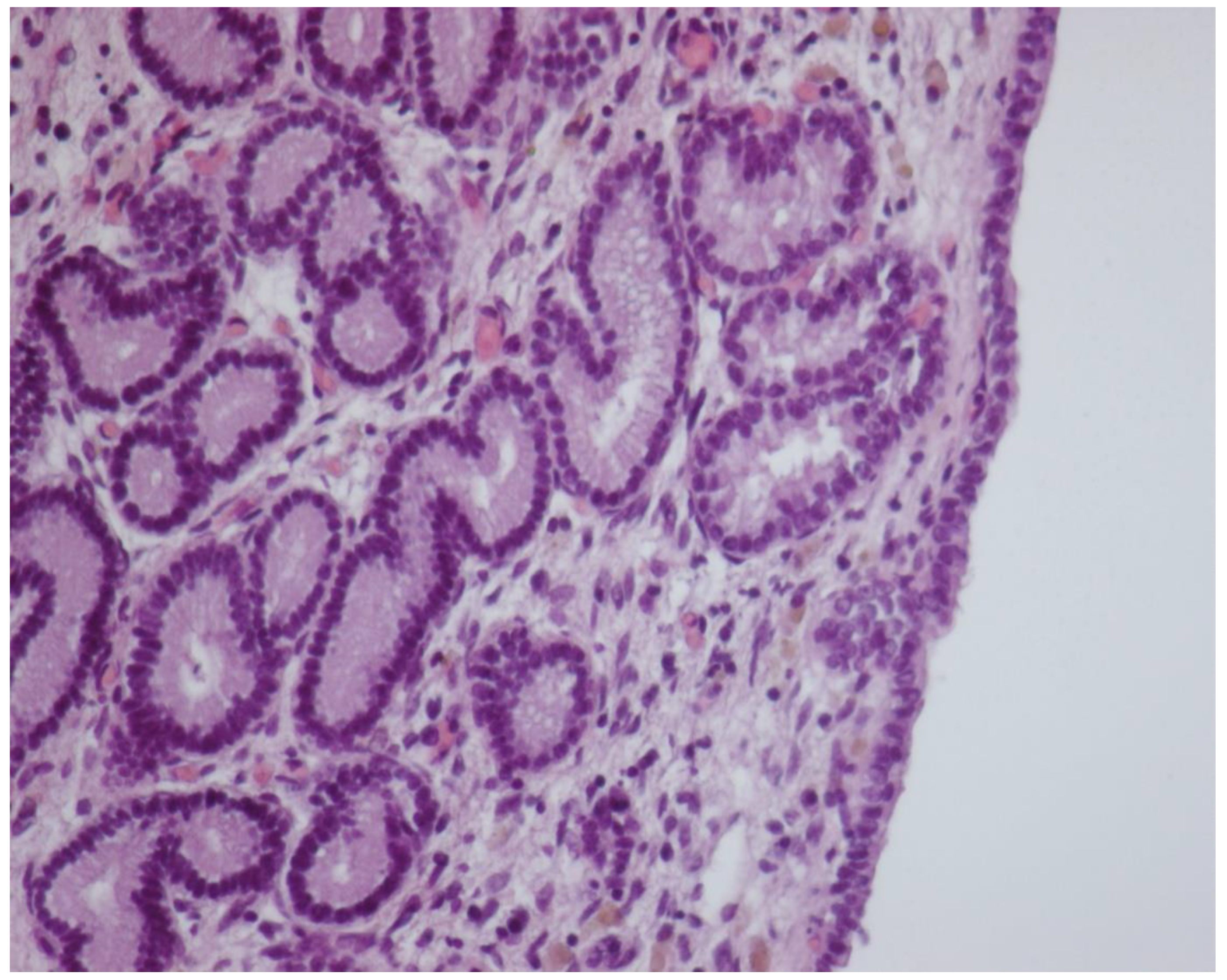
In comparison to PP Days 1–7, GL mean number showed a significant trend of increase from PP Day 14 (Figure 6), reaching the highest value on PP Day 28.
Perimeters of GL and GL area showed a significant constant trend of decrease from PP Day 1 to Day 28, failing to provide a suitable cutoff time for uterine involution. The mean ET significantly increased from PP Day 1 to Days 3–7, with a further increase, concurrently with the foal heat, at PP Day 14, followed by a decrease at PP Days 21–28, returning to a similar thickness as at PP Day 3 (Table 2). The Bonferroni post hoc test confirmed the histological data obtained by ANOVA for repeated measures.

3.4. Cytology

The number of neutrophil cells, evaluated by a score system, showed a significantly lower score between PP Day 1 and PP Days 7–28, between PP Day 3 and PP Days 7–28, and between PP Days 7–21 and PP Day 28 (Table 3).
The amount of eosinophil cells, evaluated by a score system, showed a higher significant mean score between PP Day 1 and PP Days 7–14, between PP Day 3 and PP Days 7–14, and a lower significant mean score between PP Day 7 and PP Day 21 (Table 3). Mononucleate cells were found only seldom, as small aggregates, and limited to PP Days 7–14.

4. Discussion

Uterine involution, a complex process involving anatomical, histological, and cytological changes, is pivotal to allow a new gestation. Therefore, its correct comprehension is needed for the best management of postpartum in all species, and even more in equids, in which the first postpartum heat occurs soon after parturition. Moreover, in programs for population increase, such as the endangered Martina Franca donkey breed, maximizing the pregnancy achievement plays a crucial role. Although ultrasonography is known to be the most common tool for monitoring the PP uterine involution, studies of histological and cytological changes are needed to understand the mechanisms underlying the uterine involution, providing more precise information for the better management of reproduction. In comparison to the horse mare, only few previous studies [11,12,13] investigated some aspects of uterine involution in the donkey, mainly focusing on foal heat characteristics, but the overall picture of ultrasonographical, histological, and cytological changes was lacking.
Ultrasonography, per se, does not provide precise information about the full functionality of an organ, but remains the most common tool for the management of equine female reproduction, providing immediate evaluation of ovarian and uterine structures and characteristics, especially suitable under field conditions.
In the present study, transrectal ultrasonography performed on PP Days 1–3 did not allow to obtain adequate pictures for correctly measuring the cross-sectional diameter of the CCJ regions of the PPH, differently to a previous study [11] in which measurements were performed already at Day 3 after foaling.
Altogether, the ultrasonographic findings showed that T diameters did not differ significantly between PPH and NPPH at any time, whereas the differences between the mean M and CCJ diameters of the PPH and the NPPH disappeared after PP Day 7, suggesting that uterine involution assessed by ultrasonography could be considered completed by the subsequent observation, performed on PP Day 14. These findings disagree with previous data from the same donkey breed [13], in which, in most jennies, the recovery of the normal uterine diameter was detected by PP Day 21. Even in French donkeys [11], the mean time for uterine involution was about 22 days. Those differences could be attributable to the modality and frequency of ultrasonographic monitoring (equipment, frequency, etc.) and maybe also to the different donkey breed enrolled in the study [11]. The results of the present study are more similar with data reported for the horse mare, in which by PP Days 15–16, an adequate degree of uterine involution was detectable [9,10].
Uterine biopsies showed that MC disappeared soon after foaling and they were not detectable by PP Day 3, while in pony mares, Jischa and colleagues did not find microcaruncles in specimens collected for biopsy by Day 9 [10]. Endometrial GL number increased significantly from PP Day 14, while perimeter and area decreased constantly from foaling to PP Day 28. Similarly, in pony mares, endometrial glands are reported to be less dilated at Days 9 and 16 as compared to Day 1 [10]. These findings seem to suggest that the secretive function of the endometrium is also restored as early as 14 days after foaling in Martina Franca jennies. In the mare, the glandular density was reported to show a trend of increase during the first 2 weeks after foaling [3,22]. In the present study, the ET was significantly increased at PP Day 7, further increasing at PP Day 14, concurrently with the foal heat. This agrees with the recognized increase of the height of the epithelium during estrus in the mare [8]. During postpartum, the height of the epithelium was reported to increase in the mare 5 days after foaling [23], very similar to the finding of the present study in jennies.
The assessment of eosinophil and neutrophil cells’ amount in the PPH, performed by a score system, showed a marked neutrophils reduction, of about one half–one third of the initial value, at PP Day 7, maybe indicating that, as early as Day 7 PP, in normal foaling Martina Franca jennies, the uterine mucosa is already regenerated, as recently reported in the mare [4]. In mares, in fact, the luminal epithelium was reported to become intact by Days 4–7 PP [9,24]. Moreover, in mares, the reduction of neutrophil cells in the endometrium was reported to be an indicator of undisturbed puerperal development [4]. In pony mares, this process is slightly postponed at Day 9, at which time neutrophils had largely ceased to be seen, substituted by lymphocytes and macrophages [10].
A different trend was observed for eosinophils, showing a significant, more than doubled, increase from the first 3 days after parturition to the PP Days 7–14, followed by a decrease to values similar to those recorded during the first 3 days after foaling. Eosinophils were reported to be more common on Day 5 PP in the mare [23]. The presence of eosinophils could be supposed to represent an immune response in the early postpartum period, as previously suggested for the initial interaction between the conceptus and the uterus in the mare [25]. The seldom observation of small mononucleate cell aggregates, only at PP Days 7–14, seems to suggest a minor role of these cells in the process of uterine involution in jennies.
In the present study, all the jennies showed the onset of foal heat on PP Day 6.5 ± 0.9, very similar to the PP Day 7.4 ± 2.1 previously reported for the same donkey breed [13]. In the French donkeys [11], a significant delay in uterine involution was reported for those jennies in which the foal heat occurred earlier than Day 9 after foaling, possibly because of the different donkey breed enrolled. In the present study, the evaluation of a similar possible effect on PP uterine involution was not possible, because of the small number of studied animals, and because in all the jennies, the foal heat occurred homogeneously earlier than PP Day 9. However, because uterine involution was completed in all jennies by PP Day 14, it is possible to suggest that the early occurrence of foal heat does not seem to influence the process of uterine involution in Martina Franca donkeys.

5. Conclusions

In conclusion, the present study, thanks to the combined results of ultrasonographical, histological, and cytological findings, provided a more complete evidence for the timing of PP uterine involution in normal foaling Martina Franca jennies.
Taking all the findings together, the present study showed that, in the normal foaling Martina Franca jennies investigated, the timing of PP uterine involution was 14 Days PP, similar to the results reported for the horse mare, and a bit quicker than data previously reported for another donkey breed. Besides the usefulness of ultrasonography under practical settings, in Martina Franca jennies, endometrial histology and cytology were also shown to be valuable tools for a more precise evaluation of uterine involution progression, as recently reported for the mare [4].

Author Contributions

Conceptualization, A.C.; methodology, G.M., I.D.A. and M.T.Z.; software, G.M. and I.D.A.; validation, G.M. and I.D.A.; formal analysis, I.D.A. and A.M.; investigation, G.M., I.D.A. and D.R.; data curation, I.D.A., A.M. and J.F.; writing—original draft preparation, J.F.; writing—review and editing, J.F. and D.R.; supervision, A.C. All authors have read and agreed to the published version of the manuscript.

Funding

The present study has been carried out in the framework of the Project “Demetra” (Dipartimenti di Eccellenza 2018–2022, CUP_C46C18000530001), funded by the Italian Ministry for Education, University and Research.

Institutional Review Board Statement

The project was approved by the Committee on Animal Research and Ethics of the Universities of Chieti-Pescara and Teramo (http://www.unich.it/unichieti/appmanager/federati/CEISA), Protocol #45/2013/CEISA/COM, approval date 16 July 2013.

Data Availability Statement

Data are available upon reasonable request to the authors.

Conflicts of Interest

The authors declare no conflict of interest.

References

  1. Nishikawa, Y.; Yamazaki, Y. Studies on reproduction in asses. I. Breeding season, oestrous cycle and length of heat. Jpn. J. Zootech. Sci. 1949, 19, 119–124. [Google Scholar]
  2. McKinnon, A.O. Reproductive Ultrasonography. In Equine Diagnostic Ultrasonography; Rantanen, N.R., McKinnon, A.O., Eds.; Williams and Wilkins: Baltimore, MD, USA, 1998; pp. 79–102. [Google Scholar]
  3. Blanchard, T.L.; Varner, D.D.; Brinsko, S.P.; Meyers, S.A.; Johnson, L. Effects of postparturient uterine lavage on uterine involution in the mare. Theriogenology 1989, 32, 527–535. [Google Scholar] [CrossRef]
  4. Krohn, J.; Eilenberg, R.D.; Gajewski, Z.; Failing, K.; Wehrend, A. Lochial and endometrial cytological changes during the first 10 days post-partum with special reference to the nature of foaling and puerperium in equine. Theriogenology 2019, 139, 43–48. [Google Scholar] [CrossRef] [PubMed]
  5. Meira, E.B., Jr.; Henriques, L.C.; Sá, L.R.; Gregory, L. Comparison of ultrasonography and histopathology for the diagnosis of endometritis in Holstein-Friesian cows. J. Dairy Sci. 2012, 95, 6969–6973. [Google Scholar] [CrossRef] [PubMed]
  6. Allen, W.R. Practical control of anestrus in Thoroughbred broodmares. In Endocrine Causes of Seasonal and Lactational Anestrus in Farm Animals; Ellendorff, F., Elsaesser, F., Eds.; Martinus Nijhoff Publishers: Dordrecht, The Netherlands, 1985; pp. 98–107. [Google Scholar]
  7. Noakes, D. The puerperium. In Veterinary Reproduction and Obstetrics; Noakes, D.E., Parkinson, T.J., England, G.C.W., Arthur, G.H., Eds.; Saunders Ltd.: Edinburgh, Scotland, 2009. [Google Scholar]
  8. Stanton, M.E. Uterine involution. In Equine Reproduction; Mc Kinnon, A.O., Squires, E.L., Vaala, W.E., Varner, D.D., Eds.; Wiley and Blackwell: Oxford, UK, 2011; pp. 2292–2293. [Google Scholar]
  9. Gygax, A.P.; Ganjam, V.K.; Kenney, R.M. Clinical, microbiological and histological changes associated with uterine involution in the mare. J. Reprod. Fertil. 1979, 27, 571–578. [Google Scholar]
  10. Jischa, S.; Walter, I.; Nowotny, N.; Palm, F.; Budik, S.; Kolodziejek, J.; Aurich, C. Uterine involution and endometrial function in postpartum pony mares. Am. J. Vet. Res. 2008, 69, 1525–1534. [Google Scholar] [CrossRef] [PubMed]
  11. Dadarwal, D.; Tandon, S.N.; Purohit, G.N.; Pareek, P.K. Ultrasonographic evaluation of uterine involution and postpartum follicular dynamics in French jennies (Equus asinus). Theriogenology 2004, 62, 257–264. [Google Scholar] [CrossRef] [PubMed]
  12. Tosi, U.; Bernabò, N.; Verni, F.; Valbonetti, L.; Muttini, A.; Mattioli, M.; Barboni, B. Postpartum reproductive activities and gestation length in Martina Franca jennies, an endangered Italian donkey breed. Theriogenology 2013, 80, 120–124. [Google Scholar] [CrossRef] [PubMed]
  13. Carluccio, A.; Gloria, A.; Robbe, D.; Veronesi, M.C.; De Amicis, I.; Cairoli, F.; Contri, A. Reproductive characteristics of foal heat in female donkeys. Animal 2017, 11, 461–465. [Google Scholar] [CrossRef] [PubMed]
  14. Veronesi, M.C.; Dall’Ara, P.; Gloria, A.; Servida, F.; Sala, E.; Robbe, D. IgG, IgA, and lysozyme in Martina Franca donkey jennies and their foals. Theriogenology 2014, 8, 825–831. [Google Scholar] [CrossRef] [PubMed]
  15. Carluccio, A.; Gloria, A.; Veronesi, M.C.; De Amicis, I.; Noto, F.; Contri, A. Factors affecting pregnancy length and phases of parturition in Martina Franca jennies. Theriogenology 2015, 84, 650–655. [Google Scholar] [CrossRef] [PubMed]
  16. Contri, A.; Tosi, U.; De Amicis, I.; Veronesi, M.C.; Panzani, S.; Carluccio, A. Ultrasonographic evaluation of sexual glands before and after ejaculation in the jackass. Vet. Res. Commun. 2008, 32 (Suppl. 1), S135–S137. [Google Scholar] [CrossRef] [PubMed]
  17. Veronesi, M.C.; De Amicis, I.; Panzani, S.; Kindahl, H.; Govoni, N.; Probo, M.; Carluccio, A. PGF(2α), LH, testosterone, oestrone sulphate, and cortisol plasma concentrations around sexual stimulation in jackass. Theriogenology 2011, 75, 1489–1498. [Google Scholar] [CrossRef] [PubMed]
  18. Panzani, S.; Carluccio, A.; Probo, M.; Faustini, M.; Kindahl, H.; Veronesi, M.C. Comparative study on 15-ketodihydro-PGF(2α) plasma concentrations in newborn horses, donkeys and calves. Reprod. Domest. Anim. 2012, 47, 82–86. [Google Scholar] [CrossRef] [PubMed]
  19. Panzani, S.; Carluccio, A.; Faustini, M.; Prandi, A.; Probo, M.; Veronesi, M.C. Comparative study on Insulin-Like Growth Factor I (IGF-I) plasma concentrations in new-born horse foals, donkey foals and calves. Fetal Neonatal Dev. Med. 2017, 1, 1–6. [Google Scholar] [CrossRef] [Green Version]
  20. Veronesi, M.C.; Gloria, A.; Panzani, S.; Sfirro, M.P.; Carluccio, A.; Contri, A. Blood analysis in newborn donkeys: Hematology, biochemistry, and blood gases analysis. Theriogenology 2014, 82, 294–303. [Google Scholar] [CrossRef] [PubMed]
  21. Romies, B. Färben der Schnitte. In Microskopicshe Technik; Böck, P., Ed.; Urban & Schwarzenberg: Munich, Germany, 1989; pp. 179–249. [Google Scholar]
  22. Arrott, C.; Macpherson, M.; Blanchard, T.; Varner, D.; Thompson, J.; Simpson, B.; Bruemmer, J.; Vogelsang, S.; Fernandez, M.; Fleet, T.; et al. Biodegradable estradiol microspheres do not affect uterine involution or characteristics of postpartum estrus in mares. Theriogenology 1994, 42, 371–384. [Google Scholar] [CrossRef]
  23. Katila, T. Histology of the post partum equine uterus as determined by endometrial biopsies. Acta Vet. Scand. 1988, 29, 173–180. [Google Scholar] [CrossRef] [PubMed]
  24. Bailey, J.V.; Bristol, F.M. Uterine involution in the mare after induced parturition. Am. J. Vet. Res. 1983, 44, 793–797. [Google Scholar] [PubMed]
  25. Caballeros, J.E.; Camacho, C.; Cazales, N.; Estradé, M.J.; Fiala-Rechsteiner, S.; Jobim, M.I.M.; Mattos, R.C. Ultrastructural and histological characteristics of the equine endometrium at day 5 post ovulation. Theriogenology 2019, 132, 106–112. [Google Scholar] [CrossRef] [PubMed]
Figure 1. Ultrasound image of the middle tract of the post-pregnant uterine horn, at day 7 postpartum.
Animals 11 02762 g001
Figure 2. Ultrasound image of the middle tract of the non-post-pregnant uterine horn, at day 7 postpartum.
Animals 11 02762 g002
Figure 3. Ultrasound image of the middle tract of the post-pregnant uterine horn, at day 14 postpartum.
Animals 11 02762 g003
Figure 4. Ultrasound image of the middle tract of the non-post-pregnant uterine horn, at day 14 postpartum.
Animals 11 02762 g004
Figure 5. Histologic sampling at day 1 postpartum. In the middle of the figure, a microcaruncle is visible (HE-10× magnification).
Animals 11 02762 g005
Figure 6. Histologic sampling at postpartum day 14. Cubic continuing epithelium with numerous and well-developed glandular structures (HE-20× magnification).
Animals 11 02762 g006
Table 1. Postpartum cross-sectional diameter measured (mean ± SD) at the non-post-pregnant and post-pregnant uterine horns at the level of the tip, middle, and corpora-cornual junction regions, at the different sampling times, in the 9 Martina Franca jennies.
Cross-Sectional Diameter (mm) Non-Post-Pregnant HornCross-Sectional Diameter (mm) Post-Pregnant Horn
PP Day TipMiddleCorpora-Cornual JunctionTipMiddleCorpora-Cornual Junction
141.8 ± 1.259.1 ± 0.8 A86.0 ± 1.244.0 ± 1.376.0 ± 0.8 BNot assessable
338.3 ± 1.856.7 ± 1.1 A84.5 ± 2.041.0 ± 0.871.6 ± 0.7 BNot assessable
735.5 ± 0.954.0 ± 1.4 A77.8 ± 1.4 a37.3 ± 0.862.6 ± 0.7 B96.8 ± 1.5 b
1429.0 ± 1.346.8 ± 1.567.8 ± 1.630.5 ± 1.147.1 ± 2.272.3 ± 2.4
2126.6 ± 0.643.3 ± 1.364.6 ± 1.025.7 ± 1.045.2 ± 1.364.3 ± 1.4
2824.5 ± 0.643.3 ± 0.858.1 ± 1.124.8 ± 0.742.0 ± 1.259.6 ± 1.4
A,Bp < 0.05 and a,b p < 0.05 denote within row statistical significance.
Table 2. Postpartum endometrial glandular number, perimeter, area, and epithelium thickness (mean ± SD) in the different sampling times in the 9 Martina Franca jennies.
PP DayNumber Perimeter (mm)Area (mm2)Epithelium Thickness (mm)
1129 ± 110.8 §8.8 ± 3.4 A5.3 ± 4.2 AA1.0 ± 0.3 A
3173 ± 124.6 8.6 ± 3.6 A5.2 ± 4.2 AA1.3 ± 0.4 B
7240 ± 127.0 #7.4 ± 3.2 B3.9 ± 3.1 BB1.3 ± 0.4 B
14396 ± 320.8 7.2 ± 2.9 B3.6 ± 2.5 BB1.4 ± 0.4 C
21519 ± 220.0 §6.5 ± 2.1 C2.8 ± 2.0 CC1.2 ± 0.3 B
28546 ± 240.5 #6.2 ± 2.9 C2.5 ± 2.2 CC1.2 ± 0.3 B
A,B,C Denote within column significant differences with p < 0.001. AA,BB,CC Denote within column significant differences with p < 0.001. Glandular number p < 0.05; p < 0.01; # p < 0.01; § p < 0.001; p < 0.001; p < 0.001; p < 0.001.
Table 3. Score (mean ± SD) for postpartum eosinophil and neutrophil cells’ amount evaluation at the different sampling times, in the 9 Martina Franca jennies.
PP Day
PP Day 1PP Day 3PP Day 7PP Day 14 PP Day 21PP Day 28
Eosinophil Score0.51 ± 0.42 §#0.73 ± 0.49 1.94 ± 0.82 §1.57 ± 0.82 #0.96 ± 0.87 0.94 ± 1.20
Neutrophil Score1.16 ± 0.46 §#1.51 ± 0.69 AB0.51 ± 0.30 §C0.52 ± 0.34 #D0.37 ± 0.13 AE0.17 ± 0.16 BCDE
Eosinophils: § p < 0.001; # p < 0.01; p < 0.01; p < 0.05; p < 0.05. Neutrophils: § p < 0.01; # p < 0.01; p < 0.001; p < 0.001; p < 0.01; p < 0.01; A p < 0.001; B p < 0.001; C p < 0.01; D p < 0.05; E p < 0.05.
Publisher’s Note: MDPI stays neutral with regard to jurisdictional claims in published maps and institutional affiliations.