Article

Seasonal Variation in Gut Microbiota Related to Diet in Fejervarya limnocharis

1
Key Laboratory of Southwest China Wildlife Resources Conservation (Ministry of Education), China West Normal University, Nanchong 637009, China
2
Institute of Eco-Adaptation in Amphibians and Reptiles, China West Normal University, Nanchong 637009, China
3
Institute of Evolution and Ecology, International Research Centre of Ecology and Environment, College of Life Sciences, Central China Normal University, Wuhan 430079, China
*
Author to whom correspondence should be addressed.
Animals 2021, 11(5), 1393; https://doi.org/10.3390/ani11051393
Submission received: 6 March 2021 / Revised: 7 May 2021 / Accepted: 8 May 2021 / Published: 13 May 2021
(This article belongs to the Section Wildlife)

Abstract

:

Simple Summary

Gut microbiota, such as Proteobacteria, Firmicutes, and Bacteroidetes, show chitin-degrading and/or antibiotic biosynthesis ability. To evaluate whether ecological factors (diet) and host conditions (body size, body mass, and body condition) affect the gut microbiota diversity, we analysed the diet composition and host condition in Fejervarya limnocharis among different seasons and/or sexes. The dietary difference was not significant among seasons and between males and females, but host condition seasonally changed. Using bioinformatics analysis, we observed that food variations and body mass were significantly correlated with gut microbial composition. Our findings suggest that gut microbiomes of F. limnocharis vary seasonally in response to diet variations when facing environmental changes.

Abstract

Organisms adapt to environmental fluctuations by varying their morphology and structural, physiological, and biochemical characteristics. Gut microbiome, varying rapidly in response to environmental shifts, has been proposed as a strategy for adapting to the fluctuating environment (e.g., new dietary niches). Here, we explored the adaptive mechanism of frog intestinal microbes in response to environmental changes. We collected 170 Fejervarya limnocharis during different seasons (spring, summer, autumn, and pre-hibernation) to study the compositional and functional divergence of gut microbiota and analysed the effects of seasonal feeding habits and body condition on intestinal microorganisms using 16S rRNA high-throughput sequencing, Tax4Fun function prediction analysis, and bioinformatics analysis. The results showed no significant dietary difference in various seasons and between males and females. However, a significantly positive correlation was detected between dietary diversity and food niche width. Host condition (body size, body mass, and body condition) also revealed seasonal changes. The frogs were colonised by 71 bacterial phyla and dominated by Proteobacteria, Firmicutes, and Bacteroidetes. Stenotrophomonas was the most abundant genus in the Proteobacteria. The composition, diversity, and function of intestinal microorganisms in different seasons were significantly different. Significant differences were observed in composition and function but not in the microbial diversity between sexes. Furthermore, seasonal foods and body mass were significantly correlated with gut microbial composition. Our results suggest that gut microbiomes of F. limnocharis vary seasonally in response to diet under fluctuating environments.

1. Introduction

Animals evolve a variety of behavioral and physiological strategies, including altered feeding and activity patterns and increased mobilization of stored fat to fuel energy demands, to cope with shifting demands [1]. Nutritional demands may vary in response to life-history processes, such as growth, reproduction, and hibernation. The gut microbiome has been proposed as an additional avenue by which animals can cope with changing dietary landscapes and energetic challenges [2,3] and thus play a role that has profound ecological, evolutionary, and environmental implications in amphibians for the most part [4].
Diverse and abundant microbial communities colonise vertebrate digestive systems and coevolve with the host [5,6], which affects multiple aspects of host physiology, including health, nutrition, immunity, development, and reproduction [7,8]. Chitinolytic bacteria (e.g., Clostridium) from tadpoles are advantageous in the digestion of insects, which are the main diet of anurans [4]. Important energetic gains made by tadpoles from a fermentative microbial digestion within their guts and these gains, in turn, are expected to be directly proportional to metamorphic size and/or timing [4]. In addition, the intestinal microbiota can develop a natural defense barrier exerting different protective, structural, and metabolic effects on the host epithelium [5]. Particularly, probiotics are proposed as an effective and environmentally friendly alternative to antibiotics, known as beneficial microbes. Antibiotic genes of intestinal microorganisms encode the biosynthesis of antibiotics to affect the intestinal microflora and its antimicrobial and anti-inflammatory effects [9]. However, pathogens carrying antibiotic resistance genes pose a threat to public health [10]. A typical gut microbiome can result in immunological and metabolic disorders [11,12,13]. Consequently, a growing interest focuses on the evaluation of extrinsic and intrinsic factors that manipulate gut microbial communities and diversity [14,15]. Environmental factors are particularly intriguing because they play a role as a major driver of gut microbiota communities [15,16,17,18,19,20,21] and may offer potentially simple mechanisms for treating dysbiosis to improve host health and feed utilisation [16,17].
Host diet is an important environmental factor affecting gut microbial communities [18,19,20,21,22,23,24]. Most previous studies have demonstrated that the gut microbiome seasonally reconfigures to respond to dietary fluctuations [17,24,25,26] and presumably to adapt to new dietary niches. A more diverse diet may improve microbial diversity by providing a more diverse nutrition [27]. Alternatively, diet can indirectly change the microbial composition by varying host physiology, body condition, and immune status [2,28,29,30]. However, whether gut microbiota composition and diversity of amphibians seasonally change in response to diet has not been studied.
Amphibians modify their life-history traits, physiology, behavior, and gut microbiome when they experience variation in environmental factors, such as temperature, rainfall [31,32], and diet [33]. Environmental temperature variations result in changes in food composition of frogs [34,35], thereby affecting their gut microbial composition [36]. Fejervarya limnocharis, an amphibian, is widely distributed and can represent a good system for studying the relationship between gut microbiota composition and seasonal variations in terms of adapting to fluctuating diet. Although insects are the major nutrient source of F. limnocharis [33], their gastrointestinal tract is poorly adapted to this food [37]. To compensate, F. limnocharis may depend heavily on their gut microbiota to maximise nutrient extraction from insects [4]. Additionally, given that F. limnocharis are widely distributed in farmlands, their seasonal gut microbiota may be a great system to explore the adaptation of amphibians to human activities and provide advice in the context of conservation biology. In farmlands, compared with frogs in natural environments, F. limnocharis have decreased stomach contents (abundance and number of categories) and increased levels of Firmicutes and Proteobacteria, suggesting that anthropogenic activities have not only affected the food resources of frogs, but also influenced the health and gut microbial ecosystem of wildlife [33].
Here, we investigated the gut microbiota of 161 frogs and stomach contents of 147 individuals, with samples collected over four seasons (e.g., spring, summer, autumn, and pre-hibernation). Using Tax4Fun function prediction, we predicted the function of gut microbiota. We tested for seasonal variation in gut microbiota composition, diversity, and function. We then examined seasonal changes in diet composition and diversity in the host under the four seasons. Finally, we analyzed the effects of ecological factors (e.g., diet) and host conditions on the microbiota diversity of frogs to understand how seasonal factors may drive fluctuation in the microbiome composition and function.

2. Materials and Methods

2.1. Sample Collection

A total of 170 adult frogs were collected during spring, summer, autumn, and pre-hibernation (118 males and 52 females) in Xishan from September 2018 to November 2019 in Nanchong city (106°2′ E, 30°47′ N; 310 m altitude), Sichuan Province, China (Table S1). All the frogs were collected in a cool box with two ice bags and immediately transported to the laboratory of China West Normal University for immediate experiments. The gender of all individuals was confirmed by direct observation of secondary sexual characteristics [38]. We sacrificed all individuals using single pithing. Body size (snout–vent length (SVL)) was measured to the nearest 0.01 mm with calipers, whereas body mass was measured to the nearest 0.1 mg with an electronic balance. We calculated the body condition of each individual based on the ratio between log of body mass and log of body length. The gut content was collected within 20 min after pithing. We carefully isolated the digestive tract from the body and then collected the gut and stomach. The gut contents within each individual were emptied into a sterile vial and immediately stored at −80 °C. The specimens were preserved in 4% buffered formalin, and the stomach was stored in 70% ethanol.

2.2. Diet Analyses

The prey items of 147 stomachs were identified at the order level under a stereomicroscope. Then, we estimated the percentage of frequency of each prey category. For the assessment of diet diversity, the Shannon–Weiner index was calculated based on the following model:
H = ( P i )   L n ( P i ) ,
where H represents the Shannon–Weiner index; the Pi is the proportion of individuals found in the ith species. In a sample the value of Pi is unknown but is estimated as ni/N, in which N is the total number of all species, and Ni is the number of ith species. [39]. In addition, the Simpson diversity index was used to estimate the niche width of stomach contents [39,40], and the formula is as follows:
B = 1 P i 2 ,
where B expresses the Simpson’s index; the Pi is the proportion of individuals in the ith species.
The numbers, frequency of each prey category, food diversity and ecological niche of stomach contents between sexes among different seasons were compared using non-parametric statistical methods, Pearson’s correlation analysis, and Wilcoxon rank-sum test. All statistical analyses were conducted using R 3.6.2.

2.3. DNA Extraction and Sequencing

DNA extraction and sequencing were operated by Novogene Corporation (Novogene Corporation, Beijing, China). We extracted DNA from entire intestines from 161 F. limnocharis using TIANgen DNA extraction kit DP328 (Tiangen, Beijing, China). We amplified the V4 region of 16S rRNA gene with a specific primer set 515F (5′-GTGCCAGCMGCCGCGGTAA-3′) and 806R (5′-GGACTACHVGGGTWTCTA AT-3′). All polymerase chain reaction (PCR) reactions were performed in bio-Rad T100 gradient PCR instrument with 15 μL Phusion® High-Fidelity PCR Master Mix (New England Biolabs), 0.2 µM forward and reverse primers and 10 ng template DNA per 30 μL reaction. The PCR cycle consisted of initial denaturation at 98 °C for 1 min; 30 cycles of denaturation at 98 °C for 10 s, annealing at 50 °C for 30 s, extension at 72 °C for 30 s; and extension at 72 °C for 5 min. The PCR products were mixed with same volume of 1 × loading buffer (contained SYB green) and were subsequently subjected to electrophoresis on 2% agarose gel for detection. The PCR products were mixed in equidensity ratios. Then, we obtained the purified PCR products using GeneJETTM Gel Extraction Kit (Thermo Scientific, Shanghai, China).
Sequencing libraries were generated using Ion Plus Fragment Library Kit (48 rxns) (Thermo Scientific, Shanghai, China). The library quality was assessed on the Qubit@ 2.0 Fluorometer (Thermo Scientific, Shanghai, China). Finally, the library was sequenced on an Ion S5TM XL platform and 400 bp single-end reads were generated.

2.4. Operational Taxonomic Unit (OTU) Cluster and Species Annotation

We assigned single-end reads based on their unique barcode and truncated them by cutting off the barcode and primer sequence to the samples. Quality filtering of raw reads was performed under specific filtering conditions in accordance with the Cutadapt quality-controlled process ([41]; V1.9.1, http://cutadapt.readthedocs.io/en/stable/; access date: late January to early February 2020). To detect chimera sequences, we used UCHIME algorithm (UCHIME Algorithm, http://www.drive5.com/usearch/manual/uchime_algo.html/; access date: late January to early February 2020) to compare the reads with the reference database (Silva database, https://www.arb-silva.de/; access date: late January to early February 2020) and removed the chimera sequences. Finally, we obtained the clean and effective reads using subsequent analysis.
We clustered all the effective sample data using Uparse software (v7.0.1001) and assigned the sequences with ≥97% identity to the same OTU. The representative sequence for each OTU was screened for further annotation. For each representative sequence, we used the Silva Database (https://www.arb-silva.de/; access date: late January to early February 2020) to annotate taxonomic information based on Mothur algorithm. We also used annotation analysis (threshold value: 0.8–1) to obtain taxonomic information and count the community composition of samples at all levels, such as kingdom, phylum, class, order, family, genus, and species, based on the Mothur algorithm and SSUrRNA database of SILVA132 (https://www.arb-silva.de/; access date: late January to early February 2020). OTU abundance information was normalized using a standard sequence number corresponding to the sample with the least sequences. Finally, we performed subsequent analysis of alpha and beta diversities based on this output normalized data.

2.5. Data Analysis

Alpha diversity indexes, which is used to analyse the complexity of bacterial diversity for a sample, was calculated with QIIME (Version 1.9.1) and displayed with R software (Version 2.15.3), such as observed-species index, Chao1 index, Shannon-Wiener index, Simpson’s index, ACE (Abundance Coverage-based Estimator) index, and good-coverage index (http://www.mothur.org/wiki/; access date: late January to early February 2020). To evaluate the differences among samples in terms of bacterial community complexity, we calculated the beta diversity on weighted and unweighted UniFrac using QIIME (Version 1.9.1). Moreover, to find differences among different samples (groups), we used the Unweighted Pair-group Method with Arithmetic Mean (UPGMA) and analysis of intra (inter)-group differences in beta diversity index. In particular, the NMDS (nonmetric multidimensional scaling) ordination approach was performed to visualize beta-diversity patterns and reflect the inter- and intra-group differences based on Bray–Curtis dissimilarity distances. UPGMA clustering was performed as a type of hierarchical clustering method to study the similarities between different samples using average linkage and conducted by QIIME software (Version 1.9.1).
The between- and within- group difference based on alpha and beta diversity indexes and bacterial abundance were analysed by Anosim, (un)weighted UniFrac Wilcoxon rank-sum test and linear discriminant analysis effect size (LEfSe) analysis, whereas the different bacteria was found by parametric test (t-test). Through sample clustering tree display, the differences in the community structure between different samples or groups were explored.
Data on food frequency and microbiota β-diversity were analyzed using the Procrustes routine in QIIME (Version 1.9.1). On the basis of the frequency of prey item and microbiome diversity data, weighted and un-weighted UniFrac PCoA analysis were combined with Procrustes analysis to visualize the relationship between diet and intestinal flora, and evaluated whether diet affects the gut microbiota abundance. Relationship significance was tested with Monte Carlo test in QIIME (Version 1.9.1). Generalized Linear Models (GLM) analysis with SPSS was also used to test the dependence of bacterial diversity and determine the effect of diet diversity, body size, body mass, and/or body condition on the microbiota.

2.6. Function Prediction

The function of intestinal microbiomes was predicted using Tax4Fun functional prediction analysis based on the minimum 16S rRNA sequence similarity by extracting the KEGG database prokaryotic whole genome 16S rRNA gene sequence. The predicted function was aligned to the SILVA SSU Ref NR database using BLASTN algorithm (BLAST Bitscore >1500) to establish a correction matrix. Finally, the prokaryotic whole genome functional information of the KEGG database annotated by UProC and PAUDA was mapped to the SILVA database to implement the SILVA database function annotation. The sequenced samples were clustered out of the OTU using the SILVA database sequence as a reference sequence to obtain functional annotation information.

3. Results

3.1. Gut Microbiota Composition of F. limnocharis

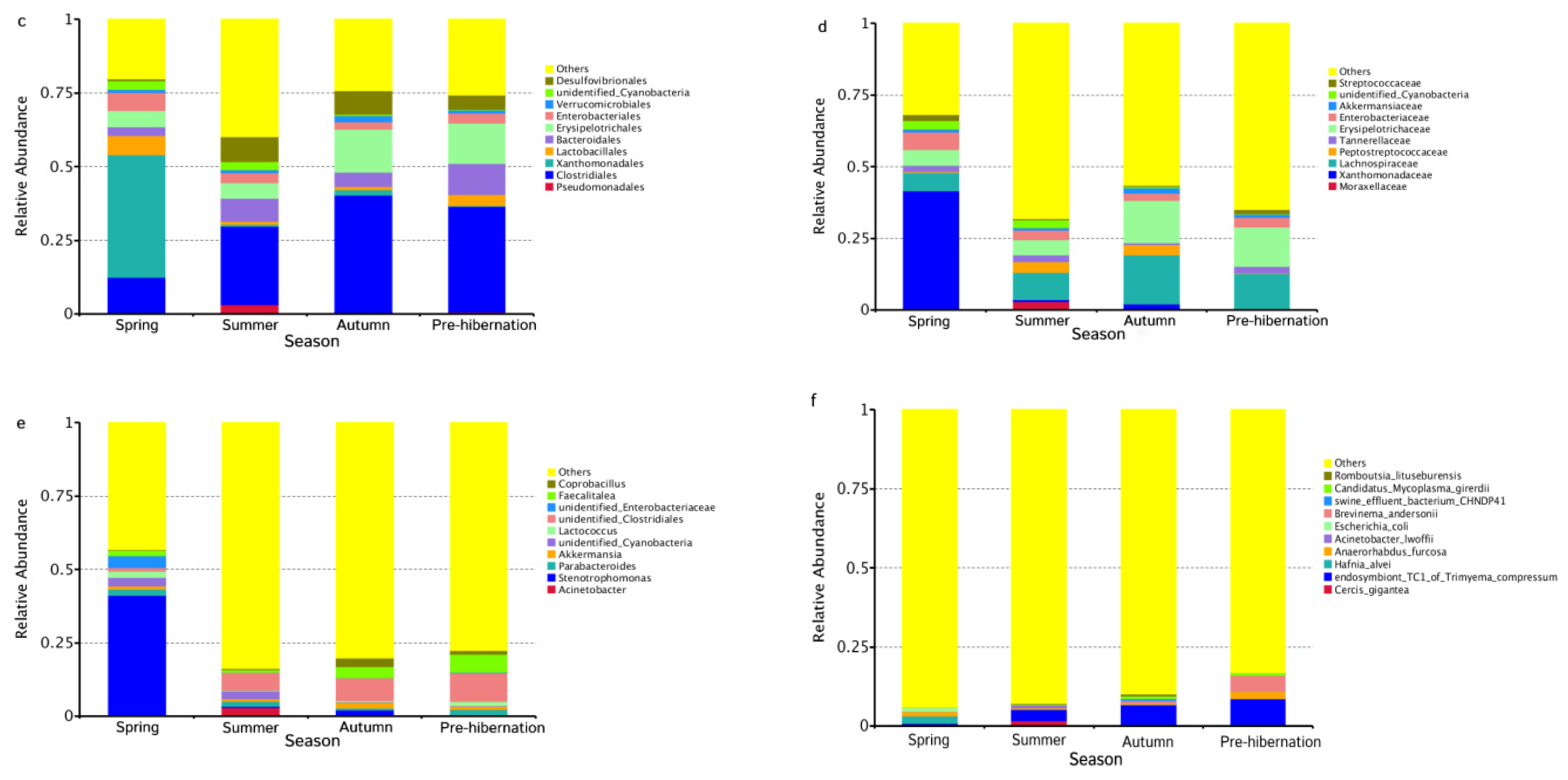
The V4 region of bacterial 16S rRNA gene in intestinal microbiota collected from 161 frogs across four seasons was sequenced to characterize microbiotas of F. limnocharis. All sequences could be identified to two boundaries, 71 phyla, 83 classes, 187 orders, 381 families, and 1301 genera. At the phylum level (Figure S1, Figure 1b), Proteobacteria, Firmicutes, and Bacteroidetes had the highest abundance and contributed 43.58%, 41.11%, and 5.78% of bacteria, respectively. At the genus level (Figure S1, Figure 1e), Stenotrophomonas (phylum: Proteobacteria), Faecalitalea (phylum: Firmicutes) and Parabacteroides (phylum: Bacteroidetes) were the most abundant and accounted for 36.69%, 11.09%, and 5.78% of bacteria, respectively.

3.2. Seasonal Changes of Intestinal Microbiota in Frogs

The seasonal variation in the abundance and diversity of intestinal microbiota was significant (ANOSIM: r > 0, p < 0.001). The unique OTUs in summer were the most abundant (Figure S2). The alpha diversity metrics (observed taxa and Chao 1) revealed the highest diversity and richness of the intestinal microbiota in summer and the least in autumn (Figure 2b,d; Table S2, for detailed information on the alpha diversity of gut in all samples, see Supplementary Table S3). Shannon diversity index and Simpson’s diversity index were the lowest in spring (Figure 2a,c). Inter- and intra-species differences in the microbiota among seasons were also explored (Anosim: r > 0, p < 0.01; Table S4). LEfSe analysis revealed that g_Stenotrophomonas was the top genus-level bacteria distinguished in frogs during different seasons (Figure S3).
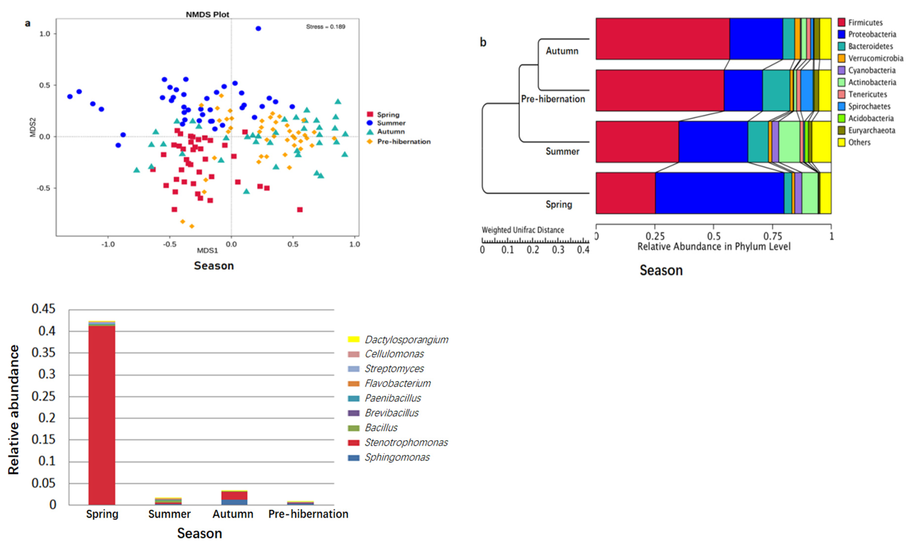
The seasonal relative abundance and distribution of bacteria may be indicative of seasonal variation in microbiota composition. Particularly, the composition of gut microbiota was seasonally changed (Figure 1a–f and Figure 3a,b). Differences were found in the specific bacteria among individuals (Table S5). The significant phylum and genera (p < 0.05) are listed in Tables S6 and S7, respectively. The abundance of chitin-digesting bacteria was found to be high in spring (Figure 3c).
After intrinsic differences in the gut microbiota composition were established between females and males within each season, significant differences were observed in Enterobacteriales (p = 0.032) order, Enterobacteriaceae (p = 0.032), and Peptococcaceae (p = 0.014), unidentified Coriobacteriales (p = 0.040), Phaselicystidaceae (p = 0.016), Aerococcaceae (p = 0.044) family, Raoultibacter (p = 0.040), Phaselicystis (p = 0.016), Variovorax (p = 0.034) genus, and Variovorax paradoxus species (p = 0.034) in autumn.

3.3. Variations of Function Prediction in Different Seasons

Functional predictions via Tax4Fun have suggested that the following three predicted functions dominated the four seasons: metabolism, genetic information processing, and environmental information processing (Figure S4). However, we found that most predicted functional categories in KEGG (Kyoto Encyclopedia of Genes and Genomes) pathways were significantly different among the four seasons, such as metabolism, environmental adaptation, and immune system (Figure 4a; t-test, p < 0.05; see Table S8). Particularly, metabolism (e.g., amino acid and lipid metabolism) and diseases (e.g., infectious diseases) were significantly higher in spring and summer than in autumn and pre-hibernation (Figure 4b; Table S8).
In order to further understand bacterial contribution to the specific environmental adaptability and diseases, we predicted that the gene function was linked to chitin-degrading ability, the biosynthesis of antibiotics, and antibiotic resistance among the four seasons. Analysis on potential chitin-degrading ability showed that the abundance of chitin-degrading enzymes, such as chitinase, β-N-acetylglucosaminidase, α-N-acetylglucosaminidase, chitin deacetylase, or putative chitinase, was significantly different among seasons (Kruskal-Wail test, p < 0.01) (Figure 5). Antibiotic genes were also significantly different between spring and summer (Mann-Whitney U test, p < 0.001), between spring and autumn (Mann-Whitney U test, p < 0.001), between spring and pre-hibernation (Mann-Whitney U test, p < 0.001), and between summer and pre-hibernation (Mann-Whitney U test, p = 0.001). In particular, the total abundance of all antibiotic genes was enriched in pre-hibernation as compared to other seasons (Wilcoxon rank-sum test, p < 0.01; Figure S5). Additionally, significant differences were also found in antibiotic resistance gene (beta-Lactam_resistance) between summer and autumn (Mann-Whitney U test, p < 0.001) and between summer and pre-hibernation (Mann-Whitney U test, p < 0.05).
The potential function of the intestinal microbiota was compared between females and males in autumn. In female frogs, nucleotide metabolism was significantly higher in autumn (t-test, p = 0.038). The total abundance of chitinase and antibiotic genes in females was higher than that in males (Figure S6a,b). Chitin-degrading enzyme (putative chitinase and bifunction chitinase/lysozyme) and antibiotic genes tetracycline biosynthesis and clavulanic acid biosynthesis) were significant different between females and males in autumn (Wilcoxon rank-sum test: putative chitinase, p = 0.036; bifunction chitinase/lysozyme, p = 0.016; tetracycline biosynthesis, p = 0.040; and clavulanic acid biosynthesis, p = 0.041). In addition, the vancomycin resistance gene was also a significant difference between females and males in autumn (Wilcoxon rank-sum test, p = 0.043).

3.4. Dietary Changes

Prey items in stomachs were sampled in 147 frogs (successfully identified for 114 frog specimens) to construct a concordance relationship between diet composition and gut microbiota. A total of 373 prey items were identified to six classes and 18 orders (Table S9). After the variability within and between seasons was established (for detailed information of stomach contents during four seasons, see Supplementary Table S10), the seasonal and sexual variations in food niche width and diet diversity were analysed. A positive correlation was found between seasons (Pearson: r = 0.97; Wilcoxon rank-sum test: p < 0.05). Among the seasons, the largest food niche width and diet diversity was found in autumn (Table S11). Furthermore, food niche width was positively related to the food diversity of females and males in summer (Pearson: r = 0.76; Wilcoxon rank-sum test: p < 0.05), and food niche width in males was larger than that in females (Table S12). However, no significant differences in food composition, food diversity, and niche width were detected among seasons and between males and females in summer (Anosim analysis: r > 0; Wilcoxon rank-sum test: p > 0.05).

3.5. Explanatory Factors of Intestinal Microbiota

Bioenv analysis based on beta diversity in bacteria was used to identify the optimal combination of ecological factors (diet diversity, humidity, and pH) and explain the seasonal variation of gut microbiota. Diet was determined to be the optimal combination (Bioenv: size = 1, correction = 0.6571).
Procrustes analysis of food frequency and microbiota β-diversity was conducted on 113 individuals during various seasons to co-visualise the data (Figure 6; Table S13). Separations based on either diet or microbiota co-segregated along the first axis of both data sets (weighted UniFrac, Figure 6a; unweighted UniFrac, Figure 6b). A strong relationship was observed between diet and intestinal microbiota (the Monte Carlo test; weighted: Counter_better = 19, M2 = 0.957, p = 0.019; unweighted: Counter_better = 2, M2 = 0.945, p = 0.020).
In addition to ecological factor (diet), the effect of body size (SVL), body mass, and body condition on gut microbiota was also explored. Body size, body condition, and body mass (converted by log 10 for the normal distribution) significantly shifted between seasons (t-test: p < 0.01, except between spring and autumn). GLM analysis was used to evaluate whether diet and body mass affects the microbiota and found a significant negative association (Pearson: r < 0; GLM: p < 0.01; Table 1)

4. Discussion

Body size, body condition, and diet are associated with gut microbiota [33,42]. Consistent with previous studies, our study found that diet (frequency of prey item and dietary diversity) and body mass were substantially associated with gut microbiota diversity. Interestingly, we rarely found an impact of temperature and humidity on gut microbial variation. These seasonal changes in bacteria could be explained by variations in seasonal food availability.
Consistent with other frogs (F. limnocharis [33], Babina adenopleura [43], Polypedates megacephalus [25], Odorrana tormota [44], and Rana dybowskii [19,45], Proteobacteria, Firmicutes, and Bacteroidetes detected in the intestinal flora were considered to be the main bacteria. This implies that the endogenous environment selects microbes that are optimally fit for the intestinal characteristics. On the other hand, the variation of community diversity found in F. limnocharis was similar with that found in R. dybowskii [46], but different from P. megacephalus [25] and O. tormota [44]. In our findings, frog communities were more diverse and abundance in summer compared to other seasons. This pattern suggested that the environmental conditions played major roles in determining the gut microbial composition.
Diet is the main ecological factor influencing bacterial community changes in frogs [33,47]. Changes in dietary habits can seriously affect the structure and composition of intestinal microbial flora [13]. Our founding revealed that the first PCA axis (PC1) explained 19.94% of the relationship between food frequency and microbiome diversity. Particularly, frogs with less digestible diet (low level of nutrient) had long digestive tract length [35,48]; this practice may indirectly affect gut microbiota composition as gut microbiota diversity was associated with intestinal pattern [27,36]. Stenotrophomonas (Gammaproteobacteria) was linked to chitin digestion [44] and is the main bacterium in F. limnocharis. In spring, when frogs eat more Scolopendromorpha and Coleoptera, the abundance of chitin-digesting bacteria increases. These results suggest that chitinolytic bacteria may confer digestive advantages to frogs. Moreover, we found more Lachnospiraceae (Firmicutes) linked to lipid metabolism in autumn and pre-hibernation as compared to that in both spring and autumn, which was likely to relate to the energy reserve (e.g., fat body formation) in preparation for hibernation [13]. The variations in dominant gut flora taxa at different seasons implied that F. limnocharis frogs acquire different bacteria due to variations in their seasonal diet. Nevertheless, intestinal microorganisms do not affect the dietary preference of frogs [49] and instead allows them adapt to their dietary niche [39].
In addition to metabolic adaption to diet, microbiome found in this frog was likely to reflect the status of the health and diseases of the host species among seasons. Pathogens (e.g., Pseudomonas and Batrachochytrium dendrobatidis) result in illness or even death in frogs [45,50,51] whereas probiotic bacteria (e.g., Bifidobacteria longun and Lactobacillus casei) and bacteria associated with pathogen resistance may promote host health [50,52]. More Enterobacteriaceae, as a common marker of gut dysbiosis, was found in frogs in spring compared to other seasons, suggesting that frogs have lower of SCFAs and higher acidic environment and diseases in spring [53]. Low gut microbiota diversity in spring supported this assumption because low gut microbiome diversity exhibited increased stress responses (high glucocorticoid levels) and reduced immune function (few cells that secrete local, strain-specific immunoglobulin A) [28,54,55].
At the function level, our study showed that many bacterial genes involved in amino acid metabolism, lipid metabolism, and chitin digestion were found during spring and summer and when the host had a low immune system, suggesting the occurrence of an energy tradeoff. By contrast, many antibiotic genes linked to antibiotic biosynthesis and gene involved in carbohydrate metabolism and cell motility was observed during autumn and pre-hibernation. This stimulation of bacterial energy metabolism and cellular activity could reflect the high SCFAs productions by gut bacteria [56] to provide frogs with additional energy in periods of pre-hibernation when nutrients are restricted. For example, in Rana dybowskii, the Firmicutes/Bacteroidetes ratio decreased from 4.23 in summer to 0.63 in winter to adapt to the challenging environment [46].
Furthermore, frog microbiota is composed additionally of functions which are related to antibiotic resistance [10]. Consistent with a previous study, we identified resistance gene (vancomycin resistance gene and beta-Lactam_resistance gene) in F. limnocharis, suggesting that wild frogs are reservoirs of multidrug-resistant bacteria [10]. The accumulation of antibiotic resistance genes in intestinal environment may increase the risk of pathogen and pose a threat to public (human) health, especially vancomycin-resistant gene [9,10]. Therefore, frogs were regarded as bioindicators when they reflected the health of their environmental changes. The emergence of antibiotic resistance genes in frogs may be resulted from the absorption of drug residues by the skin, the ingestion of invertebrates that consume drug residues, and/or the entry into the frog’s food chain of sewage that calls for antibiotics [10].
In summary, the gut microbiome of F. limnocharis exhibits seasonal variation and can respond rapidly to changes in host diet. This study suggests that the gut microbiota is an important system that provides dietary and metabolic flexibility for the host and might influence host acclimatization to fluctuating environments [57,58,59]. In addition to the profound ecological and evolutionary implications, the gut microbiota are regarded as a system contributing to host phenotypes [4,42,60] and evolution [5]. Changes in the gut microbiota community may be an important adaptive mechanism to some extent.

5. Conclusions

The gut microbiomes of F. limnocharis exhibit seasonally variations in response to changes in its diet (frequency and diversity) and body mass under fluctuating environments. These shifts may help F. limnocharis cope with seasonal changes in food available and maintain energy balance while living in a challenging environment. The findings suggest that gut microbiome is an important system that promotes metabolic flexibility and adaption to fluctuating environments. Further research on F. limnocharis could explore the influence of food nutrients/chitin content on intestinal microorganisms to uncover how the microbiota allows these frogs to occupy special dietary niches.

Supplementary Materials

The following are available online at https://www.mdpi.com/article/10.3390/ani11051393/s1, Figure S1: taxatree in 161 samples, Figure S2: Venn diagram in different seasons, Figure S3: the relative abundance of gut bacterial groups distinguishes frogs of different seasons, Figure S4: statistical map of gene prediction results based on KEGG pathway, Figure S5: comparative analysis of the abundance of antibiotic genes between different groups (Wilcoxon rank-sum test; p < 0.01 **), Figure S6: the total abundance of chitinase and antibiotic genes between females and males in autumn, Table S2: seasonal difference of alpha diversity index in gut microbial communities, Table S3: the alpha diversity of gut microbiota in all samples, Table S4: inter- and intra-species differences in the microbiota among seasons, Table S5: summary of t-test in different levels with the significant thresholds (p < 0.05), Table S6: species differences between groups below the level of phylum, Table S7: significantly different species between seasons below the level of genus, Table S8: functional differences between different seasons, Table S9: stomach contents of F. limnocharis in farmlands, Table S10: the feeding habits of F. limnocharis in different seasons (n = 114), Table S11: comparison of food diversity and niche width of F. limnocharis at different seasons, Table S12: food diversity and niche widths between males and females of F. limnocharis in different seasons, Table S13: the type and number of food in F. limnocharis (n = 114).

Author Contributions

Methodology, C.H.; formal analysis, C.H.; investigation, C.H.; writing—original draft preparation, C.H.; writing—review and editing, W.L.; funding acquisition, W.L. All authors have read and agreed to the published version of the manuscript.

Funding

This research was funded by the National Natural Sciences Foundation of China (31772451; 31970393) and the Science and Technology Youth Innovation Team of Sichuan Province (19CXTD0022).

Institutional Review Board Statement

The study was conducted according to the guidelines of the Declaration of Sichuan, and approved by Ethics Committee of China West Normal University (2020001). All the method of capturing and handling animals used in this study was approved by the Institutional Animal Care and Use Committee (IACUC) at China West Normal University (2020002).

Informed Consent Statement

Not applicable.

Data Availability Statement

The data presented in this study are available in the Supplementary Materials.

Acknowledgments

We thank Jiayu Wang, Tao Tang and Jianping Yu for the collection of frogs. The publication of this article was funded by the National Natural Sciences Foundation of China (31772451; 31970393) and the Science and Technology Youth Innovation Team of Sichuan Province (19CXTD0022).

Conflicts of Interest

The authors declare no conflict of interest. The funders had no role in the design of the study; in the collection, analyses, or interpretation of data; in the writing of the manuscript, or in the decision to publish the results.

References

  1. McNab, B.K. The Physiological Ecology of Vertebrates: A View from Energetics; Cornell University Press: Ithaca, NY, USA, 2002. [Google Scholar]
  2. Walk, S.T.; Blum, A.M.; Ewing, S.A.S.; Weinstock, J.V.; Young, V.B. Alteration of the murine gut microbiota during infection with the parasitic helminth Heligmosomoides polygyrus. Inflamm. Bowel Dis. 2010, 16, 1841–1849. [Google Scholar] [CrossRef]
  3. Linnenbrink, M.; Wang, J.; Hardouin, E.A.; Künzel, S.; Metzler, D.; Baines, J.F. The role of biogeography in shaping diversity of the intestinal microbiota in house mice. Mol. Ecol. 2013, 22, 1904–1916. [Google Scholar] [CrossRef]
  4. Pryor, G.S. Anaerobic bacteria isolated from the gastrointestinal tracts of bullfrog tadpoles (Rana catesbeiana). Herpetol. Conserv. Biol. 2008, 3, 176–181. [Google Scholar]
  5. Huang, C.H.; Yu, X.; Liao, W.B. The expensive-tissue hypothesis in vertebrates: Gut microbiota effect, a review. Int. J. Mol. Sci. 2018, 19, 1792. [Google Scholar] [CrossRef] [PubMed] [Green Version]
  6. Ley, R.E.; Lozupone, C.A.; Hamady, M.; Knight, R.; Gordon, J.I. Worlds within worlds: Evolution of the vertebrate gut microbiota. Nat. Rev. Microbiol. 2008, 6, 776. [Google Scholar] [CrossRef] [Green Version]
  7. Flint, H.J.; Scott, K.P.; Louis, P.; Duncan, S.H. The role of the gut microbiota in nutrition and health. Nat. Rev. Gastroenterol. Hepatol. 2012, 9, 577–589. [Google Scholar] [CrossRef]
  8. Sommer, F.; Bäckhed, F. The gut microbiota—Masters of host development and physiology. Nat. Rev. Microbiol. 2013, 11, 227. [Google Scholar] [CrossRef]
  9. Zhang, Y. The Community Structure and Functional Genes of the Chicken Gut Microorganism and the Chicken Growth. Ph.D. Thesis, Agricultural Genomics Institute at Shenzhen, Chinese Academy of Agricultural Sciences, Shenzhen, China, 2018. [Google Scholar]
  10. Rana, S.W.; Kumar, A.; Walia, S.K.; Berven, K.; Cumper, K.; Walia, S.K. Isolation of Tn1546-like elements in vancomycin-resistant Enterococcus faecium isolated from wood frogs: An emerging risk for zoonotic bacterial infections to humans. J. Appl. Microbiol. 2011, 110, 35–43. [Google Scholar] [CrossRef] [PubMed]
  11. Sanz, Y.; De Palma, G.; Laparra, M. Unraveling the ties between celiac disease and intestinal microbiota. Int. Rev. Immunol. 2011, 30, 207–218. [Google Scholar] [CrossRef]
  12. Koeth, R.A.; Wang, Z.; Levison, B.S.; Buffa, J.A.; Org, E.; Sheehy, B.T.; Britt, E.B.; Fu, X.M.; Wu, Y.P.; Li, L.; et al. Intestinal microbiota metabolism of L-carnitine, a nutrient in red meat, promotes atherosclerosis. Nat. Med. 2013, 19, 576–585. [Google Scholar] [CrossRef] [Green Version]
  13. Liu, R.; Zhang, Y.; Gao, J.; Li, X. Effects of octylphenol exposure on the lipid metabolism and microbiome of the intestinal tract of Rana chensinensis tadpole by RNAseq and 16s amplicon sequencing. Ecotoxicol. Environ. Saf. 2020, 197, 110650. [Google Scholar] [CrossRef] [PubMed]
  14. Benson, A.K.; Kelly, S.A.; Legge, R.; Ma, F.; Low, S.J.; Kim, J.; Zhang, M.; Oh, P.L.; Nehrenberg, D.; Hua, K.; et al. Individuality in gut microbiota composition is a complex polygenic trait shaped by multiple environmental and host genetic factors. Proc. Natl. Acad. Sci. USA 2010, 107, 18933–18938. [Google Scholar] [CrossRef] [PubMed] [Green Version]
  15. Vangay, P.; Johnson, A.J.; Ward, T.L.; Al-Ghalith, G.A.; Shields-Cutler, R.R.; Hillmann, B.M.; Lucas, S.K.; Beura, L.K.; Thompson, E.A.; Till, L.M.; et al. US immigration westernizes the human gut microbiome. Cell 2018, 175, 962–972.e10. [Google Scholar] [CrossRef] [PubMed] [Green Version]
  16. Amato, K.R.; Yeoman, C.J.; Kent, A.; Righini, N.; Carbonero, F.; Estrada, A.; Gaskins, H.R.; Stumpf, R.M.; Yildirim, S.; Torralba, M.; et al. Habitat degradation impacts black howler monkey (Alouatta pigra) gastrointestinal microbiomes. ISME J. 2013, 7, 1344. [Google Scholar] [CrossRef]
  17. Maurice, C.F.; Knowles, S.C.L.; Ladau, J.; Pollard, K.S.; Fenton, A.; Pedersen, A.B.; Turnbaugh, P.J. Marked seasonal variation in the wild mouse gut microbiota. ISME J. 2015, 9, 2423. [Google Scholar] [CrossRef] [Green Version]
  18. Bletz, M.C.; Goedbloed, D.J.; Sanchez, E.; Reinhardt, T.; Tebbe, C.C.; Bhuju, S.; Geffers, R.; Jarek, M.; Vences, M.; Steinfartz, S. Amphibian gut microbiota shifts differentially in community structure but converges on habitat-specific predicted functions. Nat. Commun. 2016, 7, 1–12. [Google Scholar] [CrossRef]
  19. Eichmiller, J.J.; Hamilton, M.J.; Staley, C.; Sadowsky, M.J.; Sorensen, P.W. Environment shapes the fecal microbiome of invasive carp species. Microbiome 2016, 4, 44. [Google Scholar] [CrossRef] [Green Version]
  20. Kohl, K.D.; Yahn, J. Effects of environmental temperature on the gut microbial communities of tadpoles. Environ. Microbiol. 2016, 18, 1561–1565. [Google Scholar] [CrossRef]
  21. Tong, Q.; Cui, L.Y.; Hu, Z.F.; Du, X.P.; Abid, H.M.; Wang, H.B. Environmental and host factors shaping the gut microbiota diversity of brown frog Rana dybowskii. Sci. Total Environ. 2020, 741, 140142. [Google Scholar] [CrossRef]
  22. Muegge, B.D.; Kuczynski, J.; Knights, D.; Clemente, J.C.; González, A.; Fontana, L.; Henrissat, B.; Knight, R.; Gordon, J.I. Diet drives convergence in gut microbiome functions across mammalian phylogeny and within humans. Science 2011, 332, 970–974. [Google Scholar] [CrossRef] [Green Version]
  23. Bolnick, D.I.; Snowberg, L.K.; Hirsch, P.E.; Lauber, C.L.; Knight, R.; Caporaso, J.G.; Svanbäck, R. Individuals’ diet diversity influences gut microbial diversity in two freshwater fish (threespine stickleback and Eurasian perch). Ecol. Lett. 2014, 17, 979–987. [Google Scholar] [CrossRef] [PubMed]
  24. Hicks, A.L.; Lee, K.J.; Couto-Rodriguez, M.; Patel, J.; Sinha, R.; Guo, C.; Olson, S.H.; Seimon, A.; Seimon, T.A.; Ondzie, A.U.; et al. Gut microbiomes of wild great apes fluctuate seasonally in response to diet. Nat. Commun. 2018, 9, 1786. [Google Scholar] [CrossRef] [PubMed] [Green Version]
  25. Weng, F.C.H.; Yang, Y.J.; Wang, D. Functional analysis for gut microbes of the brown tree frog Polypedates megacephalus in artificial hibernation. BMC Genom. 2016, 17, 1024. [Google Scholar] [CrossRef] [Green Version]
  26. Smits, S.A.; Leach, J.; Sonnenburg, E.D.; Gonzalez, C.G.; Lichtman, J.S.; Knight, P.; Manjurano, A.; Changalucha, J.; Elias, J.E.; Dominguez-Bello, M.G.; et al. Seasonal cycling in the gut microbiome of the Hadza huntergatherers of Tanzania. Science 2017, 357, 802–806. [Google Scholar] [CrossRef] [PubMed] [Green Version]
  27. Laparra, J.M.; Sanz, Y. Interactions of gut microbiota with functional food components and nutraceuticals. Pharmacol. Res. 2010, 61, 219–225. [Google Scholar] [CrossRef] [PubMed] [Green Version]
  28. Hooper, L.V.; Littman, D.R.; Macpherson, A.J. Interactions between the microbiota and the immune system. Science 2012, 336, 1268–1273. [Google Scholar] [CrossRef] [PubMed] [Green Version]
  29. Nicholson, J.K.; Holmes, E.; Kinross, J.; Burcelin, R.; Gibson, G.; Jia, W.; Pettersson, S. Host-gut microbiota metabolic interactions. Science 2012, 336, 1262–1267. [Google Scholar] [CrossRef] [Green Version]
  30. Bolnick, D.I.; Lau, O.L. Predictable patterns of disruptive selection in stickleback in postglacial lakes. Am. Nat. 2008, 172, 1–11. [Google Scholar] [CrossRef] [Green Version]
  31. Liao, W.B.; Lu, X.; Jehle, R. Altitudinal variation in maternal investment and trade-offs between egg size and clutch size in the Andrew’s toad. J. Zool. 2014, 293, 84–91. [Google Scholar] [CrossRef]
  32. Mai, C.L.; Liao, W.B.; Lüpold, S.; Kotrschal, A. Relative brain size is predicted by the intensity of intrasexual competition in frogs. Am. Nat. 2020, 196, 169–179. [Google Scholar] [CrossRef]
  33. Chang, C.W.; Huang, B.H.; Lin, S.M.; Huang, C.L.; Liao, P.C. Changes of diet and dominant intestinal microbes in farmland frogs. BMC Microbiol. 2016, 16, 33. [Google Scholar] [CrossRef] [PubMed] [Green Version]
  34. Mai, C.L.; Yu, J.P.; Liao, W.B. Ecological and geographical reasons for the variation of digestive tract length in Anurans. Asian Herpetol. Res. 2019, 10, 246. [Google Scholar]
  35. Mai, C.; Huang, J.; Liao, Q.; Liao, W.B.; Zhang, L.X. Altitudinal variation in digestive tract length in Feirana quadranus. Asian Herpetol. Res. 2019, 10, 183–189. [Google Scholar]
  36. Tong, Q. Diversity and Influencing Factors of Gut Microbiota in Rana dybowskii. Ph.D. Thesis, Northeast Forestry University, Harbin, China, 2019. [Google Scholar]
  37. Stevens, C.E. Digestive System of Amphibians, Reptiles and Birds; John Wiley & Sons, Ltd.: Chichester, West Sussex, UK, 2001. [Google Scholar]
  38. Liao, W.B.; Liu, W.C.; Merilä, J. Andrew meets Rensch: Sexual size dimorphism and the inverse of Rensch’s rule in Andrew’s toad (Bufo andrewsi). Oecologia 2015, 177, 389–399. [Google Scholar] [CrossRef] [PubMed]
  39. Magurran, A.E. Ecological Diversity and Its Measurement; Croom Helm, Ltd.: London, UK, 1988. [Google Scholar]
  40. Colwell, R.K.; Fuluyma, D.J. Measurement of niche breath and overlap. Ecology 1971, 52, 567–576. [Google Scholar] [CrossRef]
  41. David, L.A.; Maurice, C.F.; Carmody, R.N.; Gootenberg, D.B.; Button, J.E.; Wolfe, B.E.; Ling, A.V.; Devlin, A.S.; Varma, Y.; Fischbach, M.A.; et al. Diet rapidly and reproducibly alters the human gut microbiome. Nature 2014, 505, 559. [Google Scholar] [CrossRef] [PubMed] [Green Version]
  42. Li, J.; Rui, J.; Li, Y.; Tang, N.; Zhan, S.P.; Jiang, J.P.; Li, X.Z. Ambient temperature alters body size and gut microbiota of Xenopus tropicalis. Sci. China Life Sci. 2020, 63, 915–925. [Google Scholar] [CrossRef] [PubMed]
  43. Huang, B.H.; Chang, C.W.; Huang, C.W.; Gao, J.; Liao, P.C. Composition and functional specialists of the gut microbiota of frogs reflect habitat differences and agricultural activity. Front. Microbial. 2018, 8, 2670. [Google Scholar] [CrossRef]
  44. Shu, Y.; Hong, P.; Tang, D.; Qing, H.; Donde, O.O.; Wang, H.; Xiao, B.D.; Wu, H.L. Comparison of intestinal microbes in female and male Chinese concave-eared frogs (Odorrana tormota) and effect of nematode infection on gut bacterial communities. MicrobiologyOpen 2019, 8, e00749. [Google Scholar] [CrossRef] [Green Version]
  45. Tong, Q.; Zhang, J. Effects of captivity and season on the gut microbiota of the brown frog (Rana dybowskii). Front. Microbiol. 2019, 10, 1912. [Google Scholar]
  46. Tong, Q.; Hu, Z.F.; Du, X.P.; Bie, J.; Wang, H.B. Effects of seasonal hibernation on the similarities between the skin microbiota and gut microbiota of an amphibian (Rana dybowskii). Microb. Ecol. 2019, 79, 898–909. [Google Scholar] [CrossRef]
  47. Antwis, R.E.; Haworth, R.L.; Engelmoer, D.J.; Ogilvy, V.; Fidgett, A.L.; Preziosi, R.F. Ex situ diet influences the bacterial community associated with the skin of red-eyed tree frogs (Agalychnis callidryas). PLoS ONE 2014, 9, e85563. [Google Scholar] [CrossRef] [PubMed]
  48. Henneron, L.; Bernard, L.; Hedde, M.; Pelosi, C.; Villenave, C.; Chenu, C.; Bertrand, M.; Girardin, C.; Blanchart, E. Fourteen years of evidence for positive effects of conservation agriculture and organic farming on soil life. Agron. Sustain. Dev. 2015, 35, 169–181. [Google Scholar] [CrossRef] [Green Version]
  49. Rice, J.A. Does Gut Microbiota Affect the Diet Preference in Anurans? University of Southern Mississippi: Hattiesburg, MS, USA, 2015. [Google Scholar]
  50. Glorioso, J.C.; Amborski, R.L.; Amborski, G.F.; Culley, D.D. Microbiological studies on septicemic bullfrogs (Rana catesbeiana). Am. J. Vet. Res. 1974, 35, 1241–1245. [Google Scholar]
  51. Bresciano, J.C.; Salvador, C.A.; Paz-Y-Miño, C.; Parody-Merino, A.M.; Bosch, J.; Woodhams, D.C. Variation in the presence of anti-Batrachochytrium dendrobatidis bacteria of amphibians across life stages and elevations in Ecuador. Ecohealth 2015, 12, 310–319. [Google Scholar] [CrossRef] [Green Version]
  52. Round, J.L.; Mazmanian, S.K. The gut microbiota shapes intestinal immune responses during health and disease. Nat. Rev. Immunol. 2009, 9, 313–323. [Google Scholar] [CrossRef]
  53. Yoo, J.Y.; Groer, M.; Dutra, S.V.O.; Sarkar, A.; McSkimming, D.I. Gut Microbiota and Immune System Interactions. Microorganisms 2020, 8, 1587. [Google Scholar] [CrossRef]
  54. Macpherson, A.J.; McCoy, K.D.; Johansen, F.E.; Brandtzaeg, P. The immune geography of IgA induction and function. Nat. Rev. 2008, 1, 11–22. [Google Scholar] [CrossRef] [Green Version]
  55. Neish, A.S. Microbes in gastrointestinal health and disease. Gastroenterology 2009, 136, 65–80. [Google Scholar] [CrossRef] [Green Version]
  56. Russell, J.B.; Rychlik, J.L. Factors that alter rumen microbial ecology. Science 2001, 292, 1119–1122. [Google Scholar] [CrossRef]
  57. Candela, M.; Biagi, E.; Maccaferri, S.; Turroni, S.; Brigidi, P. Intestinal microbiota is a plastic factor responding to environmental changes. Trends Microbiol. 2012, 20, 385–391. [Google Scholar] [CrossRef] [PubMed]
  58. Alberdi, A.; Aizpurua, O.; Bohmann, K.; Zepeda-Mendoza, M.L.; Gilbert, M.T.P. Do vertebrate gut metagenomes confer rapid ecological adaptation? Trends Ecol. Evol. 2016, 31, 689–699. [Google Scholar] [CrossRef] [PubMed]
  59. Macke, E.; Tasiemski, A.; Massol, F.; Callens, M.; Decaestecker, E. Life history and eco-evolutionary dynamics in light of the gut microbiota. Oikos 2017, 126, 508–531. [Google Scholar] [CrossRef] [Green Version]
  60. De La Cuesta-Zuluaga, J.; Corrales-Agudelo, V.; Carmona, J.A.; Abad, J.M.; Escobar, J.S. Body size phenotypes comprehensively assess cardiometabolic risk and refine the association between obesity and gut microbiota. Int. J. Obes. 2018, 42, 424–432. [Google Scholar] [CrossRef] [PubMed]
Figure 1. The relative abundance of bacterial compositions in different seasonal groups at different levels, phylum (a), class (b), order (c), family (d), genus (e), and species (f).
Animals 11 01393 g001aAnimals 11 01393 g001b
Figure 2. Comparison of alpha diversity of the gut microbiota in F. limnocharis at different seasons (Wilcoxon rank-sum test; p < 0.01 **; and p < 0.05 *). Simpson (a), Chao1 (b), Shannon (c) and observed taxa (d).
Animals 11 01393 g002
Figure 3. Comparation of gut microbiotal composition across four seasons. (a) NMDS analysis between different individuals based on OTUs. Each point in the graph represents a sample, the distance between points indicates the degree of difference, samples in the same group are represented in the same color, when the stress <0.2, NMDS can accurately reflect the differences between groups and within groups of samples. (b) The UPGMA cluster analysis of different seasonal samples in weighted UniFrac distances. The UPGMA cluster tree structure is shown on the left side of the figure; the relative abundance distribution of each sample at the phylum level is shown on the right side. Others represent the sum of the relative abundances of all the other phyla outside the 10 phyla in the graph. (c) Comparison of chitin-digesting bacteria in F. limnocharis at different seasons.
Animals 11 01393 g003
Figure 4. Cluster heatmap of annotated function by Tax4Fun. (a) The main function in level 1. (b) The metabolism functions in level 2. In the figure, the horizontal ordinate represents the sample information and species annotation information; the cluster trees on the left and the top are species clustering and sample clustering, respectively; middle heat map matching is the Z-value. Z-value is obtained after standardized treatment of relative abundance of species. It is the difference between the relative abundance of a sample in this classification and the average relative abundance of all samples in this classification divided by the standard deviation of all samples in this classification.
Animals 11 01393 g004
Figure 5. Abundance of Tax4Fun-predicted reads annotated to genes for chitin-degrading enzyme.
Animals 11 01393 g005
Figure 6. The relationship between food types and gut microbes. (a) Procrustes analysis combining weighted UniFrac PCoA; (b) Procrustes analysis combining unweighted UniFrac PCoA. The abscissa is the first principal component and the percentage represents the contribution of the first principal component to the sample difference; The ordinate is the second principal component and the percentage represents the contribution of the second principal component to the sample difference; Each point in the figure represents an individual and samples in the same group are represented by the same color.
Animals 11 01393 g006
Table 1. Results of a GLM (Generalized Linear Models) analysis testing whether microbial diversity depends on diet and body mass in F. limnocharis.
CoefficientsWald Chisqdfp
Intercept1306.9361<0.0001
Diet diversity14.27920.001
Body mass1597.758880.0001
df is the degree of freedom; and p is the probability value, which is used to show the level of significance. When p < 0.05, it means there is a significant difference.
Publisher’s Note: MDPI stays neutral with regard to jurisdictional claims in published maps and institutional affiliations.