Article

An N-ethyl-N-Nitrosourea Mutagenesis Screen in Mice Reveals a Mutation in Nuclear Respiratory Factor 1 (Nrf1) Altering the DNA Methylation State and Correct Embryonic Development

1
Research Group of Cancer Biomarkers, Lleida Institute for Biomedical Research Dr. Pifarré Foundation IRBLleida, 25198 Lleida, Spain
2
Department of Medicine, University of Lleida, 25198 Lleida, Spain
3
Epigenetics Laboratory, QIMR Berghofer Medical Research Institute, Herston, Brisbane, QLD 4006, Australia
*
Author to whom correspondence should be addressed.
Animals 2021, 11(7), 2103; https://doi.org/10.3390/ani11072103
Submission received: 25 May 2021 / Revised: 30 June 2021 / Accepted: 12 July 2021 / Published: 15 July 2021
(This article belongs to the Section Animal Genetics and Genomics)

Abstract

:

Simple Summary

In this work, we aimed to discover unknown genes that are important in the regulation of other genes. These genes often play an important role during the development of the embryo. By screening thousands of mice, we found a gene, namely, Nuclear Respiratory Factor 1 (Nrf1), that controls the switching on and off of other genes. Mice with a defective Nrf1 present lesser levels of the gene and embryonic delay. When the mutation is in both chains of the DNA, mice are not born and die in the uterus. Our work unveils a novel, previously unknown functionality of Nrf1 and provides a new mice model for the study of diseases caused by a defective Nrf1.

Abstract

We have established a genome-wide N-ethyl-N-nitrosourea (ENU) mutagenesis screen to identify novel genes playing a role in epigenetic regulation in mammals. We hypothesize that the ENU mutagenesis screen will lead to the discovery of unknown genes responsible of the maintenance of the epigenetic state as the genes found are modifiers of variegation of the transgene green fluorescent protein (GFP) expression in erythrocytes, which are named MommeD. Here we report the generation of a novel mutant mouse line, MommeD46, that carries a new missense mutation producing an amino acid transversion (L71P) in the dimerization domain of Nuclear Respiratory Factor 1 (Nrf1). The molecular characterization of the mutation reveals a decrease in the Nrf1 mRNA levels and a novel role of Nrf1 in the maintenance of the DNA hypomethylation in vivo. The heritability of the mutation is consistent with paternal imprinting and haploinsufficiency. Homozygous mutants display embryonic lethality at 14.5 days post-coitum and developmental delay. This work adds a new epi-regulatory role to Nrf1 and uncovers unknown phenotypical defects of the Nrf1 hypomorph. The generated mouse line represents a valuable resource for studying NRF1-related diseases.

1. Introduction

Mutagenesis screens for the identification of modifiers of position-effect variegation (PEV) in Drosophila melanogaster have been pivotal for the development of the field of epigenetics [1]. These screens, performed in X-ray irradiated flies, have identified PEV alleles able to modify the expression of various genes presenting variegated expression, including the white (w) locus which leads to an easily-identifiable mosaicism of white and red patches in the eyes [1]. The identified alleles have been classified as dominant enhancers or suppressors of variegation, namely E(var)s or Su(var)s [2,3], depending on whether they enhance or suppress heterochromatin-mediated gene silencing. Similarly, mutagenesis screens have been performed in mammals for the identification of PEV modifiers. For this purpose, our group has established an N-ethyl-N-nitrosourea (ENU) forward dominant mutagenesis screen in transgenic mice (Line3) expressing green fluorescent protein (GFP) in erythrocytes in a variegated manner [4,5]. The transgene is composed by the human α1-globin gene promoter and the human α-globin locus enhancer region (HS-40) which is linked to the human GFP (reporter gene) [5]. Such a construct is expressed in approximately 55% of the erythrocytes of Line3 mice [6]. In this screen, enhancers and suppressors of variegation are easily identified by flow cytometry with a drop of blood [7,8], and are denominated as MommeDs (modifiers of murine metastable epialleles, Dominant). In our laboratory, this screen has successfully identified more than 40 unknown and known genes playing a role in fundamental epigenetic processes in mammals [4,6,7,8,9,10,11,12,13,14]. Nearly all these genes are also required for correct embryonic development [6,7,8,10,13] and have been linked to a broad range of syndromes and diseases. For instance, reduced levels of Structural Maintenance of Chromosomes Flexible Hinge Domain Containing 1 (SMCHD1) have been associated to the human facioscapulohumeral dystrophy type 2 and cancer [15,16], a loss-of-function mutation in Wiz has been associated with anxiety-like behavior in mice [14] and loss of Rearranged L-Myc fusion (RLF) has been shown to cause heart defects in mice [17].
Herein, we report the identification and characterization of a novel mutation in the Nuclear Respiratory Factor 1 gene (Nrf1). The mice line is named MommeD46. NRF1 is a transcription factor initially found to bind the cytochrome c promoter and activate transcription [18]. NRF1 regulates respiration, mitochondrial biogenesis and mitochondrial DNA transcription and replication [19,20,21]. Consistent with being identified in our mutagenesis screen, Nrf1 plays a role in the maintenance of DNA hypomethylation, because the under-functioning Nrf1 induces hypermethylation. Moreover, the novel Nrf1 mutation is homozygous lethal and presents a characteristic hereditability consistent with paternal imprinting and haploinsufficiency. This work adds a new epi-regulatory role to Nrf1 and discovers unknown embryonic effects of a novel Nrf1 hypomorph. The generated mouse line could be used for modeling and studying diseases caused by an underfunctioning NRF1.

2. Materials and Methods

2.1. Animals

All animal experiments were performed in accordance with protocols approved by the Animal Ethics Committee of QIMR Berghofer Medical Research Institute. All the animal experiments comply with the ARRIVE guidelines. Pups were weaned at three-weeks-old and bred when males and females were eight and six-weeks-old, respectively. The ENU screen was performed in FVB/NJ inbred transgenic mice, namely Line3, homozygous for the green fluorescence protein (GFP) transgene under the control of the α-globin promoter, as previously described [4]. MommeD46 was maintained in this mice line and crossed for at least five generations with wild-type FVB/NJ mice, down from the mutant founder. Line3C mice were used for the linkage analysis (Figure 1a). Line3C was obtained by crossing Line3 with mice of strand C57BL/6J for ten generations and selecting for the offspring carrying the GFP transgene by flow cytometry. C57BL/6J mice were purchased from the Animal Resources Centre (Perth, WA, Australia). Sperm of MommeD46 is cryopreserved at the Australian Phenome Bank at the Australian National University (2601 ACT, Australia).

2.2. Embryo Dissections

Heterozygous MommeD46 mice were crossed and females were daily monitored for vaginal plugs. A vaginal plug was counted as 0.5 days post-coitum (dpc). Pregnant females were sacrificed by CO2 inhalation, dissected and embryos isolated and visualized under a binocular dissecting microscope.

Determination of GFP Expression in Erythrocytes

A tiny cut in the tail tip was performed in manually strained (three-weeks-old) mice to extract a drop of blood of three-weeks-old mice to be then dissolved in Osmosol buffer and analyzed using the benchtop flow cytometer Guava easyCyte HT (Merck/Millipore, Darmstadt, Germany). A gating was set to exclude 99.9% of erythrocytes not expressing the GFP.

2.3. Linkage Analysis by SNP Chip

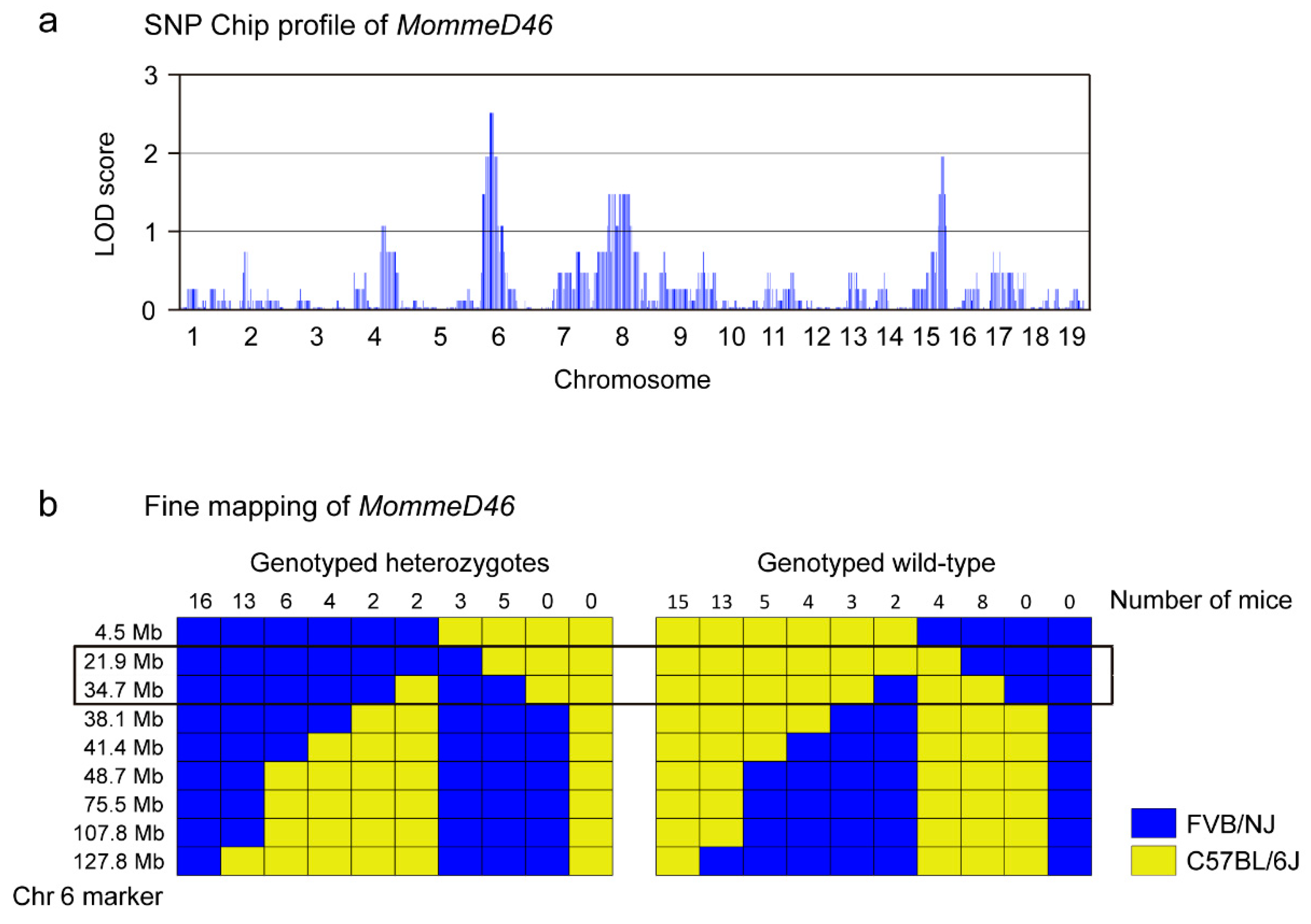
DNA from tail tips from 15 mice phenotypically expressing GFP in 55% of the erythrocytes (wild-type) and 15 expressing lower GFP percentages (MommeD46) from the backcross between heterozygous MommeD46 with Line3 background (FVB/NJ strain) and Line3C (C57BL/6J strain) mice were included in the linkage analysis (Figure 1a). Mice were at least three-weeks-old. The Mouse Medium Density Linkage Panel from Illumina (San Diego, CA, USA) was used for single nucleotide polymorphism (SNP) genotyping. The panel contains 766 measurable differential SNPs between C57BL/6J and FVB/NJ strains. Wild-type mice for the mutation should only have C57BL/6J-specific SNPs in the linked region while heterozygous MommeD46 mice can have both C57BL/6J and FVB/NJ-specific SNPs. SNP Chip was performed according to Illumina instructions and SNP calling was carried out using the Genotyping module of the GenomeStudio v1.1 software (Illumina). Only call rates higher than 95 were accepted. Logarithm of odds (LOD) scores across the entire mouse genome were calculated and linked intervals identified (LOD scores ˃ 2, Figure 1a and Figure 2). Further fine mapping using additional SNP markers (Figure S1) was performed to reduce the linked interval.

2.4. Whole Exome Sequencing

Whole exome sequencing for the identification of the underlying mutation in MommeD46 was carried out as previously described [7].

2.5. Methylation Analysis in the Transgene HS-40 Enhancer

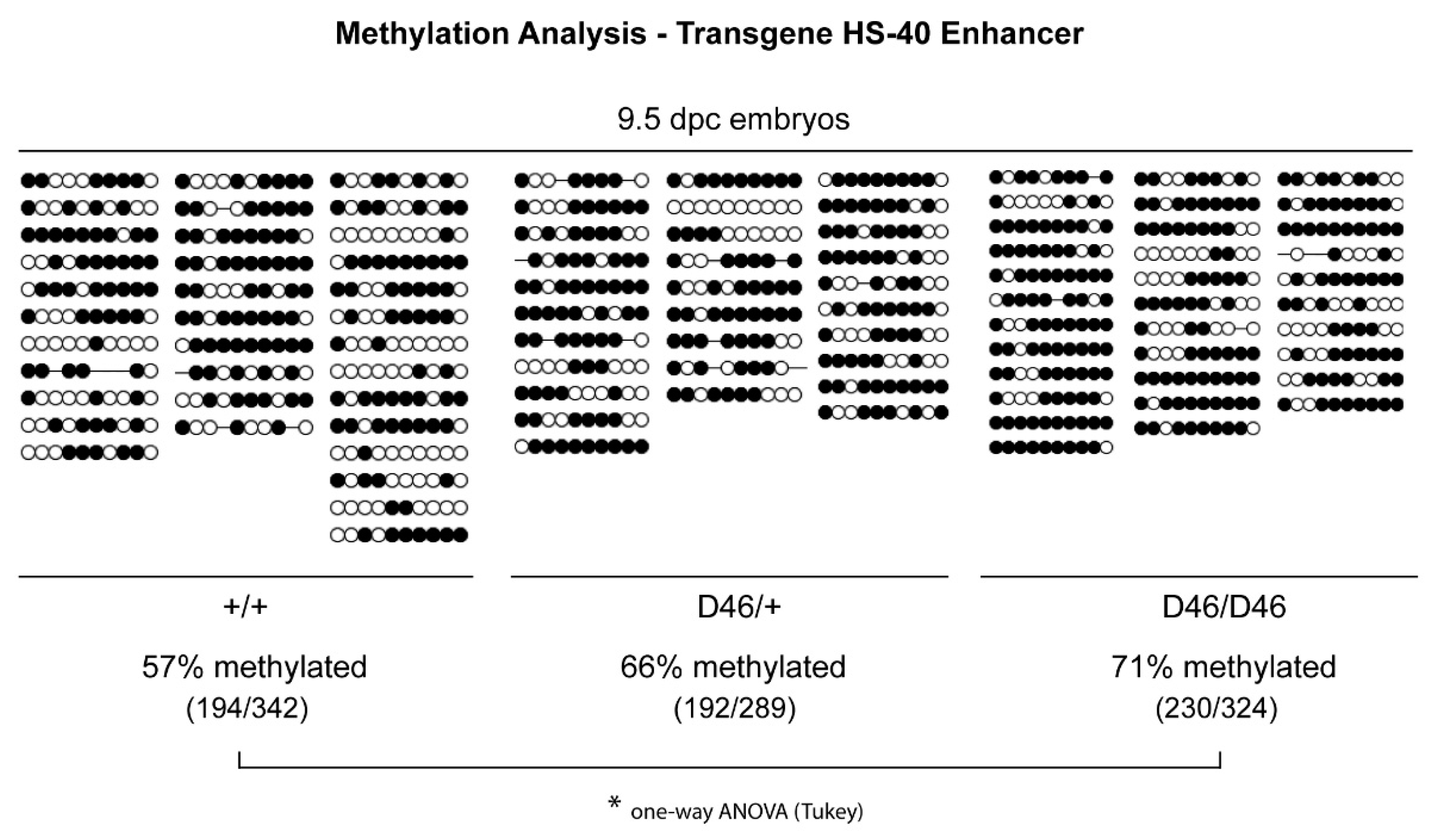
DNA samples from three wild-type, three heterozygous and three homozygous MommeD46 9.5 dpc embryos were converted with bisulfite using the EpiTect Bisulfite Kit (Qiagen, Doncaster, VIC, Australia) in accordance with the manufacturer’s instructions. Bisulfite-converted DNA was amplified (first PCR) using specific oligonucleotides (Integrated DNA Technologies, IA, USA) for the HS-40 enhancer region, as follows: 5′-AAAATAAAATTTTTGGATTGTTATTATTATAA-3′ (Forward 1), 5′-ATATTTGTAATTTTAGTATTTTGGGAGGTT-3′ (Forward 2) and 5′-AATCTCTACTCACTACAAACTCCATCTC-3′ (Reverse) with the following cycling conditions: one cycle at 94 °C for 2 min; 35 cycles at 94 °C for 30 s, 60 °C for 30 s, 72 °C for 45 s; one cycle at 72 °C for 6 min. Next, a semi-nested PCR was performed with the first PCR product using the oligonucleotides Forward 2 and Reverse. The product was then ligated into a pGEM®-T Vector (Promega, Madison, WI, USA) as per Promega instructions. DH5α competent E. coli were transformed using the heat shock method and plated in LB agar plates containing ampicillin, x-gal and IPTG. Positive colonies (white) were picked and DNA was amplified to determine transgene insertion using the following oligonucleotides: 5′-ATTTAGGTGACACTATAG-3′ (Forward, SP6) and 5′-TAATACGACTCACTATAGGG-3′ (Reverse, T7) and PCR conditions according to the MangoTaq DNA polymerase protocol (Bioline Reagents Limited, London, UK). Plasmid DNA from positive colonies was then extracted with Qiagen Miniprep Kit (Germantown, MD, USA) and sequenced. The BIQ Analyser software was used to determine the quality of the conversion and for the analysis. Sequences with <98% of conversion rate and <80% of sequence matching as well as identical clones were excluded from the analysis. White and dark circles-containing diagrams, obtained from introducing the BIQ results’matrix to the software freshteapot, were used to represent the unmethylated and methylated CpG sites in the HS-40 enhancer, respectively.

2.6. Genotyping

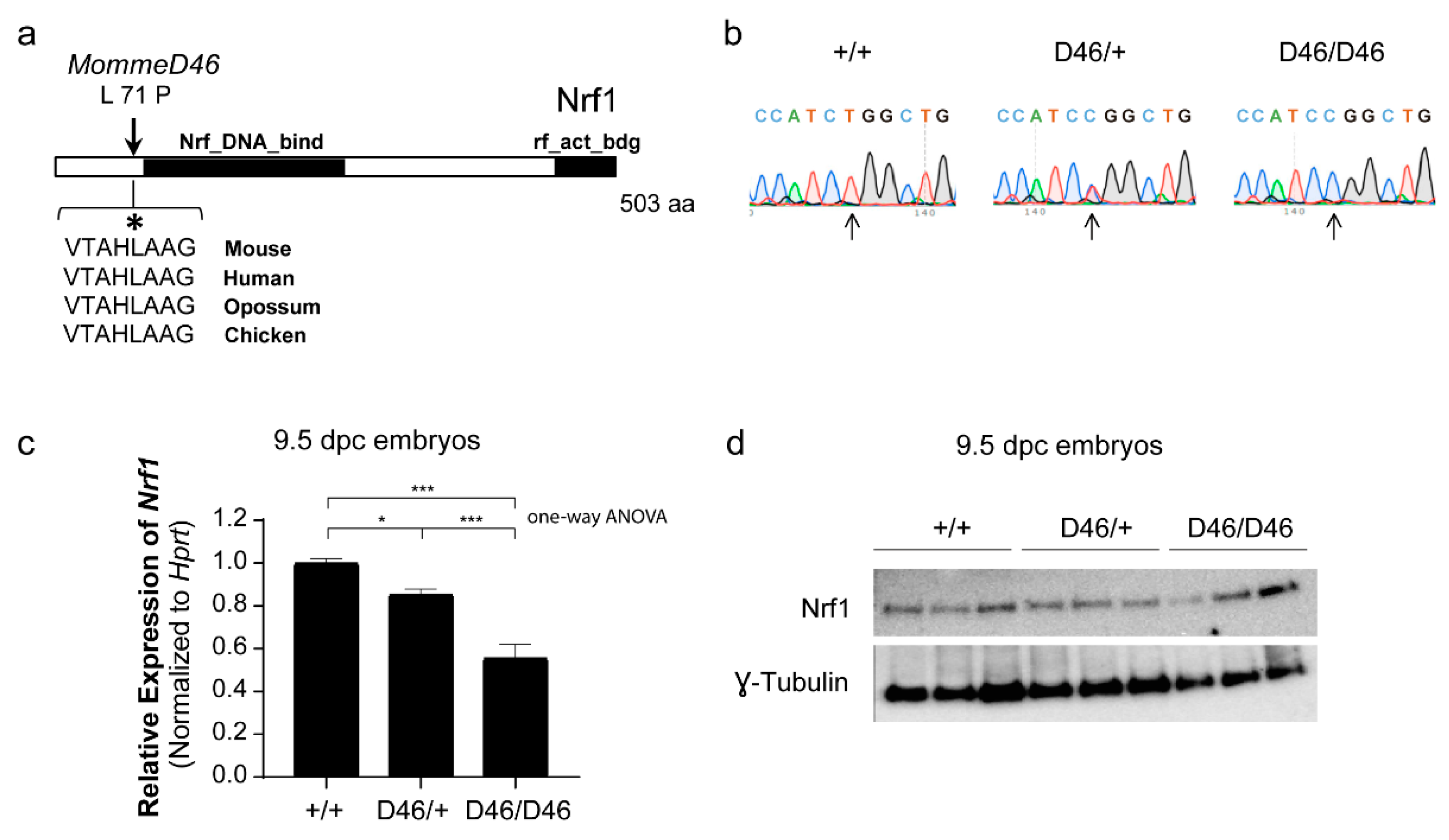
Genotyping of MommeD46 was performed by Sanger sequencing or by restriction enzyme digestion followed by gel electrophoresis. For both genotyping methods, DNA from tail tips (at least three-weeks-old) and embryos was extracted using a saturated salt solution containing 50 mM Tris pH 6.8, 20 mM EDTA pH 8, 2% SDS and 6.25 µg/mL proteinase K. DNA was precipitated with isopropanol and washed with 75% and 100% ethanol. DNA pellets were air dried and treated with 200 µL of RnaseA (10 mg/mL). Sanger sequencing was performed for 9.5 dpc embryos genotyping (Figure 3b). Wild-type, heterozygous and homozygous MommeD46 embryos (Figure 3b) were homogenized. DNA was isolated and a genomic region of 321 base pairs (bp) covering the causative mutation in Nrf1 was amplified by PCR using the oligonucleotides 5′-GGCTCAGCCAGTGTTTTCTTA-3′ (forward) and 5′-TTTGATCCCCAGCAGTGAA-3′ (reverse). The PCR product was then sequenced using the BigDye Terminator Sequencing Kit (Applied Biosystems, Foster City, CA, USA) and the previous forward oligonucleotide. The restriction enzyme digestion was performed for both adult and embryo genotyping. To do so, the same obtained above PCR product was subjected to digestion with HpaII (New England BioLabs, Ipswich, MA, USA). In wild-type MommeD46 the enzyme did not cut, in heterozygous MommeD46 the enzyme only cut one DNA strand, producing fragments of 321 bp, 173 bp and 148 bp, and in homozygous MommeD46 the enzyme cut both DNA strands, producing fragments of 173 bp and 148 bp.

2.7. Determination of Nrf1 mRNA Levels by Quantitative Real-Time PCR

The determination of Nrf1 mRNA levels was performed in three wild-type, three heterozygous and three homozygous MommeD46 of at least three weeks of age, following a three-step process which included RNA isolation, cDNA conversion and quantitative real-time (qRT)-PCR. For RNA isolation, 9.5 dpc embryos were mechanically homogenized and treated with Trizol® (Qiagen, Doncaster, VIC, Australia) and chloroform. RNA was precipitated with isopropanol and high salt solution (0.8 M NaCitrate and 1.2 M NaCl). RNA was washed with 75% ethanol, air dried and dissolved in DEPC water. RNA was converted into cDNA using SuperScriptIII reverse transcriptase and random hexamer primers (Invitrogen, Carlsbad, CA, USA). For the qRT-PCR, the forward oligonucleotide for Nrf1 was designed at the boundary of the first exon and the second exon (5′-TGAGGTCGAATGGTATGTGGT-3′), and the reverse oligonucleotide 78 bp downstream (5′-ACACCCATGTTCATAGCAGCA-3′), leaving a product of 121 bp. The DNA amplification was performed using the Platinum SYBR Green qPCR Super Mix—UDG (Invitrogen, Carlsbad, CA, USA). The PCR was run in the Viia7 (Applied Biosystems, Mulgrave, VIC, Australia) and analyzed using the Viia7 software. Hprt was used as housekeeping gene, using the qRT-PCR oligonucleotides 5′-GGCCAGACTTTGTTGGATTT-3′ (forward) and 5′-ACTGGCAACATCAACAGGACT-3′ (reverse).

2.8. Determination of Nrf1 Protein Levels by Western Blot

Whole-protein extracts were prepared from three wild-type, three heterozygous and three homozygous MommeD46 9.5 dpc embryos. Embryo tissue was mechanically homogenized and lysed with lysis buffer containing urea 8 M, 1% sodium dodecyl sulfate, 10% glycerol, 0.5 mM dithiothreitol and 10 mM Tris pH 6.8, complemented with the serine protease inhibitor phenylmethylsulfonyl fluoride and half tablet of the protease inhibitor cocktail cOmplete® from Roche (Swedesboro, NJ, USA) for 5 mL of lysis buffer. Next, samples were sonicated and quantified using the Pierce BCA Protein Assay Kit (ThermoFisher Scientific Australia Pty Ltd., Scoresby, VIC, Australia). Equal amounts of protein (30 µg) were loaded in a Mini-Protean precast 7.5% polyacrylamide gel from Bio-Rad (Hercules, CA, USA) and run at 100 V. Proteins were transferred into PVDF membranes following the wet transfer protocol, overnight at 4 °C at 20 V. Membranes were blocked with 5% of skim milk and 1% BSA in PBS-T. The anti-Nrf1 antibody from Novus Biologicals (Littleton, CO, USA) was incubated in the same solution overnight at 4 °C, 1:1000 dilution. A goat anti-rabbit secondary antibody conjugated to horseradish peroxidase (HRP) from DAKO, 1:5000 dilution in PBS-T, (Santa Clara, CA, USA) was added for one hour at room temperature. Clarity Western ECL substrate (Bio-Rad) was added in the membrane and the chemiluminescence signal was captured with the MF Chemi-Bis (DNR Bio-imaging systems). The γ-Tubulin antibody from Sigma-Aldrich (St Louis, MO, USA), 1:1000 dilution, was used as loading control.

2.9. Statistical Analysis

All the experiments were done in at least three biological replicates. Normality of data was assessed by Saphiro–Wilk or Kolmogorov–Smirnov tests, depending on the sample size. When the data were satisfied with normality assumption, statistical significance between mean differences of the groups was assessed by two-tailed Student’s t-test or one-way ANOVA and p < 0.05 was considered significant. Otherwise, the non-parametric Mann–Whitney U test was applied and p < 0.05 was considered significant. Chi–Square test (χ2) or Fisher’s exact test (F) were used to compare the observed proportion of genotypes with the expected Mendelian ratios when the number of events in a cell was ≥5 or <5, respectively, and a p value < 0.05 was deemed as significant. Levene’s test or F-test of equality of variances was used to determine variance differences between wild-type and heterozygous distributions, holding significance when p < 0.05. In this figure, two-way ANOVA was also employed. All analyses were performed using the software IBM SPSS Statistics version 24.0 (Chicago, IL, USA).

3. Results

3.1. MommeD46 Is an Enhancer of Variegation

The causative mutation in MommeD46 induces a change in the GFP expression in erythrocytes. Wild-type MommeD46 mice express the GFP in approximately 55% of red blood cells (Figure 1b), as previously shown for Line3 mice [7]. Heterozygous MommeD46 mutants present 44 ± 5% of GFP-expressing erythrocytes, which is significantly less (p = 2.15 × 10⁻²⁸) than the percentage observed in their wild-type counterparts (53 ± 4%). Figure 1c shows the flow cytometry GFP profiles of wild-type and heterozygous mutant MommeD46. Mutant mice show a lesser count. Therefore, MommeD46 is an enhancer of variegation.

3.2. MommeD46 Carries a Point Mutation in the Nrf1 Gene

To elucidate the underlying mutation in MommeD46, we followed different strategies. First, we performed SNP Chip to determine the linked genomic interval where the mutation was located. We identified two linked regions with LOD score of 2 or higher in Chromosome (Chr) 6 and Chr 15 (Figure 2a). As the highest LOD score was in Chr 6, we narrowed down the linked interval in that Chr, comprising a region of 123.3 Mb, from the genomic location 4.5 Mb to 127.8 Mb (Figure S1). We exploited nine differential SNP markers between FVB/NJ and C57BL/6J at 4.5, 21.9, 34.7, 38.1, 41.4, 48.7, 75.5, 107.8, and 127.8 Mb. We found a number of supporting genotypically heterozygous mice, and thus, with C57BL/6J-specific SNPs, concentrated at 21.9 and 34.7 Mb. At the same locations, we also found genotypically wild-type mice (with FVB/NJ-specific SNPs) (Figure 2b). This observation indicated that the underlying mutation in MommeD46 was located somewhere between 21.9 and 34.7 Mb. Later, we performed exome deep sequencing and we identified the causative mutation to be a missense mutation at the end of the third exon of Nrf1 (Chr 6:30,040,135; USC Genome Browser) (Figure S2). This was confirmed by Sanger sequencing in 9.5 dpc embryos (Figure 3b). The mutation is a T ˃ C substitution that induces a Leucine (L) ˃ Proline (P) change at position 71 of the amino acid sequence of Nrf1, located before the predicted Nrf1 DNA binding domain. The affected amino acid is conserved between species (Figure 3a). The point mutation in Nrf1 causes a decrease in the Nrf1 mRNA levels as seen by qRT-PCR. In particular, Nrf1MommeD46/+ and Nrf1MommeD46/MommeD46 9.5 dpc embryos showed a 15% (p = 0.014) and a 44% reduction (p = 0.0004) in Nrf1 mRNA levels, respectively, compared to Nrf1+/+ (Figure 3c). In contrast, the mutation did not induce a noticeable change in the Nrf1 protein levels as observed by Western blot (Figure 3d). In addition, the band quantification of the Western Blot did not show significant differences of band intensity between the groups when normalized to γ-Tubulin (Figure S3).

3.3. Nrf1 Mutants Display Enhanced DNA Methylation

To further prove a role of Nrf1 as epigenetic regulator in mammals, we performed bisulfite sequencing of the HS-40 enhancer region of the GFP transgene in wild-type, heterozygous and homozygous MommeD46 9.5 dpc embryos. We observed that Nrf1+/+ embryos presented 57% of the CpG islands methylated. This percentage increased up to 66% in Nrf1MommeD46/+ embryos, and up to 71% in Nrf1MommeD46/MommeD46 embryos (p = 0.049) (Figure 4). This result suggests that Nrf1 is involved in DNA methylation and a functional Nrf1 is essential to maintain a hypomethylated state, which is consistent with the fact that Nrf1 is an enhancer of variegation.

3.4. The Observed Hereditability of the Nrf1 Suggests Paternal Imprinting and Haploinsufficiency, and Normal Nrf1 Is Required for Normal Embryonic Development

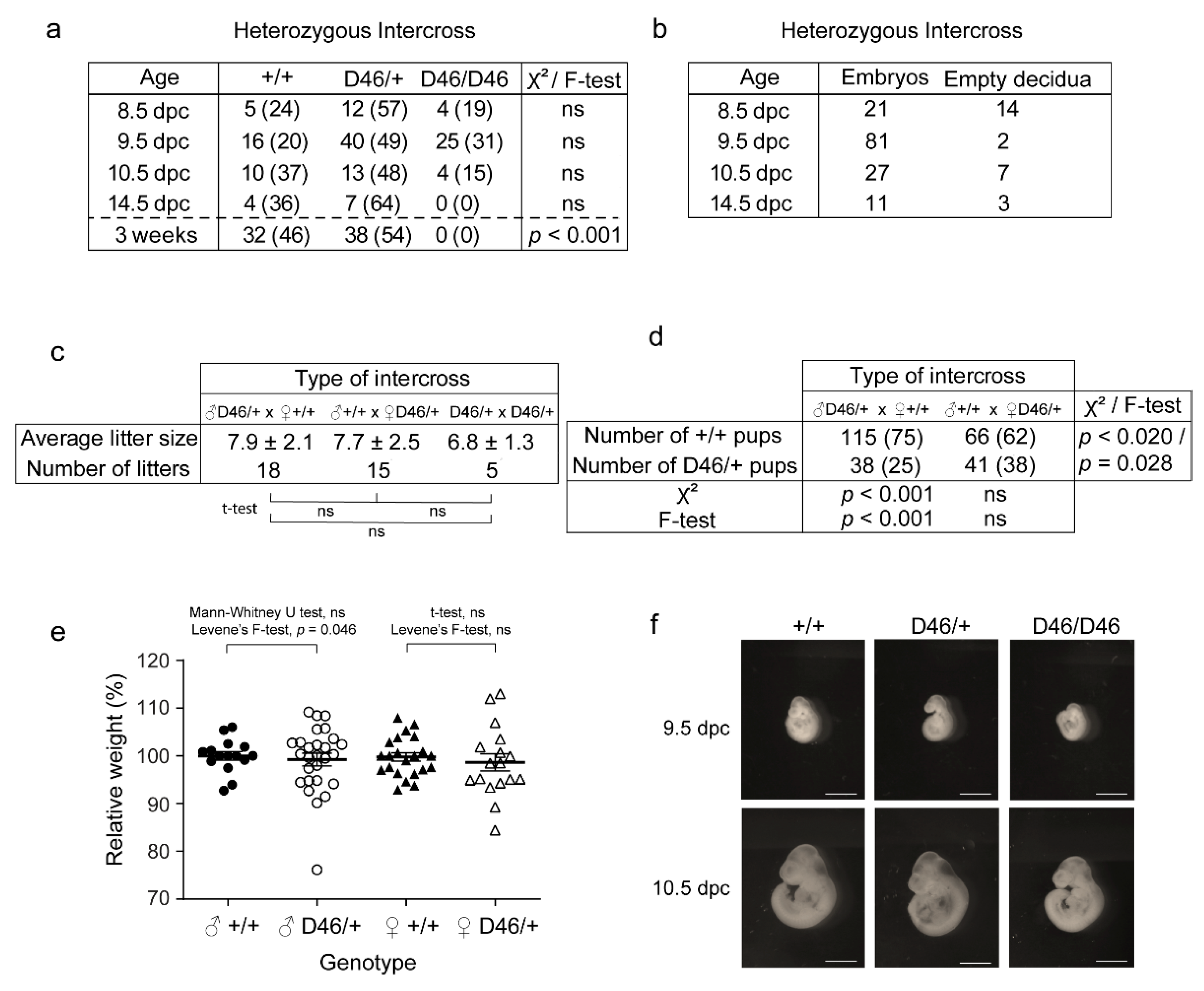
Herein, we wanted to determine whether the newly discovered mutation in Nrf1 was responsible for any phenotypic change or defects in mice. To do so, we performed different intercrosses between adult Nrf1MommeD46/+ mice. We observed proportions of Nrf1+/+, Nrf1MommeD46/+ and Nrf1MommeD46/MommeD46 to be significantly different than that expected by Mendel at weaning (p < 0.001) (Figure 5a). Interestingly, neither homozygous mice were observed at weaning nor at the embryonic stage of 14.5 dpc indicating an earlier embryonic lethality of the mutation in homozygosis. In the earlier embryonic stages of 8.5, 9.5 and 10.5 dpc, homozygous embryos were observed at decreased ratios than expected (not significant). When further analyzing the homozygous embryos, we detected the presence of empty decidua in some of them, all presumably being Nrf1MommeD46/MommeD46, while the rest resembled embryos (Figure 5b). However, some of them looked abnormal. Male and female heterozygous mice showed similar fertility, producing an average of 7.9 ± 2.1 pups per litter when the heterozygous progenitor was a male, and 7.7 ± 2.5 pups when the heterozygous progenitor was a female. As expected, the heterozygous intercross produced less pups per litter, specifically 6.8 ± 1.3 (Figure 5c). To determine any possible progenitor effect in the hereditability of the mutation, we genotyped the offspring of two types of intercrosses: a heterozygous mutant male crossed with a wild-type female, and a wild-type male crossed with a heterozygous mutant female. The expected Mendelian proportions for both intercrosses were 50% wild-type and 50% heterozygous mutants. However, we observed different proportions in both intercrosses, with the ratios significantly different than expected (p < 0.001) for the intercross including a female wild-type. Moreover, the percentage of wild-type pups obtained from the intercross containing the female wild-type was significantly greater (75%) than the percentage obtained from the intercross having the male wild-type (62%) (p < 0.020) (Figure 5d). Altogether, these results suggest an accentuated maternal hereditability and some degree of haploinsufficiency of the described mutation in Nrf1 which is further discussed in the next section. Finally, to ascertain any possible embryonic or adult defects caused by the mutation in Nrf1, we determined the body weights of Nrf1+/+ and Nrf1MommeD46/+ mice at weaning and explored the morphology of Nrf1+/+, Nrf1MommeD46/+ and Nrf1MommeD46/MommeD46 embryos at different embryonic stages. As seen in Figure 5e, heterozygous mutant mice weighed less than their wild-type counterparts in both sexes (not significant), with significantly different variances found in males (Levene’s F-test, p < 0.05). When examining the embryos at 8.5 and 9.5 dpc, we observed that homozygous mutant embryos were clearly smaller and less developed than the wild-type or heterozygous mutants (Figure 5f). However, no differences were detected between wild-type and heterozygous mutant embryos. This observation highlights the importance of Nrf1 for correct embryonic development.

4. Discussion

By deploying a forward dominant ENU mutagenesis screen in mice, we have discovered a novel missense mutation in Nrf1 capable of altering the epigenetic state. Additional mutations were found in the link obtained by SNP-Chip but outside the fine mapped interval (Figure S4), which reassured us that the mutation in Nrf1 was causative of the phenotype. Nrf1 is an enhancer of variegation as seen by the decrease in the percentage of GFP positive erythrocytes in heterozygous mutant mice. The exonic mutation in Nrf1 is responsible for the amino acid (aa) change L ˃ P at the position 71 of the Nrf1 protein (total length: 503 aas) and is located in the aa region responsible for Nrf1 dimerization. This region ranges between 1 and 78 aas as the previously generated 1-to-78-deletion mutant has been shown to be entirely monomeric [22]. This same mutant also binds DNA less robustly compared to the wild-type protein [22], consistent with a possible overlap with the Nrf1 DNA binding domain, predicted to be between 79 and 172 in the chicken Nrf1 [23]. This suggests that the 71L ˃ P substitution of MommeD46 could perturb both the homodimerization and DNA binding ability of Nrf1. Nrf1MommeD46/+ and Nrf1MommeD46/MommeD46 embryos show substantial reduction in the Nrf1 mRNA levels indicating that the mutation could affect mRNA synthesis and processing. In contrast, Nrf1 protein levels do not seem to be altered in the mutant embryos. This could be due to increases in the half-life of the mutated Nrf1 protein or an increase in the Nrf1MommeD46 mRNA translation, processes that have been reviewed in Liu et al. [24]. As for studies with Nrf1, Dhar et al. found a drastic reduction in Nrf1 mRNA levels by qRT-PCR accompanied with a not so accentuated decrease in protein levels by Western blot in murine neuroblastoma cells [25].
We are the first to demonstrate a role of Nrf1 in maintaining DNA hypomethylation in vivo as seen by the significant hypermethylation of the α-globin HS-40 enhancer region observed in Nrf1MommeD46/+ and Nrf1MommeD46/MommeD46 embryos in comparison with wild-type embryos. Interestingly, only this present MommeD46 and the previously published MommeD28 (Rlf null mutant) [7] show hypermethylation in the HS-40 enhancer. This is consistent with the decreased expression of the GFP transgene. Although the preference of NRF1 to bind un-methylated regions [26,27] and methylated sequences [28] has been studied in depth, little is known about the role of Nrf1 in directly controlling DNA methylation. In this line, one study has found that Nrf1 transcriptionally activates Euchromatic histone methyltransferase 1 (EHMT1) during in vitro meiosis, and that its DNA binding ability is negatively regulated by CDK2-mediated phosphorylation [29]. However, the authors did not assess DNA methylation changes. In another work, the authors did not find changes in DNA methylation in the gene promoters of the six Nrf1 target genes analyzed in CD9+ spermatogonia from Nrf1 germ cell-conditional knockout (Nrf1f/f) mice [27]. In contrast, in our study, Nrf1 was isolated from a genome-wide genetic screen in search of epigenetic modifiers and we found methylation changes in the HS-40 enhancer in mutants with dysfunctional Nrf1. Besides all this, it could be that NRF1 indirectly maintains DNA hypomethylation. The mutation L71P in Nrf1 could reduce the ability of Nrf1 to bind the DNA, such that Nrf1 is then unable to protect the CpG sites from methylation, leading to a methylation gain as previously described [26].
According to our embryonic and adult examinations of MommeD46, Nrf1MommeD46/MommeD46 mice are not viable at birth and die before 14.5 dpc. Also, the offspring of the heterozygous intercross present significantly altered Mendelian ratios at weaning. Similarly, the Nrf1 full knockout has shown to be embryonic lethal between 3.5 and 6.5 dpc [30]. Regarding mice fertility, our mutation in Nrf1 does not seem to affect fertility as seen by the similar average litter sizes obtained from the cross Nrf1+/+ × Nrf1MommeD46/+ for both progenitor genders. However, in the literature, conditional ablation of Nrf1 in gonocytes has been shown to lead to infertility in male mice [27]. This contrasting evidence could be due to the subtle nonobvious effect that our mutation has on fertility. We observed that the wild-type Nrf1 allele is significantly more present than expected in the offspring when the progenitor is a wild-type female rather than when it is a wild-type male (75% versus 62%). This could be explained by paternal imprinting of the mutated allele in the mating ♀Nrf1+/+ × ♂Nrf1MommeD46/+, by maternal imprinting of the mutated allele in the mating ♀Nrf1MommeD46/+ × ♂Nrf1+/+, by haploinsufficiency or by the existence of an additional ENU mutation having a cis- or trans-acting effect on the wild-type or the mutated allele. Maternal imprinting or inhibitory cis-/trans-acting effects on the mutated allele does not occur because the proportion of Nrf1MommeD46/+ progeny from the mating ♀Nrf1MommeD46/+ × ♂Nrf1+/+ is not significantly different from the expected Mendelian ratios, 38% versus 50%. In contrast, the possibility of activating cis- or trans-activating effects on the wildtype allele due to another ENU mutation cannot be ruled out, but, at the same time, is very difficult to prove. Regarding Nrf1 inheritance, the literature shows that there is no maternal inheritance of Nrf1 gene products as wild-type embryo eggs (Nrf1+) from the Nrf1null/+ heterozygous intercross were seen at the stage before the first meiotic division [30]. Regarding paternal imprinting, NRF1 has previously been shown to positively regulate the small nuclear ribonucleoprotein polypeptide N (SNRPN) locus which is associated with the imprinting center that regulates the Prader–Willi syndrome domain (PWS-IC), suggesting a relevant contribution of NRF1 in PWS-IC activity [31,32]. In other words, NRF1 orchestrates the characteristic loss of expression of the paternally-inherited allele of an imprinted gene cluster, that includes SNRPN, in the PWS. This suggests that the mutation in MommeD46, which affects the dimerization and DNA binding capabilities of Nrf1, could generate imprinting defects and be responsible for the increase in the expression of the maternal wild-type allele observed. Another possibility that could explain the greater presence of the wild-type allele in the offspring when the progenitor is a wild-type female is the haploinsufficiency caused by the mutated Nrf1 allele in the heterozygous mice. The early lethality of the homozygous mutants supports this notion.
Regarding the effects of the mutation in embryos and adult phenotypes, we observed a decrease in size and developmental delay in 9.5 and 10.5 Nrf1MommeD46/MommeD46 embryos compared to wild-type embryos. Moreover, both male and female Nrf1MommeD46/+ mice display a slight decrease in body weight compared to wild-type mice at weaning, with significantly different variances in males. Similarly, a previous study demonstrated defective development in vitro of all Nrf1-null and some Nrf1 heterozygous blastocysts as opposed to their wild-type counterparts [30]. In mice, Nrf1 is highly expressed in thymocytes, B-cells and retina, and in humans, in natural killer cells, T cells and skeletal muscle. Consistent with that, retinal progenitor cell and postmitotic rod photoreceptor cell-specific Nrf1 conditional knockout mice exhibit rod photoreceptor cell degeneration as a result of mitochondrial impairment [33]. In addition, Drosophila erect wing (ewg) (Nrf1 homologue) mutants show aberrant axonal projection and absent or reduced indirect flight muscles [34], and zebrafish nrf (Nrf1 homologue) mutants exhibit loss of retinal photoreceptors and reduced brain size [35]. Also in the neural context, reduced transcriptional activity of NRF1 has been associated with fragile X mental retardation syndrome [36] and with GABA-associated neuronal disorders [37], and functional NRF1 has been shown to be necessary for neurite outgrowth of human neuroblastoma cells [38,39]. Other non-neuronal roles attributed to NRF1 are the regulation of oncogenesis [40,41,42,43], stem cell aging [44], telomere transcription [45] and oxidative stress protection and lipid homeostasis in the liver; liver-specific Nrf1 conditional knockout mice develop steatohepatitis and spontaneous liver cancer [43]. In addition, low levels of (mRNA) Nrf1 have been observed in Cystathionine β synthase (CBS) heterozygous-null mice modeling hyperhomocysteinemia, a syndrome characterized by skeletal muscle weakness and fatigability [46]. However, it remains unknown whether Nrf1 is a causative factor or a consequence of the disease. Interestingly, CBS+/− mice exhibit significant weight loss [47], and homocysteine treatment, as a way to model the disease in vitro, increases the expression of DNA methyltransferase (DNMT)3a and DNMT3b and consequent global methylation levels in muscle C2C12 cells [46], which is in agreement with our findings of Nrf1 controlling the epigenetic state and the decrease in body weight observed in Nrf1MommeD46/+ mice. Alternatively, given that Nrf1 controls muscle energy production [48], it could be that the decrease in body weight in the heterozygous Nrf1 mutants is due to the reduction in muscle function and subsequent muscle mass. Further investigations will be undertaken to unravel other phenotypic traits or defects caused by the 71L ˃ P substitution in Nrf1.

5. Conclusions

By using a genome-wide mutagenesis screen, we have identified Nrf1 acting as a controller of the epigenetic landscape in mammals, through the maintenance of DNA hypomethylation in vivo. This novel MommeD46 causes a reduction in the Nrf1 mRNA levels and severe embryonic developmental defects. Additionally, we noted a peculiar hereditability of the mutation which is consistent with a paternal imprinting of the Nrf1MommeD46 allele and haploinsufficiency. Our study unveils an important new epi-role of NRF1 in addition to its extensive transcriptional regulation throughout the animal life. Furthermore, the variant mouse produced in our laboratory could be used to model or elucidate unknown syndromes driven by a reduced function of NRF1.

Supplementary Materials

The following are available online at https://www.mdpi.com/article/10.3390/ani11072103/s1, Figure S1: fine mapping of MommeD46, Figure S2: exome sequencing in MommeD46, Figure S3: Full Western Blot images and band quantification corresponding to Figure 3d. Full Western Blot images of (a) Nrf1 and (b) γ-Tubulin, Figure S4: Deep genome sequencing of the linked interval in MommeD46.

Author Contributions

Conceptualization, A.S.; methodology, M.A.S. and M.M.; software, M.A.S. and E.P.; formal analysis, A.S.; investigation, M.A.S., M.M. and A.S.; resources, A.S.; writing—original draft preparation, A.S.; writing—review and editing, M.A.S., E.P. and A.S.; supervision, A.S.; project administration, A.S.; funding acquisition, A.S. All authors have read and agreed to the published version of the manuscript.

Funding

This research was funded by the National Health and Medical Research Council of Australia under Grant APP0552445, along with the 2020 Marie Skłodowska-Curie Individual Fellowship from the European Commission under Grant 893384, 2020 Miguel Servet fellowship from the Instituto de Salud Carlos III (Spanish National Institute of Health) under Grant CP20/00039 and European Social Fund (ESF) “Investing in your future”, and the Raine Medical Research Foundation under Grant RPG-004-19 given to A.S., and the Emergent Research Group Recognition Award from the University and Research Grants Management Agency of Catalonia (Spain) under Grant 2017SRG1620 given to M.A.S. The research was supported by CERCA Programme of Generalitat de Catalunya and the IRBLleida—Fundació Dr. Pifarré.

Institutional Review Board Statement

The study was conducted according to the guidelines of the Declaration of Helsinki and approved by the Institutional Review Board (or Ethics Committee) of the Queensland Institute of Medical Research (RA3/100/1336) with date of approval of 12/12/2009.

Acknowledgments

We would like to thank Harald Oey (University of Queensland) for performing the whole exome sequencing and the bioinformatic analysis.

Conflicts of Interest

The authors declare no conflict of interest. The funders had no role in the design of the study; in the collection, analyses, or interpretation of data; in the writing of the manuscript, or in the decision to publish the results.

References

  1. Henikoff, S. Position-effect variegation after 60 years. Trends Genet. 1990, 6, 422–426. [Google Scholar] [CrossRef]
  2. Dorn, R.; Szidonya, J.; Korge, G.; Sehnert, M.; Taubert, H.; Archoukieh, E.; Tschiersch, B.; Morawietz, H.; Wustmann, G.; Hoffmann, G.; et al. P transposon-induced dominant enhancer mutations of position-effect variegation in Drosophila melanogaster. Genetics 1993, 133, 279–290. [Google Scholar] [CrossRef] [PubMed]
  3. Reuter, G.; Wolff, I. Isolation of dominant suppressor mutations for position-effect variegation in Drosophila melanogaster. Mol. Gen. Genet. 1981, 182, 516–519. [Google Scholar] [CrossRef] [PubMed]
  4. Blewitt, M.E.; Vickaryous, N.K.; Hemley, S.J.; Ashe, A.; Bruxner, T.J.; Preis, J.I.; Arkell, R.; Whitelaw, E. An N-ethyl-N-nitrosourea screen for genes involved in variegation in the mouse. Proc. Natl. Acad. Sci. USA 2005, 102, 7629–7634. [Google Scholar] [CrossRef] [PubMed] [Green Version]
  5. Preis, J.I.; Downes, M.; Oates, N.A.; Rasko, J.E.; Whitelaw, E. Sensitive flow cytometric analysis reveals a novel type of parent-of-origin effect in the mouse genome. Curr. Biol. 2003, 13, 955–959. [Google Scholar] [CrossRef] [Green Version]
  6. Sorolla, A.; Tallack, M.R.; Oey, H.; Harten, S.K.; Daxinger, L.C.; Magor, G.W.; Combes, A.N.; Ilsley, M.; Whitelaw, E.; Perkins, A.C. Identification of novel hypomorphic and null mutations in Klf1 derived from a genetic screen for modifiers of alpha-globin transgene variegation. Genomics 2015, 105, 116–122. [Google Scholar] [CrossRef]
  7. Daxinger, L.; Harten, S.K.; Oey, H.; Epp, T.; Isbel, L.; Huang, E.; Whitelaw, N.; Apedaile, A.; Sorolla, A.; Yong, J.; et al. An ENU mutagenesis screen identifies novel and known genes involved in epigenetic processes in the mouse. Genome Biol. 2013, 14, R96. [Google Scholar] [CrossRef] [Green Version]
  8. Blewitt, M.E.; Gendrel, A.V.; Pang, Z.; Sparrow, D.B.; Whitelaw, N.; Craig, J.M.; Apedaile, A.; Hilton, D.J.; Dunwoodie, S.L.; Brockdorff, N.; et al. SmcHD1, containing a structural-maintenance-of-chromosomes hinge domain, has a critical role in X inactivation. Nat. Genet. 2008, 40, 663–669. [Google Scholar] [CrossRef]
  9. Chong, S.; Vickaryous, N.; Ashe, A.; Zamudio, N.; Youngson, N.; Hemley, S.; Stopka, T.; Skoultchi, A.; Matthews, J.; Scott, H.S.; et al. Modifiers of epigenetic reprogramming show paternal effects in the mouse. Nat. Genet. 2007, 39, 614–622. [Google Scholar] [CrossRef] [Green Version]
  10. Ashe, A.; Morgan, D.K.; Whitelaw, N.C.; Bruxner, T.J.; Vickaryous, N.K.; Cox, L.L.; Butterfield, N.C.; Wicking, C.; Blewitt, M.E.; Wilkins, S.J.; et al. A genome-wide screen for modifiers of transgene variegation identifies genes with critical roles in development. Genome Biol. 2008, 9, R182. [Google Scholar] [CrossRef] [Green Version]
  11. Harten, S.K.; Bruxner, T.J.; Bharti, V.; Blewitt, M.; Nguyen, T.M.; Whitelaw, E.; Epp, T. The first mouse mutants of D14Abb1e (Fam208a) show that it is critical for early development. Mamm. Genome 2014, 25, 293–303. [Google Scholar] [CrossRef] [PubMed] [Green Version]
  12. Harten, S.K.; Oey, H.; Bourke, L.M.; Bharti, V.; Isbel, L.; Daxinger, L.; Faou, P.; Robertson, N.; Matthews, J.M.; Whitelaw, E. The recently identified modifier of murine metastable epialleles, Rearranged L-Myc Fusion, is involved in maintaining epigenetic marks at CpG island shores and enhancers. BMC Biol. 2015, 13, 21. [Google Scholar] [CrossRef] [PubMed] [Green Version]
  13. Isbel, L.; Srivastava, R.; Oey, H.; Spurling, A.; Daxinger, L.; Puthalakath, H.; Whitelaw, E. Trim33 Binds and Silences a Class of Young Endogenous Retroviruses in the Mouse Testis; a Novel Component of the Arms Race between Retrotransposons and the Host Genome. PLoS Genet. 2015, 11, e1005693. [Google Scholar] [CrossRef] [PubMed] [Green Version]
  14. Isbel, L.; Prokopuk, L.; Wu, H.; Daxinger, L.; Oey, H.; Spurling, A.; Lawther, A.J.; Hale, M.W.; Whitelaw, E. Wiz binds active promoters and CTCF-binding sites and is required for normal behaviour in the mouse. eLife 2016, 5. [Google Scholar] [CrossRef] [PubMed] [Green Version]
  15. Lemmers, R.J.; Tawil, R.; Petek, L.M.; Balog, J.; Block, G.J.; Santen, G.W.; Amell, A.M.; van der Vliet, P.J.; Almomani, R.; Straasheijm, K.R.; et al. Digenic inheritance of an SMCHD1 mutation and an FSHD-permissive D4Z4 allele causes facioscapulohumeral muscular dystrophy type 2. Nat. Genet. 2012, 44, 1370–1374. [Google Scholar] [CrossRef] [Green Version]
  16. Leong, H.S.; Chen, K.; Hu, Y.; Lee, S.; Corbin, J.; Pakusch, M.; Murphy, J.M.; Majewski, I.J.; Smyth, G.K.; Alexander, W.S.; et al. Epigenetic regulator Smchd1 functions as a tumor suppressor. Cancer Res. 2013, 73, 1591–1599. [Google Scholar] [CrossRef] [PubMed] [Green Version]
  17. Bourke, L.M.; Del Monte-Nieto, G.; Outhwaite, J.E.; Bharti, V.; Pollock, P.M.; Simmons, D.G.; Adam, A.; Hur, S.S.; Maghzal, G.J.; Whitelaw, E.; et al. Loss of Rearranged L-Myc Fusion (RLF) results in defects in heart development in the mouse. Differentiation 2017, 94, 8–20. [Google Scholar] [CrossRef] [PubMed]
  18. Evans, M.J.; Scarpulla, R.C. Interaction of nuclear factors with multiple sites in the somatic cytochrome c promoter. Characterization of upstream NRF-1, ATF, and intron Sp1 recognition sequences. J. Biol. Chem. 1989, 264, 14361–14368. [Google Scholar] [CrossRef]
  19. Ramachandran, B.; Yu, G.; Gulick, T. Nuclear respiratory factor 1 controls myocyte enhancer factor 2A transcription to provide a mechanism for coordinate expression of respiratory chain subunits. J. Biol. Chem. 2008, 283, 11935–11946. [Google Scholar] [CrossRef] [PubMed] [Green Version]
  20. Virbasius, J.V.; Scarpulla, R.C. Activation of the human mitochondrial transcription factor A gene by nuclear respiratory factors: A potential regulatory link between nuclear and mitochondrial gene expression in organelle biogenesis. Proc. Natl. Acad. Sci. USA 1994, 91, 1309–1313. [Google Scholar] [CrossRef] [PubMed] [Green Version]
  21. Wu, Z.; Puigserver, P.; Andersson, U.; Zhang, C.; Adelmant, G.; Mootha, V.; Troy, A.; Cinti, S.; Lowell, B.; Scarpulla, R.C.; et al. Mechanisms controlling mitochondrial biogenesis and respiration through the thermogenic coactivator PGC-1. Cell 1999, 98, 115–124. [Google Scholar] [CrossRef] [Green Version]
  22. Gugneja, S.; Scarpulla, R.C. Serine phosphorylation within a concise amino-terminal domain in nuclear respiratory factor 1 enhances DNA binding. J. Biol. Chem. 1997, 272, 18732–18739. [Google Scholar] [CrossRef] [Green Version]
  23. Gómez-Cuadrado, A.; Martín, M.; Noël, M.; Ruiz-Carrillo, A. Initiation binding repressor, a factor that binds to the transcription initiation site of the histone h5 gene, is a glycosylated member of a family of cell growth regulators [corrected]. Mol. Cell. Biol. 1995, 15, 6670–6685. [Google Scholar] [CrossRef] [Green Version]
  24. Liu, Y.; Beyer, A.; Aebersold, R. On the Dependency of Cellular Protein Levels on mRNA Abundance. Cell 2016, 165, 535–550. [Google Scholar] [CrossRef] [PubMed] [Green Version]
  25. Dhar, S.S.; Ongwijitwat, S.; Wong-Riley, M.T. Nuclear respiratory factor 1 regulates all ten nuclear-encoded subunits of cytochrome c oxidase in neurons. J. Biol. Chem. 2008, 283, 3120–3129. [Google Scholar] [CrossRef] [PubMed] [Green Version]
  26. Domcke, S.; Bardet, A.F.; Adrian Ginno, P.; Hartl, D.; Burger, L.; Schubeler, D. Competition between DNA methylation and transcription factors determines binding of NRF1. Nature 2015, 528, 575–579. [Google Scholar] [CrossRef] [PubMed]
  27. Wang, J.; Tang, C.; Wang, Q.; Su, J.; Ni, T.; Yang, W.; Wang, Y.; Chen, W.; Liu, X.; Wang, S.; et al. NRF1 coordinates with DNA methylation to regulate spermatogenesis. FASEB J. 2017, 31, 4959–4970. [Google Scholar] [CrossRef] [PubMed] [Green Version]
  28. Hu, S.; Wan, J.; Su, Y.; Song, Q.; Zeng, Y.; Nguyen, H.N.; Shin, J.; Cox, E.; Rho, H.S.; Woodard, C.; et al. DNA methylation presents distinct binding sites for human transcription factors. eLife 2013, 2, e00726. [Google Scholar] [CrossRef] [PubMed]
  29. Palmer, N.; Talib, S.Z.A.; Ratnacaram, C.K.; Low, D.; Bisteau, X.; Lee, J.H.S.; Pfeiffenberger, E.; Wollmann, H.; Tan, J.H.L.; Wee, S.; et al. CDK2 regulates the NRF1/Ehmt1 axis during meiotic prophase I. J. Cell. Biol. 2019, 218, 2896–2918. [Google Scholar] [CrossRef] [PubMed] [Green Version]
  30. Huo, L.; Scarpulla, R.C. Mitochondrial DNA instability and peri-implantation lethality associated with targeted disruption of nuclear respiratory factor 1 in mice. Mol. Cell. Biol. 2001, 21, 644–654. [Google Scholar] [CrossRef] [PubMed] [Green Version]
  31. Rodriguez-Jato, S.; Nicholls, R.D.; Driscoll, D.J.; Yang, T.P. Characterization of cis- and trans-acting elements in the imprinted human SNURF-SNRPN locus. Nucleic Acids Res. 2005, 33, 4740–4753. [Google Scholar] [CrossRef] [PubMed] [Green Version]
  32. Rodriguez-Jato, S.; Shan, J.; Khadake, J.; Heggestad, A.D.; Ma, X.; Johnstone, K.A.; Resnick, J.L.; Yang, T.P. Regulatory elements associated with paternally-expressed genes in the imprinted murine Angelman/Prader-Willi syndrome domain. PLoS ONE 2013, 8, e52390. [Google Scholar] [CrossRef] [PubMed]
  33. Kiyama, T.; Chen, C.K.; Wang, S.W.; Pan, P.; Ju, Z.; Wang, J.; Takada, S.; Klein, W.H.; Mao, C.A. Essential roles of mitochondrial biogenesis regulator Nrf1 in retinal development and homeostasis. Mol. Neurodegener. 2018, 13, 56. [Google Scholar] [CrossRef] [PubMed]
  34. DeSimone, S.M.; White, K. The Drosophila erect wing gene, which is important for both neuronal and muscle development, encodes a protein which is similar to the sea urchin P3A2 DNA binding protein. Mol. Cell. Biol. 1993, 13, 3641–3649. [Google Scholar] [CrossRef] [PubMed] [Green Version]
  35. Becker, T.S.; Burgess, S.M.; Amsterdam, A.H.; Allende, M.L.; Hopkins, N. not really finished is crucial for development of the zebrafish outer retina and encodes a transcription factor highly homologous to human Nuclear Respiratory Factor-1 and avian Initiation Binding Repressor. Development 1998, 125, 4369–4378. [Google Scholar] [CrossRef]
  36. Kumari, D.; Usdin, K. Interaction of the transcription factors USF1, USF2, and alpha -Pal/Nrf-1 with the FMR1 promoter. Implications for Fragile X mental retardation syndrome. J. Biol. Chem. 2001, 276, 4357–4364. [Google Scholar] [CrossRef] [Green Version]
  37. Li, Z.; Cogswell, M.; Hixson, K.; Brooks-Kayal, A.R.; Russek, S.J. Nuclear Respiratory Factor 1 (NRF-1) Controls the Activity Dependent Transcription of the GABA-A Receptor Beta 1 Subunit Gene in Neurons. Front. Mol. Neurosci. 2018, 11. [Google Scholar] [CrossRef] [Green Version]
  38. Chang, W.T.; Chen, H.I.; Chiou, R.J.; Chen, C.Y.; Huang, A.M. A novel function of transcription factor alpha-Pal/NRF-1: Increasing neurite outgrowth. Biochem. Biophys. Res. Commun. 2005, 334, 199–206. [Google Scholar] [CrossRef]
  39. Tong, C.W.; Wang, J.L.; Jiang, M.S.; Hsu, C.H.; Chang, W.T.; Huang, A.M. Novel genes that mediate nuclear respiratory factor 1-regualted neurite outgrowth in neuroblastoma IMR-32 cells. Gene 2013, 515, 62–70. [Google Scholar] [CrossRef]
  40. Lee, C.H.; Wu, S.B.; Hong, C.H.; Liao, W.T.; Wu, C.Y.; Chen, G.S.; Wei, Y.H.; Yu, H.S. Aberrant cell proliferation by enhanced mitochondrial biogenesis via mtTFA in arsenical skin cancers. Am. J. Pathol. 2011, 178, 2066–2076. [Google Scholar] [CrossRef] [Green Version]
  41. Zhou, Y.; Xu, Z.; Quan, D.; Zhang, F.; Zhang, H.; Xiao, T.; Hou, S.; Qiao, H.; Harismendy, O.; Wang, J.Y.J.; et al. Nuclear respiratory factor 1 promotes spheroid survival and mesenchymal transition in mammary epithelial cells. Oncogene 2018, 37, 6152–6165. [Google Scholar] [CrossRef] [PubMed]
  42. Kumar, R.; Bhat, T.A.; Walsh, E.M.; Chaudhary, A.K.; O’Malley, J.; Rhim, J.S.; Wang, J.; Morrison, C.D.; Attwood, K.; Bshara, W.; et al. Cytochrome c Deficiency Confers Apoptosome and Mitochondrial Dysfunction in African-American Men with Prostate Cancer. Cancer Res. 2019, 79, 1353–1368. [Google Scholar] [CrossRef] [PubMed] [Green Version]
  43. Xu, Z.; Chen, L.; Leung, L.; Yen, T.S.; Lee, C.; Chan, J.Y. Liver-specific inactivation of the Nrf1 gene in adult mouse leads to nonalcoholic steatohepatitis and hepatic neoplasia. Proc. Natl. Acad. Sci. USA 2005, 102, 4120–4125. [Google Scholar] [CrossRef] [PubMed] [Green Version]
  44. Mohrin, M.; Shin, J.; Liu, Y.; Brown, K.; Luo, H.; Xi, Y.; Haynes, C.M.; Chen, D. Stem cell aging. A mitochondrial UPR-mediated metabolic checkpoint regulates hematopoietic stem cell aging. Science 2015, 347, 1374–1377. [Google Scholar] [CrossRef] [Green Version]
  45. Diman, A.; Boros, J.; Poulain, F.; Rodriguez, J.; Purnelle, M.; Episkopou, H.; Bertrand, L.; Francaux, M.; Deldicque, L.; Decottignies, A. Nuclear respiratory factor 1 and endurance exercise promote human telomere transcription. Sci. Adv. 2016, 2, e1600031. [Google Scholar] [CrossRef] [Green Version]
  46. Veeranki, S.; Winchester, L.J.; Tyagi, S.C. Hyperhomocysteinemia associated skeletal muscle weakness involves mitochondrial dysfunction and epigenetic modifications. Biochim. Biophys. Acta 2015, 1852, 732–741. [Google Scholar] [CrossRef] [PubMed] [Green Version]
  47. Narayanan, N.; Pushpakumar, S.B.; Givvimani, S.; Kundu, S.; Metreveli, N.; James, D.; Bratcher, A.P.; Tyagi, S.C. Epigenetic regulation of aortic remodeling in hyperhomocysteinemia. FASEB J. 2014, 28, 3411–3422. [Google Scholar] [CrossRef] [PubMed] [Green Version]
  48. Baar, K.; Song, Z.; Semenkovich, C.F.; Jones, T.E.; Han, D.H.; Nolte, L.A.; Ojuka, E.O.; Chen, M.; Holloszy, J.O. Skeletal muscle overexpression of nuclear respiratory factor 1 increases glucose transport capacity. FASEB J. 2003, 17, 1666–1673. [Google Scholar] [CrossRef] [PubMed] [Green Version]
Figure 1. MommeD46 is an enhancer of variegation. (a) Schematic representation of the ENU mutagenesis gene discovery pipeline. (b) Table indicating the average percentage of GFP positive erythrocytes ± standard deviation (SD) for Nrf1+/+ and Nrf1MommeD46/+. Nrf1MommeD46/MommeD46 were not viable at weaning. (c) Graph of the GFP fluorescence profile obtained by flow cytometry from a drop of blood of Nrf1+/+ (wild-type) and Nrf1MommeD46/+ (heterozygote). Statistical significance was determined by two-tailed Student’s t-test, **** indicates p < 0.00005.
Animals 11 02103 g001
Figure 2. The point mutation in the MommeD46 is localized in Chromosome 6. (a) Manhattan plot showing the linked intervals identified by SNP Chip in MommeD46. The y-axis represents the logarithm of the odds (LOD) score and the x-axis represents the Chr number. (b) Scheme of the fine mapping performed in the linked interval identified by SNP Chip in Chr 6 comprising a genomic region ranging from 4.5 Mb to 127.8 Mb. The numbers indicate the number of mice having a heterozygous or wild-type genotype in the genomic regions indicated. The blue boxes represent the existence of FVB/NJ-specific SNPs and the yellow boxes the existence of C57BL/6J-specific SNPs at the given markers.
Animals 11 02103 g002
Figure 3. MommeD46 harbors a point mutation in the third exon of Nrf1 (a) Schematic representation of the mutation in the NRF1 protein and conservation of the indicated amino acid sequence across different species. (b) Representative Sanger sequences traces of amplicons derived from Nrf1+/−, Nrf1MommeD46/+ and Nrf1MommeD46/MommeD46 9.5 days post-coitum (dpc) embryos. (c) Graph of qRT-PCR relative expression values of Nrf1 ± SD in Nrf1+/+, Nrf1MommeD46/+ and Nrf1MommeD46/MommeD46 9.5 dpc embryos. Hprt is used as normalizer. (d) Western Blot of Nrf1 using whole protein extracts from Nrf1+/+, Nrf1MommeD46/+ and Nrf1MommeD46/MommeD46 9.5 dpc embryos. γ-Tubulin is used as loading control. D46 is an abbreviation of MommeD46. Statistical significance was determined by one-way ANOVA and post hoc Tukey test, * indicates p < 0.05 and *** indicates p < 0.0005.
Animals 11 02103 g003
Figure 4. Nrf1 is involved in DNA methylation. Methylation of the CpG sites in the HS-40 enhancer region of the GFP transgene in Nrf1+/+, Nrf1MommeD46/+ and Nrf1MommeD46/MommeD46 9.5 dpc embryos. Three embryos per group were analyzed. Dark circles indicate methylated sites and white circles represent unmethylated sites. Statistical differences between the groups were determined by one-way ANOVA and post hoc Tukey test, * indicates p < 0.05.
Animals 11 02103 g004
Figure 5. MommeD46 shows developmental defects, reduced weight and non-Mendelian inheritance. (a) Table showing the number of Nrf1+/−, Nrf1MommeD46/+ and Nrf1MommeD46/MommeD46 offspring at 8.5 dpc, 9.5 dpc, 10.5 dpc, 14.5 dpc and 3 weeks after birth obtained from the heterozygous intercross. The respective percentages are between parentheses. (b) Table showing the number of normal and abnormal (empty decidua) offspring at 8.5 dpc, 9.5 dpc, 10.5 dpc and 14.5 dpc obtained from the heterozygous intercross. (c) Table including the average litter size ± SD obtained from the intercrosses between male Nrf1MommeD46/+ and female Nrf1+/+, male Nrf1+/+ and female Nrf1MommeD46/+, and between male Nrf1MommeD46/+ and female Nrf1MommeD46/+. The number of litters analyzed for each intercross are indicated. (d) Table showing the number of pups obtained with genotypes Nrf1+/+ and Nrf1MommeD46/+ from the intercrosses between male Nrf1MommeD46/+ and female Nrf1+/+ and between male Nrf1+/+ and female Nrf1MommeD46/+. The respective percentages appear in parenthesis. (e) Whisker plots indicating the relative weight ± SD of Nrf1+/+ and Nrf1MommeD46/+ mice at 3 weeks after birth, for each sex. (f) Representative images Nrf1+/−, Nrf1MommeD46/+ and Nrf1MommeD46/MommeD46 embryos of 9.5 and 10.5 dpc. Scale bars indicate 1mm. ♂ and ♀ mean male and female, respectively. Statistical significance in (a,d) was determined by Chi–Square test and/or Fisher’s exact test, and in (c,e) by two-tailed Student’s t-test (for females) or Mann–Whitney U test (for males). Levene’s F-test determined significant differences on variances when p < 0.05. ns means not significant.
Animals 11 02103 g005
Publisher’s Note: MDPI stays neutral with regard to jurisdictional claims in published maps and institutional affiliations.