Article

High β-Lactam and Quinolone Resistance of Enterobacteriaceae from the Respiratory Tract of Sheep and Goat with Respiratory Disease

1
Department of Infectious Diseases, Graduate School of Medicine, International University of Health and Welfare, Narita 286-8686, Japan
2
Department of Pharmacology, Faculty of Veterinary Medicine, Kafrelsheikh University, Kafr El-Sheikh 33516, Egypt
3
Division of Clinical Research, Medical Mycology Research Center, Chiba University, Chiba 260-8673, Japan
4
Department of Animal Medicine (Infectious Diseases), Faculty of Veterinary Medicine, Kafrelsheikh University, Kafr El-Sheikh 33516, Egypt
5
Pharmacology and Experimental Oncology Unit, Cancer Biology Department, National Cancer Institute, Cairo University, Cairo 12613, Egypt
6
Department of Cellular and Molecular Medicine, School of Medicine, University of California, San Diego, CA 92093-0651, USA
7
Department of Microbiology, St. Marianna University School of Medicine, Sugao, Kawasaki 216-8511, Japan
*
Author to whom correspondence should be addressed.
Animals 2021, 11(8), 2258; https://doi.org/10.3390/ani11082258
Submission received: 10 June 2021 / Revised: 25 July 2021 / Accepted: 28 July 2021 / Published: 30 July 2021
(This article belongs to the Section Veterinary Clinical Studies)

Abstract

:

Simple Summary

β-lactams and quinolones are major groups of antibiotics that are commonly used for the treatment of severe infection both in animals and humans. Little is known about their resistance mechanisms in animals. Our results revealed high resistance rates against both groups in Gram-negative bacteria recovered from small ruminants suffering from respiratory disease. Phenotypically, 9.2% of the isolates were multidrug-resistant, and 11.8% and 6.6% of the isolates were positive for AmpC and ESBL production, respectively. Genetic characterization identified different β-lactamase-encoding genes such as blaTEM, blaSHV, and blaCTX-M which are responsible for β-lactam resistance. Furthermore, the plasmid-mediated quinolone resistance gene, qnrS, was identified to be associated with quinolone resistance. Our results regenerate interest in the wise use of antimicrobials in animal fields as well as to apply a One Health approach to prevent and/or mitigate their dissemination to the human environment.

Abstract

During the last decade’s increase of antimicrobial resistance (AMR) in animals, animal-human transmission has become a major threat. Therefore, the present study aimed to evaluate the genetic basis of AMR in Gram-negative bacteria recovered from sheep and goats with respiratory disease. Nasal and ocular swabs were collected from 69 diseased animals, and 76 Gram-negative bacterial isolates were identified from 59 animals. All isolates were checked phenotypically for resistance and genotypically for different resistance mechanisms, including β-lactam, quinolone, and aminoglycoside resistance. Our results demonstrated that 9.2% (95% CI 4.5–17.8%) of the isolates were multidrug-resistant, with high resistance rates to β-lactams and quinolones, and 11.8% (95% CI 6.4–21%) and 6.6% (95% CI 2.8–14.5%) of the isolates were phenotypically positive for AmpC and ESBL, respectively. Genotypically, blaTEM was the most identified β-lactamase encoding gene in 29% (95% CI 20–40%) of the isolates, followed by blaSHV (14.5%, 95% CI 8.3–24.1%) and blaCTX-M (4%, 95% CI 1.4–11%). Furthermore, 7.9% (95% CI 3.7–16.2%) of the isolates harbored plasmid-mediated quinolone resistance gene qnrS. Our study revealed for the first time to our knowledge high β-lactam and quinolone resistance associated with the bacteria recovered from sheep and one goat with respiratory disease. Furthermore, different antimicrobial resistant determinants were identified for the first time from animals in Africa, such as blaLEN-13/55, blaTEM-176 and blaTEM-198/214. This study highlights the potential role of sheep and goats in disseminating AMR determinants and/or resistant bacteria to humans. The study regenerates interest for the development of a One Health approach to combat this formidable problem.

1. Introduction

In recent years, the worldwide accretion of antimicrobial resistance (AMR) has gained great attention. Increased AMR is associated with the heavy and imprudent use of antimicrobials in humans and animals, for therapeutic and nontherapeutic implications [1]. The dilemma of AMR has extended its health burden to also include the economic costs. According to a recent appraisal, AMR is responsible for GBP 3–11 billion to USD 100 trillion in economic losses [2]. Recently, there have been growing reports that elucidate and support the potential link between the emergence of AMR in humans and AMR in livestock populations [2,3]. Gram-negative bacteria, especially Enterobacteriacea, are of grave concern as their emergence and transmission, particularly extended-spectrum beta-lactamase (ESBL) and AmpC-producers from animals, have become a critical worldwide community concern [4]. The uncontrolled use of antibiotics in humans and animals, coupled with the use of human antibiotics in veterinary medicine has resulted in increasing the rates of AMR, especially in developing countries including Egypt [5,6,7,8,9,10,11,12].
β-lactam and quinolones are classes of antibiotics widely used and classified as “critically important” for human use by the WHO [13]. However, their extensive use in humans and animals results in the emergence and spread of their resistance [6,7]. β-lactam resistance is mediated by the ability of bacteria, especially Gram-negative bacteria that produce β-lactamases [6,7]. Among β-lactamases, extended-spectrum and AmpC beta-lactamases (ESBL/AmpC) have drawn much attention [6,7]. These enzymes hydrolyze a wide range of β-lactams and induce resistance to different antimicrobial drugs, which is consequently associated with treatment failure in humans and animals. Furthermore, recent poor therapeutic outcomes with substantially high mortality rates were associated with ESBL in humans [7].
Quinolone resistance is attributed to mutations in the drug’s target enzymes DNA gyrase and DNA topoisomerase IV and mutations in bacterial membrane efflux pump regulatory genes [14]. Furthermore, the bacterial acquisition of resistance genes, such as plasmid-mediated resistance gene qnr, are also responsible for low resistance levels [6,7,14]. However, these genes have gained a special interest owing to their rapid bacterial dissemination by plasmid mobility and their presence might potentiate the incidence of high resistance levels [6,7,14].
Enterobacteriaceae acquire ESBL genes as well as other antimicrobial resistance determinates by mutation or horizontal gene transfer, which allows the rapid transmission and dissemination of antimicrobial resistance [4]. The emergence of ESBL-producing Enterobacteriaceae has been well documented in animals and/or animal products in Europe and Asia [1,2,3,4]. However, little is known about their prevalence and their genetic basis of AMR in animals in Egypt. Therefore, the present study was designed to evaluate the prevalence and genetic basis of AMR in Enterobacteriaceae recovered from the respiratory tracts of sheep and goats infected with respiratory infections in Egypt. To the best of our knowledge, this is the first African study elucidating AMR genetic mechanisms associated with Enterobacteriaceae recovered from sheep and goats with respiratory infections.

2. Materials and Methods

2.1. Animals and Sampling

In this study, between November and December 2017 from seven different private and governmental farms in Kafrelsheikh city, Egypt, a total of 69 deep nasal swabs and three ocular swabs were collected from 69 diseased animals (65 sheep and 4 goats) after careful clinical examination for respiratory signs. The diseased animals were suffering from different manifestations such as cough, pneumonia, and fever. Strict aseptic conditions were applied for collection and transmission of the swabs to the lab at 4 °C for further microbiological and molecular analyses.

2.2. Bacterial Isolation and Identification

For Enterobacteriaceae isolation and identification, the swabs were enriched under aseptic conditions in 5 mL tryptic soya broth at 37 °C for 16–18 h, followed by streaking on MacConkey agar plates. After 24 h incubation at 37 °C, suspected colonies were isolated and preserved at 20% glycerol stocks for further investigation. A total of 76 non-duplicate Gram-negative bacteria were recovered from the nasal cavity of 59 diseased animals (58 sheep and 1 goat), with 43 animals harboring a single bacterial infection and 16 animals harboring multiple bacterial infections. Matrix assisted laser desorption ionization-time of flight mass spectrometry (MALDI-TOF-MS) was used for isolate identification.

2.3. Antimicrobial Sensitivity Testing

Antimicrobial susceptibility of the isolates was tested by the Kirby–Bauer disc diffusion method according to the Clinical and Laboratory Standards Institute (CLSI) [15]. In this study, different classes of antibiotics were tested, including β-lactams (amoxicillin–clavulanic acid [AMC], 20–10 µg; ampicillin [AMP], 10 µg; cefoperazone [CFP], 75 µg; ceftriaxone [CRO], 30 µg; cefoxitin [FOX], 30 µg; imipenem [IPM], 10 µg; and meropenem [MEM]; 10 µg); quinolones (nalidixic acid [NAL], 30 µg and ciprofloxacin [CIP], 5 µg); aminoglycosides (amikacin [AMK], 30 µg and gentamicin [GEN],10 µg); chloramphenicol (CHL), 30 µg; and tetracycline (TET), 30 µg. All discs were purchased from Becton Dickinson and Company Sparks (Sparks, MD 21152, USA).

2.4. Detection of Carbapenemase Production

A modified carbapenem inactivation method was applied for the detection of carbapenemase production in isolates as previously described [16]. In brief, under aseptic conditions, 1 μL inoculation loop of the tested isolate previously cultured overnight on a nutrient agar (Eiken Chemical Co., LTD., Tochigi, Japan) at 37° C was added to a tube containing 2 mL of tryptic soy broth (Becton, Dickinson and Company, Sparks, MD 21152 USA). The bacterial suspension was vortexed for 15 sec, then a 10 μg MEM disc was added, followed by 4 h incubation at 37 °C. E. coli ATCC 25922 at a concentration of 0.5 McFarland standard was used as an indicator organism and thoroughly spread on a Muller Hinton agar (MHA, Becton, Dickinson and Company, Sparks, MD 21152 USA) plate using a sterile cotton swab before the end of the incubation time. After 4 h incubation with the MEM disk and bacterial suspension, the disk was aseptically removed and placed on the inoculated MHA plate, followed by incubation for 18–24 h at 37 °C. Inhibition zone diameters around the MEM discs were measured after the incubation time. E. coli ATCC 25922 was used as a negative control, and carbapenemase-producing isolates from our previous reports served as positive controls [5,8,9].

2.5. Detection of ESBL and AmpC production

In this study, D68C AmpC and ESBL detection set (Mast Diagnostics, Mast Group Ltd., Merseyside, UK) were used to detect ESBL and AmpC production in bacterial isolates as per the manufacturer’s instructions and as previously reported [6]. Briefly, the tested isolates at concentrations of 0.5 McFarland standard were inoculated on MHA plates after overnight culture at 37 °C. The four discs included in the set were aseptically placed on the inoculated MHA plates and incubated for 24 h at 37 °C. After the incubation time, inhibition zone diameters around the discs were measured, and the results were evaluated by comparing the inhibition zone diameters of the four discs. Of note, the four discs included in the D68C AmpC and ESBL detection set were: disc A that contains cephalosporin (10 μg cefpodoxime), disc B that contains cephalosporin and ESBL inhibitor (clavulanate), disc C that contains cephalosporin and AmpC inhibitor (cloxacillin), and disc D that contains cephalosporin and both AmpC and ESBL inhibitors. In this test, E. coli ATCC 25922 was used as a non-ESBL and non-AmpC producer, while ESBL and AmpC producers from our previous reports were used as positive controls [6].

2.6. DNA Preparation for PCR Experiments

The DNA was prepared using DNA extraction kits (Cica Geneus DNA extraction reagent, KANTO CHEMICAL CO., INC., Tokyo, Japan) according to manufacturer’s instructions. Briefly, 10 μL of overnight cultured bacterial suspension at a concentration of 1– 3 McFarland standard was mixed with 100 µL of kit reagents, followed by incubation at 72 °C for 6 min and at 94 °C for 4 min. The bacterial suspension was thoroughly mixed and centrifuged at 15,000× g rpm for 1 min, and the supernatant was used as a template for PCR experiments.

2.7. Molecular Screening for ESBL and AmpC-Encoding Genes

Multiplex PCR followed by single PCR for positive isolates and sequencing were employed to detect ESBL and/or AmpC-encoding genes and other β-lactamases as previously described [17,18,19] (Table 1). The isolates were tested for the presence of blaCTX-M, blaSHV, and blaTEM to confirm the β-lactams resistance and ESBL production. Furthermore, all the isolates were assessed for the presence of blaACC, blaLAT, blaCMY, blaBIL, blaMOX, blaDHA, blaMIR, blaACT, and blaFOX to confirm AmpC production.

2.8. Molecular Screening for Quinolone and Aminoglycoside Resistance

Quinolone resistance was evaluated by testing all the isolates for the presence of plasmid-mediated quinolone resistance genes (qnrA, qnrB, and qnrS) and quinolone efflux pump determinant, qepA, as previously described [20,21] (Table 1). Aminoglycoside resistance was assessed by determining the presence of 16S rRNA methylases (rmtA, rmtB, rmtC, rmtD, armA, and npmA) as previously described [22] (Table 1).

2.9. Sequencing and Sequence Data Analysis

PCR reaction products were analyzed by 1.0–2.0% agarose gel electrophoresis; then, gels were stained with ethidium bromide and visualized under ultraviolet light. The Cica Geneus PCR & Gel Prep Kit (KANTO CHEMICAL CO., INC., Tokyo, Japan) was used for PCR product purification according to the manufacturer’s instructions. The sequencing of PCR products was performed by ABI 3130xl Genetic Analyzer and the BigDye Terminator Cycle Sequencing Kit, v3 as previously described [23,24]. After sequencing, BLAST analysis (http://blast.ncbi.nlm.nih.gov/Blast.cgi (accessed on 10–26 January 2021)) was performed to assess similarities among the sequenced data.

2.10. Statistical Analysis

The correlation between the bacterial isolates, phenotypic, and genotypic data were analyzed by Fisher’s exact test, followed by a post hoc pairwise comparisons test using R-statistical software. The confidence interval of the proportion of all the findings among examined animals was calculated as follows: p ± z√((p(1 − p))/n). Where p is the proportion of animals showing the special findings, n is the number of examined animals and z is the level of confidence and it equals 1.96 for 95% confidence.

3. Results

3.1. Isolates Identification

Out of the 1050 animals, there were 69 disease cases, with a sampling fraction of 6.6% (95% CI 5.2–8.3%). Seventy-six Gram-negative bacteria were recovered from 59 disease cases with a percentage of 85.5% (95% CI 75.3–92.8%). The identified isolates were: 36 Escherichia coli isolates (47.4%, 95% CI 36.54–58.45%), 20 Klebsiella spp. isolates (26.3%, 95% CI 16.9–37.7%), 18 Enterobacter spp. isolates (23.7%, 95% CI 14.7–24.8%), and a single Citrobacter koseri, and Serratia marcescens isolates (1.3%, 95% CI 0.2–7.1% each) (Figure 1). Klebsiella spp. include 12 K. pneumoniae isolates, six K. aerogenes isolates, a single isolate of K. oxytoca, and a single isolate K. varciicola. Enterobacter spp. include 11 E. cloacae isolates, four E. cancerogenus isolates, a single isolate of either E. kobei, E. hormaechei or E. asburia.

3.2. Phenotypic Characterization of Isolates

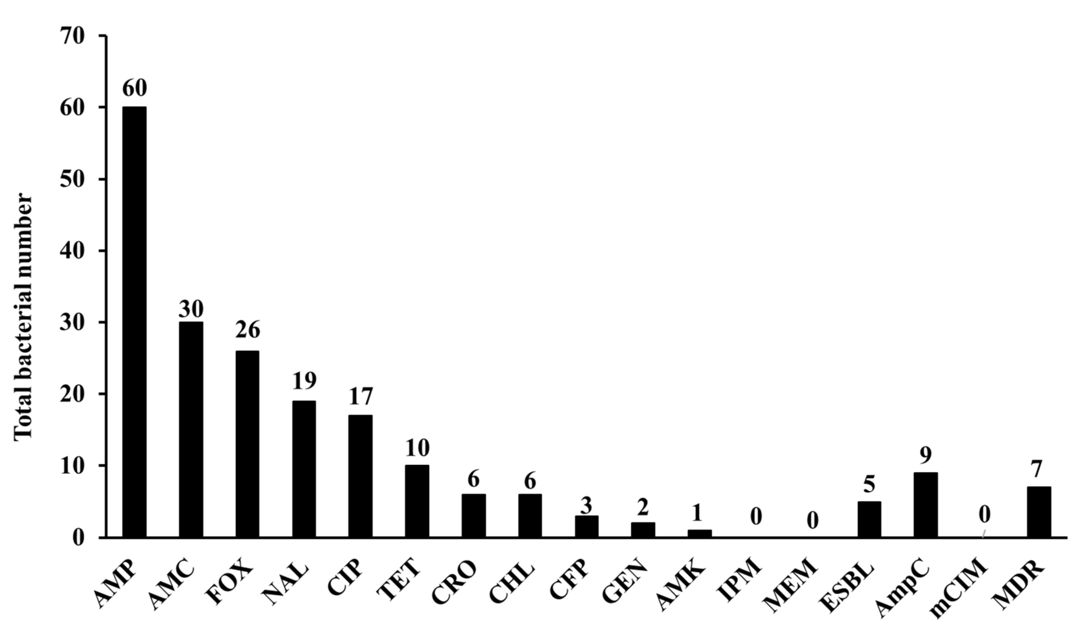
In this study, 9.2% (95% CI 4.5–17.8%) of the isolates showed multidrug-resistant profiles attributed to the resistance to three or more antimicrobial classes (Figure 2, Supplementary Table S1). The isolates showed high resistance rates to AMP, AMC, FOX, NAL, and CIP, 78.9% (95% CI 68.5–86.6%), 39.5% (95% CI 29.2–50.7%), 34.2% (95% CI 24.5–45.4%), 25% (95% CI 16.6–35.8%), and 22.4% (95% CI 14.5–33%), respectively. On the other hand, the isolates showed low resistance rates to TET (13.2%, 95% CI 7.3–22.6%), CRO (7.9%, 95% CI 3.7–16.2%), CHL (7.9%, 95% CI 3.7–16.2%), CFP (3.9%, 95% CI 1.4–11%), GEN (2.6%, 95% CI 0.7–9.1%), and AMK (1.3%, 95% CI 0.2–7.1%). All the isolates were sensitive to IPM and MEM (Figure 2, Supplementary Table S1).
Phenotypic AmpC production was identified in 11.8% (95% CI 6.4–21%) of the isolates, and 6.6% (95% CI 2.8–14.5%) of the isolates produced ESBL. None of the isolates produced carbapenemases as determined by the mCIM assay (Figure 2, Supplementary Table S1).

3.3. Prevalence of β-Lactamase Encoding Genes

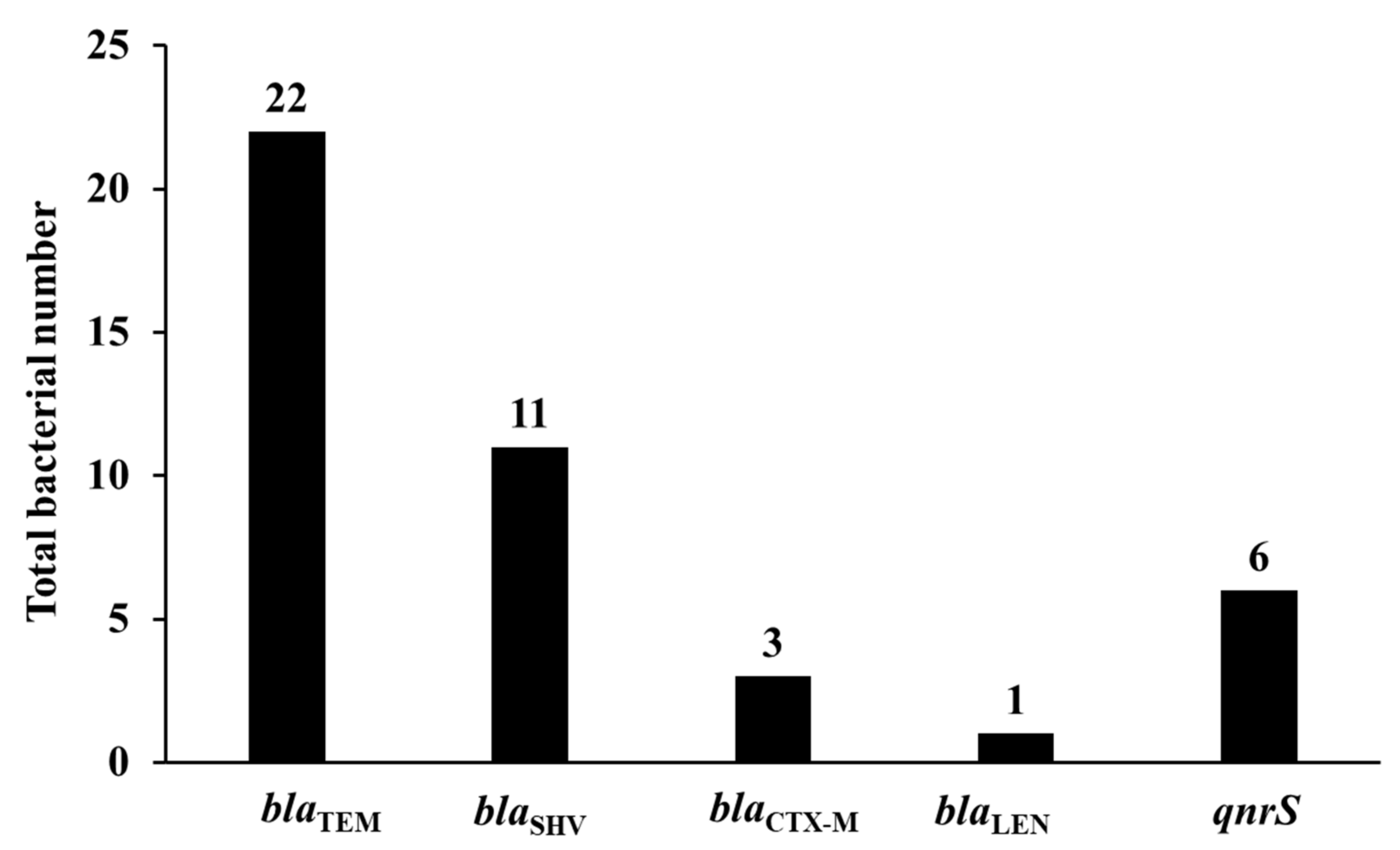
Our results confirmed that blaTEM was the most prevalent β-lactamase encoding gene identified in 29% (95% CI 20–40%) of the isolates with 17 isolates harboring blaTEM-1, three isolates harboring blaTEM-176, and two isolates harboring blaTEM-198/214. blaSHV was the second most identified β-lactamase encoding genes in 14.5% (95% CI 8.3–24.1%) of the isolates with 10 isolates harboring blaSHV-1 and a single isolate harboring blaSHV-1/11. blaCTX-M was identified only in three isolates (4%, 95% CI 1.4–11%), with two isolates harboring blaCTX-M-15-like and a single isolate harboring blaCTX-M-14 (Figure 3, Supplementary Table S1). Interestingly, a single isolate was positive for blaTEM by PCR using TEM primers, but after sequencing it was confirmed that it harbored blaLEN-13/55. Despite of using different sets of primers, conditions, PCR reagents, and repeating the experiments all the isolates were genotypically negative for plasmid AmpC-encoding genes.

3.4. Prevalence of Plasmid-Mediated Quinolone Resistance Genes and Other Resistance Genes

In our study, only qnrS was the identified PMQR in 7.9% (95% CI 3.7–16.2%) of the isolates (Figure 3, Supplementary Table S1). Multiplex and/or single PCR confirmed that none of the isolates harbored qepA and 16S rRNA methylases genes.

3.5. Relation between the Recovered Isolates and Resistant Determinants

There was no relation between the recovered bacterial isolate and incidence of multidrug resistance infection (p = 0.4), incidence of infection with ESBL-producers (p = 0.6), identification of blaCTX-M (p = 0.8), or qnrS identification (p = 0.6). On the other hand, the recovered bacterial isolates were statistically related to the incidence of infection with AmpC-producing isolates (p = 0.01). Furthermore, there was a statistical relationship between the recovered bacterial isolates and identification of blaSHV gene (p = 0.000008) with most blaSHV genes identified in K. pneumoniae, and the identification of blaTEM gene (p = 0.0002), with most blaTEM gene identified in E. coli isolates.

4. Discussion

The extensive use of antimicrobials in the veterinary field for the treatment and prevention of infectious diseases and to increase animal production has led to the dissemination of antimicrobial resistance. Furthermore, the close contact between animals and humans facilitates cross-transmission, especially members of the Enterobacterales which are closely related normal intestinal flora that rarely cause disease in normal hosts. However, they can cause serious disease in immunocompromised patients due to other diseases [4], and have the ability to acquire and disseminate several unique resistance mechanisms [25,26]. The emergence and spread of ESBL- and Amp-producing Enterobacteriaceae from animals has become an important community concern worldwide. Unfortunately, data are lacking about the molecular resistance mechanisms associated with Enterobacteriaceae from animals, especially sheep and goats in Egypt. Therefore, this study heightened the genetic mechanisms governing these isolates in sheep and goats.
In this study, 9.2% of the isolates were multidrug-resistant, which is comparable or lower than previous findings of primary respiratory pathogens belonging to the Pasteurellaceae family in Canada that ranged from 11.8% to 38.1% [27]. On the other hand, our results showed very high resistance levels for different β-lactams, including AMP (78.9%), AMC (39.5%), and FOX (34.2%). Our results were higher than those from previous reports of sheep and goat respiratory tract pathogens in Canada [27] and the USA [28]. Interestingly, our results for the Enterobacteriaceae showed a high resistance rate also for quinolones, with 25% and 22.4% of isolates resistant to NAL and CIP, respectively. These were higher frequencies than those previously reported from North American countries [27,28] and some Asian and African countries [29,30]. Most of the isolates were susceptible to tetracycline, aminoglycosides, and chloramphenicol. Similar tetracycline antimicrobial susceptibility patterns have been reported in other studies from Pasteurellaceae in North America [27,28]. However, high tetracycline resistance has been identified in Mycoplasma mycoides subspecies capri in Pakistan and respiratory tract bacterial flora in Nigeria [29,30]. The high prevalence of β-lactam and quinolone resistance in Egypt is an issue of great clinical consideration as these drugs are commonly used for the treatment of severe infections in both animals and humans [6,31]. Although a high prevalence of carbapenem resistance and carbapenemase production has been observed in clinical settings in Egypt [8,9], all isolates were sensitive to carbapenem antibiotics and phenotypically negative for carbapenemase production (Figure 2, Supplementary Table S1). This may be attributed to the restrictions of carbapenem use in the veterinary field in Egypt. Carbapenems are clinically important lifesaving β-lactams, and this study rejuvenated the concern for their constraint in veterinary and agriculture fields.
To understand the genetic basis of β-lactam resistance, the isolates were assessed phenotypically for ESBL and AmpC production and genotypically different classes of β-lactamases. Although ESBL and AmpC production are considered as major mechanisms of β-lactams resistance [6,7,31], only 11.8% and 6.6% of the isolates were phenotypically positive for AmpC and ESBL production, respectively. On the other hand, with the exception of the identified blaCTX-M, genotypic examination identified different narrow-spectrum β-lactamases such as blaSHV and blaTEM. The high prevalence of β-lactamases in our study explaining the high levels of resistance AMP, AMC, and FOX. Our study showed that blaTEM and blaSHV were the most abundant β-lactamases which was in agreement with the previous reports from other developing countries such as ESBL-producing E. coli in Turkey [32]. More interestingly, in this study, several antibiotic resistant determinants were identified for the first time from animals in Egypt or even in Africa, such as blaLEN-13/55, blaTEM-176 and blaTEM-198/214. Furthermore, this is the first report of blaCTX-M identification in small ruminants in Egypt. Notably, CTX-M enzymes have recently emerged in different isolates from food, food-producing animals, and companion animals [5,31,32,33,34], triggering their potential as a public health hazard. For instance, different variants of blaCTX-M, including blaCTX-M-1, blaCTX-M-2, blaCTX-M-3, blaCTX-M-9, blaCTX-M-14, blaCTX-M-15, and blaCTX-M-28, have been identified from diseased and healthy animals and/or food products in Spain, Denmark, France, Netherlands, UK, China, and Japan [31,35]. Furthermore, blaCTX-M was the most identified ESBL gene in Egypt from clinical settings and food [7,34], indicating potential transmission among animals, humans, and food. Although 11.8% (95% CI 6.4–21%) of the isolates were phenotypically positive for AmpC production, no plasmid-mediated AmpC beta-lactamase genes were detected, despite repeating the PCR experiment with using different sets of primers and different conditions (data not shown). Other mechanisms, especially the chromosomal AmpC which are intrinsic in certain bacterial isolates such as Enterobacter and Citrobacter spp. might be responsible for the phenotypic resistance to AmpC. Confirming our findings, plasmid AmpC was recently identified in only 1.1% of sheep meat samples in Egypt [36].
In this study, 7.9% (95% CI 3.7–16.2%) of the isolates harbored the qnrS gene, confirming our previous results that qnrS is the major circulating PMQR gene in Egypt in clinical, veterinary, and food production settings [7,34]. Much higher than our results, qnrS was the only detected PMQR gene identified in 44% of ESBL-producing E. coli from companion and domestic farm animals in Tanzania [33]. Furthermore, qnrS was only detected in sheep in Italy and was the most prevalent among Salmonella and E. coli isolates from animals, humans, food, and the environment from 13 European countries [37]. qnrS was also the only PMQR gene detected in 80% of ESBL-producing Enterobacteriaceae recovered from sheep in Abidjan, Ivory Coast [38]. On the other hand, qnrB, qnrA, and qnrS were recently identified in E. coli from healthy and diarrheic sheep and goats in Saudi Arabia by 62.1, 56, and 52.4%, respectively [39]. In a recent study from Tunisia, although qnrS1 and qnrB1 were identified among the E. coli recovered from poultry and turkeys, no PMQR genes were identified from sheep [40]. The prevalence of qnrS identified in this study is of considerable clinical impact, as PMQR genes not only potentiate quinolone resistance, they also disseminate quinolone resistance through their plasmid mobility [6,13]. Our results confirmed the limited role of qepA and 16S rRNA methylases to mediate quinolone and aminoglycoside resistance, respectively, which is also in agreement with our previous report from food and pets in Egypt [6,34].
As a limitation of this study that only four goats were checked, and a single isolate was recovered from one goat and wild small ruminants were not included. Furthermore, this study was performed in a single city in Egypt. Therefore, it is of critical importance to perform other large-scale nationwide studies on a wide range of small ruminants to evaluate the molecular resistance mechanisms all over Egypt. Even so, this study is considered to be the first African study to evaluate the AMR associated with Gram-negative bacteria recovered from the respiratory tract of sheep and goats with respiratory infections.

5. Conclusions

This study identified Enterobacteriaceae recovered from sheep and one goat suffering from respiratory infections in Egypt, highlighted the prevalence of β-lactam and quinolone resistance. The high prevalence of antimicrobial resistance in sheep and goats is an issue of grave concern given the opportunity of human transmission and development of severe, untreatable infections. Therefore, strict regulations such as prudent antibiotic use, large scale surveillance, and prompt infection control measures are necessary to combat this problem.

Supplementary Materials

The following are available online at https://www.mdpi.com/article/10.3390/ani11082258/s1, Table S1: Full phenotypic and genotypic characterization of the isolates tested in this study.

Author Contributions

Conceptualization, H.O.K.; methodology, H.O.K., A.O., A.A.A.E.-H., A.A.E.L. and T.O.; software, H.O.K. and A.A.A.E.-H.; validation, H.O.K. and T.M.; formal analysis, H.O.K.; investigation, H.O.K., A.O., A.A.A.E.-H. and T.O.; resources, H.O.K., Y.K. and T.M.; data curation, H.O.K.; writing—original draft preparation, H.O.K.; writing—review and editing, H.O.K., A.O., A.A.E.L., A.A.A.E.-H., T.O., Y.K. and T.M.; visualization, H.O.K.; project administration, H.O.K., T.M. All authors have read and agreed to the published version of the manuscript.

Funding

This research received no external funding.

Institutional Review Board Statement

Ethical review and approval was waived for this study, as we did not perform any animal experiments and nasal and ocular swabs from diseased animals were carried out as a part of the routine activity for laboratory examination, diagnosis and for animal treatment. All the animal research activities in this study were performed in compliance with local laws and regulations.

Data Availability Statement

All the data are presented in this study and in the Supplementary File.

Acknowledgments

We are deeply grateful to Katsuhiko Kamei and Akira Watanabe, Division of Clinical Research, Medical Mycology Research Centre, Chiba University, Japan for using their lab resources to perform some experiments. H.O.K. is supported by a postdoctoral fellowship from the Egypt-Japan Education Partnership (EJEP).

Conflicts of Interest

The authors declare no conflict of interest.

References

  1. Wall, B.A.; Mateus, A.L.P.; Marshall, L.; Pfeiffer, D.U.; Lubroth, J.; Ormel, H.J.; Otto, P.; Patriarchi, A. Drivers, Dynamics and Epidemiology of Antimicrobial Resistance in Animal Production; Food and Agriculture Organization of the United Nations: Rome, Italy, 2016. [Google Scholar]
  2. Wozniak, T.M.; Barnsbee, L.; Lee, X.J.; Pacella, R.E. Using the best available data to estimate the cost of antimicrobial resistance: A systematic review. Antimicrob. Resist. Infect. Control. 2019, 8, 26. [Google Scholar] [CrossRef] [PubMed] [Green Version]
  3. Ecdc/Efsa/EmEa. ECDC/EFSA/EMA First joint report on the integrated analysis of the consumption of antimicrobial agents and occurrence of antimicrobial resistance in bacteria from humans and food-producing animals. EFSA J. 2015, 13, 114. [Google Scholar]
  4. Ejaz, H.; Younas, S.; Abosalif, K.O.; Junaid, K.; Alzahrani, B.; Alsrhani, A.; Abdalla, A.E.; Ullah, M.I.; Qamar, M.U.; Hamam, S.S. Molecular analysis of blaSHV, blaTEM, and blaCTX-M in extended-spectrum β-lactamase producing Enterobacteriaceae recovered from fecal specimens of animals. PLoS ONE 2021, 16, e0245126. [Google Scholar] [CrossRef] [PubMed]
  5. Khalifa, H.O.; Oreiby, A.F.; Abd El-Hafeez, A.A.; Okanda, T.; Haque, A.; Anwar, K.S.; Tanaka, M.; Miyako, K.; Tsuji, S.; Kato, Y.; et al. First report of multidrug-resistant carbapenemase-producing bacteria co-harboring mcr-9 associated with pets’ respiratory disease complex: A potential of animal–human transmission. Antimicrob. Agents Chemother. 2020, 65, e01890-20. [Google Scholar] [CrossRef]
  6. Khalifa, H.O.; Oreiby, A.F.; Okanda, T.; Kato, Y.; Matsumoto, T. High β-lactam resistance in Gram-negative bacteria associated with kennel cough and cat flu in Egypt. Sci. Rep. 2021, 11, 1–9. [Google Scholar] [CrossRef]
  7. Khalifa, H.O.; Soliman, A.M.; Ahmed, A.M.; Shimamoto, T.; Nariya, H.; Matsumoto, T.; Shimamoto, T. High prevalence of antimicrobial resistance in gram-negative bacteria isolated from clinical settings in Egypt: Recalling for judicious use of conventional antimicrobials in developing nations. Microb. Drug Resist. 2019, 25, 371–385. [Google Scholar] [CrossRef]
  8. Khalifa, H.O.; Soliman, A.M.; Ahmed, A.M.; Shimamoto, T.; Hara, T.; Ikeda, M.; Kuroo, Y.; Kayama, S.; Sugai, M.; Shimamoto, T. High carbapenem resistance in clinical Gram-negative pathogens isolated in Egypt. Microb. Drug Resist. 2017, 23, 838–844. [Google Scholar] [CrossRef]
  9. Khalifa, H.O.; Soliman, A.M.; Ahmed, A.M.; Shimamoto, T.; Shimamoto, T. NDM-4-and NDM-5-producing Klebsiella pneumoniae coinfection in a 6-month-old infant. Antimicrob. Agents Chemother. 2016, 60, 4416–4417. [Google Scholar] [CrossRef] [Green Version]
  10. Khalifa, H.O.; Ahmed, A.M.; Oreiby, A.F.; Eid, A.M.; Shimamoto, T.; Shimamoto, T. Characterization of the plasmid-mediated colistin resistance gene mcr-1 in Escherichia coli isolated from animals in Egypt. Int. J. Antimicrob. Agent 2016, 5, 413–414. [Google Scholar] [CrossRef]
  11. Elnahriry, S.S.; Khalifa, H.O.; Soliman, A.M.; Ahmed, A.M.; Hussein, A.M.; Shimamoto, T.; Shimamoto, T. Emergence of plasmid-mediated colistin resistance gene mcr-1 in a clinical Escherichia coli isolate from Egypt. Antimicrob. Agents Chemother. 2016, 60, 3249–3250. [Google Scholar] [CrossRef] [Green Version]
  12. Oreiby, A.; Khalifa, H.; Eid, A.; Ahmed, A.; Shimamoto, T. Staphylococcus aureus and bovine mastitis: Molecular typing of methicillin resistance and clinical description of infected quarters. J. Hell. Vet. Med. Soc. 2019, 70, 1511–1516. [Google Scholar] [CrossRef] [Green Version]
  13. World Health Organization. Critically Important Antimicrobials for Human Medicine, 6th ed.; World Health Organization: Geneva, Switzerland, 2018; Available online: https://apps.who.int/iris/bitstream/handle/10665/312266/9789241515528-eng.pdf (accessed on 20 May 2021).
  14. Hooper, D.C.; Jacoby, G.A. Mechanisms of drug resistance: Quinolone resistance. Ann. N. Y. Acad. Sci. 2015, 1354, 12–31. [Google Scholar] [CrossRef] [Green Version]
  15. Clinical and Laboratory Standard Institute. Performance Standard for Antimicrobial Susceptibility Testing; Twenty Second Informational Supplement; Clinical and Laboratory Standard Institute: Wayne, PA, USA, 2012; Volume 32, M100-S22. [Google Scholar]
  16. Khalifa, H.O.; Okanda, T.; Abd El-Hafeez, A.A.; Abd El Latif, A.; Habib, A.G.; Yano, H.; Kato, Y.; Matsumoto, T. Comparative evaluation of five assays for detection of carbapenemases with a proposed scheme for their precise application. J. Mol. Diagn. 2020, 22, 1129–1138. [Google Scholar] [CrossRef] [PubMed]
  17. Xu, L.; Ensor, V.; Gossain, S.; Nye, K.; Hawkey, P. Rapid and simple detection of blaCTX-M genes by multiplex PCR assay. J. Med. Microbiol. 2005, 54, 1183–1187. [Google Scholar] [CrossRef]
  18. Caroline, D.; Anaelle, D.C.; Dominique, D.; Christine, F.; Guillaume, A. Development of a set of multiplex PCR assays for the detection of genes encoding important β-lactamases in Enterobacteriaceae. J. Antimicrob. Chemother. 2010, 65, 490–495. [Google Scholar]
  19. Perez-Perez, F.J.; Hanson, N.D. Detection of plasmid-mediated AmpC beta-lactamase genes in clinical isolates by using multiplex PCR. J. Clin. Microbiol. 2002, 40, 2153–2162. [Google Scholar] [CrossRef] [Green Version]
  20. Jacoby, G.A.; Gacharna, N.; Black, T.A.; Miller, G.H.; Hooper, D.C. Temporal appearance of plasmid-mediated quinolone resistance genes. Antimicrob. Agents Chemother. 2009, 53, 1665–1666. [Google Scholar] [CrossRef] [Green Version]
  21. Kim, H.B.; Park, C.H.; Kim, C.J.; Kim, E.C.; Jacoby, G.A.; Hooper, D.C. Prevalence of plasmid-mediated quinolone resistance determinants over a 9-year period. Antimicrob. Agents Chemother. 2009, 53, 639–645. [Google Scholar] [CrossRef] [Green Version]
  22. Wangkheimayum, J.; Paul, D.; Dhar, D.; Nepram, R.; Chetri, S.; Bhowmik, D.; Chakravarty, A.; Bhattacharjee, A. Occurrence of acquired 16S rRNA methyltransferase-mediated aminoglycoside resistance in clinical isolates of Enterobacteriaceae within a tertiary referral hospital of Northeast India. Antimicrob. Agents Chemother. 2017, 61, e01037-16. [Google Scholar] [CrossRef] [Green Version]
  23. Khalifa, H.O.; Arai, T.; Majima, H.; Watanabe, A.; Kamei, K. Genetic basis of azole and echinocandin resistance in clinical Candida glabrata in Japan. Antimicrob. Agents Chemother. 2020, 64, e00783-20. [Google Scholar] [CrossRef]
  24. Khalifa, H.O.; Arai, T.; Majima, H.; Watanabe, A.; Kamei, K. Evaluation of Surveyor nuclease for rapid identification of FKS genes mutations in Candida glabrata. J. Infect. Chemother. 2021, 27, 834–839. [Google Scholar] [CrossRef]
  25. Khalifa, H.O.; Soliman, A.M.; Saito, T.; Kayama, S.; Yu, L.; Hisatsune, J.; Sugai, M.; Nariya, H.; Ahmed, A.M.; Shimamoto, T.; et al. First report of foodborne Klebsiella pneumoniae co-harboring blaVIM-1, blaNDM-1, and mcr-9. Antimicrob. Agents Chemother. 2020, 64, e00882-20. [Google Scholar] [CrossRef]
  26. Soliman, A.M.; Khalifa, H.O.; Ahmed, A.M.; Shimamoto, T.; Shimamoto, T. Emergence of an NDM-5-producing clinical Escherichia coli isolate in Egypt. Int. J. Infect. Dis. 2016, 48, 46–48. [Google Scholar] [CrossRef] [PubMed] [Green Version]
  27. Awosile, B.B.; Heider, L.C.; Saab, M.E.; McClure, J.T. Antimicrobial resistance in mastitis, respiratory and enteric bacteria isolated from ruminant animals from the Atlantic Provinces of Canada from 1994–2013. Can. Vet. J. 2018, 59, 1099–1104. [Google Scholar]
  28. Berge, A.C.; Sischo, W.M.; Craigmill, A.L. Antimicrobial susceptibility patterns of respiratory tract pathogens from sheep and goats. J. Am. Vet. Med. Assoc. 2006, 229, 1279–1281. [Google Scholar] [CrossRef] [PubMed]
  29. Shah, M.K.; Saddique, U.; Ahmad, S.; Hayat, Y.; ur Rahman, S.; Hassan, M.F.; Ali, T. Isolation rate and antimicrobial susceptibility profiles of Mycoplasma mycoides subspecies capri field isolates from sheep and goats in Pakistan. Small Rumin. Res. 2017, 153, 118–122. [Google Scholar] [CrossRef]
  30. Emikpe, B.O.; Oyero, O.G.; Akpavie, S.O. Isolation and antibiogram of aerobic nasal bacterial flora of apparently healthy West African dwarf goats. Rev. D’elevage Med. Vet. Pays Trop. 2009, 62, 17–21. [Google Scholar] [CrossRef] [Green Version]
  31. Trott, D. β-lactam resistance in Gram-negative pathogens isolated from animals. Curr. Pharm. Des. 2013, 19, 239–249. [Google Scholar] [CrossRef]
  32. Pehlivanoglu, F.; Turutoglu, H.; Ozturk, D.; Yardimci, H. Molecular characterization of ESBL-producing Escherichia coli isolated from healthy cattle and sheep. Acta Vet. 2016, 66, 520–533. [Google Scholar] [CrossRef]
  33. Seni, J.; Falgenhauer, L.; Simeo, N.; Mirambo, M.M.; Imirzalioglu, C.; Matee, M.; Rweyemamu, M.; Chakraborty, T.; Mshana, S.E. Multiple ESBL-producing Escherichia coli sequence types carrying quinolone and aminoglycoside resistance genes circulating in companion and domestic farm animals in Mwanza, Tanzania, harbor commonly occurring plasmids. Front. Microbiol. 2016, 7, 142. [Google Scholar] [CrossRef]
  34. Elafify, M.; Khalifa, H.O.; Al-Ashmawy, M.; Elsherbini, M.; El Latif, A.A.; Okanda, T.; Matsumoto, T.; Koseki, S.; Abdelkhalek, A. Prevalence and antimicrobial resistance of Shiga toxin-producing Escherichia coli in milk and dairy products in Egypt. J. Environ. Sci. Health B 2020, 55, 265–272. [Google Scholar] [CrossRef]
  35. Li, X.Z.; Mehrotra, M.; Ghimire, S.; Adewoye, L. β-Lactam resistance and β-lactamases in bacteria of animal origin. Vet. Microbiol. 2007, 121, 197–214. [Google Scholar] [CrossRef]
  36. Rensing, K.L.; Abdallah, H.M.; Koek, A.; Elmowalid, G.A.; Vandenbroucke-Grauls, C.M.; Al Naiemi, N.; van Dijk, K. Prevalence of plasmid-mediated AmpC in Enterobacteriaceae isolated from humans and from retail meat in Zagazig, Egypt. Antimicrob. Resist. Infect. Control. 2019, 8, 45. [Google Scholar] [CrossRef] [PubMed] [Green Version]
  37. Veldman, K.; Cavaco, L.M.; Mevius, D.; Battisti, A.; Franco, A.; Botteldoorn, N.; Bruneau, M.; Perrin-Guyomard, A.; Cerny, T.; De Frutos Escobar, C.; et al. International collaborative study on the occurrence of plasmid-mediated quinolone resistance in Salmonella enterica and Escherichia coli isolated from animals, humans, food and the environment in 13 European countries. J. Antimicrob. Chemother. 2011, 66, 1278–1286. [Google Scholar] [CrossRef] [PubMed]
  38. Baguy, O.M.; Nathalie, G.K.; David, C.N.G.; Daniel, S.N.G.; Julien, C.K.; Rose, K.N.; Djénéba, O.G.; Valérie, G.; Bertin, T.K.; Mireille, D. First report of qnr genes in multidrugs resistant (ESBL) enterobacteria isolated from different ecosystems in Abidjan, Ivory Coast. Int. J. Biol. Sci. Appl. 2014, 1, 170–175. [Google Scholar]
  39. Shabana, I.I.; Al-Enazi, A.T. Investigation of plasmid-mediated resistance in E. coli isolated from healthy and diarrheic sheep and goats. Saudi. J. Biol. Sci. 2020, 27, 788–796. [Google Scholar] [CrossRef]
  40. Kilani, H.; Ferjani, S.; Mansouri, R.; Boutiba-Benboubaker, I.; Abbassi, M.S. Occurrence of plasmid-mediated quinolone resistance determinants among Escherichia coli strains isolated from animals in Tunisia: Specific pathovars acquired qnr genes. J. Glob. Antimicrobe. Resist. 2020, 20, 50–55. [Google Scholar] [CrossRef] [PubMed]
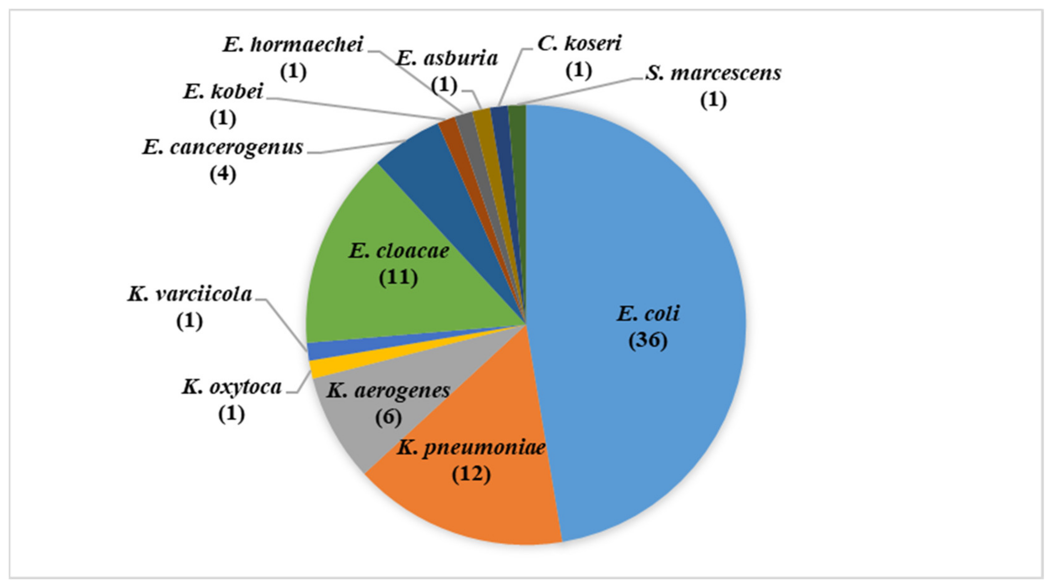
Figure 1. Bacterial isolates identified and tested in this study.
Animals 11 02258 g001
Figure 2. Level of antimicrobial resistance to different antibiotics and phenotypic characteristics of the isolates. Antibiotics: ampicillin (AMP), amoxicillin–clavulanic acid (AMC), cefoxitin (FOX), nalidixic acid (NAL), ciprofloxacin (CIP), tetracycline (TET), ceftriaxone (CRO), chloramphenicol (CHL), cefoperazone (CFP), gentamicin (GEN), amikacin (AMK), imipenem (IPM), and meropenem (MEM).
Animals 11 02258 g002
Figure 3. Detected antimicrobial resistance genes. Regarding β-lactamase encoding genes, blaTEM was identified in 22 isolates (29% 95% CI 20–40%) of the isolates, followed by blaSHV identified in 11 isolates (14.5%, 95% CI 8.3–24.1%), blaCTX-M identified in three isolates (4%, 95% CI 1.4–11%), and finally, a single isolate harbored blaLEN-13/55 (1.3%, 95% CI 0.2–7.1%). qnrS was the only identified PMQR in six isolates (7.9%, 95% CI 3.7–16.2%).
Animals 11 02258 g003
Table 1. Oligonucleotides used in this study.
Primer NameSequence (5’-3’)Amplicon Size (bp)TargetReference
β-lactamases
CTXM7
CTXM8
GCGTGATACCACTTCACC TC
TGAAGTAAGTGACCAGAA TC
260blaCTX-M-1group[17]
CTXM17
CTXM18
TGATACCACCACGCCGCT C
TATTGCATCAGAAACCGTGGG
341blaCTX-M-2group[17]
CTXM19
CTXM20
CAATCTGACGTTGGGCAATG
ATAACCGTCGGTGACAATT
207blaCTX-M-8/25/26group[17]
CTXM11
CTXM12
ATCAAGCCTGCCGATCTGGTTA
GTAAGCTGACGCAACGTCTGC
293blaCTX-M-9group[17]
SHV_F
SHV_R
AGCCGCTTGAGCAAATTAAAC
ATCCCGCAGATAAATCACCAC
713blaSHV-1/variant[18]
TEM-F
TEM-R
CATTTCCGTGTCGCCCTTATTC
CGTTCATCCATAGTTGCCTGAC
800blaTEM-1/-2/variant[18]
MOXMF
MOXMR
GCT GCTCAAGGAGCACAG GAT
CAC ATT GAC ATA GGT GTG GTG C
520blaMOX-1,blaMOX-2, and blaCMY-1,
CMY-8 to CMY-11
[19]
CITMF
CITMR
TGGCCAGAACTGACAGGCAAA
TTTCTCCTGAACGTGGCTGGC
462blaLAT-1 to LAT-4,blaCMY-2
to CMY-7, and blaBIL-1
[19]
DHAMF
DHAMR
AACTTTCACAGGTGTGCTGGGT
CCGTACGCATACTGGCTTTGC
405blaDHA[19]
ACCMF
ACCMR
AACAGCCTCAGCAGCCGGTTA
TTCGCCGCAATCATCCCTAGC
346blaACC[19]
EBCMF
EBCMR
TCGGTAAAGCCGATGTTGCGG
CTTCCACTGCGGCTGCCAGTT
302blaMIR-1 and blaACT-1[19]
FOXMF
FOXMR
AACATGGGGTATCAGGGAGATG
CAAAGCGCGTAACCGGATTGG
190blaFOX-1 to FOX-5b[19]
Plasmid-mediated quinolone resistance
qnrA-FATTTCTCACGCCAGGATTTG468qnrA[20]
qnrA-RTGCCAGGCACAGATCTTGAC
qnrB-FCGACCTKAGCGGCACTGAAT513qnrB[20]
qnrB-RGAGCAACGAYGCCTGGTAGYTG
qnrS-FACTGCAAGTTCATTGAACAG431qnrS[20]
qnrS-RGATCTAAACCGTCGAGTTCG
Quinolone efflux pump determinant
qepA-F
qepA-R
AACTGCTTGAGCCCGTAGAT
GTCTACGCCATGGACCTCAC
596qepA[21]
16S rRNA methylases
armA-F
armA-R
GGTGCGAAAACAGTCGTAGT
TCCTCAAATATCCTCTATGT
1153armA[22]
npmA-F
npmA-R
CGGGATCCAAGCACTTTCATACTGACG
CGGAATTCCAATTTTGTTCTTATTAGC
981npmA[22]
rmtA-F
rmtA-R
CTAGCGTCCATCCTTTCCTC
TTTGCTTCCATGCCCTTGCC
635rmtA[22]
rmtB-F
rmtB-R
GGAATTCCATATGAACATCAACGATGCC
CCGCTCGAGTCCATTCTTTTTTATCAAGT
756rmtB[22]
rmtC-F
rmtC-R
CGAAGAAGTAACAGCCAAAG
GCTAGAGTCAAGCCAGAAAA
1000rmtC[22]
rmtD-F
rmtD-R
TCATTTTCGTTTCAGCAC
AAACATGAGCGAACTGAAGG
744rmtD[22]
Publisher’s Note: MDPI stays neutral with regard to jurisdictional claims in published maps and institutional affiliations.