Article

Prevalence, Antimicrobial Resistance, and Characterization of Staphylococcus aureus Isolated from Subclinical Bovine Mastitis in East Coast Malaysia

by
Shamsaldeen Ibrahim Saeed
1,2,*,
Khairun Anisa Mat Yazid
1,
Hidayatul Athirah Hashimy
1,
Siti Khadijah Dzulkifli
1,
Fatihah Nordin
1,
Nik Azmi Nik Him
3,
Mohd Fikry Fahmi bin Omar
4,
Erkihun Aklilu
1,
Maizan Mohamad
1,
Che Wan Salma Zalati
1 and
Nor Fadhilah Kamaruzzaman
1,*
1
Faculty of Veterinary Medicine, University Malaysia Kelantan, Pengkalan Chepa 16100, Kelantan, Malaysia
2
Faculty of Veterinary Science, University of Nyala, P.O. Box 155, Nyala 63311, South Darfur State, Sudan
3
Pusat Perkhidmatan Industri Tenusu, Jabatan Perkhidmatan Veterinar, Pasir Puteh 16800, Kelantan, Malaysia
4
Pusat Perkhidmatan Industri Tenusu, Jalan Chamang, Bentong 28700, Pahang, Malaysia
*
Authors to whom correspondence should be addressed.
Animals 2022, 12(13), 1680; https://doi.org/10.3390/ani12131680
Submission received: 20 April 2022 / Revised: 10 June 2022 / Accepted: 14 June 2022 / Published: 29 June 2022
(This article belongs to the Section Animal System and Management)

Abstract

:

Simple Summary

Subclinical mastitis remains a burden in the dairy industry due to difficulties in its treatment and the economic losses that are associated with it. This study reported the prevalence and characterization of Staphylococcus aureus (S. aureus) that was isolated from bovine mastitis in dairy farms in East Coast Peninsular Malaysia. The data from this study showed the presence of subclinical mastitis and antimicrobial resistance (AMR) in S. aureus that was isolated from milk samples. In addition, S. aureus isolates could invade and survive within the bovine mammary epithelial cell in vitro and form biofilms. This feature provides an additional challenge for mastitis treatment.

Abstract

S. aureus is the pathogen that is commonly associated with subclinical mastitis, causing significant economic losses to dairy farms. This infection responds poorly to antimicrobial treatment, which could be due to the development of AMR, biofilm formation, and the intracellular invasion of S. aureus into bovine mammary cells leading to treatment failure. Thus, it is important to understand the challenge of this problem. Therefore, the present study aims to determine the prevalence, antimicrobial resistance, and characterization of S. aureus that was isolated from subclinical bovine mastitis in East Coast Malaysia. A total of 235 milk samples from dairy cows were collected from selected farms in Kelantan and Pahang. The samples were subjected to a somatic cell analysis to identify subclinical mastitis, followed by bacteria isolation and antimicrobial susceptibility testing. The isolated S. aureus were further analyzed for their ability to form biofilms and invade the bovine mammary epithelial cells (MAC-T cells) in in vitro infections modeling using a gentamicin protection assay. The overall total of 74/235 (31.4%; 95% CI = 0.31; 0.32) of the milk samples demonstrated >200,000 somatic cells/mL, suggesting the presence of subclinical mastitis in the animals. A total of 39/235 (16.5%; 95% CI = 0.16, 0.17) of the milk samples harbored S. aureus which demonstrated resistance towards the following antimicrobials: penicillin (18/39, 46%), ampicillin (17/39, 43.6%), oxacillin (12/39, 31%), tetracycline (10/39, 26%), and erythromycin (7/39, 18%). AMR was recorded for a total of (17/39, 43.6%) of S. aureus isolates. All isolates formed biofilms, with (8/30, 27%) strongly biofilm-forming, (18/30, 60%) moderately biofilm-forming, and the remaining (4/30, 13%) of isolates weakly biofilm-forming. Interestingly, the AMR isolates appear to produce weak and moderate biofilm. Moreover, (6/20, 30%) of the S. aureus isolates were invasive towards MAC-T cells, as indicated by their ability to evade gentamicin treatment. The study demonstrated the presence of AMR, invasiveness, and biofilm formation in S. aureus that was isolated from subclinical mastitis. This characteristic presents additional challenges to existing antimicrobial therapy.

1. Introduction

Mastitis is a significant disease affecting the dairy industry, reducing animals’ health and milk quality and subsequently reducing farmers’ income [1,2]. S. aureus is the most frequent bacteria to be associated with intramammary infection resulting in a persistent chronic infection [3]. Mastitis is the most common reason for antimicrobial use in dairy cattle, and it can be administered mainly via intramammary and intramuscular routes [4]. However, the treatment has become less effective, with only a 10–30% cure rate [5]. The antimicrobials lose their efficacy in mastitis treatment, due, in part to the development of antimicrobial resistance bacteria (AMR) [6,7,8,9,10,11]. In addition to developing resistance to antimicrobials, S. aureus that is isolated from mastitis is also known to form biofilms and is able to invade and survive in host cells. This may present an additional challenge for the antimicrobial treatment of mastitis. The presence of S. aureus in the bovine mammary epithelial cells therefore represents a privileged reservoir from which re-infection can occur [12], leading to long-term disease progression and recurrent infections [13].
Thus, it is important to understand the challenge of AMR in S. aureus that is associated with mastitis. Therefore, the present study aims to determine the prevalence, antimicrobial resistance, and characterization of S. aureus that is isolated from subclinical bovine mastitis in East Coast Malaysia.

2. Material and Methods

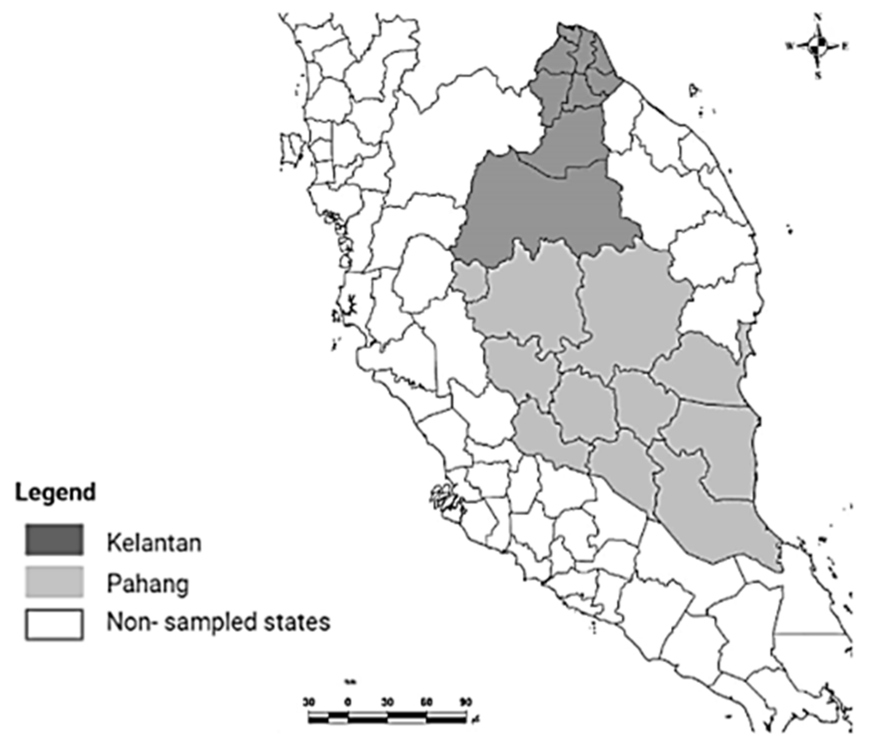
2.1. Study Area and Sampling

The study was conducted in five districts (Pasir Mas, Pasir Puteh, and Kota Bharu, Raub, and Bentong) in Kelantan and Pahang, which are located on the East Coast of Peninsular Malaysia (Figure 1). These areas have a tropical climate that is characterized by humidity, high rainfall, and uniform temperatures between 25 °C and 37 °C [14]. Ten small commercial dairy farms were included in this research. A total of 235 milk samples were collected, according to the guidelines by the National Mastitis Council [15]. Briefly, the quarter was washed with tap water and dried, then the teat end was swabbed with cotton that was soaked in 70% ethyl alcohol. To avoid environmental contamination, the first three milk streams were discarded. Following that, approximately 10 mL of milk were collected aseptically into sterile universal bottles. The samples were transported on ice to the Zoonotic laboratory, Faculty of Veterinary Medicine, University Malaysia Kelantan. The samples were processed within 24 h of arrival at the laboratory.

2.2. Determining the Somatic Cell Count (SCC)

To detect the subclinical mastitis, an SCC analysis was performed for all milk samples using the Fossomatic TM FC 5000 machine (Foss Analytical A/S, Foss Alle 1, DK-3400, Hillerød, Denmark). For each analysis, 7 mL of milk samples were used. A cutoff value of 200,000 cells/mL was used as a guideline to indicate that milk comes from an animal with subclinical mastitis [16].

2.3. Isolation and Identification of S. aureus

The isolation and identification of S. aureus from the milk were conducted using microbiological and serological methods. Briefly, 1 mL of milk sample was added into 9 mL of tryptic soy broth (TSB, Oxoid, Hampshire, UK) and incubated at 37 °C for 24 h. Two loopfuls from each broth sample were plated on mannitol salt agar (MSA, Oxoid, Hampshire, UK) and 5% sheep blood agar (Oxoid Ltd., Hampshire, UK) and incubated aerobically at 37 °C for 24 h. The colonies were further examined by Gram-stain biochemical methods and finally confirmed using the latex agglutination test using a Staphytect Plus kit (Oxoid, UK). The confirmed isolates of S. aureus were preserved at −80 °C in Luria-Bertani (LB medium) containing 20% glycerol until further use.

2.4. Antimicrobial Susceptibility Tests

S. aureus antimicrobial susceptibility tests were conducted using a disc diffusion method, according to the method by the Clinical Laboratory Standard Institute (CLSI). Briefly, S. aureus were cultivated on nutrient agar and incubated at 37 °C for 18 h. Following that, bacterial colonies were suspended in 5 mL of sterile saline and the suspensions were adjusted to a density that was approximately equal to 1.5 × 10⁸ CFU/mL, according to the McFarland standard. The bacterial suspension was inoculated onto Mueller–Hinton agar (MHA) and tested for susceptibility towards the following antimicrobials disc: ampicillin (AMP; 10 μg); penicillin-G (10 units); tetracycline (TE; 30 μg); chloramphenicol (C; 30 μg); streptomycin (S; 10 μg); gentamicin (CN; 10 μg); sulfamethoxazole/trimethoprim (SXT; 25 μg); ciprofloxacin (5 μg); and oxacillin (1 µg). All antimicrobials’ discs were obtained from Oxoid, England. The discs were dispensed on the surface of the medium with a disc dispenser and the bacterial plate was incubated aerobically at 37 °C for 18 h. The results were recorded and interpreted based on the measurement of the inhibition zone diameter (ZD) using caliper, according to the interpretive standards of CLSI [17]. To determine the level of antimicrobial resistance for each individual bacteria isolate, the MAR index was calculated as the ratio between the number of antibiotics to which the isolate showed resistance to the number of antibiotics to which the isolate had been exposed. A MAR index of >0.2 was indicative of multiple antimicrobial resistance bacteria [18].

2.5. Mammary Epithelial Cell Cultures (MAC-T Cells) and Growth Condition

MAC-T cells were obtained from Prof Liam Good, Royal Veterinary College, London. The cells were maintained in freezing medium (90% FBS + 10% DMSO) and kept in −80 °C. for experimental purposes. The cells were maintained in 14 mL of Dulbecco’s modified Eagle’s medium (DMEM, Sigma-Aldrich, Hampshire, UK) that was supplemented with 10% fetal bovine serum (FBS, Sigma-Aldrich, UK) and 5% penicillin-streptomycin (Sigma-Aldrich, Hampshire, UK). The cells were maintained at 37 °C in 5% CO2. All cell culture work was performed under an aseptic technique in a biological safety cabinet.

2.6. Intracellular Invasions of MAC-T Cells by S. aureus

Intracellular invasions of the MAC-T cells (host cells) by S. aureus isolates were established using a gentamicin protection procedure, as described by Kamaruzzaman et al. [19]. Briefly, the MAC-T cells were seeded at 1.2 × 10⁵ cells/well in a 12-well plate and cultured overnight in DMEM with 10% FBS, without antimicrobials. In parallel, 1 mL of overnight S. aureus culture was centrifuged at 8000 rpm, and the pellet was washed with PBS to remove the bacterial toxin. A total of 1 mL of 1 × 10⁷ CFU/mL of bacteria that was diluted in cell culture media without antimicrobials was added into the cell culture plate for 3 h to allow bacterial invasion. Following that, gentamicin (200 mg/L) was added for another 3 h to kill the extracellular bacteria. Finally, the gentamicin was removed and the cells were washed with PBS to remove the residual antimicrobials. A total of 1 mL of 0.5% Triton X-100 that was diluted in PBS was added to lyse the host cells. The lysed cells were serially diluted in PBS and plated on nutrient agar for enumeration of the intracellular bacteria. The uninfected cells were subjected to the lysis procedure to confirm their sterility.

2.7. Biofilm Formation

Biofilm formation assays were performed with S. aureus isolates (n = 30) following the protocol that was described in our previous work [19]. Briefly, the bacteria were grown overnight at 37 °C in 5 mL of tryptic soy broth (TSB; Oxoid) for 18 h. Following 18 h of incubation, the bacteria were diluted in TSB that was supplemented with 20% glucose to reach the final concentration of 107 CFU/mL. Aliquots of bacteria (1 mL, 107 CFU/mL) were placed in a 12-well cell culture plate and incubated at 37 °C for 48 h. After incubation, the non-adherent cells were removed and the produced biofilms on the surface of the wells were fixed at 60 °C for 1 h. The biofilm was stained with 0.1% crystal violet solution and rinsed with PBS. The OD was measured at 550 mm using a spectrophotometer. Each assay was performed in triplicate. S. aureus 15 AL was used as positive control.

2.8. Statistical Analysis

A statistical analysis was performed using a one-way analysis of variance (ANOVA). The data are presented as means ± standard deviation (SD). The level of significance was accepted as p ≤ 0.05. For the graph, error bars represent the standard deviations. * p ≤ 0.05; *** p ≤ 0.001; **** p ≤ 0.000; and NS, not significant. All the experiments were performed at least three times.

3. Results

3.1. Prevalance of Subclinical Mastitis and S. aureus

To determine the prevalence of subclinical mastitis in the dairy farm, milk samples were analyzed for their somatic cells count (SCC). Of 235 samples, 74 (31.4%; 95% CI = 0.31; 0.32) showed a number of cells of more than 200,000 cells/mL, suggesting that the animal contracted subclinical mastitis when the sampling was carried out. The highest subclinical mastitis was recorded in Pahang state from the Bentong and Raub area with a 45.2% and 25.2% occurrence rate, respectively. Meanwhile, the milk samples that were collected from Pasir Mas, Kota Bharu, and Pasir Putih in Kelantan state had lower rates of subclinical mastitis at 9%, 16.6%, and 27%, respectively. The milk samples were further subjected to a microbiological analysis for S. aureus isolation. A total of 39/235 (16.5%; 95% CI = 0.16, 0.17) milk samples were positive for S. aureus (Table 1).

3.2. Antimicrobial Susceptibility Testing

Kirby–Bauer methods on Mueller–Hinton agar were performed to determine the susceptibility of S. aureus to the antimicrobials that were tested. S. aureus showed resistance to the following antimicrobials: penicillin (18/39, 46%); ampicillin (17/39, 43.6%); oxacillin (12/39, 31%); and tetracycline (10/39, 26%), and (7/39, 18%) were resistant towards erythromycin. In contrast, all S. aureus were sensitive (39/39, 100%) towards chloramphenicol, ciprofloxacin, sulfamethoxazole/trimethoprim, and gentamicin. Table 2 summarized the antimicrobial susceptibility of S. aureus.

3.3. S. aureus AMR Profile and Multiple Antimicrobial Resistance (MAR) Indexes

The AMR profile and MAR index of S. aureus isolates are summarized in Table 3. A total of (17/39, 43.6%) of S. aureus showed an antimicrobial resistance profile (resistance to >1 antimicrobials). This profile included 17.9% of isolates that were resistant to five antimicrobials; 7.7% with a resistance to four antimicrobials; 5.1% with a resistance to three antimicrobials; and 12.8% with a resistance to two antimicrobials. The most predominant S. aureus AMR profile was AMP, E, P, TE, OXA and AMP, P. The MAR index analysis indicated that 41% of AMR isolates had a MAR index > 0.2, indicating that the isolates were multiple antimicrobial-resistant bacteria.

3.4. S. aureus Biofilm Formation and Their Association with AMR Profile

The biofilm formation of S. aureus (n = 30) showed that all isolates could produce biofilms. A total of 27% of isolates were substantial, 60% were moderate, and the remaining 13% were weak biofilm producers (Figure 2). Table 4 shows the biofilm formation ability of S. aureus and its relationship with antimicrobial resistance. Our study showed that 75% and 61% of weak and moderate biofilm producers were AMR, respectively, compared to 12.5% of strong biofilm producers, suggesting that AMR isolates tend to develop weak and moderate biofilms.

3.5. Intracellular Invasion of MAC-T Cell by S. aureus

A gentamicin protection assay was performed to determine the S. aureus invasion activities toward the bovine mammary epithelial cells. A total of 20 isolates were selected based on their susceptibility to the leading antimicrobial group and subjected to a gentamicin protection assay. Six isolates were found to be invasive towards MAC-T cells as evidenced by their ability to escape gentamicin treatment by infecting the host cells. Following cell lysis, the bacteria were enumerated. These isolates were labeled F31D, F41A, F41B, F51B, F53D, and PBF1 (Figure 3).

4. Discussion

Subclinical mastitis remains a burden in the dairy industry due to difficulties in the treatment and the economic losses that are associated with it. This study reported the prevalence of subclinical mastitis and antimicrobial resistance of S. aureus that was isolated from bovine subclinical mastitis in East Coast Malaysia. This study also investigated the intracellular invasion and biofilm formation ability of the isolates. Our results showed the high prevalence of AMR in S. aureus that was isolated from bovine subclinical mastitis, and the isolates were able to invade bovine mammary epithelial cells in in vitro infection modeling. The isolates were also able to form a biofilm, and the AMR isolates tended to form weak and moderate biofilms.
Subclinical mastitis is inherently difficult to detect because the animal shows no clinical signs, and no abnormalities are visible in the milk. The current diagnostic method relies heavily on the CMT test and somatic cell analysis using a laboratory that is usually available in reference laboratories, which limits the effectiveness of early detection of the disease. Several new methods have been developed to allow the early detection of subclinical mastitis. For example, ELISA and lateral flow have been developed for the early detection of the enzyme myeloperoxidase of milk neutrophils, a biomarker for subclinical mastitis [20,21].
In this study, a high prevalence of subclinical mastitis was found in the milk sample based on somatic cell count overall (31.4%; 95% CI = 0.31; 0.32). The prevalence rate differs from one province to another, and this could be due to the farm management and implementation of a mastitis control strategy, which includes the rapid identification and treatment of mastitis cases, the isolation and culling of infected cows, practicing the routine dry cow therapy, and post-milking teat disinfection. A similar finding was reported in the state of Selangor on the West Coast of Peninsular Malaysia. The prevalence of mastitis ranged from 70 to 90% in each farm [22]. Another study that was conducted in farms in Selangor and Johor showed a prevalence of 81% of subclinical mastitis in the animals [23]. The prevalence of S. aureus that was associated with mastitis was also found in other countries, with 4.8% in Brazil, 12.5% in Iran, 25.8% in China, 28.6% in Turkey, and 55% in Malaysia [24,25,26,27].
A systematic review and analysis revealed that the overall estimate of worldwide S. aureus resistance from 1969 to 2000 was to penicillin, clindamycin, erythromycin, and gentamicin [6,28]. In our study, a high prevalence of AMR to penicillin, ampicillin, oxacillin, tetracycline, and erythromycin was found, consistent with previous studies from the Malaysian states of Johor and Terengganu [29]. In another study in Penang state, S. aureus was found to be resistant to the following antimicrobials: penicillin, ampicillin, trimethoprim, cefoxitin, linezolid, clindamycin, erythromycin, and tetracycline [9], and the same trend was found elsewhere. For example, a study that was conducted in China showed that 61.1% of S. aureus isolates had multidrug resistance [24]. In another study that was conducted in Ethiopia, S. aureus was highly resistant to penicillin and tetracycline [30].
In this study, S. aureus that was isolated from milk was able to invade bovine mammary epithelial cells, as evidenced by their ability to evade treatment with gentamicin. This study was supported by the results of other studies reporting the invasion activities of S. aureus in bovine mammary cells [31]. It has been reported that the uptake of S. aureus into mammalian cells is mediated by a zipper-type mechanism. The fibronectin-binding proteins of the bacterium can bridge to the à5ss1 integrin on mammalian cells to induce its zipper uptake mechanism into host cells [32]. S. aureus-related subclinical mastitis has been associated with a weak udder immune response. In general, no solid systemic response has been detected due to activation of the Wnt/B-catenin cascades, leading to the active suppression of NF-KB signaling [33]. These weak immune system responses following S. aureus infection resulted in persistent infections in udders.
In this study, all S. aureus that were isolated from milk were capable of forming biofilms. Several studies have found that S. aureus causing bovine mastitis can form biofilms, which are a critical factor in treatment failure [34,35,36,37]. Bacteria in biofilm structures are inherently difficult to treat with antimicrobials, compared to single cells, because the structure is less permeable to conventional antimicrobials. In addition, the extracellular DNA on the surface of the biofilm could interact with the small molecules of antibiotics and intercept them, preventing them from reaching the individual cells. In addition, the bacteria could exist within the biofilm structure as small colony variants and have better tolerance to antimicrobials, compared to the wild type [38].
Biofilm formation is considered to be part of the bacterial survival strategy. Exposure of bacterial strains to subinhibitory concentrations of certain antimicrobial agents is thought to provoke and induce biofilm formation. Ranieri et al. discussed the proposed mechanism of biofilm formation that is induced by subinhibitory concentrations of antimicrobials, including beta-lactams and quinolone antibiotics, to a range of bacteria, including S. aureus [38].

5. Conclusions

In conclusion, this study shows that subclinical mastitis is a problem in dairy cows in East Coast Malaysia. The high prevalence of AMR, invasiveness, and ability of S. aureus to form biofilms poses additional challenges to existing therapy. Further improvement of the current treatment is needed to improve animal recovery, increase milk production, and thus increase dairy production in Malaysia.

Author Contributions

S.I.S. performed the experiments and wrote the manuscript. N.A.N.H. and M.F.F.b.O. helped during the sampling process. C.W.S.Z., K.A.M.Y., H.A.H., S.K.D. and F.N. helped during the laboratory experiment. E.A., M.M. and N.F.K. supervised the work, and edited and reviewed the manuscript. All authors have read and agreed to the published version of the manuscript.

Funding

This study was funded by the Malaysia Ministry of Higher Education Fundamental Research Grant Scheme [R/FRGS/A06.00/00686A/001/2017/000427].

Institutional Review Board Statement

This study was approved by the institutional animal care and use committee of Universiti Malaysia Kelantan (UMK/FPV/ACUE/PG/1/2019).

Informed Consent Statement

Not applicable.

Data Availability Statement

All obtained data from this study were included in the manuscript.

Conflicts of Interest

The authors declare no conflict of interest.

References

  1. Wang, J.; Li, H.; Pan, J.; Dong, J.; Zhou, X.; Niu, X.; Deng, X. Oligopeptide targeting sortase a as potential anti-infective therapy for Staphylococcus aureus. Front. Microbiol. 2018, 9, 1–10. [Google Scholar] [CrossRef]
  2. Zhou, K.; Li, C.; Chen, D.; Pan, Y.; Tao, Y.; Qu, W.; Liu, Z.; Wang, X.; Xie, S. A review on nanosystems as an effective approach against infections of Staphylococcus aureus. Int. J. Nanomed. 2018, 13, 7333. [Google Scholar] [CrossRef] [PubMed] [Green Version]
  3. Grunert, T.; Stessl, B.; Wolf, F.; Sordelli, D.O.; Buzzola, F.R.; Ehling-Schulz, M. Distinct phenotypic traits of Staphylococcus aureus are associated with persistent, contagious bovine intramammary infections. Sci. Rep. 2018, 8, 1–10. [Google Scholar] [CrossRef] [PubMed] [Green Version]
  4. Abdi, R.D.; Gillespie, B.E.; Ivey, S.; Pighetti, G.M.; Almeida, R.A.; Dego, O.K. Antimicrobial resistance of major bacterial pathogens from dairy cows with high somatic cell count and clinical mastitis. Animals 2021, 11, 131. [Google Scholar] [CrossRef] [PubMed]
  5. Gomes, F.; Henriques, M. Control of Bovine Mastitis: Old and Recent Therapeutic Approaches. Curr. Microbiol. 2016, 72, 377–382. [Google Scholar] [CrossRef] [Green Version]
  6. Molineri, A.I.; Camussone, C.; Zbrun, M.V.; Suárez Archilla, G.; Cristiani, M.; Neder, V.; Calvinho, L.; Signorini, M. Antimicrobial resistance of Staphylococcus aureus isolated from bovine mastitis: Systematic review and meta-analysis. Prev. Vet. Med. 2021, 188, 105261. [Google Scholar] [CrossRef]
  7. Jamali, H.; Paydar, M.; Radmehr, B.; Ismail, S.; Dadrasnia, A. Prevalence and antimicrobial resistance of Staphylococcus aureus isolated from raw milk and dairy products. Food Control. 2015, 54, 383–388. [Google Scholar] [CrossRef]
  8. Varela-Ortiz, D.F.; Barboza-Corona, J.E.; González-Marrero, J.; León-Galván, M.F.; Valencia-Posadas, M.; Lechuga-Arana, A.A.; Sánchez-Felipe, C.G.; Ledezma-García, F.; Gutiérrez-Chávez, A.J. Antibiotic susceptibility of Staphylococcus aureus isolated from subclinical bovine mastitis cases and in vitro efficacy of bacteriophage. Vet. Res. Commun. 2018, 42, 243–250. [Google Scholar] [CrossRef]
  9. Sasidharan, S.; Prema, B.; Latha, L.Y. Antimicrobial drug resistance of Staphylococcus aureus in dairy products. Asian Pac. J. Trop. Biomed. 2011, 1, 130–132. [Google Scholar] [CrossRef] [Green Version]
  10. León-Galván, M.F.; Barboza-Corona, J.E.; Lechuga-Arana, A.A.; Valencia-Posadas, M.; Aguayo, D.D.; Cedillo-Pelaez, C.; Martínez-Ortega, E.A.; Gutierrez-Chavez, A.J. Molecular detection and sensitivity to antibiotics and bacteriocins of pathogens isolated from bovine mastitis in family dairy herds of central mexico. Biomed. Res. Int. 2015, 2015, 9. [Google Scholar] [CrossRef]
  11. Dai, J.; Wu, S.; Huang, J.; Wu, Q.; Zhang, F.; Zhang, J.; Wang, J.; Ding, Y.; Zhang, S.; Yang, X.; et al. Prevalence and Characterization of Staphylococcus aureus Isolated from Pasteurized Milk in China. Front. Microbiol. 2019, 10, 641. [Google Scholar] [CrossRef] [PubMed]
  12. Clement, S.; Vaudaux, P.; Francois, P.; Schrenzel, J.; Huggler, E.; Kampf, S.; Chaponnier, C.; Lew, D.; Lacroix, J.S. Evidence of an intracellular reservoir in the nasal mucosa of patients with recurrent Staphylococcus aureus rhinosinusitis. J. Infect. Dis. 2005, 192, 1023–1028. [Google Scholar] [CrossRef] [PubMed] [Green Version]
  13. Monack, D.M.; Mueller, A.; Falkow, S. Persistent bacterial infections: The interface of the pathogen and the host immune system. Nat. Rev. Microbiol. 2004, 2, 747–765. [Google Scholar] [CrossRef] [PubMed]
  14. Mayowa, O.O.; Pour, S.H.; Shahid, S.; Mohsenipour, M.; Harun, S.B.; Heryansyah, A.; Ismail, T. Trends in rainfall and rainfall-related extremes in the east coast of peninsular Malaysia. J. Earth Syst. Sci. 2015, 124, 1609–1622. [Google Scholar] [CrossRef] [Green Version]
  15. Oliver, S.P.; Gonzalez, R.N.; Hogan, J.S.; Jayarao, B.M.; Owens, W.E. Microbiological procedures for the diagnosis of bovine udder infection and determination of milk quality. Verona Natl. Mastit. Counc. 2004, 47. [Google Scholar]
  16. Damm, M.; Holm, C.; Blaabjerg, M.; Bro, M.N.; Schwarz, D. Differential somatic cell count—A novel method for routine mastitis screening in the frame of Dairy Herd Improvement testing programs. J. Dairy Sci. 2017, 100, 4926–4940. [Google Scholar] [CrossRef] [PubMed] [Green Version]
  17. Wayne, P.A. Clinical and Laboratory Standards Institute. Performance standards for antimicrobial susceptibility testing. Inf. Suppl. 2021, 31, 100–121. [Google Scholar]
  18. Davis, R.; Brown, P.D. Multiple antibiotic resistance index, fitness and virulence potential in respiratory Pseudomonas aeruginosa from Jamaica. J. Med. Microbiol. 2016, 65, 261–271. [Google Scholar] [CrossRef]
  19. Kamaruzzaman, N.F.; Chong, S.Q.; Edmondson-Brown, K.M.; Ntow-Boahene, W.; Bardiau, M.; Good, L. Bactericidal and anti-biofilm effects of polyhexamethylene Biguanide in models of intracellular and biofilm of Staphylococcus aureus isolated from bovine mastitis. Front. Microbiol. 2017, 11, 1518. [Google Scholar] [CrossRef]
  20. Kamal, R.M.; Bayoumi, M.A.; Abd El Aal, S.F. Correlation between some direct and indirect tests for screen detection of subclinical mastitis. Int. Food Res. J. 2014, 1, 1249–1254. [Google Scholar]
  21. Sharma, N.; Pandey, V.; Sudhan, N.A. Comparison of some indirect screening tests for detection of subclinical mastitis in dairy cows. Bulg. J. Vet. Med. 2010, 1, 13. [Google Scholar]
  22. Marimuthu, M.; Abdullah, F.F.J.; Mohammed, K.; Poshpum, S.S.; Adamu, L.; Osman, A.Y.; Abba, Y.; Tijjani, A. Prevalence and antimicrobial resistance assessment of subclinical mastitis in milk samples from selected dairy farms. Am. J. Anim. Vet. Sci. 2014, 9, 65–70. [Google Scholar] [CrossRef] [Green Version]
  23. Othman, N.; Bahaman, A.R. Prevalence of Subclinical Mastitis and Antibiotic Resistant Bacteria in Three. Malaysian J. Vet. Res. 2005, 17, 27–31. [Google Scholar]
  24. Liu, H.; Li, S.; Meng, L.; Dong, L.; Zhao, S.; Lan, X.; Wang, J.; Zheng, N. Prevalence, antimicrobial susceptibility, and molecular characterization of Staphylococcus aureus isolated from dairy herds in northern China. J. Dairy Sci. 2017, 100, 8796–8803. [Google Scholar] [CrossRef] [PubMed] [Green Version]
  25. Rall, V.L.M.; Miranda, E.S.; Castilho, I.G.; Camargo, C.H.; Langoni, H.; Guimarães, F.F.; Júnior, J.A.; Júnior, A.F. Diversity of Staphylococcus species and prevalence of enterotoxin genes isolated from milk of healthy cows and cows with subclinical mastitis. J. Dairy Sci. 2014, 97, 829–837. [Google Scholar] [CrossRef]
  26. Murat, K.; Mehmet, N.; Burhan, C. Investigation of Toxin Genes by Polymerase Chain Reaction in Staphylococcus aureus Strains Isolated from Bovine Mastitis in Turkey. Foodborne Pathog. Dis. 2009, 6, 1029–1035. [Google Scholar]
  27. Wang, D.; Jia, D.; He, R.; Lian, S.; Wang, J.; Wu, R. Association between serum selenium level and subclinical mastitis in dairy cattle. Biol. Trace Elem. Res. 2021, 199, 1389–1396. [Google Scholar] [CrossRef]
  28. El-Sayed, A.; Kamel, M. Bovine mastitis prevention and control in the post-antibiotic era. Trop. Anim. Health Prod. 2021, 53, 236. [Google Scholar] [CrossRef]
  29. Chai, M.H.; Faiq, T.A.; Ariffin, S.M.; Suhaili, Z.; Sukiman, M.Z.; Ghazali, M.F. Prevalence of methicillin resistant Staphylococcus aureus in raw goat milks from selected farms in Terengganu, Malaysia. Trop. Anim. Sci. J. 2020, 43, 64–69. [Google Scholar] [CrossRef] [Green Version]
  30. Elemo, K.K.; Sisay, T.; Shiferaw, A.; Fato, M.A. Prevalence, risk factors and multidrug resistance profile of Staphylococcus aureus isolated from bovine mastitis in selected dairy farms in and around Asella town, Arsi Zone, South Eastern Ethiopia. Afr. J. Microbiol. Res. 2017, 11, 1632–1642. [Google Scholar]
  31. Anaya-López, J.L.; Contreras-Guzmán, O.E.; Cárabez-Trejo, A.; Baizabal-Aguirre, V.M.; López-Meza, J.E.; Valdez-Alarcón, J.J.; Ochoa-Zarzosa, A. Invasive potential of bacterial isolates associated with subclinical bovine mastitis. Res. Vet. Sci. 2006, 81, 358–361. [Google Scholar] [CrossRef] [PubMed]
  32. Fraunholz, M.; Sinha, B. Intracellular Staphylococcus aureus: Live-in and let die. Front. Cell. Infect. Microbiol. 2012, 24, 43. [Google Scholar] [CrossRef] [Green Version]
  33. Günther, J.; Petzl, W.; Bauer, I.; Ponsuksili, S.; Zerbe, H.; Schuberth, H.J.; Brunner, R.M.; Seyfert, H.M. Differentiating Staphylococcus aureus from Escherichia coli mastitis: S. aureus triggers unbalanced immune-dampening and host cell invasion immediately after udder infection. Sci. Rep. 2017, 6, 4811. [Google Scholar] [CrossRef] [PubMed]
  34. Notcovich, S.; DeNicolo, G.; Flint, S.H.; Williamson, N.B.; Gedye, K.; Grinberg, A.; Lopez-Villalobos, N. Biofilm-forming potential of Staphylococcus aureus isolated from bovine mastitis in New Zealand. Vet. Sci. 2018, 5, 8. [Google Scholar] [CrossRef] [PubMed] [Green Version]
  35. Gomes, F.; Saavedra, M.J.; Henriques, M. Bovine mastitis disease/pathogenicity: Evidence of the potential role of microbial biofilms. Pathog Dis. 2016, 74, 74. [Google Scholar] [CrossRef] [Green Version]
  36. Fox, L.K.; Zadoks, R.N.; Gaskins, C.T. Biofilm production by Staphylococcus aureus associated with intramammary infection. Vet. Microbiol. 2005, 107, 295–299. [Google Scholar] [CrossRef]
  37. Kamaruzzaman, N.F.; Tan, L.P.; Yazid, K.A.M.; Saeed, S.I.; Hamdan, R.H.; Choong, S.S.; Wong, W.K.; Chivu, A.; Gibson, A.J. Targeting the Bacterial protective armour; challenges and novel strategies in the treatment of microbial biofilm. Materials 2018, 11, 1705. [Google Scholar] [CrossRef] [Green Version]
  38. Ranieri, M.R.; Whitchurch, C.B.; Burrows, L.L. Mechanisms of biofilm stimulation by subinhibitory concentrations of antimicrobials. Curr. Opin. Opin. Opin. Microbiol. 2018, 1, 164–169. [Google Scholar] [CrossRef]
Figure 1. Map of the study area. The samples were collected from dairy farms in Kelantan and Pahang, East Coast of Peninsular Malaysia. The map was created using ArcGIS v. 7 (Esri Inc., Redlands, CA, USA).
Animals 12 01680 g001
Figure 2. Biofilm formation of S. aureus isolates. Based on OD reader the biofilm was classified into 3 categories: strong (OD550 > 2.0), moderate (2.0 > OD550 > 0.4), weak (0.4 > OD550 > 0.1), or negative (OD550 < 0.1). OD 0.1 was set as a cut-off point to distinguish between biofilm producer and non-biofilm producer. Error bars represent standard deviation of triplicates.
Animals 12 01680 g002
Figure 3. S. aureus invasion of MAC-T cells. Each graph (af) shows the survival of invasive isolates of S. aureus in MAC-T cells. Lysis of MAC-T cells after gentamicin exposure released approximately 106 CFU/mL of S. aureus F41B (d), 105 CFU/mL of F41A (a) and PBF1 (f), 104 CFU/mL of F31D (d), and 103 CFU/mL of F53D (e), and F51B(b). The graphs were generated using GraphPad Prism 8 (San Diego, CA, USA).
Animals 12 01680 g003
Table 1. The prevalence of subclinical mastitis in different areas in Kelantan and Pahang, Malaysia.
State/localityNo. of samplesSubclinical Mastitis (%)S. aureus isolates (%)
Kelantan
Pasir Putih3710 (27%)14 (37.8%)
Kota Bharu122 (16.6%)1(8.3%)
Pasir Mas111 (9.0%)3 (27%)
Total6013 (21.6%)18 (30%)
Pahang
Bentong8438 (45.2%)13 (15.5%)
Raub9123 (25.2%)8 (8.7%)
Total17561 (34.8)21 (12%)
Overall 23574 (31.4%)39 (16.5%)
Table 2. Antimicrobial susceptibility of S. aureus isolated from bovine subclinical mastitis.
AntimicrobialsDose (µg)Breakpoint
S (mm)R (mm)* n/39R (%)I (%)S (%)
P10≥29≤281846054
AMP10≥29≤281743.6056.4
CN10≥23≤17000100
E15≥23≤13718082
OX1≥13≤101231069
SXT25≥16≤10000100
TE30≥23≤171026074
CIP5≥21≤15000100
C30≥18≤12000100
* n: No. of resistance S. aureus, S: susceptible, I: intermediate, R: resistance, P: penicillin, AMP: ampicillin, CN: gentamicin, E: erythromycin, OX: oxacillin, SXT: sulfamethoxazole/trimethoprim, TE: tetracycline, CIP: ciprofloxacin, C: chloramphenicol.
Table 3. Antimicrobial resistance profile of S. aureus isolates from bovine subclinical mastitis.
S. aureus Isolates CodeAMR ProfileNo. of Antimicrobial ClassMAR Index
F51BAMP, P10.11
F02BAMP, P10.11
F30BAMP, P10.11
F32AAMP, P10.11
PBF14CAMP, P10.11
F31AAMP, P, OXA10.11
PBF38CAMP, P, OXA10.11
F04BAMP, P, TE, OXA20.22
PBF39CAMP, P, TE, OXA20.22
PBF18AAMP, P, TE, OXA20.22
F31DAMP, E, P, TE, OXA 30.33
F32CAMP, E, P, TE, OXA 30.33
F32BAMP, E, P, TE, OXA 30.33
F41AAMP, E, P, TE, OXA 30.33
F41BAMP, E, P, TE, OXA 30.33
PBF2IBAMP, E, P, TE, OXA 30.33
PBF33CAMP, E, P, TE, OXA 30.33
MAR: multiple antibiotics resistance, P: penicillin, AMP: ampicillin, CN: gentamicin, E: erythromycin, OX: oxacillin, SXT: sulfamethoxazole/trimethoprim, TE: tetracycline, CIP: ciprofloxacin, C: chloramphenicol.
Table 4. Association between S. aureus biofilm formation and antimicrobial susceptibility profile.
NOS. aureus IsolatesBiofilm Production AssayBiofilm Production AbilityAntimicrobial Susceptibility
PAMPCNEOXSXTTECIPC
1.F04B0.17+RRSSRSRSS
2.PBF38C0.2+RRSSRSSSS
3.F31D0.4+RRSRRSRSS
4.PBF18A0.4+RRSSRSRSS
5.F51B0.82++RRSSSSSSS
6.F11A0.94++SSSSSSSSS
7.F10A0.94++SSSSSSSSS
8.F31A1.0++RRSRSSSSS
9.F13C1.1++SSSSSSSSS
10.PBF39C1.15++RRSSRSRSS
11.F10B1.4++SSSSSSSSS
12.PBF14C1.4++RRSSSSSSS
13.F32B1.6++RRSRRSRSS
14.F32C1.6++RRSRRSRSS
15.F41A1.8++RRSRRSRSS
16.F13A1.8++SSSSSSSSS
17.F02B2.0++RRSSSSSSS
18.F30B2.0++RRSSSSSSS
19.F32A2.0++RRSSSSSSS
20.PBF21B2.0++RRSRRSRSS
21.PBF13B2.0++SSSSSSSSS
22.PBF22B2.0++SSSSSSSSS
23.PBF33C2.1+++RRSRRSRSS
24.F11C2.2+++SSSSSSSSS
25.F11C2.2+++SSSSSSSSS
26.F31C2.3+++SSSSSSSSS
27.PBF39A2.4+++SSSSSSSSS
28.F41B2.4+++RRSRRSRSS
29.F03B2.5+++SSSSSSSSS
30.F53D2.5+++SSSSSSSSS
P: penicillin, AMP: ampicillin, CN: gentamicin, E: erythromycin, OX: oxacillin, SXT: sulfamethoxazole/trimethoprim, TE: tetracycline, CIP: ciprofloxacin, C: chloramphenicol, S: sensitive, R: resistance, +++: strong biofilm, ++ moderate biofilm, + weak biofilm.
Publisher’s Note: MDPI stays neutral with regard to jurisdictional claims in published maps and institutional affiliations.

