Article

Corynebacterium conjunctivae: A New Corynebacterium Species Isolated from the Ocular Surface of Healthy Horses

by
José F. Fernández-Garayzábal
1,2,
Stacey LaFrentz
3,
Almudena Casamayor
1,
Eva Abarca
4,
Haitham H. Mohammed
5,
Rosemary S. Cuming
6,
Cova R. Arias
7,†,
Lucas Domínguez
1,2 and
Ana I. Vela
1,2,*
1
Centro de Vigilancia Sanitaria Veterinaria (VISAVET), Universidad Complutense, 28040 Madrid, Spain
2
Department of Animal Health, Veterinary School, Complutense University, 28040 Madrid, Spain
3
Department of Biological Sciences, Auburn University, Auburn, AL 36894, USA
4
Servei d’Oftalmologia, ARS Veterinaria, 08034 Barcelona, Spain
5
Department of Aquatic Animal Medicine and Management, Faculty of Veterinary Medicine, Assiut University, Assiut 71526, Egypt
6
Scone Equine Hospital, Scone, NSW 2337, Australia
7
School of Fisheries, Aquaculture and Aquatic Sciences, Auburn University, Auburn, AL 36894, USA
*
Author to whom correspondence should be addressed.
Decease.
Animals 2022, 12(14), 1827; https://doi.org/10.3390/ani12141827
Submission received: 23 June 2022 / Revised: 15 July 2022 / Accepted: 15 July 2022 / Published: 18 July 2022
(This article belongs to the Section Equids)

Abstract

:

Simple Summary

The identification of commensal bacteria in normal eyes is relevant because they contribute to ocular defense mechanisms. Studies focused on identifying the normal ocular flora in horses confirm that Gram-positive bacteria are predominant, with the genus Corynebacterium being among the most frequently identified. However, identification at the species level is uncommon, which limits precise knowledge about the diversity of the corynebacterial species in equine eyes. The purpose of this study was to characterize some commensal Corynebacterium-like organisms recovered from the conjunctival sac of healthy adult horses using phenotypic, chemotaxonomic and molecular genetic methodologies. Based on the results obtained, we propose a new Corynebacterium species, Corynebacterium conjunctivae, as a commensal organism of the horse eye.

Abstract

Twenty-two unidentified Gram-positive, rod-shaped organisms were recovered from the conjunctival surface of apparently healthy horses and subjected to a polyphasic taxonomic analysis. Based on cellular morphology and biochemical criteria, the isolates were tentatively assigned to the genus Corynebacterium, although they did not match any recognized species. Comparative 16S rRNA gene sequencing studies demonstrated that all of the isolates were phylogenetically members of the genus Corynebacterium. The isolates shared 99.4 to 100% 16S rRNA gene sequence similarity among the strains and 96.5% similarity with Corynebacterium tapiri 2385/12T, which was the closest phylogenetically related species. The DNA G+C content was 58.4 mol%. The major fatty acids were C15:0, C16:0, C17:1 ω8c and C18:1 ω9c, while the predominant mycolic acids consisted of C30:0, C32:0 and C34:0. The isolates were distinguished from related Corynebacterium species by a number of phenotypic properties. On the basis of phenotypic and phylogenetic evidence, it is proposed that the unknown isolates from horses be classified in the genus Corynebacterium as Corynebacterium conjunctivae sp. nov. The type strain of C. conjunctivae is ICM19-01138T (DSM 109759T = CCUG 73728T).

1. Introduction

Eye surfaces are continually exposed to the environment, providing a habitat for different bacterial and fungal microorganisms. This microbiota contributes to the ocular defense mechanism by producing antibacterial substances that prevent colonization by pathogenic microorganisms [1,2]. Despite this, eye infections are very common in horses. On many occasions, these infections are caused by members of the conjunctiva commensal microbiota that act as opportunistic pathogens after damage to the integrity of the ocular surface or surrounding areas [3]. Therefore, better knowledge of the normal equine conjunctiva microbiota may reveal its role in the pathogenesis of eye infections and help clinicians to prevent complications arising from the normal conjunctiva microbiota and possibly identify better treatment measures. Various studies have focused on identifying the normal ocular microbiota in horses, and most of them confirm that Gram-positive bacteria are predominant, with the genus Corynebacterium among the most frequently identified [4,5,6,7,8,9,10]. With the exception of those species associated with ocular pathology, mainly in humans, such as Corynebacterium bovis, Corynebacterium lowii, Corynebacterium oculi, Corynebacterium accolens, Corynebacterium macginleyi and Corynebacterium minutissimum [11,12,13,14], corynebacteria isolated from normal ocular flora are commonly identified only to the genus level. Only the isolation of Corynebacterium pseudotuberculosis and Corynebacterium mastitidis from normal cattle and mouse eyes has been reported [15,16]. This drawback limits the availability of information on the diversity of Corynebacterium species present in the normal flora of the eye surface. The presented work was carried out in healthy animals from the John Thomas Vaughan Large Animal Teaching Hospital at Auburn University. The purpose of this study was to characterize some commensal Corynebacterium-like organisms isolated from the conjunctival sac of healthy adult horses using a polyphasic approach. Based on the results of this study, we propose a new Corynebacterium species, Corynebacterium conjunctivae, as a commensal organism of the equine eye.

2. Materials and Methods

2.1. Animal Care

All animal procedures were conducted in accordance with the ARVO Statement for the Use of Animals in Ophthalmic and Vision Research and approved by the Animal Care and Use Committee of Auburn University (protocol#2013–2252). Eight healthy horses were included in this study [17]. The sampling period was between June and September 2014. All animals were free of ocular diseases after routine ophthalmic examination and had no reported history of recent antibiotic treatment.

2.2. Sample Collection

Bilateral conjunctival swabs were collected using routine procedures in 8 horses. One eye of one horse was excluded due to the presence of corneal scars. Samples were obtained along the surface of the ventral conjunctival fornix, taking special care not to contaminate the swabs with contact to the vibrissae, eyelids or eyelashes. The samples were placed in individual sterile vials on ice and processed immediately in the laboratory.

2.3. Bacterial Sampling

Swabs were plated onto tryptic soy agar (Becton Dickinson, El Paso, TX, USA) with 5% sheep blood (Hardy Diagnostics, Santa Maria, CA, USA) and then incubated at 35 °C for 18–24 h. After incubation, colonies that were morphologically different (a maximum of 5 colonies per plate) were picked and sub-cultured on TSB (Becton Dickinson, El Paso, TX, USA) under the same conditions. Individual purified isolates were cryopreserved in 20% glycerol at −80 °C until further use.

2.4. 16S rRNA Gene Sequencing

The phylogenetic similarity of twenty-two isolates was established by the sequencing of their 16S rRNA genes. Approximately 1400 bases of the 16S rRNA gene of the isolates were determined using PCR-amplified products and further sequenced as described previously [18]. The EzTaxon server (http:/eztaxon-e.ezbiocloud.net, accessed on 12 December 2021) was used to identify the phylogenetic neighbors and calculate pair-wise 16S rRNA gene sequence similarities [19]. The newly determined sequences were aligned with the sequences of the most closely related species and other representative species within the genus Corynebacterium retrieved from GenBank, using the program SeqTools Version 8.4.053 [20]. Three different algorithms were used to reconstruct the phylogenetic trees: neighbor-joining [21] using the programs SeqTools and treeview [20,22], as well as maximum-parsimony and maximum-likelihood using the software package MEGA (Molecular Evolutionary Genetics Analysis) version 5 [23]. Kimura’s two-parameter method was used to calculate the genetic distances for the neighbor-joining and maximum-likelihood algorithms [24], and close-neighbor-interchange (search level = 2, random additions = 100) was applied in the maximum-parsimony analysis. The stabilities of the groupings were estimated by bootstrap analysis (1000 replications).

2.5. DNA G+C Content

The HPLC method of Mesbah et al. [25] was used to determine the G+C content of the DNA of a representative isolate (strain ICM19-01138T) at the DSMZ (Braunschweig, Germany).

2.6. Fatty Acid Composition and Cell Wall Analyses

The cell fatty acid–fatty acid methyl ester (CFA–FAME) profile and the presence of wall murein acid and mycolic acids of the type strain (ICM19-01138T) were determined by the identification service of the DSMZ (DSMZ, Braunschweig, Germany; https://www.dsmz.de/services/microorganisms/biochemical-analysis, accessed on 9 October 2021).

2.7. Morphological, Physiological and Biochemical Characteristics

The API Coryne (version 2.0), API 50CH and API ZYM systems (bioMérieux España S.A, Madrid, Spain) were used to biochemically characterize the equine isolates following the manufacturer’s instructions. The CAMP test, with Staphylococcus aureus ATCC 25923 and lipophilic requirements, was conducted according to standard procedures [26].

2.8. PFGE Typing

Isolates were characterized by pulsed-field gel electrophoresis (PFGE) according to the specifications of Galán-Relaño et al. [27], but the restriction enzyme used was XbaI (MBI Fermentans), and the pulse times ranged from 0.1 to 20 s for a period of 22 h. Visual comparisons of the band patterns of isolates run in the same gel were used to determine the similarities between the restriction endonuclease digestion profiles. Strains were considered different when they differed in at least one band.

2.9. Susceptibility Testing

The equine (EQUIN1F) antimicrobial susceptibility panel for veterinary organisms (Trek Sensititre™, Thermo Fisher Scientific, Oakwood Village, OH, USA) was used to determine the antimicrobial susceptibility of the isolates. The following antimicrobials were included: amikacin, ampicillin, azithromycin, ceftiofur, chloramphenicol, doxycycline, enrofloxacin, erythromycin, gentamicin, imipenem, oxacillin, penicillin, rifampicin, tetracycline, ticarcillin, ticarcillin-clavulanic acid and trimethoprim-sulfamethoxazole. The concentrations for each antimicrobial included in the commercial panel are available at https://assets.thermofisher.com/TFS-Assets/MBD/Specification-Sheets/Sensititre-Plate-Layout-EQUIN1F.pdf, accessed on 17 July 2022). The breakpoints recommended by the Clinical and Laboratory Standards Institute were used to determine the antimicrobial susceptibility [28,29]. Staphylococcus aureus ATCC 29,213 was included as a quality control.

3. Results and Discussion

3.1. Phenotypic Characteristics

The 22 isolates consisted of Gram-positive, short rod-shaped cells, approximately 0.5 µm in width and 1 µm in length. The isolates were non-motile and did not produce spores. When cultured aerobically on Columbia blood agar plates, the isolates formed small colonies (approximately 1–2 mm diameter after 24 h incubation at 37 °C) that were circular, convex, dull, cream, and non-hemolytic. All isolates were catalase-positive and oxidase-negative and grew under anaerobic conditions. They were non-lipophilic and did not display a positive CAMP reaction with S. aureus after 48 h. Using the commercial API Coryne system, the equine isolates produced the numerical biochemical profile 0101304, corresponding to Corynebacterium pseudotuberculosis; however, the equine isolates could be easily differentiated because they did not produce acid from the maltose (C. pseudotuberculosis is positive) and additionally did not produce a reverse CAMP test [12]. Moreover, the colonies of equine isolates were not dry and opaque like those of C. pseudotuberculosis [30]. All equine isolates were also biochemically very homogeneous when they were characterized by the API 50CH and API ZYM systems. The biochemical characteristics are detailed in the species descriptions below. Three strains did not give a positive reaction for lipase C14 (ICM19-01138T, ICM19-01139 and ICM19-01141).

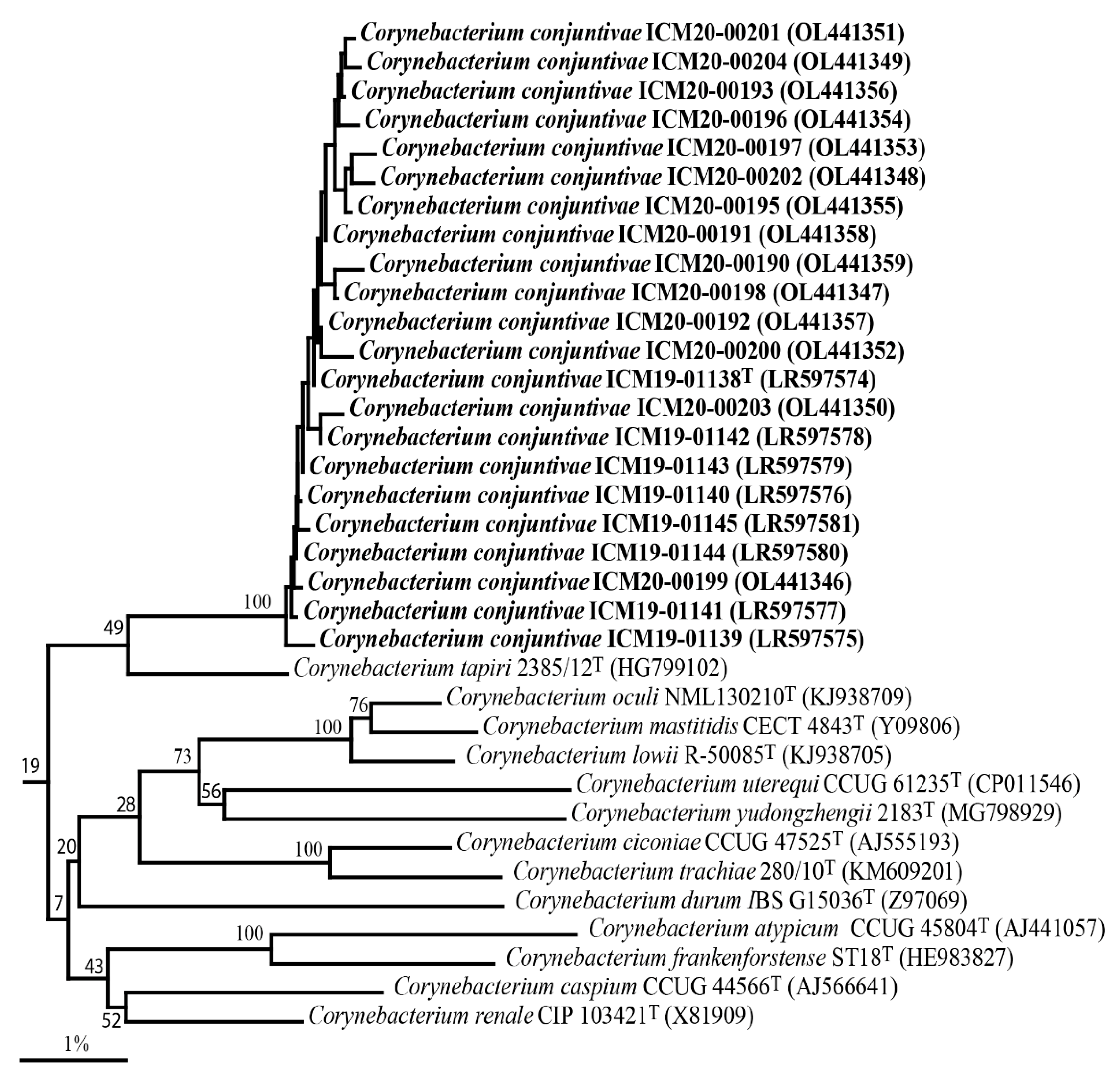
3.2. Phylogenetic Analysis

Sequence searches of GenBank based on their 16S rRNA gene sequences showed that the equine isolates were members of the genus Corynebacterium, exhibiting 99.4 to 100% similarity among them, thereby demonstrating high genealogical homogeneity. The 16S rRNA gene sequence of the strain ICM19-01138T exhibited 96.5% sequence similarity with the type strain of Corynebacterium tapiri 2385/12T and less than 96.0% with all other Corynebacterium species. The 16S rRNA sequence divergence values of > 3% indicate that the isolates from the horses represent a hitherto unknown species [31]. The phylogenetic relationship of the equine isolates within the genus Corynebacterium, based on the neighbor-joining algorithm, revealed that the 22 equine isolates clustered together and formed a distinct lineage to C. tapiri 2385/12T (Figure 1). The equine isolates exhibited weak associations (<70% bootstrap values) with C. tapiri and other validated Corynebacterium species (Figure 1). The GenBank accession numbers for the 16S rRNA gene sequences of the equine isolates are shown in Figure 1.

3.3. Chemotaxonomic and G+C Analysis

The wall murein acid hydrolysates of ICM19-01138T showed the presence of meso-diaminopimelic acid. The main mycolic acids of this strain consisted of short-chain mycolic acids (C28:0, 5%; C30:0, 28 %; C32:0, 53 % and C34:0, 14 %). The predominant cellular fatty acids (>1%) of the novel bacterium consisted of straight-chain saturated (C15:0, 23.4%; C16:0, 17.6%; C17:0, 11.6% and C18:0, 1.7%) and monounsaturated (C16:1 ω9c, 1.2%; C17:1 ω8c, 21.7% and C18:1 ω9c, 21.5%) fatty acid types. These chemotaxonomic characteristics resembled those of corynebacteria [4], supporting the phylogenetic results. The G+C content of the type strain ICM19-01138T was 58.4 mol%, which is also consistent with the values reported for members of the genus Corynebacterium (46–74 mol%) [12].

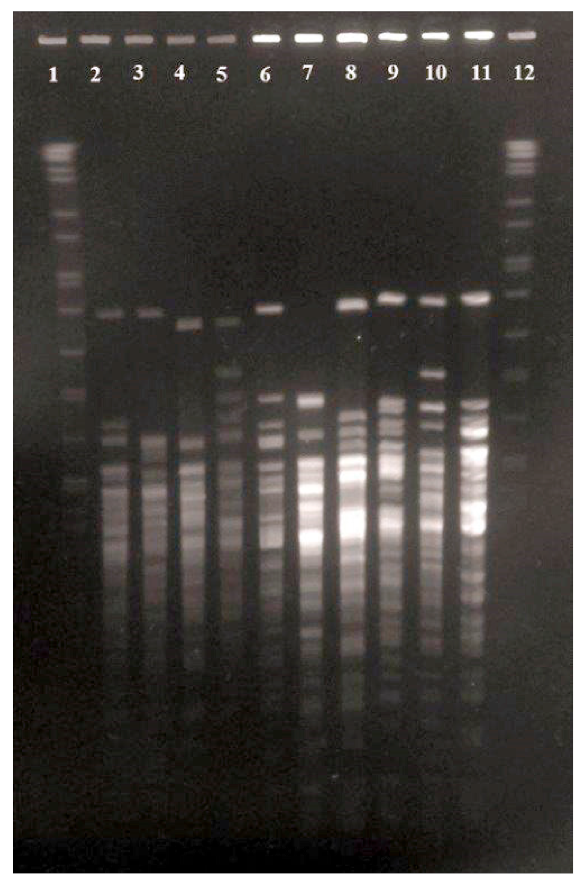
3.4. Antimicrobial Susceptibility and PFGE Analysis

All isolates were susceptible to amikacin, azithromycin, chloramphenicol, enrofloxacin, gentamicin, cefazolin, doxycycline, imipenem and rifampicin, whereas a few isolates were resistant to trimethoprim/sulfamethoxazole (9%), oxacillin (4.5%), penicillin (4.5%), erythromycin (4.5%) and tetracycline (9%). These results are in line with the high susceptibility detected to most of the commonly used veterinary antimicrobials in Corynebacterium isolates previously isolated from the conjunctiva of healthy horses [32].
A visual comparison of the restriction endonuclease digestion profiles generated by the 22 equine isolates revealed 10 different profiles represented by isolates ICM19/01138, ICM19/01139, ICM19/01141, ICM19/01144, ICM20/00190, ICM20/00191, ICM20/00193, ICM20/00195, ICM20/00199 and ICM20/00202 (Figure 2; lines 2–23). Corynebacterium isolates were recovered from all animals included in the study, supporting its high frequency of isolation from normal equine ocular flora [23]. These data, together with the relatively high genetic diversity detected by PFGE analysis and commonly observed in widespread microorganisms [33], suggest that the new Corynebacterium species identified in this study may be part of the commensal flora present in the conjunctival sac of equids. The formal description of Corynebacterium conjunctivae will allow its recognition and identification in the future and will improve knowledge of its distribution on ocular surfaces.

4. Conclusions

The polyphasic analyses based on phenotypic, chemotaxonomic and phylogenetic data indicated that the unidentified isolates from the ocular surface of apparently healthy horses belong to a distinct genomic species of the genus Corynebacterium, for which the name Corynebacterium conjunctivae sp. nov. is proposed, with strain ICM19-01138T (DSM 109759T = CCUG 60112T) as the type strain. C. conjunctivae can be differentiated from its nearest phylogenetic relative by the characteristics shown in Table 1.

Description of Corynebacterium conjunctivae sp. nov.

Corynebacterium conjunctivae (con.junc.ti’vae. N.L. n. conjunctiva, the membrane joining the eyeball to the eyelids; N.L. gen. n. conjunctivae, of conjunctiva).
Cells are Gram-positive, non-spore-forming, non-motile rods, approximately 0.5 µm in width and 1 µm in length. They are facultatively anaerobic, non-lipophilic and CAMP-negative with S. aureus. On Columbia blood agar, after 24 h incubation at 37 °C, colonies are cream, circular, convex, dull, approximately 1–2 mm in diameter and non-hemolytic. Urea is hydrolyzed but not aesculin and gelatin. Nitrate is not reduced. Acid is produced from glucose, D-fructose, D-mannose, ribose and trehalose but not from maltose, lactose, sucrose, glycogen, D-xylose, mannitol, N-acetyl-glucosamine, erythritol, glycerol, L-arabinose, L-xylose, adonitol, β-methyl-xyloside, galactose, dulcitol, inositol, sorbitol, L-rhamnose, sorbose, α-methyl-D-mannoside, α-methyl-D-glucoside, melibiose, inulin, melezitose, D-raffinose, xylitol, amygdalin, arbutin, salicin, cellobiose, β-gentibiose, D-turanose, D-lyxose, D-tagatose, D-arabinose, L-fucose, gentiobiose, D-fucose, D-arabitol, L-arabitol, 2-ketogluconate or 5-ketogluconate. Activity for alkaline and acid phosphatases, esterase C4, lipase C14, ester lipase C8, naphthol-AS-BI-phosphohydrolase, leucine arylamidase and β-glucosidase is detected. Pyrrolidonyl arylamidase, α-mannosidase, α-galactosidase, α-fucosidase, β-glucuronidase, pyrazinamidase, β-galactosidase, α-glucosidase, N-acetyl-β-glucosaminidase, chymotrypsin, trypsin, valine arylamidase and cystine arylamidase are not produced. Activity for lipase C14 is variable. Long-chain cellular acids are of the straight-chain saturated and monounsaturated types, with C15:0, C16:0, C17:1 ω8c and C18:1 ω9c predominating. Cell-wall murein is based on meso-diaminopimelic acid. Corynomycolic acids are present (C28–C34).
The type strain, ICM19-01138T (DSM 109759T = CCUG 60112T), was isolated from the ocular surface of an apparently healthy horse.

Author Contributions

J.F.F.-G.: data curation, supervision, writing—review and editing. S.L.: resources, supervision. A.C.: methodology, software. E.A.: resources, investigation. H.H.M.: resources, investigation. R.S.C.: resources, investigation. C.R.A.: conceptualization of the study. L.D.: visualization, investigation. A.I.V.: software, formal analysis, writing—original draft preparation. All authors have read and agreed to the published version of the manuscript.

Funding

This research was partially supported by the Spanish Ministry of Science, Innovation, and Universities [RTI2018-098530-B-I00].

Institutional Review Board Statement

All animal procedures were conducted in accordance with the ARVO Statement for the Use of Animals in Ophthalmic and Vision Research and were approved by the Animal Care and Use Committee of Auburn University (protocol#2013-2252).

Informed Consent Statement

Not applicable.

Data Availability Statement

The data presented in this study are available in this article.

Acknowledgments

The authors would like to dedicate this work to the memory of Cova Arias, who started and worked through the initial stages of this study.

Conflicts of Interest

The authors declare no conflict of interest.

References

  1. Leigue, L.; Montiani-Ferreirm, F.; Moore, B.A. Antimicrobial susceptibility and minimal inhibitory concentration of Pseudomonas aeruginosa isolated from septic ocular surface disease in different animal species. Open Vet. J. 2016, 6, 215–222. [Google Scholar] [CrossRef] [Green Version]
  2. Willcox, M.D. Characterization of the normal microbiota of the ocular surface. Exp. Eye Res. 2013, 117, 99–105. [Google Scholar] [CrossRef]
  3. Kugadas, A.; Gadjeva, M. Impact of microbiome in ocular health. Ocul. Surf. 2016, 14, 342–349. [Google Scholar] [CrossRef] [Green Version]
  4. Andrew, S.E.; Nguyen, A.; Jones, G.L.; Brooks, D.E. Seasonal effects on the aerobic bacterial and fungal conjunctival flora of normal thoroughbred brood mares in Florida. Vet. Ophthalmol. 2003, 6, 45–50. [Google Scholar] [CrossRef]
  5. Gemensky-Metzler, A.J.; Wilkie, D.A.; Kowalski, J.J.; Schmall, L.M.; Willis, A.M.; Yamagata, M. Changes in bacterial and fungal ocular flora of clinically normal horses following experimental application of topical antimicrobial or antimicrobial-corticosteroid ophthalmic preparations. Am. J. Vet. Res. 2005, 66, 800–811. [Google Scholar] [CrossRef] [PubMed]
  6. Hampson, E.C.G.M.; Gibson, J.S.; Barot, M.; Shapter, F.M.; Greer, R.M. Identification of bacterial and fungi samples from the conjunctival surface of normal horses in South-East Queensland, Australia. Vet. Ophthalmol. 2019, 22, 265–275. [Google Scholar] [CrossRef] [PubMed]
  7. Johns, I.C.; Baxter, K.; Booler, H.; Hicks, C.; Menzies-Gow, N. Conjunctival bacterial and fungal flora in healthy horses in the UK. Vet. Ophthalmol. 2011, 14, 195–199. [Google Scholar] [CrossRef] [PubMed]
  8. Moore, C.P.; Heller, N.; Majors, L.J.; Whitley, R.D.; Burgess, E.C.; Weber, J. Prevalence of ocular microorganisms in hospitalized and stabled horses. Am. J. Vet. Res. 1988, 49, 773–777. [Google Scholar]
  9. Whitley, R.D.; Moore, C.P. Microbiology of the equine eye in health and disease. Vet. Clin. North Am. Large Anim. Pract. 1984, 6, 451–466. [Google Scholar] [CrossRef]
  10. Whitley, R.D.; Burgess, E.C.; Moore, C.P. Microbial isolates of the normal equine eye. Equine Vet. J. 1983, 15, 138–140. [Google Scholar] [CrossRef]
  11. Arsan, A.K.; Sizmaz, S.; Ozkan, S.B.; Duman, S. Corynebacterium minutissimum endophthalmitis: Management with antibiotic irrigation of the capsular bag. Int. Ophthalmol. 1995, 19, 313–316. [Google Scholar] [CrossRef]
  12. Bernard, K.A.; Funke, G. Corynebacterium. In Bergey’s Manual of Systematics of Archaea and Bacteria (BMSAB); Bergey’s Manual Trust: East Lansing, MI, USA, 2015; Available online: https://onlinelibrary.wiley.com/doi/pdf/10.1002/9781118960608.gbm00026 (accessed on 18 April 2022). [CrossRef] [Green Version]
  13. Bernard, K.A.; Pacheco, A.L.; Loomer, C.; Burdz, T.; Wiebe, D.; Huynh, C.; Kaplen, B.; Olson, A.B.; Cnockaert, M.; Eguchi, H.; et al. Corynebacterium lowii sp. nov. and Corynebacterium oculi sp. nov., derived from human clinical disease and an emended description of Corynebacterium mastitidis. Int. J. Syst. Evol. Microbiol. 2016, 66, 2803–2812. [Google Scholar] [CrossRef] [Green Version]
  14. Chow, S.K.; Bui, U.; Clarridge, J.E. Corynebacterium bovis eye infections, Washington, USA, 2013. Emerg. Infect. Dis. 2015, 21, 1687–1689. [Google Scholar] [CrossRef] [Green Version]
  15. Kojouri, G.A.; Ebrahimi, A.; Nikookhah, F. Systemic dexamethasone and its effect on normal aerobic bacterial flora of cow. Pak. J. Biol. Sci. 2007, 10, 2095–2097. [Google Scholar]
  16. St. Leger, A.J.; Desai, J.V.; Drummond, R.A.; Kugadas, A.; Almaghrabi, F.; Silver, P.; Raychaudhuri, K.; Gadjeva, M.; Iwakura, Y.; Lionakis, M.S.; et al. An ocular commensal protects against corneal infection by driving an Interleukin-17 response from mucosal γδ T cells. Immunity 2017, 47, 148–158. [Google Scholar] [CrossRef] [Green Version]
  17. LaFrentz, S.; Abarca, E.; Mohammed, H.H.; Cuming, R.; Arias, C.R. Characterization of the normal equine conjunctival bacterial community using culture-independent methods. Vet. Ophthalmol. 2020, 23, 480–488. [Google Scholar] [CrossRef]
  18. Vela, A.I.; Mentaberre, G.; Lavín, S.; Domínguez, L.; Fernández-Garayzábal, J.F. Streptococcus caprae sp. nov., isolated from iberian ibex (Capra pyrenaica hispanica). Int. J. Syst. Evol. Microbiol. 2016, 66, 196–200. [Google Scholar] [CrossRef]
  19. Yoon, S.H.; Ha, S.M.; Kwon, S.; Lim, J.; Kim, Y.; Seo, H.; Chun, J. Introducing EzBio-Cloud: A taxonomically united database of 16S rRNA gene sequences and whole-genome assemblies. Int. J. Syst. Evol. Microbiol. 2017, 67, 1613–1617. [Google Scholar] [CrossRef]
  20. Rasmussen, S.W. SeqTools, a Program Suite for Sequence Analysis; Carlsberg Laboratory: Copenhagen, Denmark, 2002. [Google Scholar]
  21. Saitou, N.; Nei, M. The neighbor-joining method: A new method for reconstructing phylogenetic trees. Mol. Biol. Evol. 1987, 4, 406–425. [Google Scholar]
  22. Page, R.D. TreeView: An application to display phylogenetic trees on personal computers. Comput. Appl. Biosci. 1996, 12, 357–358. [Google Scholar]
  23. Tamura, K.; Peterson, D.; Peterson, N.; Stecher, G.; Nei, M.; Kumar, S. MEGA5: Molecular evolutionary genetics analysis using maximum likelihood, evolutionary distance, and maximum parsimony methods. Mol. Biol. Evol. 2011, 28, 2731–2739. [Google Scholar] [CrossRef] [Green Version]
  24. Kimura, M. A simple method for estimating evolutionary rates of base substitutions through comparative studies of nucleotide sequences. J. Mol. Evol. 1980, 16, 111–120. [Google Scholar] [CrossRef]
  25. Mesbah, M.; Premachandran, U.; Whitman, W.B. Precise measurement of the G-C content of deoxyribonucleic acid by high-performance liquid chromatography. Int. J. Syst. Bacteriol. 1989, 39, 159–167. [Google Scholar] [CrossRef] [Green Version]
  26. Funke, G.; von Graevenitz, A.; Clarridge, J.E.; Bernard, K.A. Clinical microbiology of coryneform bacteria. Clin. Microbiol. Rev. 1997, 10, 125–159. [Google Scholar] [CrossRef]
  27. Galán-Relaño, A.; Gómez-Gascón, L.; Luque, I.; Barrero-Domínguez, B.; Casamayor, A.; Cardoso-Toset, F.l; Vela, A.I.; Fernández-Garayzábal, J.F.; Tarradas, C. Antimicrobial susceptibility and genetic characterization of Trueperella pyogenes isolates from pigs reared under intensive and extensive farming practices. Vet. Microbiol. 2019, 232, 89–95. [Google Scholar] [CrossRef]
  28. Clinical and Laboratory Standard Institute. Performance Standards for Antimicrobial Disk and Dilution Susceptibility Tests for Bacteria Isolated from Animals—Approved Standard, 2nd ed.; Document M31-A2; Clinical and Laboratory Standard Institute: Wayne, PA, USA, 2002; pp. 1–86. [Google Scholar]
  29. Clinical and Laboratory Standard Institute. Methods for Antimicrobial Susceptibility Testing of Infrequently Isolated or Fastidious Bacteria Isolated from Animals—Approved Standard, 1st ed.; Document VET06; Clinical and Laboratory Standard Institute: Wayne, PA, USA, 2017; pp. 1–100. [Google Scholar]
  30. Dorella, F.A.; Carvalho Pacheco, L.G.; Costa Oliveira, S.; Miyoshi, A.; Azevedo, V. Corynebacterium pseudotuberculosis: Microbiology, biochemical properties, pathogenesis and molecular studies of virulence. Vet. Res. 2006, 37, 201–218. [Google Scholar] [CrossRef] [Green Version]
  31. Stackebrandt, E.; Goebel, B.M. Taxonomic note: A place for DNA-DNA reassociation and 16S rRNA sequence analysis in the present species definition in bacteriology. Int. J. Syst. Bacteriol. 1994, 44, 846–849. [Google Scholar] [CrossRef] [Green Version]
  32. de Araújo Ferreira, A.R.; Santana, A.F.; da Veiga Rodarte de Almeida, A.C.; Sousa, R.F.; Perecmanis, S.; Galera, P.D. Bacterial culture and antibiotic sensitivity from the ocular conjunctiva of horses. Cienc. Rural 2017, 47, e20160753. [Google Scholar]
  33. Pors, S.E.; Hansen, M.S.; Christensen, H.; Jensen, H.E.; Petersen, A.; Bisgaard, M. Genetic diversity and associated pathology of Pasteurella multocida isolated from porcine pneumonia. Vet. Microbiol. 2011, 150, 354–361. [Google Scholar] [CrossRef] [Green Version]
  34. Baumgardt, S.; Loncaric, I.; Kämpfer, P.; Busse, H.J. Corynebacterium tapiri sp. nov. and Corynebacterium nasicanis sp. nov., isolated from a tapir and a dog, respectively. Int. J. Syst. Evol. Microbiol. 2015, 65, 3885–3893. [Google Scholar] [CrossRef]
Figure 1. Neighbor-joining tree based on 16S rRNA showing the phylogenetic relationships within the genus Corynebacterium of Corynebacterium conjunctivae sp. nov. Mycobacterium smegmatis MC2 155T was used as an out-group. Bootstrap values (expressed as a percentage of 1000 replications) higher than 70% are given at the branching points. The nodes (groupings) also obtained in the maximum-likelihood and parsimony trees are indicated by open circles. The corresponding nodes also obtained in the parsimony tree are indicated by filled circles. Bar, 1% sequence divergence.
Animals 12 01827 g001
Figure 2. PFGE patterns of Corynebacterium conjunctivae sp. nov generated after XbaI macrorestriction. Lanes 1 and 12, Salmonella serotype Branderup strain H9812; Lanes 2 to 11, strains ICM19/01138T (four isolates), ICM19/01139 (four isolates), ICM19/01141 (five isolates), ICM19/01144 (two isolates), ICM20/00190, ICM20/00191, ICM20/00193, ICM20/00195 (two isolates), ICM20/00199 and ICM20/00202, respectively.
Animals 12 01827 g002
Table 1. Characteristics that differentiate Corynebacterium conjunctivae sp. nov. from its closest phylogenetic relative Corynebacterium tapiri.
CharacteristicC. conjunctivae
(22 Isolates)
C. tapiri 2385/12Tb
Nitrate reduction+
Hydrolysis of:
Esculin
Urea++
Production of:
Pyrazinamidase+
Esterase C4+
Ester lipase C8+
β-glucuronidase+
α-glucosidase+
Alkaline phosphatase++
Acid phosphatase+
Acid production from:
Glucose++
Ribose++
Maltose+
Saccharose+
Data obtained from Baumgardt et al. [34].
Publisher’s Note: MDPI stays neutral with regard to jurisdictional claims in published maps and institutional affiliations.

