Article

New Data on Pterygodermatites (Pterygodermatites) plagiostoma Wedl, 1861 (Nematoda, Rictulariidae) Parasite of the Algerian Hedgehog Atelerix algirus Linnaeus, 1758 (Eulipotyphla: Erinaceidae) from the Canary Islands †

by
Jordi Miquel
1,2,*,
Alexis Ribas
1,2,
Román Pino-Vera
3,
Elena Izquierdo-Rodríguez
4,
Natalia Martín-Carrillo
4,
Carlos Feliu
1 and
Pilar Foronda
3,4
1
Secció de Parasitologia, Departament de Biologia, Sanitat i Medi Ambient, Facultat de Farmàcia i Ciències de l’Alimentació, Universitat de Barcelona, Avgda. Joan XXIII, sn, 08028 Barcelona, Spain
2
Institut de Recerca de la Biodiversitat (IRBio), Universitat de Barcelona, Avgda. Diagonal, 645, 08028 Barcelona, Spain
3
Departamento Obstetricia y Ginecología, Pediatría, Medicina Preventiva y Salud Pública, Toxicología, Medicina Legal y Forense y Parasitología, Facultad de Farmacia, Universidad de La Laguna, Avda. Astrofísico F. Sánchez, sn, 38203 La Laguna, Canary Islands, Spain
4
Instituto Universitario de Enfermedades Tropicales y Salud Pública de Canarias, Universidad de La Laguna, Avda. Astrofísico F. Sánchez, sn, 38203 La Laguna, Canary Islands, Spain
*
Author to whom correspondence should be addressed.
In memory of Professor Jean-Claude Quentin (1938–2021).
Animals 2022, 12(15), 1991; https://doi.org/10.3390/ani12151991
Submission received: 29 June 2022 / Revised: 30 July 2022 / Accepted: 4 August 2022 / Published: 5 August 2022

Abstract

:

Simple Summary

A redescription of Pterygodermatites (Pterygodermatites) plagiostoma (Nematoda, Rictulariidae) is made by means of light and scanning electron microscopy, including the first data on male specimens. The morphologic study was based on specimens recovered from two Algerian hedgehogs from Tenerife and Gran Canaria islands (Canary Archipelago, Spain). The main differential characteristics of P. (P.) plagiostoma males are the number of cuticular projection pairs, the number of precloacal fans, and the size of spicules. The cloacal papillae are arranged according to the Ascaridida type, with two precloacal pairs, an unpaired precloacal papilla, one pair lateral to the cloaca, six postcloacal pairs, and a pair of phasmids. Females are mainly characterized and differentiated by the number of prevulvar pairs of cuticular projections, by the total number of cuticular projection pairs, by the level of differentiation from combs to spines and by the position of the vulva in relation to the esophagus–intestine junction. The comparison with species of the subgenus P. (Pterygodermatites) shows P. (P.) plagiostoma as a species clearly differentiated from the remaining species of this subgenus.

Abstract

A redescription of the rictulariid nematode Pterygodermatites (Pterygodermatites) plagiostoma Wedl, 1861, is made by means of light and scanning electron microscopy, including the first data on male specimens. The morphologic study was based on specimens recovered from two Algerian hedgehogs (Atelerix algirus) from Tenerife and Gran Canaria islands (Canary Archipelago, Spain). The main characteristics of P. (P.) plagiostoma males are the presence of 49–53 pairs of cuticular projections, the presence of one or two midventral precloacal fans (generally one), and the size of two unequal spicules, measuring 98–123 µm (right spicule) and 185–236 µm (left spicule). The cloacal papillae are arranged according to the Ascaridida type. They include two precloacal pairs, an unpaired precloacal papilla, one pair lateral to the cloaca, six postcloacal pairs, and a pair of phasmids near the tail tip. Females are mainly characterized by the presence of 71–77 pairs of cuticular projections, with 43–46 pairs of prevulvar combs, by the differentiation from combs to spines at the level of or slightly posterior to the vulva and by the position of the vulva, located posteriorly to the esophagus–intestine junction. Clear differences were found between P. (P.) plagiostoma and related species of the subgenus P. (Pterygodermatites).

Graphical Abstract

1. Introduction

Pterygodermatites (Pterygodermatites) plagiostoma is a rictulariid nematode frequently found parasitizing hedgehogs. It has been recorded in Atelerix algirus, Erinaceus europaeus, Hemiechinus auritus, and Paraechinus aethiopicus [1,2,3,4,5,6,7,8]. It has also been cited parasitizing the bat Vespertilio mystacinus (=Myotis mystacinus) (Vespertilionidae), the rodent Sciurus melanogaster (=Callosciurus melanogaster) (Sciuridae), and the carnivores Vulpes vulpes niloticus (Canidae) and Paguma larvata (Viverridae) [9,10,11,12]. However, the P. plagiostoma specimens of Parona [10] in C. melanogaster from Mentawei (Sumatra) were later studied by Jägerskiöld [3], who identified them as a new species Rictularia fallax, currently P. (Mesopectines) fallax after Quentin [13]. Moreover, the record of Sonsino [11] in the fox from Egypt was later considered Rictularia affinis (=Pterygodermatites (Multipectines) affinis) by Jägerskiöld [3]. The report of Willemoes-Suhm [12] in M. mystacinus from Germany, described as Ophiostomum spinosum and later identified as Rictularia plagiostoma [14,15], was finally considered as P. (P.) spinosa by Quentin [13]. Finally, the finding of Leiper [9] in the palm-civet P. larvata obtained from the London Zoo is probably a misidentification or an accidental parasitism [7] and, therefore, it must be considered with caution.
Pterygodermatites (P.) plagiostoma is the type species of both genus and nominal subgenus. The subgenus P. (Pterygodermatites) has only six species, namely P. (P.) plagiostoma, P. (P.) aethechini, P. (P.) atlanticaensis, P. (P.) mexicana, P. (P.) shaldibini, and P. (P.) spinosa [13,16,17,18]. In the original description of P. (P.) plagiostoma, Wedl [8] illustrates the caudal extremity of a male specimen showing two unequal spicules, but no other morphologic characteristic or measurements are presented. Despite the numerous posterior findings [1,2,3,4,5,6,7], to date, the detailed morphology of the male remains unknown [13,19].
The aim of the present study is to describe for the first time the male of P. (P.) plagiostoma and contribute with new data on females from a hedgehog A. algirus from the Canary Islands. In addition, the sequence of the mitochondrial cytochrome c oxidase subunit I gene (MT-CO1) of P. (P.) plagiostoma is provided.

2. Materials and Methods
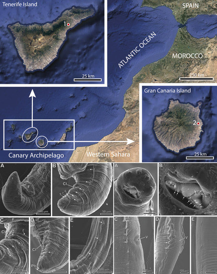
2.1. Specimens

Two Atelerix algirus Linnaeus, 1758 (Eulipotyphla: Erinaceidae), were found dead on the road in El Rosario (Tenerife Island) on 3 June 2021 and in Jinámar (Gran Canaria Island) (Canary Archipelago, Spain) on 9 October 2019 and then they were scanned for intestinal helminths. A total of 149 rictulariid nematodes (38 males and 111 females) were found in the gastrointestinal tract of the two hedgehogs. They were identified as P. (P.) plagiostoma following the available literature [7,13].

2.2. Scanning Electron Microscopy Study

Six P. (P.) plagiostoma males and eight females were preserved for scanning electron microscopy (SEM) examination, fixed in 70% ethanol and posteriorly dehydrated in an ethanol series and critical point dried with carbon dioxide in an Emitech K850X (Quorum Technologies Ltd., Laughton, East Sussex, UK). Finally, specimens were mounted on stubs with conductive adhesive tape and colloidal silver, coated with carbon in an Emitech K950X (Quorum Technologies Ltd., Laughton, East Sussex, UK) evaporator, and examined using a Field Emission SEM JSM-7001F (Jeol) (Jeol Ltd., Tokyo, Japan) at 10 kV in the “Centres Científics i Tecnològics” of the University of Barcelona (CCiTUB).

2.3. Molecular Analyses

Genomic DNA samples were isolated from the mid-section fragment of P. (P.) plagiostoma following López et al. [20]. The DNA extraction procedure was checked using DeNovix DS-11+ Spectrophotometer (DeNovix Inc., Wilmington, DE, USA).
DNA amplification by PCR was conducted using the primer cocktail as described by Prosser et al. [21], for the barcode region of the mitochondrial cytochrome c oxidase subunit I gene (MT-CO1). The PCR amplification contained 1X Buffer (Bioline, London, UK), 0.2 mM of each dNTP (Bioline), 0.5 µL of each primer cocktail (10 µM of a three-forward-primers mix, and 10 µM of a three-reverse-primers mix), 1U of Taq DNA polymerase (Bioline), 1.5 mM MgCl2 (Bioline), and 20–30 ng of total genomic DNA in a total volume of 50 µL. Amplification was conducted with XP Cycler (Bioer Technology) using the following parameters: 94 °C for 1 min; five cycles at 94 °C for 40 s, 45 °C for 40 s, 72 °C for 1 min; followed by 35 cycles at 94 °C for 40 s, 51 °C for 40 s, 72 °C for 1 min; and a final extension at 72 °C for 5 min [21]. The resulting amplifications were visualized on 1.2% agarose gel at 100 V for 45 min.
The product of PCR was sequenced in Macrogen (Madrid, Spain) with primers NemF1_t1 and NemR1_t1 [21]. The analysis of the sequences was carried out with software MEGA X [22], using the multiple alignment program ClustalW included in MEGA X, and minor corrections were made by hand.
A phylogenetic analysis based on the MT-CO1 gene sequences of P. (P.) plagiostoma and other Pterygodermatites species available in GenBank was performed using Neighbor-Joining distance method (NJ) with the p-distance model [23] and Maximum-Likelihood (ML) method with Tamura–Nei model [24], both with at least 1000 bootstrap replications in MEGA X [22]. The sequence Plectus aquatilis KX017524 was used as the outgroup.

3. Results

3.1. Taxonomic Summary

Family Rictulariidae Hall, 1913.
Genus Pterygodermatites Wedl, 1861.
Subgenus Pterygodermatites (Pterygodermatites) Quentin, 1969.
Pterygodermatites (Pterygodermatites) plagiostoma Wedl, 1861 (Figures 2A–C, 3A–E, 4A–E, 5A,B and 6A–E).
Type host: Atelerix algirus Linnaeus, 1758 (Eulipotyphla: Erinaceidae).
Type locality: El Rosario (Tenerife Island, Canary Archipelago, Spain) (28°25′57.15″ N, 16°22′6.328″ W).
Other localities: Jinámar (Gran Canaria Island, Canary Archipelago, Spain) (28°1′58.457″ N, 15°25′8.994″ W).
Site of infection: small intestine.
Prevalence and intensity: two A. algirus studied with an intensity of 49 worms (15 males and 34 females) in the hedgehog from El Rosario and 100 worms (23 males and 77 females) in the hedgehog from Jinámar.
Type specimens: deposited in Muséum National d’Histoire Naturelle (Paris, France), under accession nos. MNHN HEL 1822 (8 males) and MNHN HEL 1823 (7 females).
Mitochondrial cytochrome c oxidase subunit I gene (MT-CO1) sequence: a fragment of 700 bp was obtained for the MT-CO1 of P. (P.) plagiostoma isolated from A. algirus in El Rosario, Tenerife. A 551 bp fragment was successfully sequenced and submitted to the GenBank database under the accession number ON502379.

3.2. Phylogenetic Tree

The phylogenetic trees created using NJ and ML (Figure 1) methods based on the MT-CO1 gene showed similar results. P. (P.) plagiostoma is included in a clade together with P. (Paucipectines) zygodontomis and P. (Pa.) jägerskiöldi with a high bootstrap value (100%), and clearly separated from these two species. In the other clade P. (Mesopectines) whartoni and P. (Me.) nycticebi are included.

3.3. Description

General: medium-sized nematodes. Dorsal oral opening, surrounded by 6 internal labial papillae, 4 external pairs of cephalic papillae, and two lateral amphids (Figure 2A, Figure 3A and Figure 6A). Oral opening with irregularly distributed oral denticles of different sizes (Figure 4B); 3 internal esophageal teeth at the bottom of the buccal capsule, 1 dorsal and 2 lateroventral (Figure 2A, Figure 3A,B and Figure 5A). Well-developed buccal capsule. Two subventral rows of cuticular projections along the body, in the form of combs and spines both in males and females (Figure 3A,B, Figure 4E, Figure 5A and Figure 6C–E).
Male (17 specimens measured, mean in parentheses): Well-developed buccal capsule, oriented dorsally (Figure 2A and Figure 3B). Oral opening surrounded by irregular denticles. Presence of three esophageal teeth (Figure 2A and Figure 3A,B). Total pairs of combs 49–53 (50). Body length 5.36–7.67 mm (6.23 mm); width at the level of the esophagus basis 413–619 µm (498 µm). Esophagus length 2.09–2.87 mm (2.45 mm); width at base 111–180 µm (148 µm). Nerve ring located at 206–375 µm (269 µm) from the cephalic extremity; at the level of 3–4 pairs of cuticular combs. Deirids located at 516–795 µm (654 µm) from the cephalic extremity; at the level of 6–8 pairs of cuticular combs. Posterior end of body strongly curved ventrally (Figure 2B, Figure 3C,D and Figure 4A). Distance between the last cuticular spine and the tail tip 702–1060 µm (870 µm). The pericloacal surface is ornamented with cuticular markings (Figure 4B–D). Total of 19 caudal papillae which are sessile; 2 pairs of precloacal papillae (pairs 1 and 2), 1 unpaired precloacal papilla, 1 pair of papillae lateral to cloaca (pair 4), and 6 pairs of postcloacal papillae (pairs 3 and 5–9) (Figure 2C and Figure 4B,C). Pairs of papillae 1, 4, and 8 in a dorsolateral position, particularly the pair 8 that is located outside the ornamented area (Figure 2C and Figure 4B–D). A pair of phasmids near the tip of tail (Figure 2C and Figure 4D). One or two (generally one) precloacal midventral fans immediately before the first pair of precloacal papillae (Figure 2C, Figure 3C,D and Figure 4A,B). Spicules unequal in size; right spicule 98–123 µm (110 µm); left spicule 185–236 µm (217 µm) (Figure 2B and Figure 3C–E). Gubernaculum 28–39 µm (34 µm) (Figure 3E).
Figure 2. Pterygodermatites (Pterygodermatites) plagiostoma male. (A) Cephalic extremity, dorsal view. (B) Caudal extremity, lateral view. (C) Cloacal papillae. (*) Unpaired precloacal papilla; (Ph) phasmids.
Animals 12 01991 g002
Figure 3. Pterygodermatites (Pterygodermatites) plagiostoma male, light microscopy. (A) Cephalic extremity, dorsal view showing the two lateroventral esophageal teeth (lvT) and four internal labial papillae (arrows). (B) Cephalic extremity, lateral view showing dorsal (dT) and lateroventral (lvT) esophageal teeth. (C) Caudal extremity of a male with one midventral fan (F). (D) Caudal extremity of a male with two midventral fans. (E) Detail of the right spicule (rSp), left spicule (lSp), and gubernaculum (G). (C) Cuticular combs; (O) esophagus; (Sp) spicules.
Animals 12 01991 g003
Figure 4. Pterygodermatites (Pterygodermatites) plagiostoma male’s caudal extremity, scanning electron microscopy. (A) Precloacal position of the midventral fan (F). (B) Lateral view showing the distribution cloacal papillae (pairs 1–9). (C) Cloacal region showing the unpaired precloacal papilla (*). (D) Position of phasmids (Ph) near the tail tip. (E) Lateral view illustrating the last cuticular spines (arrows). (Cl) Cloaca.
Animals 12 01991 g004
Female (17 gravid specimens measured, mean in parentheses): Well-developed buccal capsule, oriented dorsally (Figure 5A and Figure 6A). Oral opening surrounded by irregular denticles (Figure 6B). Presence of three esophageal teeth. Body length 12.07–17.54 mm (14.52 mm); width at the level of the vulva 423–712 µm (539 µm). Esophagus length 3.11–4.13 mm (3.71 mm); width at base 129–180 µm (154 µm). Nerve ring located at 285–463 µm (349 µm) from the cephalic extremity; at the level of 2–4 pairs of cuticular combs. Deirids located at 578–753 µm (644 µm) from the cephalic extremity; at the level of 6–9 pairs of cuticular combs. Total pairs of combs and spines 71–77 (74); prevulvar pairs of combs 43–46 (44); prevulvar combs in close contact to one another (Figure 5A,B and Figure 6C,D); at the level of vulva combs transform into two small pairs of combs and later into spines, and become more spaced from each other (Figure 5B and Figure 6C–E); postvulvar pairs of spines 28–32 (30); comb transformation to spines at the vulva or immediately posterior to the vulva (Figure 6C,D). Vulva located at 3.20–4.67 mm (3.99 mm) from the cephalic extremity; posterior to the esophagus–intestine junction (Figure 3B). Tail 177–350 µm (236 µm); with a terminal spine. Eggs oval, with a thick eggshell; embryonated 51.4–56.6 × 38.6–41.7 µm (55.6 × 42.1 µm); unembryonated 46.3–48.9 × 30.9–36.0 µm (47.5 × 33.1 µm).
Figure 5. Pterygodermatites (Pterygodermatites) plagiostoma female, light microscopy. (A) Cephalic extremity, lateral view showing the dorsal esophageal tooth (dT). (B) Vulvar region. Note the posterior position of the vulva (V) in relation to the esophageal–intestinal junction (arrow). (C) Cuticular combs; (I) intestine; (O) esophagus.
Animals 12 01991 g005
Figure 6. Pterygodermatites (Pterygodermatites) plagiostoma female, scanning electron microscopy. (A) Apical view showing the internal circle of labial papillae (*). (B) Detail of oral opening showing the irregular peribuccal denticles (arrows). (C,D) Vulvar region of two females showing the transition from combs to spines at a vulvar level or slightly posterior to the vulva (V), respectively. Note the presence of two reduced combs (arrows) before the appearance of spines. (E) Morphology of the most terminal spines.
Animals 12 01991 g006

4. Discussion

Within the subgenus P. (Pterygodermatites) there are only six described species (see Table 1). Considering male specimens, there is data for only three species, namely P. (P.) aethechini, P. (P.) mexicana, and P. (P.) shaldybini [13,16,17]. In the original description of P. (P.) plagiostoma [8], there was no additional data on males other than a caudal extremity showing the presence of two unequal spicules. In the present study, for the first time, we extensively describe the male of P. (P.) plagiostoma.
In the Rictulariidae, the number of cloacal papillae of males is constant (19 papillae arranged in nine pairs of papillae and an unpaired papilla [13]). Thus, there are two precloacal pairs, an unpaired precloacal papilla, either another precloacal pair or a pair located laterally to the cloaca and six postcloacal pairs. Additionally, near the tail tip there is a pair of phasmids. Quentin [13] analyzed the disposition of male cloacal papillae in numerous rictulariids and described three types of arrangement of papillae: the type Ascaridida presenting some pairs of papillae not aligned and slightly dorsolateral, the type Spirurida with all pairs of papillae aligned, and a third type with pedunculated and grouped papillae. In males of P. (P.) plagiostoma, the cloacal papillae arrangement corresponds to the type Ascaridida [13,27] having the pairs 1, 4, and 8 in a dorsolateral position, particularly the pair 8. According to Quentin [13], the type Ascaridida, which corresponds to the most primitive arrangement of cloacal papillae, is present in rictulariids included in both genera Pterygodermatites and Rictularia, e.g., P. (Paucipectines) coloradensis, P. (Pa.) microti, P. (Pa.) ondatrae, P. (Pa.) zygodontomis, P. (Neopaucipectines) desportesi, R. citelli, R. dhanra, R. halli, R. lucifugus, R. macdonaldi, and R. proni, among others. After the review of Quentin [13], the Ascaridida type of arrangement of cloacal papillae has been described in other species, particularly those belonging to the subgenus P. (Paucipectines), e.g., P. (Pa.) andyraicola, P. (Pa.) argentinensis, P. (Pa.) baiomydis, P. (Pa.) chaetophracti, and P. (Pa.) hispanica [28,29,30,31]. With respect to the subgenus P. (Pterygodermatites), Le Roux [17] reported only three precloacal and five postcloacal pairs of cloacal papillae in P. (P.) aethechini. However, the original description of Le Roux was illustrated with a lateral view of the male caudal extremity showing probably the unpaired precloacal papilla. In a posterior study concerning P. (P.) mexicana, Caspeta-Mandujano et al. [16] described in five sublateral pairs of cloacal papillae (pairs 1, 2, 4, 8, and 9) and four subventral pairs (pairs 3, 5, 6, and 7) but no unpaired precloacal papilla are mentioned.
Within the P. (Pterygodermatites) subgenus, males present three characters that are useful to discriminate between species: the number of cuticular projection pairs (combs and spines), the number of midventral fans, and the size of spicules [13,19,32]. Considering the species of the subgenus P. (Pterygodermatites) for which information on males is available, there are differences in the male of P. (P.) plagiostoma in the characters spicules size, number of fans, and number of cuticular projection pairs (see Table 1). In fact, in comparison to P. (P.) aethechini [17], the spicule sizes, particularly the right spicule is 98–123 µm in P. (P.) plagiostoma vs. 118–132 µm in P. (P.) aethechini. Additionally, in comparison to P. (P.) mexicana [16], P. (P.) plagiostoma presents a higher number of cuticular projection pairs (49–53 vs. 40 in P. (P.) mexicana), larger spicules (98–123 µm (right) and 185–236 µm (left) vs. 30–50 µm (right) and 83–111 µm (left) in P. (P.) mexicana) and lower number of midventral fans (1–2 vs. 3–4 in P. (P.) mexicana). Finally, in comparison to P. (P.) shaldybini [13], P. (P.) plagiostoma presents larger spicules (98–123 µm (right) and 185–236 µm (left in) vs. 70 µm (right) and 140 µm (left) in P. (P.) shaldybini).
Concerning females, the most discriminant characters between species are the prevulvar and total number of cuticular projection pairs, the body level where the transition from combs to spines occurs, and the position of the vulva in relation to the esophagus–intestine junction [13,19,32]. In fact, the differentiation of cuticular projections (from combs to spines) occurs at the level of the vulva in P. (P.) plagiostoma whereas it occurs at a posterior level in P. (P.) aethechini [17]. On the other hand, the vulva position is posterior to the esophagus–intestine junction in P. (P.) plagiostoma whereas it is anterior in P. (P.) atlanticaensis [18]. There is also a higher number of both prevulvar and total pairs of cuticular projections (combs and spines) (43–46/71–77) in P. (P.) plagiostoma in comparison to the number of these characters (40/66) in P. (P.) mexicana [16] although the number of cuticular projection pairs in P. (P.) plagiostoma (71–77) is surpassed by the 84 pairs described in P. (P.) shaldybini [13]. However, none of these discriminant characters of females differ between P. (P.) plagiostoma and P. (P.) spinosa [13].
Pterygodermatites (P.) plagiostoma is present in several hedgehog species (A. algirus, E. europaeus, H. auritus, and P. aethiopicus), and has a wide geographical distribution, including North Africa (Algeria, Tunisia and Egypt), Saudi Arabia, and Spain [1,2,3,4,5,6,7,8]. As for the remaining species of the subgenus, P. (P.) atlanticaensis and P. (P.) mexicana are parasites of bats in Brazil and Mexico [16,18], P. (P.) spinosa were detected in bats from Baviera (Germany) [12], P. (P.) shaldybini were found in bats and hedgehogs from Asia (Kazakstan, Turkmenistan, and Mongolia) [13,25,26], and P. (P.) aethechini were recorded in the hedgehog Atelerix frontalis from South Africa [16]. The present study enlarges the distribution of P. (P.) plagiostoma to the Tenerife and Gran Canaria islands (Canary Archipelago, Spain).

5. Conclusions

The present work on the rictulariid nematode P. (P.) plagiostoma contributes with the first morphoanatomical study of male specimens since its original description and provides further information on females. Moreover, the current finding in Tenerife and Gran Canaria islands (Canary Archipelago, Spain) enlarges the geographical distribution of this rictulariid.
The most useful characteristics to differentiate P. (P.) plagiostoma from the remaining species of the subgenus P. (Pterygodermatites) are the total number of pairs of cuticular projections, the size of spicules and the number of midventral fans for males, and the number of prevulvar and total cuticular projection pairs and the position of the vulva in relation to the esophagus–intestine junction for females. The parasitized hosts and the geographical distribution are also useful criteria. The arrangement of cloacal papillae in males of P. (P.) plagiostoma follows the type Ascaridida and this type seems to be a characteristic of the subgenera P. (Pterygodermatites), P. (Paucipectines), and P. (Neopaucipectines).
The analysis of available data on the species of P. (Pterygodermatites) emphasizes the need for more research, particularly in respect to male specimens, for which data are scarce or unknown for several species.

Author Contributions

Conceptualization, J.M., A.R., C.F. and P.F.; methodology, J.M., R.P.-V.; E.I.-R., N.M.-C. and P.F.; investigation, J.M., A.R., R.P.-V., E.I.-R., N.M.-C. and P.F.; resources, J.M., A.R., R.P.-V., E.I.-R., N.M.-C. and P.F.; writing—original draft preparation, J.M., N.M.-C. and P.F.; writing—review and editing, J.M., A.R., C.F. and P.F.; supervision, J.M., C.F. and P.F.; project administration, C.F. and P.F.; funding acquisition, P.F. All authors have read and agreed to the published version of the manuscript.

Funding

This study was funded by “Gobierno de Canarias” and FEDER Canarias 2014–2020 grant number ProID2021010013; and Gobierno de Canarias-ULL agreement (“Estudio de patógenos en aves migratorias y en especies exóticas en un escenario de cambio climático”).

Institutional Review Board Statement

Animal work was approved in accordance with the Spanish Government Laws 42/2007 and RD 630/2013, the Canary Government law 151/2001 (references FYF141/10, FYF205/09, EEI-007/2019, ADL/mjb, MRR/rsh, A/EST-030/2016, AFF115/16, and EEI-007/2019), and the Ethic Committees of Research and Animal Wellness of Universidad de La Laguna (CEIBA2018-0330).

Informed Consent Statement

Not applicable.

Data Availability Statement

The type specimens are available upon request from MNHN Paris. Additional specimens are available upon request from the corresponding author.

Acknowledgments

The authors wish to thank the staff of the “Centres Científics i Tecnològics” of the University of Barcelona (CCiTUB) for their assistance in the preparation of SEM samples. A.R. and J.M. are members of the 2017-SGR-1008 research group.

Conflicts of Interest

The authors declare no conflict of interest.

References

  1. Amin, O.M.; Heckmann, R.A. Nematodes and cestodes from the desert hedgehog, Paraechinus aethiopicus (Ehrenberg) in Central Saudi Arabia, revealed by SEM and microscopy, with notes on histopathology. Sci. Parasitol. 2016, 17, 26–35. [Google Scholar]
  2. Feliu, C.; Blasco, S.; Torres, J.; Miquel, J.; Casanova, J.C. On the helminthfauna of Erinaceus europaeus Linnaeus, 1758 (Insectivora, Erinaceidae) in the Iberian Peninsula. Rev. Ibér. Parasitol. 2001, 61, 31–37. [Google Scholar]
  3. Jägerskiöld, L.A. Nematodem aus Ägypten und dem Sudan (eingesammelt von der Schwedischen Zoologischen Expedition). I. Rictularia und Dichelyne. In Results of the Swedish Zoological Expedition to Egypt and the White Nile 1901 under the Direction of LA Jägerskiöld; Part I. K.W.; Appelberg: Upsala, Sweden, 1904; pp. 1–66. [Google Scholar]
  4. Jrijer, J.; Bordes, F.; Morand, S.; Neifar, L. A survey of nematode parasites of small mammals in Tunisia, North Africa: Diversity of species and zoonotic implications. Comp. Parasitol. 2015, 82, 204–210. [Google Scholar] [CrossRef]
  5. Khaldi, M.; Torres, J.; Samsó, B.; Miquel, J.; Biche, M.; Benyettou, M.; Barech, G.; Benelkadi, H.A.; Ribas, A. Endoparasites (helminths and coccidians) in the hedgehogs Atelerix algirus and Paraechinus aethiopicus from Algeria. Afr. Zool. 2012, 47, 48–54. [Google Scholar] [CrossRef]
  6. Mas-Coma, S.; Esteban, J.G.; Fuentes, M.V.; Bargues, M.D.; Valero, M.A.; Galán-Puchades, M.T. Helminth parasites of small mammals (insectivores and rodents) on the Pityusic Island of Eivissa (Balearic Archipelago). Res. Rev. Parasitol. 2000, 60, 41–49. [Google Scholar]
  7. Miquel, J.; Blasco, S.; Marchand, B.; Torres, J.; Feliu, C. Étude en microscopie électronique à balayage de la femelle de Pterygodermatites (Pterygodermatites) plagiostoma (Nematoda: Rictulariidae) chez un nouvel hôte. Vie Milieu 1997, 47, 213–220. [Google Scholar]
  8. Wedl, K. Zur Helminthenfauna Äegyptens. III Nematoda. Sitz. Akad. Wiss. Math-Naturwiss. Kl. 1861, 44, 463–482. [Google Scholar]
  9. Leiper, R.T. Demonstration or Nematode Parasites obtained from animals in the Zoological gardens during the year ending November 1910. Proc. Zool. Soc. Lond. 1911, XLIV, 620–621. [Google Scholar]
  10. Parona, C. Sopra alcuni elminti di Vertebrati Birmani raccolti da Leonardo Fea. Ann. Mus. Civ. Stor. Nat. Genova Ser. 2 1889, 7, 765–780. [Google Scholar]
  11. Sonsino, P. Notices helminthologiques. II. Rictularia plagiostoma et espèces semblables. Arch. Ital. De Biol. 1888, 10, 192–196. [Google Scholar]
  12. von Willemoes-Suhm, R. Helminlhologische Notizen. Z. Wiss. Zool. 1869, XIX, 469–475. [Google Scholar]
  13. Quentin, J.-C. Essai de classification des Nématodes Rictulaires. Mém. Mus. Natn. Hist. Nat. Sér. A, Zool. 1969, 54, 55–115. [Google Scholar]
  14. von Willemoes-Suhm, R. Helminlhologische Notizen III. Z. Wiss. Zool. 1873, XXIII, 331–345. [Google Scholar]
  15. Blanchard, R. Notices helminthologiques (Première série). Bull. Soc. Zool. Fr. 1886, XI, 294–304. [Google Scholar]
  16. Caspeta-Mandujano, J.M.; Jiménez, F.A.; Peralta-Rodríguez, J.L.; Guerrero, J.A. Pterygodermatites (Pterygodermatites) mexicana n. sp. (Nematoda: Rictulariidae), a parasite of Balantiopteryx plicata (Chiroptera) in Mexico. Parasite 2013, 20, 47. [Google Scholar] [CrossRef] [PubMed] [Green Version]
  17. Le Roux, P.L. A New Nematode (Rictularia Aethechini sp. nov.) a Physaloptera and an Acanthocephala from the Hedgehog (Aethechinus Frontalis); XVIth Report Director Veterinary Services and Animal Industry: Pretoria, South Africa, 1930; pp. 217–232. [Google Scholar]
  18. Simões, M.B.; Moreira, N.I.B.; Leite, Y.L. First record of Pterygodermatites (Pterygodermatites) (Nematoda: Rictulariidae) in South America, with the description of a new species from the Atlantic Forest, southeast Brazil. Zootaxa 2019, 4629, 96–108. [Google Scholar] [CrossRef]
  19. Simões, M.B.; Pinto, H.A.; Moreira, N.I.B. An annotated checklist of the genus Pterygodermatites Wedl, 1861 (Nematoda: Rictulariidae), with notes on hosts and geographical distribution. Syst. Parasitol. 2022, 99, 253–283. [Google Scholar] [CrossRef]
  20. López, C.; Clemente, S.; Almeida, C.; Brito, A.; Hernández, M. A genetic approach to the origin of Millepora sp. in the eastern Atlantic. Coral Reefs 2015, 34, 631–638. [Google Scholar] [CrossRef]
  21. Prosser, S.W.J.; Velarde-Aguilar, M.G.; León-Règagnon, V.; Hebert, P.D.N. Advancing nematode barcoding: A primer cocktail for the cytochrome c oxidase subunit I gene from vertebrate parasitic nematodes. Mol. Ecol. Resour. 2013, 13, 1108–1115. [Google Scholar] [CrossRef]
  22. Kumar, S.; Stecher, G.; Li, M.; Knyaz, C.; Tamura, K. MEGA X: Molecular evolutionary genetics analysis across computing platforms. Mol. Biol. Evol. 2018, 35, 1547–1549. [Google Scholar] [CrossRef]
  23. Saitou, N.; Nei, M. The neighbor-joining method: A new method for reconstructing phylogenetic trees. Mol. Biol. Evol. 1987, 4, 406–425. [Google Scholar] [PubMed]
  24. Tamura, K.; Nei, M. Estimation of the number of nucleotide substitutions in the control region of mitochondrial DNA in humans and chimpanzees. Mol. Biol. Evol. 1993, 10, 512–526. [Google Scholar] [PubMed]
  25. Babaev, Y.A.; Kolodenko, A.I. Studies on helminth fauna of insectivorous mammals from the Turkmen-SSR USSR. Izv. Akad. Nauk Turkm. SSR Ser. Biol. Nauk 1975, 4, 71–75. [Google Scholar]
  26. Tinnin, D.S.; Ganzorig, S.; Gardner, S.L. Helminths of small mammals (Erinaceomorpha, Soricomorpha, Chiroptera, Rodentia, and Lagomorpha) of Mongolia. Fac. Publ. Harold W. Manter Lab. Parasitol. Special Publ. Mus. Texas Tech. Univ. 2011, 59, 1–50. [Google Scholar]
  27. Chabaud, A.G.; Petter, A.J. Remarques sur l’évolution des papilles cloacales chez les Nématodes phasmidiens parasites de Vertébrés. Parassitologia 1961, 3, 51–70. [Google Scholar]
  28. Cardia, D.F.; Tebaldi, J.H.; Fornazari, F.; Menozzi, B.D.; Langoni, H.; Nascimento, A.A.; Bresciani, K.D.S.; Hoppe, E.G.L. Pterygodermatites (Paucipectines) andyraicola n. sp. (Spirurida: Rictulariidae), an intestinal nematode of Neotropical Molossidae bats from Brazil. Comp. Parasitol. 2015, 82, 296–300. [Google Scholar] [CrossRef]
  29. Ezquiaga, M.C.; Rios, T.A.; Abba, A.M.; Navone, G.T. A new Rictulariid (Nematoda: Spirurida) in xenarthrans from Argentina and new morphological data of Pterygodermatites (Paucipectines) chaetophracti. J. Parasitol. 2017, 103, 727–735. [Google Scholar] [CrossRef] [PubMed]
  30. Lynggaard, C.; García-Prieto, L.; Guzmán-Cornejo, C.; Osorio-Sarabia, D. Pterygodermatites (Paucipectines) baiomydis n. sp. (Nematoda: Rictulariidae), a parasite of Baiomys taylori (Cricetidae). Parasite 2014, 21, 58. [Google Scholar] [CrossRef] [PubMed] [Green Version]
  31. Quentin, J.-C. Un nouveau nématode Rictulaire Pterygodermatites hispanica n. sp., parasite de rongeurs en Espagne. Bull. Mus. Natn. Hist. Nat. 1973, 183, 1395–1401. [Google Scholar]
  32. Dollfus, R.P.; Desportes, C. Sur le genre Rictularia Froelich 1802 (Nématodes Spiruroidea). Ann. Parasitol. Hum. Comp. 1945, 20, 6–34. [Google Scholar] [CrossRef] [Green Version]
Figure 1. Phylogenetic analysis using the Maximum Likelihood method with p-distance and 1000 bootstrap replications based on the MT-CO1 gene sequences exploring the relationships among Pterygodermatites species including the nucleotide sequences obtained in this study (shown in bold). Plectus aquatilis was used as the outgroup.
Animals 12 01991 g001
Table 1. Main morphological characteristics, host group, and geographical distribution of the species of the subgenus Pterygodermatites (Pterygodermatites) Quentin, 1969.
SpeciesMalesFemalesHost GroupGeographical DistributionReferences
CPSpicule length
right/left (in µm)
FansCP diff. *VP #Prevulvar CP/
Total CP
P. (P.) aethechini50Unequal
118–132/190–225
1PostPost42/75EulipotyphlaSouth Africa[13,17]
P. (P.) atlanticaensis PostAnt44–47/56–72ChiropteraBrazil[18]
P. (P.) mexicana 40Unequal
30–50/83–111
3–4PostPost40/66ChiropteraMexico[16]
P. (P.) plagiostoma49–53 1Unequal
98–123/185–236 1
1–2 1Vulva-Post 1Post 143–46/71–77 1
43–46/72–75 2
43–44/74–77 3
Eulipotyphla


Carnivora?
Saudi Arabia
Mainland Spain
Egypt
Tunisia
Algeria
Eivissa Island (Spain)
Canary Islands (Spain)
London Zoo
[1]
[2,7]
[3,8,13]
[4]
[5]
[6]
[Present study]
[9,13]
P. (P.) shaldybiniUnequal
70/140
42/84Chiroptera
Eulipotyphla
 
Kazakhstan
Turkmenistan
Mongolia
[13]
[25]
[26]
P. (P.) spinosa43–44/77ChiropteraGermany[12,13]
(*) Cuticular projections differentiation (combs to spines) in relation to the vulva; (#) position of vulva in relation to esophagus–intestine junction; (Ant) anterior; (CP) cuticular projections; (Post) posterior; (VP) position of the vulva; (–) unknown data; (?) doubtful data.1 Present study in Atelerix algirus from Tenerife and Gran Canaria islands (Canary Archipelago, Spain); 2 according to Miquel et al. [7] in Erinaceus europaeus from mainland Spain; 3 according to Jägerskiöld [3] in Erinaceus auritus lybicus (=Hemiechinus auritus) from Egypt.
Publisher’s Note: MDPI stays neutral with regard to jurisdictional claims in published maps and institutional affiliations.

