Article

Characterization of Gut Microbiome in the Mud Snail Cipangopaludina cathayensis in Response to High-Temperature Stress

1
Key Laboratory of Ecology of Rare and Endangered Species and Environmental Protection, Guangxi Normal University, Ministry of Education, Guilin 541004, China
2
College of Animal Science and Technology, Guangxi University, 100 Daxue Road, Nanning 530004, China
*
Authors to whom correspondence should be addressed.
These authors contributed equally to this work.
Animals 2022, 12(18), 2361; https://doi.org/10.3390/ani12182361
Submission received: 1 July 2022 / Revised: 30 August 2022 / Accepted: 7 September 2022 / Published: 9 September 2022
(This article belongs to the Section Animal Physiology)

Abstract

:

Simple Summary

This study investigated the effects of high-temperature stress on the intestinal microbiome of Cipangopaludina cathayensis. High-temperature exposure significantly changed the intestinal microbiota structure of C. cathayensis. The relative abundance of putatively beneficial bacteria decreased, whereas the relative abundance of putatively pathogenic bacteria increased after thermal stress. Consistent with the trends of change in the intestinal microbiota, the high-temperature treatment inhibited some carbohydrate metabolism pathways and induced certain disease-related pathways. Thermal stress disrupts the homeostasis of gut microbiota, which may lead to disease outbreak in C. cathayensis.

Abstract

The mud snail Cipangopaludina cathayensis is a widely distributed species in China. Particularly in Guangxi province, mud snail farming contributes significantly to the economic development. However, global warming in recent decades poses a serious threat to global aquaculture production. The rising water temperature is harmful to aquatic animals. The present study explored the effects of high temperature on the intestinal microbiota of C. cathayensis. Snail intestinal samples were collected from the control and high-temperature groups on days 3 and 7 to determine the gut microbiota composition and diversity. Gut bacterial community composition was investigated using high-throughput sequencing of the V3–V4 region of bacterial 16S rRNA genes. Our results suggested that thermal stress altered the gut microbiome structure of C. cathayensis. At the phylum level, Proteobacteria, Bacteroidetes, and Firmicutes were dominant in C. cathayensis gut microbiota. The T2 treatment (32 ± 1 °C, day 7) significantly decreased the relative abundance of Firmicutes, Actinobacteria, and Deinococcus-Thermus. In T2, the abundance of several genera of putatively beneficial bacteria (Pseudomonas, Aeromonas, Rhodobacter, and Bacteroides) decreased, whereas the abundance of Halomonas—a pathogenic bacterial genus—increased. The functional prediction results indicated that T2 treatment inhibited some carbohydrate metabolism pathways and induced certain disease-related pathways (e.g., those related to systemic lupus erythematosus, Vibrio cholerae infection, hypertrophic cardiomyopathy, and shigellosis). Thus, high temperature profoundly affected the community structure and function of C. cathayensis gut microbiota. The results provide insights into the mechanisms associated with response of C. cathayensis intestinal microbiota to global warming.

1. Introduction

Temperature is an important environmental factor that influences the survival and growth of aquatic organisms [1,2]. In recent decades, human activities and the burning of fossil fuels have increased the level of atmospheric CO2, which is estimated to result in an increase of 0.9–5.4 °C in the global mean surface temperatures by the end of the century [3]. Indeed, global warming poses a serious threat to global aquaculture production [4]. Rising water temperature has a series of deleterious effects on the immunity [2,5], metabolism [6,7], reproduction and development [8,9], and behavioral patterns [10,11] of aquatic organisms.
The mud snail Cipangopaludina cathayensis (phylum Mollusca, Gastropoda, Prosobranchia, Mesogastropoda, Viviparidae) [12] is a widely distributed species in rivers, lakes, ponds, and other water bodies in China. C. cathayensis has high nutritional value, is rich in protein, and contains a balanced proportion of amino acids, particularly umami amino acids [13]. In addition, C. cathayensis contains active fibrinolytic proteins [14] and polysaccharides [15] that have biological activity and thus may be used in tumor suppression [14,15]. Indeed, C. cathayensis can be used not only as a food source but also for human disease prevention and treatment [14]. C. cathayensis is one of the most common cultured aquatic animals in China [13]. In particular, in Guangxi province, mud snail farming plays an important role in the economic development of Guangxi, as mud snails in the family Viviparidae are key components of a popular snack “snail rice noodles” [13]. In recent years, with the rapid increase in the consumption of snail rice noodles, the demand for mud snails has increased [16].
Freshwater ecosystems are particularly fragile under the scenario of global change [17]. Previous studies have found that high-temperature stress has many adverse effects on freshwater mollusks [5,18,19,20,21] and is a key factor that causes mass mortality [21]. To date, few studies have focused on the adverse impacts of thermal stress on C. cathayensis [22]. Temperature is a crucial factor affecting the metabolism in C. cathayensis [22], and data show that 25 °C is the optimal temperature for C. cathayensis. The increase in average water temperatures greatly increases the likelihood of exposure of mud snails to high-temperature stress. As C. cathayensis is one of the most important aquaculture species, it is crucial to investigate the effect of thermal stress on C. cathayensis.
Gut bacterial communities are important to the host and are considered to constitute an additional organ [23]. The gut microbiome of aquatic animals not only contributes to digestion but also affects nutrition, growth, reproduction, immunity, and vulnerability of the host to diseases [24,25]. Intestinal microbiome homeostasis plays an important role in the health of aquatic animals [26]. Previous studies have shown that thermal stress leads to an imbalance of intestinal microflora [27], provides an ecological niche for invasion by pathogenic flora [28,29,30,31], and increases the risk of pathogen infection [32]. However, the effects of high-temperature stress on the intestinal microbiota of C. cathayensis remain unclear. To address this gap, in this study, the effect of high-temperature stress on the gut microbiota composition and diversity in C. cathayensis was investigated by exposing C. cathayensis over 7 days to two different temperatures (control, 25 ± 1 °C; high temperature, 32 ± 1 °C) [22].

2. Materials and Methods

2.1. Snail Preparation and Experimental Design

In total, 240 adult snails (C. cathayensis) were obtained from Juhe agricultural development cooperatives (25.75909N, 109.386421E), Sanjiang District, Liuzhou City, China. The snails were immediately transported to the laboratory and acclimated in four 50 L tanks (65 × 41 × 20 cm) aerated with an air pump (ACO-318, Hailea, Guangzhou, Guangdong, China) for at least 14 days. The pH value and dissolved oxygen level were 7.5 ± 0.5 and 6.0 ± 0.5 mg/L, respectively [33]. During the acclimation period, specimens were fed with commercial ground fish food (Tongwei, Chengdu, China) once a day at 0.5% of their body weight [34]. The tank water was partially (30%) replaced every day by adding aerated tap water [34].
After acclimation, 180 snails were randomly divided into two groups at 25 ± 1 °C (control group, C) and 32 ± 1 °C (high-temperature group, T). For the treatment group at 32 ± 1 °C, the temperature was gradually increased by 1 °C/day from 25 ± 1 to 32 ± 1 °C to minimize heat shock [29]. Each group had three replicates (30 snails per tank, random allocation). Other conditions in this experiment were consistent with those in acclimation. During the experimental period, one-third of the water in the tank was replaced every day by adding aerated tap water.

2.2. Sample Collection

In total, 36 snails’ intestinal samples were collected from the control and high-temperature groups on day 3 (C1 and T1, nine snails, respectively) and day 7 (C2 and T2, nine snails, respectively) to determine the gut microbiota composition and diversity. The intestines of three snails were pooled into a single sample to ensure sample adequacy, and three biological replicates were established per group. In brief, the shells of sampled snails were wiped with 75% ethanol before the snails were removed from the shell. Then, the snails were dissected and the guts were extracted and rinsed with sterile water three times. The gut samples were flash-frozen in liquid nitrogen and stored at 80 °C for subsequent use.

2.3. DNA Extraction, Bacterial 16S rRNA Amplification, and Sequencing

Total genomic DNA (gDNA) of the gut microbiota was extracted using a Fast DNA SPIN Extraction Kit (MP Biomedicals, Santa Ana, CA, USA) following the manufacturer’s protocol. The V3–V4 regions of bacterial 16S rRNA genes were amplified with polymerase chain reaction (PCR) using universal bacterial primers (338F: 5′-ACTCCTACGGGAGGGAGCA-3′, 806R: 5′-GGACTACHVGGGTWTCTAAT-3′). The PCR thermal cycling conditions for each sample were as follows: an initial denaturation at 95 °C for 5 min; 25 cycles of denaturation at 95 °C for 30 s, annealing at 55 °C for 30 s, and extension at 72 °C for 30 s; and then a final extension at 72 °C for 5 min. PCR products were purified and quantified using an AxyPrep DNA Gel Extraction Kit (Axygen, New York, CA, USA) and a Quant-iT PicoGreen dsDNA Assay Kit (Invitrogen, Waltham, MA, USA), respectively. After the individual quantification step, amplicons were pooled in equal amounts, and pair-end 2 × 250 bp sequencing was performed using the Illlumina NovaSeq platform with NovaSeq 6000 SP Reagent Kit (500 cycles) at Shanghai Personal Biotechnology Co., Ltd. (Shanghai, China). The raw reads were deposited in the NCBI Sequence Read Archive database (PRJNA847054).

2.4. Sequencing Data Processing

Quantitative Insights into Microbial Ecology (QIIME2, 2019.4) [35] was employed to process the sequencing data. Briefly, raw sequence data were demultiplexed using the demux plugin following by primers cutting with cutadapt plugin [36]. Sequences were then quality filtered, denoised, merged, and chimera removed using the DADA2 plugin [37]. Non-singleton amplicon sequence variants (ASVs) were aligned with mafft [38] and used to construct a phylogeny with fasttree2 [39]. Taxonomy was assigned to ASVs using the classify-sklearn naive Bayes taxonomy classifier in feature-classifier plugin against the Greengenes database (Rlease 13.8, http://greengenes.secondgenome.com/) (accessed on 27 February 2022).

2.5. Data Analysis

All steps of sequencing data analysis were performed using QIIME2 and R packages (version 3.2.0). The rarefaction curve was generated by ASVs at a 97% similarity cut-off level. For alpha diversity analyses, Good’s coverage, Shannon diversity, and Simpson indexes were calculated using QIIME2 (for calculation methods, see http://scikit-bio.org/docs/latest/generated/skbio.diversity.alpha.html#module-skbio.diversity.alpha) (accessed on 27 February 2022), and visualized as box plots. Beta diversity was calculated using a weighted Bray–Curtis distance matrix and visualized with principal coordinates analysis (PCoA). Hierarchical clustering using Jaccard distances based on the relative abundances of species was performed to cluster the data set. A Venn diagram was generated to visualize the shared and unique ASVs among samples or groups using R package “VennDiagram”, based on the occurrence of ASVs across samples/groups regardless of their relative abundance. The significance of differentiation of microbiota structure among groups was assessed using PERMANOVA (permutational multivariate analysis of variance) [40], ANOSIM (analysis of similarities) [41], Permdisp [42] using QIIME2. PICRUSt2 (Phylogenetic Investigation of Communities by Reconstruction of Unobserved States; https://github.com/picrust/picrust2) (accessed on 2 March 2022) was used for functional profiling of the intestinal microflora. In brief, Gappa was applied for phylogenetic placement of reads, Castor for hidden state prediction, and MinPath for pathway inference. The Kyoto Encyclopedia of Genes and Genomes (KEGG) pathway database (http://www.genome.jp/kegg/pathway.html) (accessed on 2 March 2022) was used to analyze the differential expression of gut microbial functional pathways under thermal stress. For this, the PICRUSt2 output file pred_metagenome_unstrat.tsv was unzipped and then combined with the appropriate KEGG file using the function inner_join (from dplyr). Differences between populations were analyzed using a t-test (SPSS 23.0). Statistical significance was accepted at p < 0.05. Data were expressed as mean ± standard deviation (SD).

3. Results

3.1. High-Throughput Sequencing Data

The microbial communities of C. cathayensis were determined using 16S rRNA (V3–V4 region) sequencing analysis. In total, 1,392,723 raw sequences were obtained from the 12 samples, and 1,041,438 effective sequences were obtained after quality processing (Table S1). In total, 19,225 ASVs were derived from all samples; control treatment groups C1 (day 3) and C2 (day 7) and high-temperature treatment groups T1 (day 3) and T2 (day 7) had 4664, 3260, 6589, and 4712 ASVs per sample, respectively. In addition, 297 ASVs were common among the four treatment groups, whereas 2164, 1322, 3204, and 2004 ASVs were unique to the C1, C2, T1, and T2 treatments, respectively (Figure 1). Rarefaction curves showed that the curves reached a plateau as the number of identified sequences increased, suggesting that the identified sequences could sufficiently cover the bacteria in samples and could be used for further analysis (Figure S1).

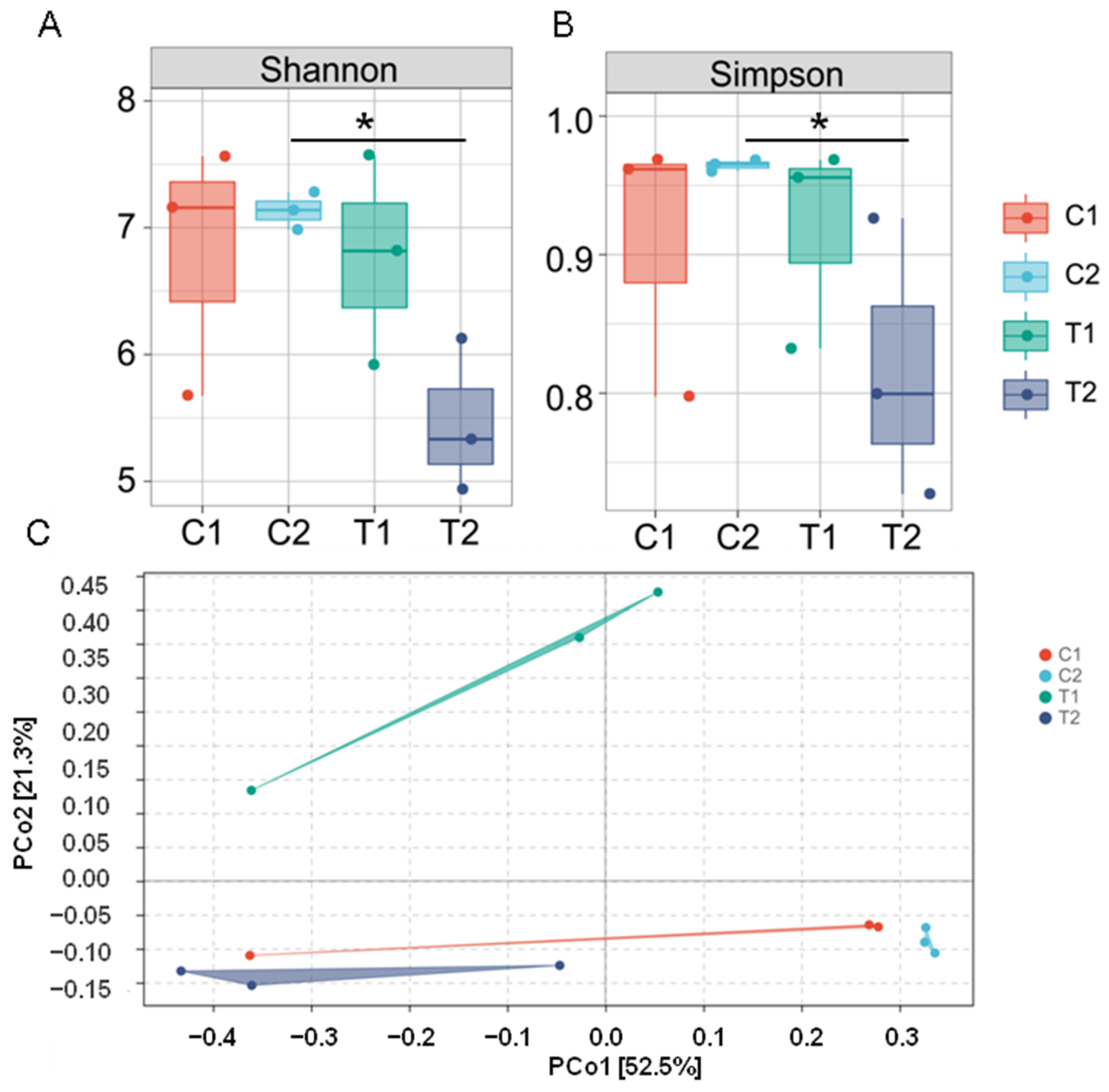
3.2. Diversity of Intestinal Microflora

Alpha and beta diversities were estimated across different groups to evaluate the effects of high-temperature stress on the structure of C. cathayensis intestinal microflora. There was no significant difference in the Shannon and Simpson indexes between the C1 and T1 groups (Figure 2A,B). However, with the extension of the treatment time to 7 days, the Shannon and Simpson indexes decreased significantly in the T2 group compared with the C2 group (p < 0.05). These two alpha-diversity indices were also lower (although not significantly lower) in the T2 group than in the T1 group (Figure 2A,B; Table S2). Similarly, the beta diversity analysis (PCoA based on the Bray–Curtis distance matrix) showed a clear separation in bacterial community composition between high-temperature stress and control treatments (excluding one control sample), with the following main principal component (PC) scores: PC1 = 52.5%, PC2 = 21.3% (Figure 2C). Apparently, T1 and C1 could be separated by PC1, and T2 and C2 could be separated by PC2. Moreover, the results of the PERMANOVA analysis of microbial community structure showed a significant different among all groups (p < 0.05, Tables S3 and S4). In addition, a hierarchical clustering tree (Figure S1) revealed that microbial communities among the four treatment groups were clustered into two groups, ASVs from the C1 and C2 groups were clustered in one group based on similarity (excluding one control sample), while the T1 and T2 groups clustered into one independent group. Overall, these results indicate that thermal stress significantly affected the diversity of the gut microbiome of C. cathayensis.

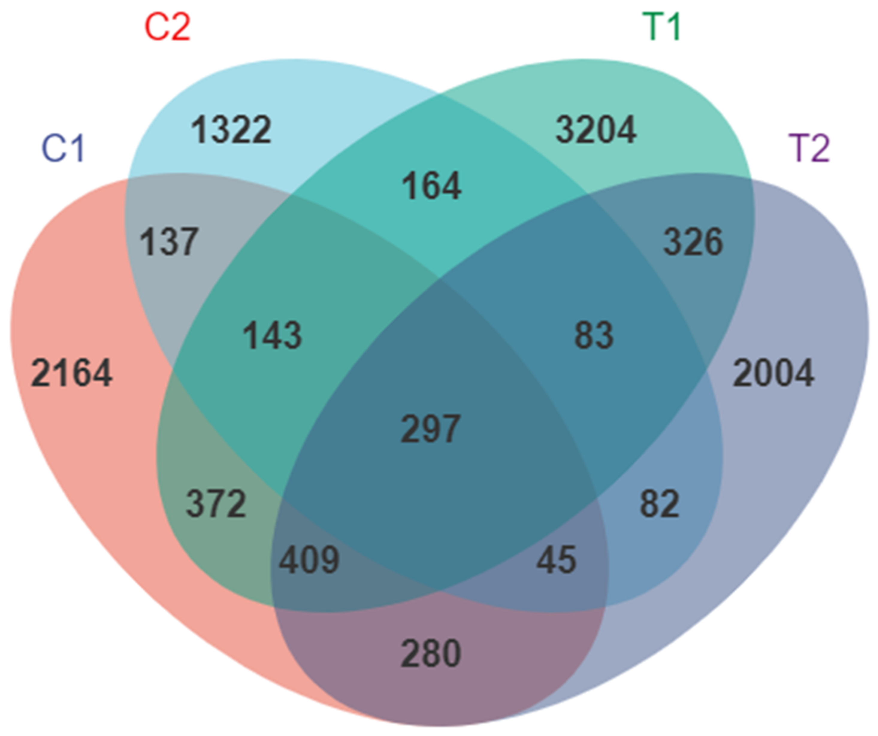
3.3. Effect of High Temperature on the Overall Community Structure of Intestinal Microbiota

In total, 28 phyla, 60 classes, 133 orders, 217 families, 396 genera, and 115 species were identified. At the phylum level, Proteobacteria was the predominant phylum in all intestinal samples (65.04% in C1, 63.62% in C2, 46.26% in T1, and 43.59% in T2). Proteobacteria, Bacteroidetes, Firmicutes, Actinobacteria, and Deinococcus-Thermus together with Chlamydiae accounted for 71.1% of the total abundance of intestinal microbiota (Figure 3A; Table S5). The relative abundance of Proteobacteria decreased in T1 and T2 groups, however, the difference was not statistically significant (Figure 3B). The relative abundance of Firmicutes was significantly higher in T1 than in C1 (p = 0.021; Figure 3C). In contrast, the relative abundance of Firmicutes (p = 0.001), Actinobacteria (p = 0.001), and Deinococcus-Thermus (p = 0.025) was significantly lower in T2 than in C2 (Figure 3C–E). Notably, the Firmicutes/Bacteroidetes ratio in the T2 group was lower than that in the C2 group, but the difference was not statistically significant (Figure 3F).
At the genus level, the dominant genera were Pseudomonas, Acinetobacter, Pelomonas, Bacteroides, Aeromonas, Aquabacterium, Rhodobacter, Halomonas, Muribaculaceae, and Brevundimonas and accounted for 41.67% of the total abundance of intestinal microbiota (Figure 4A; Table S6). With the increase in treatment time, the relative abundance of Bacteroides (p = 0.027; Figure 4B), Pseudomonas (p = 0.008; Figure 4C), Aeromonas (p = 0.001; Figure 4D), and Rhodobacter (p = 0.008; Figure 4E) was lower in the T2 treatment than in the C2 treatment. Remarkably, the relative abundance of the genus Bacteroides significantly increased in the T1 group (p = 0.05) and then decreased in the T2 group (p = 0.027). In contrast, the relative abundance of Halomonas increased in the T2 group (p = 0.015; Figure 4F, Table S6).

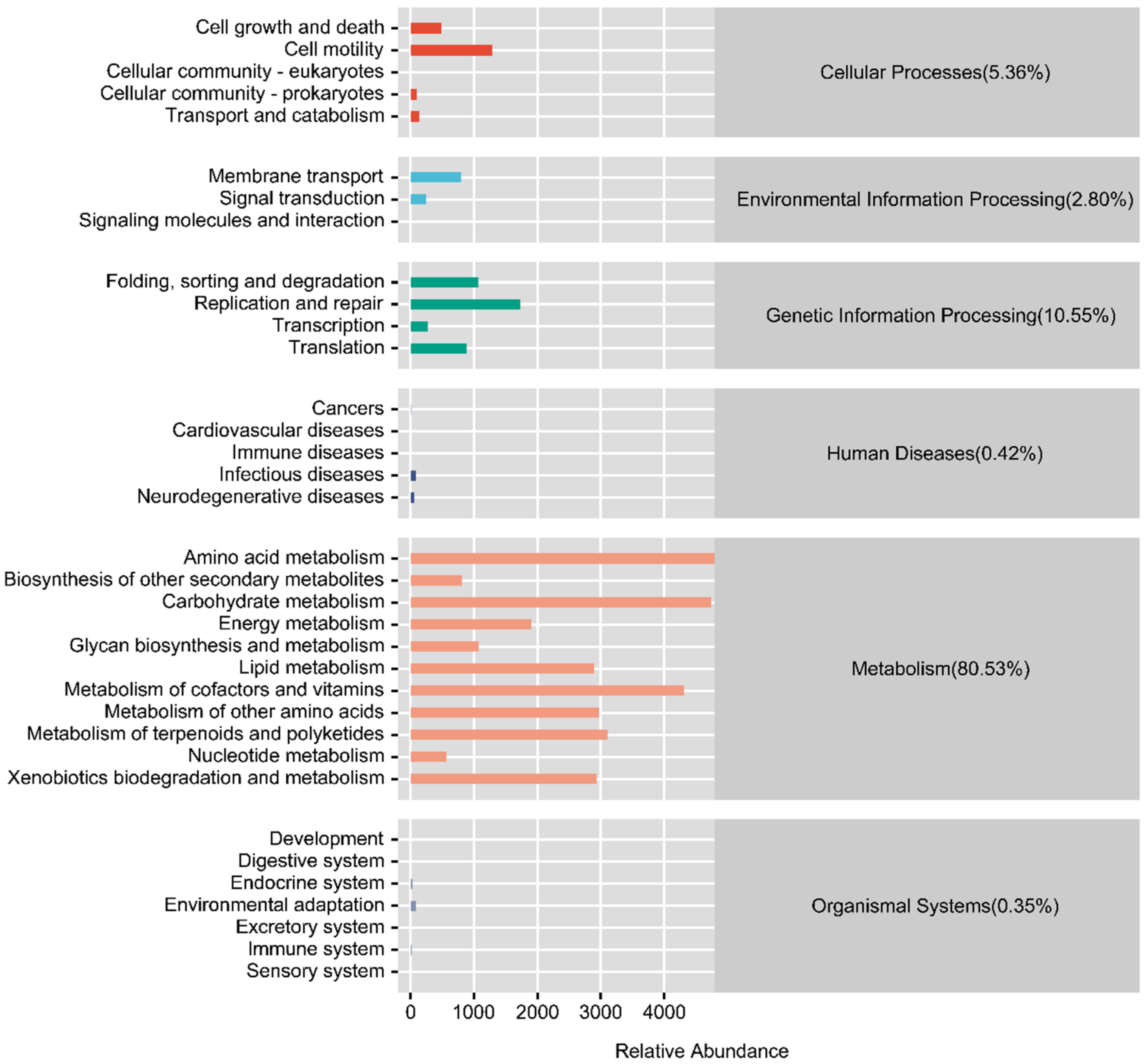
3.4. Functional Prediction of the Intestinal Microflora

To further evaluate the function of gut microbiota in response to high-temperature stress in C. cathayensis, we predicted the gene function of the gut microbial community. The PICRUSt functional predictions and the KEGG pathway enrichment results showed that six functional groups (pathway level 1) were identified, including metabolism (80.53%), genetic information processing (10.55%), cellular processes (5.63%), environmental information processing (2.80%), human diseases (0.42%), and organismal systems (0.35%; Figure 5). We found that some carbohydrate metabolism pathways were enriched in the T1 treatment compared to the control group (Figure 6A); moreover, one potentially disease-related pathway (bacterial invasion of epithelial cells) was also elevated in the T1 treatment (Figure 6A). Notably, carbohydrate metabolism pathways were found to be inhibited in the T2 group relative to the T1 group (Figure 6C). Furthermore, other disease-related pathways (e.g., those related to systemic lupus erythematosus, Vibrio cholerae infection, hypertrophic cardiomyopathy, and shigellosis) were increased in the T2 treatment (Figure 6B,C). Taken together, these findings suggest that thermal exposure affects the metabolism of the gut microbiota and in turn the health of C. cathayensis.

4. Discussion

The gut microbiome often serves as an additional organ [23] that plays an important role in nutrient digestion and absorption, immunity, and development of the host [24,25]. Environmental shift is an important abiotic factor affecting the intestinal microflora structure in aquatic animals, including mollusks [28,29,30,32,43]. Indeed, temperature affects the composition and function of mollusk intestinal microbiota [28,29,30,32,43]. Changes in the gut microbiome were found to be an important process through which animal hosts acclimate to temperature shifts [44]. However, the effects of thermal stress on the intestinal microbiota of C. cathayensis remained unclear. In the present study, C. cathayensis were exposed over 7 days to two different temperatures (control, 25 ± 1 °C; high temperature, 32 ± 1 °C). 16S rRNA gene sequencing technology was used to analyze the effects of high-temperature stress on the intestinal microflora structure and function in C. cathayensis.
In the present study, although there was no significant difference in alpha diversity between C1 and T1, a reduction was observed in the T2 group with increased exposure time. Similarly, previous studies reported decreased alpha diversity in some mollusks [28,30,45] exposed to high-temperature stress. In contrast, some studies detected an increase in the alpha diversity metrics in mollusks [29,43] and crustaceans [46]. Apparently, the response strategies of the intestinal microbiome under thermal stress are related to host taxonomy. Indeed, different microbiome responses to temperature have been observed in closely related species [28,29,30]. In addition, PCoA and hierarchical clustering results showed that the gut microbial community structure in the control groups, excluding one control sample, was distinct from that in the T1 and T2 groups; the latter two treatments also showed significantly different gut microbial community structures from each other, implying that high-temperature exposure significantly changed the intestinal microbiota structure in C. cathayensis. Gut microbiota are beneficial for their hosts [24,25]; thus, the decrease in alpha diversity and changes in the gut microbial community structure following high-temperature exposure observed in the present study may have adverse effects on C. cathayensis [29,30].
In this study, Proteobacteria, Bacteroidetes, and Firmicutes were the three dominant phyla across all treatment groups, which is consistent with previous findings in mollusks [47,48]. However, Bacteroidetes and Firmicutes were not the dominant phyla in a closely related species, Cipangopaludina chinensis [48,49]. This inconsistency implies that although the two species are closely related, they may have different strategies of responding to high-temperature stress [28,29,30]. Proteobacteria, widely used as probiotics in aquaculture [50], provide many benefits to aquaculture animals [50,51,52]. In the present study, the relative abundance of Proteobacteria slightly decreased in the T1 and T2 treatment groups, suggesting that high-temperature exposure has adverse effects on C. cathayensis health [43,46].
Studies have shown that Firmicutes plays an important role in maintaining the gut homeostasis [53] and actively responds to temperature changes [54]. Notably, in the present study, the relative abundance of Firmicutes increased significantly in T1 and the decreased in T2, suggesting that thermal stress affects the gut homeostasis of C. cathayensis [54]. In addition, Firmicutes is known to be positively correlated with intestinal nutrient metabolism [32,46], and a reduction in Firmicutes abundance may hinder intestinal nutrient absorption [32]. It is also worth noting that the Firmicutes/Bacteroidetes ratio slightly decreased in T1 and T2. The Firmicutes/Bacteroidetes ratio is an important biomarker of gut dysbiosis [55]; a decreased Firmicutes/Bacteroidetes ratio affects nutrient metabolism and restricts the growth of aquaculture animals [32,56]. Thus, a decrease in the Firmicutes/Bacteroidetes ratio may have adverse effects on C. cathayensis growth [56].
Similarly, Actinobacteria and Deinococcus-Thermus were both found to be decreased in our study. Actinobacteria, widely present in soil, freshwater, and marine ecosystems and found in plants and animals [57,58,59], are endowed with the ability to biosynthesize and produce secondary metabolites that play a vital role in nutrient recycling and regulation of a wide variety of diseases [60]. However, the abundance of Actinobacteria decreased significantly in T2, implying that the synthesis of bioactive compounds is disrupted under high-temperature stress, potentially harming the host. The phylum Deinococcus-Thermus comprises several thermophilic species, which are highly tolerant to high temperatures [61], and their relative abundance in mussel Mytilus coruscus showed significant differences in response to temperature [28]. In addition, this phylum was also found to include extremophiles, which are associated with the production of carotenoids [62] and exhibit resistance to extreme environmental stressors such as radiation, oxidation, and heavy metals [62,63]. A decreased relative abundance of Deinococcus-Thermus in the present study suggests a decreased adaptation potential to high temperature.
At the genus level, the abundance of Pseudomonas, Aeromonas, Rhodobacter, Bacteroides, and Halomonas was found to be significantly different among different treatments. Pseudomonas is widely distributed in the gut of aquatic animals [25,64] and has been identified as pathogenic in teleosts [25,65]. However, Pseudomonas is also used as a probiotic in aquaculture [66]. In the present study, the relative abundance of Pseudomonas significantly decreased in the T2 treatment, implying that thermal stress exerts adverse effects on this genus. Further research is required to verify whether Pseudomonas acts as a probiotic in C. cathayensis. Similarly, the relative abundance of Aeromonas—which is potentially beneficial to aquatic animals in many aspects, including growth, immunity, and disease resistance—was decreased in T2 [52,67], suggesting that high temperatures disrupt the homeostatic balance of probiotics and adversely affect the health of the host. Moreover, the abundance of Rhodobacter also decreased significantly in T2. Rhodobacter is a candidate probiotic for fish [68], which can improve growth, reduce oxidative damage, and stimulate the immune system [69]. Decreased Rhodobacter abundance has been reported to reduce growth [70] and have adverse effects on fish innate immunity [71], resulting in increased vulnerability to disease [72]. Bacteroides species are highly adapted to the gut environment, and they live and grow exclusively in the gastrointestinal tracts of mammals [73] and aquatic animals [74]. Although members of this group are known to be opportunistic pathogens [28], most of them actively enhance the gut environment, establish stable and long-term associations with their hosts, and confer numerous health benefits [73]. Notably, Bacteroides release commensal molecules (e.g., polysaccharide A and sphingolipids) that play an important role in maintaining intestinal homeostasis and modulating the host immune system in health and disease [75,76]. A reduction in the abundance of Bacteroides has been associated with inflammatory bowel disease [77]. In our study, the relative abundance of Bacteroides significantly increased in T1 and decreased in T2, implying that Bacteroides respond positively to high temperatures; however, extended thermal stress disrupts the homeostasis of Bacteroides, which may lead to disease outbreak in C. cathayensis [77].
In contrast, under high-temperature stress, the abundance of Halomonas was elevated in T2. Halomonas species, commonly found in saline environments, are halotolerant or halophilic [78] and cause infections in humans [79]. Halomonas strains are also considered to be pathogenic in fish [80,81]. Indeed, Halomonas are found to be elevated in the intestine of zebrafish (Danio rerio) after pathogen invasion [80] or exposure to environmental stress (e.g., Microcystis aeruginosa and phthalate esters), inducing intestinal innate immune responses [80], which results in intestinal injury [82]. These findings illustrate that the health of C. cathayensis exposed to thermal stress may be impaired.
To further assess the effect of high temperature on C. cathayensis, we examined the functional pathways encoded by C. cathayensis gut microbiota. The functional prediction analysis showed that most genes of the C. cathayensis gut microbiota were related to metabolism. Gut microbiota contributes to host nutrition, absorption, and metabolism [54]; thus, thermal stress may affect C. cathayensis metabolism [54]. Indeed, in the present study, some carbohydrate metabolism pathways were found to be enriched in the T1 group but decreased in the T2 group compared to the T1 group. These results are consistent with the change trend of Firmicutes abundance observed in different treatment groups, which is known to be positively correlated with intestinal nutrient metabolism and nutrient absorption [32]. A reduction in the abundance of Firmicutes was reported to hamper growth in aquaculture animals [32,56]. Therefore, we speculate that increasing temperatures disrupt Firmicutes homeostasis and inhibit carbohydrate metabolism pathways, thus, inhibiting the growth of C. cathayensis. Gut microbiota homeostasis also plays an important role in host health [80,82]. Under thermal stress, one disease-related pathway was found to be enriched in T1, whereas more disease-related pathways were detected in T2. This finding is consistent with the results that thermal stress significantly decreased the abundance of some functionally important taxa including the phyla Firmicutes, Actinobacteria, Deinococcus-Thermus and the genera Pseudomonas, Aeromonas, Rhodobacter, and Bacteroides and increased the abundance of Halomonas, a potentially pathogenic genus, in T2. The functional changes in the intestinal microflora are associated with microflora composition in mud snails under high-temperature stress and the deficiency of functionally important gut microbial taxa may increase their susceptibility to disease [32].

5. Conclusions

Our results revealed that the structure and function of intestinal microbial communities in C. cathayensis differed between the control treatment and high-temperature treatment. High-temperature stress disrupted the homeostasis of the intestinal microbiota of C. cathayensis, as it decreased the abundance of putatively beneficial bacteria and increased the abundance of putatively pathogenic bacteria. Moreover, thermal stress profoundly influenced the metabolic functions of gut microbiota, which may increase the disease susceptibility of C. cathayensis. To the best of our knowledge, this is the first study to explore the effects of high-temperature stress on the intestinal microbiota of C. cathayensis. The results obtained in this study provide insights into the mechanisms associated with the response of the intestinal microbiota of C. cathayensis to global warming. However, due to the small sample size and obtaining the 16S rRNA gene sequences through the Illumina HiSeq platform our study may have limitations. In the present study, we did not isolate and identify the putatively pathogenic and putatively beneficial bacteria, which warrants further research.

Supplementary Materials

The following supporting information can be downloaded at: https://www.mdpi.com/article/10.3390/ani12182361/s1, Figure S1. Rarefaction curves for all analyzed samples. Different samples are represented with different colors; Figure S2. Hierarchical cluster analysis of the Jaccard distances generated from taxa tables showed Amplicon Sequence Variant (ASV) similarity across microbial communities among Cipan-gopaludina cathayensis treatment groups C1 (control treatment; 25 ± 1 ℃, day 3), C2 (control treatment; 25 ± 1 ℃, day 7), T1 (high-temperature treatment; 32 ± 1 ℃, day 3), and T2 (high-temperature treatment; 32 ± 1 ℃, day 7); Table S1. Number of sequences derived from 12 samples; Table S2. Alpha-diversity indexes of different samples under a 97% similarity cut-off; Table S3. Results of PERMAVOVA of Bray-Curtis dissimilarity between samples using ANOSIM test; Table S4. Results of PERMAVOVA of Bray-Curtis dissimilarity between samples using Adonis test; Table S5. Relative abundance of intestinal bacteria at the phylum level; Table S6. Relative abundance of intestinal bacteria at the genus level.

Author Contributions

Conceptualization, J.-Y.J. and W.-H.L.; methodology, J.-Y.J.; software, J.-Y.J.; validation, J.-Y.J. and W.-H.L.; formal analysis, Y.-Y.W. and C.-X.C.; resources, J.-Y.J. and Q.-Q.Y.; data curation, L.Y., Q.-Q.Y. and J.-Y.J.; writing—original draft preparation, Y.-Y.W., C.-X.C. and J.-Y.J.; writing—review and editing, J.-Y.J., Y.-Y.W. and C.-X.C.; visualization, supervision, project administration, funding acquisition, J.-Y.J. and W.-H.L. All authors have read and agreed to the published version of the manuscript.

Funding

The study was supported by the National Key R&D Program of China (2021YFD1100106), and the Key Laboratory of Ecology of Rare and Endangered Species and Environmental Protection (Guangxi Normal University), Ministry of Education, China (grant no. ERESEP2021Z10).

Institutional Review Board Statement

The experimental procedures in this study complied with the animal welfare and research laws in China and were approved by the Ethics Committee of Guangxi Normal University (No.202207−01).

Informed Consent Statement

Not applicable.

Data Availability Statement

The raw sequencing reads that support the findings of the present study are publicly available in the NCBI Sequence Read Archive (SRA) database at (https://www.ncbi.nlm.nih.gov, accessed on 8 June 2022) under accession number PRJNA847054.

Acknowledgments

The authors would like to thank all participants in this work, including the staff at the Key Laboratory of Ecology of Rare and Endangered Species and Environmental Protection (Guangxi Normal University) for their help in snail culture and sample collection during the research, and would like to thank the anonymous reviewers and the editor of the journal for their valuable comments.

Conflicts of Interest

The authors declare no competing interest.

References

  1. Dalon, P.W.; David, H.W. Growth and physiological responses in largemouth bass populations to environmental warming: Effects of inhabiting chronically heated environments. J. Therm. Biol. 2020, 88, 102467. [Google Scholar]
  2. Yang, T.Y.; Zhang, Y.; Meng, W.; Zhong, X.; Shan, Y.; Gao, T.X. Comparative transcriptomic analysis brings new insights into the response to acute temperature acclimation in burbot (Lota lota lota). Aquacult. Rep. 2021, 20, 100657. [Google Scholar] [CrossRef]
  3. Meredith, M.; Sommerkorn, M.; Cassotta, S.; Derksen, C.; Ekaykin, A.; Hollowed, A.; Kofinas, G.; Mackintosh, A.; Melbourne-Thomas, J.; Muelbert, M.M.C.; et al. (Eds.) IPCC Special Report on the Ocean and Cryosphere in a Changing Climate; IPCC: Geneva, Switzerland, 2019; Available online: https://www.ipcc.ch/srocc/chapter/chapter-3-2/ (accessed on 18 June 2022).
  4. Ahmed, N.; Thompson, S.; Glaser, M. Global aquaculture productivity, environmental sustainability, and climate change adaptability. Environ. Manag. 2019, 63, 159–172. [Google Scholar] [CrossRef] [PubMed]
  5. Sun, Y.; Liu, Y.; Yu, X.B.; Wang, T.; Wang, Q.; Yao, W.Z.; Wu, Z.L. Combined effects of ration levels and temperature on immune responses of the triangle sail mussel Hyriopsis cumingii. Aquac. Res. 2022, 53, 440–452. [Google Scholar] [CrossRef]
  6. Zhao, T.T.; Ma, A.J.; Huang, Z.H.; Liu, Z.F.; Sun, Z.B.; Zhu, C.Y.; Yang, J.K.; Li, Y.D.; Wang, Q.M.; Qiao, X.W. Transcriptome analysis reveals that high temperatures alter modes of lipid metabolism in juvenile turbot (Scophthalmus maximus) liver. Comp. Biochem. Physiol. Part D Genom. Proteom. 2021, 40, 100887. [Google Scholar] [CrossRef] [PubMed]
  7. Zhang, H.; Jia, H.X.; Xiong, P.P.; Yao, G.Y.; He, M.X. Transcriptome and enzyme activity analyses of tolerance mechanisms in pearl oyster (Pinctada fucata) under high-temperature stress. Aquaculture 2022, 550, 737888. [Google Scholar] [CrossRef]
  8. Cannaday, J.D.; Farmer, T.M. Assessing the effects of sub-tropical winter thermal conditions on coolwater fish reproduction. Ecol. Freshw. Fish 2022, 3, 300–316. [Google Scholar] [CrossRef]
  9. Hue, T.; Chateau, O.; Lecellier, G.; Kayal, M.; Lanos, N.; Gossuin, H.; Adjeroud, M.; Dumas, P. Temperature affects the reproductive outputs of coral-eating starfish Acanthaster spp. after adult exposure to near-future ocean warming and acidification. Mar. Environ. Res. 2020, 162, 105164. [Google Scholar] [CrossRef]
  10. Watson, S.A.; Allan, B.J.M.; McQueen, D.E.; Nicol, S.; Parsons, D.M.; Pether, S.M.J.; Pope, S.; Setiawan, A.N.; Smith, N.; Wilson, C.; et al. Ocean warming has a greater effect than acidification on the early life history development and swimming performance of a large circumglobal pelagic fish. Glob. Chang. Biol. 2018, 24, 4368–4385. [Google Scholar] [CrossRef]
  11. Kong, H.; Clements, J.C.; Dupont, S.; Wang, T.; Huang, X.; Shang, Y.; Huang, W.; Chen, J.; Hu, M.; Wang, Y. Seawater acidification and temperature modulate anti-predator defenses in two co-existing Mytilus species. Mar. Pollut. Bull. 2019, 145, 118–125. [Google Scholar] [CrossRef]
  12. Lu, H.F.; Du, L.N.; Li, Z.Q.; Chen, X.Y.; Yang, J.X. Morphological analysis of the Chinese Cipangopaludina species (Gastropoda; Caenogastropoda: Viviparidae). Zool. Res. 2014, 35, 510–527. [Google Scholar]
  13. Luo, H.; Chen, L.T.; Jing, T.S.; Sun, W.B.; Li, Z.; Zhou, M.R.; Qin, J.Q.; Du, X.S.; Wen, L.T.; Pan, X.H.; et al. Muscle nutrition analysis of four snail species. J. Fish. China. 2021. in press. Available online: https://kns.cnki.net/kcms/detail/31.1283.S.20211018.1713.002.html (accessed on 20 June 2022).
  14. Zhao, T.; Xiong, J.Q.; Chen, W.; Xu, A.H.; Zhu, D.; Liu, J.T. Purification and characterization of a novel fibrinolytic enzyme from Cipangopaludina cahayensis. Iran. J. Biotechnol. 2021, 19, 121–127. [Google Scholar]
  15. Wang, C.; Liu, J.; Huang, Y.; Zhang, X. In vitro polysaccharide extraction from Cipangopaludina cathayensis and its pharmacological potential. J. Environ. Biol. 2016, 37, 1069–1072. [Google Scholar]
  16. Li, W.F.; Song, Y.J.; Wen, Y.H.; Luo, F.G.; Wang, W.M.; Li, Y.H. Investigation of current status of freshwater snail industry in China. Cult. Feed Sci. 2019, 11, 131–138. [Google Scholar]
  17. Woodward, G.; Perkins, D.M.; Brown, L.E. Climate change and freshwater ecosystems: Impacts across multiple levels of organization. Phil. Trans. R. Soc. B 2010, 365, 2093–2106. [Google Scholar] [CrossRef] [Green Version]
  18. Sangsawang, A.; Kovitvadhi, U.; Kovitvadhi, S. The effect of water temperature on the early-life development, growth and survival of the freshwater mussel Hyriopsis bialata. Aquaculture 2019, 510, 311–317. [Google Scholar] [CrossRef]
  19. Allan, E.R.O.; Blouin, M.S. Heat shock increases hydrogen peroxide release from circulating hemocytes of the snail Biomphalaria glabrata. Fish Shellfish Immun. 2020, 105, 203–208. [Google Scholar] [CrossRef]
  20. Hraoui, G.; Bettinazzi, S.; Gendron, A.D.; Boisclair, D.; Breton, S. Mitochondrial thermo-sensitivity in invasive and native freshwater mussels. J. Exp. Biol. 2020, 223, jeb215921. [Google Scholar] [CrossRef]
  21. Said, R.M.; Nassar, S.E. Mortality, energy reserves and oxidative stress responses of three native fresh water mussels to temperature as indicator of the potential impacts of climate change: A laboratory experiment approach. J. Therm. Biol. 2022, 104, 103154. [Google Scholar] [CrossRef]
  22. Sun, L.Y.; Wen, X.M.; Yu, N.; Chen, L.Q. Influence of water temperature and salinity on standard metabolism of Cipangopaludina cathayensis and Bellamya aeruginosa. J. Fish. Sci. China 2012, 19, 275–282. [Google Scholar] [CrossRef]
  23. Jiang, P.; Yu, X.J.; Yu, L.L.; Tian, F.W.; Zhao, J.X.; Zhang, H.; Chen, W.; Zhai, Q.X. The influence of gut microbiome on bone health and related dietary strategies against bone dysfunctions. Food Res. Int. 2021, 144, 110331. [Google Scholar]
  24. Talwar, C.; Nagar, S.; Lal, R.; Negi, R.K. Fish gut microbiome: Current approaches and future perspectives. Indian J. Microbiol. 2018, 58, 397–414. [Google Scholar] [CrossRef]
  25. Diwan, A.D.; Harke, S.N.; Gopalkrishna; Panche, A.N. Aquaculture industry prospective from gut microbiome of fish and shellfish: An overview. J. Anim. Physiol. Anim. Nutr. 2021, 106, 441–469. [Google Scholar] [CrossRef]
  26. Wang, N.; Jiang, M.; Zhang, P.J.; Shu, H.; Li, Y.R.; Guo, Z.Y.; Li, Y.H. Amelioration of Cd-induced bioaccumulation, oxidative stress and intestinal microbiota by Bacillus cereus in Carassius auratus gibelio. Chemosphere 2020, 245, 125613. [Google Scholar] [CrossRef] [PubMed]
  27. Wang, Z.L.; Zhou, J.; Li, J.Y.; Lv, W.; Zou, J.X.; Fan, L.F. A new insight into the intestine of pacific white shrimp: Regulation of intestinal homeostasis and regeneration in litopenaeus vannamei during temperature fluctuation. Comp. Biochem. Physiol. Part D Genom. Proteom. 2020, 35, 100687. [Google Scholar] [CrossRef] [PubMed]
  28. Li, Y.F.; Yang, N.; Liang, X.; Yoshida, A.; Osatomi, K.; Power, D.; Batista, F.M.; Yang, J.L. Elevated seawater temperatures decrease microbial diversity in the gut of Mytilus coruscus. Front. Physiol. 2018, 9, 839. [Google Scholar] [CrossRef] [PubMed] [Green Version]
  29. Li, Y.F.; Xu, J.K.; Chen, Y.W.; Ding, W.Y.; Shao, A.Q.; Liang, X.; Zhu, Y.T.; Yang, J.L. Characterization of gut microbiome in the mussel Mytilus galloprovincialis in response to thermal stress. Front. Physiol. 2019, 10, 1086. [Google Scholar] [CrossRef] [PubMed]
  30. Li, Y.F.; Chen, Y.W.; Xu, J.K.; Ding, W.Y.; Shao, A.Q.; Zhu, Y.T.; Wang, C.; Liang, X.; Yang, J.L. Temperature elevation and Vibrio cyclitrophicus infection reduce the diversity of haemolymph microbiome of the mussel Mytilus coruscus. Sci. Rep. 2019, 9, 16391. [Google Scholar] [CrossRef]
  31. Huang, Z.J.; Zeng, S.Z.; Xiong, J.H.; Hou, D.W.; Zhou, R.J.; Xing, C.G.; Wei, D.D.; Deng, X.S.; Yu, L.F.; Wang, H.; et al. Microecological Koch’s postulates reveal that intestinal microbiota dysbiosis contributes to shrimp white feces syndrome. Microbiome 2020, 8, 32. [Google Scholar] [CrossRef]
  32. Kong, N.; Han, S.; Fu, Q.; Yu, Z.C.; Wang, L.L.; Song, L.S. Impact of ocean acidification on the intestinal microflora of the pacific oyster Crassostrea gigas. Aquaculture 2022, 546, 737365. [Google Scholar] [CrossRef]
  33. Chen, J.J.; Cao, J.L.; Li, X.; Yin, Q.L. Effects of fluoride on the activities of antioxidant enzymes and MDA levels in hepatopancreas of Cipangopaludina cahayensis. Asian J. Ecotoxicol. 2018, 13, 268–273. [Google Scholar]
  34. Jiang, J.J.; Li, W.H.; Wu, Y.Y.; Cheng, C.X.; Ye, Q.Q.; Feng, J.X.; Xie, Z.X. Effects of cadmium exposure on intestinal microflora of Cipangopaludina cathayensis. Front. Microbiol. 2022, 13, 984757. [Google Scholar] [CrossRef]
  35. Bolyen, E.; Rideout, J.R.; Dillon, M.R.; Bokulich, N.A.; Abnet, C.C.; Al-Ghalith, G.A.; Alexander, H.; Alm, E.J.; Arumugam, M.; Asnicar, F.; et al. QIIME 2: Reproducible, interactive, scalable, and extensible microbiome data science. PeerJ Prepr. 2018, 6, e27295v2. [Google Scholar] [CrossRef]
  36. Martin, M. Cutadapt removes adapter sequences from high-throughput sequencing reads. EMBnet 2011, 17, 10–12. [Google Scholar] [CrossRef]
  37. Callahan, B.J.; McMurdie, P.J.; Rosen, M.J.; Han, A.W.; Johnson, A.J.A.; Holmes, S.P. DADA2: High-resolution sample inference from Illumina amplicon data. Nat. Methods 2016, 13, 581–583. [Google Scholar] [CrossRef]
  38. Katoh, K.; Misawa, K.; Kuma, K.I.; Miyata, T. Mafft: A novel method for rapid multiple sequence alignment based on fast fourier transform. Nucleic Acids Res. 2002, 30, 3059–3066. [Google Scholar] [CrossRef]
  39. Price, M.N.; Dehal, P.S.; Arkin, A.P. FastTree: Computing large minimum evolution trees with profiles instead of a distance matrix. Mol. Biol. Evol. 2009, 26, 1641–1650. [Google Scholar] [CrossRef]
  40. McArdle, B.H.; Anderson, M.J. Fitting multivariate models to community data: A comment on distance-based redundancy analysis. Ecology 2001, 82, 290–297. [Google Scholar] [CrossRef]
  41. Warton, D.I.; Wright, S.T.; Wang, Y. Distance-based multivariate analyses confound location and dispersion effects. Methods Ecolo. Evol. 2012, 3, 89–101. [Google Scholar] [CrossRef]
  42. Anderson, M.J.; Ellingsen, K.E.; McArdle, B.H. Multivariate dispersion as a measure of beta diversity. Ecol. Lett. 2006, 9, 683–693. [Google Scholar] [CrossRef]
  43. Scanes, E.; Parker, L.M.; Seymour, J.R.; Siboni, N.; King, W.L.; Danckert, N.P.; Wegner, K.M.; Dove, M.C.; O’Connor, W.A.; Ross, P.M. Climate change alters the haemolymph microbiome of oysters. Mar. Pollut. Bull. 2021, 164, 111991. [Google Scholar] [CrossRef] [PubMed]
  44. Sepulveda, J.; Moeller, A.H. The Effects of temperature on animal gut microbiomes. Front. Microbiol. 2020, 11, 384. [Google Scholar] [CrossRef] [PubMed]
  45. Wang, X.Z.; Tang, B.; Luo, X.; Ke, C.H.; Huang, M.Q.; You, W.W.; Wang, Y. Effects of temperature, diet and genotype-induced variations on the gut microbiota of abalone. Aquaculture 2020, 524, 735269. [Google Scholar] [CrossRef]
  46. Duan, Y.F.; Xiong, D.L.; Wang, Y.; Li, H.; Dong, H.B.; Zhang, J.S. Toxic effects of ammonia and thermal stress on the intestinal microbiota and transcriptomic and metabolomic responses of Litopenaeus vannamei. Sci. Total Environ. 2021, 754, 141867. [Google Scholar] [CrossRef]
  47. Musella, M.; Wathsala, R.; Tavella, T.; Rampelli, S.; Barone, M.; Palladino, G.; Biagi, E.; Brigidi, P.; Turroni, S.; Franzellitti, S.; et al. Tissue-scale microbiota of the Mediterranean mussel (Mytilus galloprovincialis) and its relationship with the environment. Sci. Total Environ. 2020, 717, 137209. [Google Scholar] [CrossRef]
  48. Zhou, Z.H.; Wu, H.Y.; Li, D.H.; Zeng, W.L.; Huang, J.L.; Wu, Z.J. Comparison of gut microbiome in the Chinese mud snail (Cipangopaludina chinensis) and the invasive golden apple snail (Pomacea canaliculata). PeerJ 2022, 10, e13245. [Google Scholar] [CrossRef]
  49. Zhou, K.Q.; Qin, J.Q.; Pang, H.F.; Chen, Z.; Huang, Y.; Li, W.H.; Du, X.S.; Wen, L.T.; Pan, X.H.; Lin, Y. Comparison of the composition and function of gut microbes between adult and juvenile Cipangopaludina chinensis in the rice snail system. PeerJ 2022, 10, e13042. [Google Scholar] [CrossRef]
  50. Wang, A.R.; Ran, C.; Wang, Y.B.; Zhang, Z.; Ding, Q.W.; Yang, Y.L.; Olsen, R.E.; Ringo, E.; Bindelle, J.; Zhou, Z.G. Use of probiotics in aquaculture of china-a review of the past decade. Fish Shellfish Immunol. 2019, 86, 734–755. [Google Scholar] [CrossRef]
  51. Guo, Y.B.; Pan, Q.; Yan, S.Q.; Chen, Y.H.; Li, M.J.; Chen, D.; Han, H.C.; Wu, B.; Cai, J.P. Bdellovibrio and like organisms promoted growth and survival of juvenile abalone haliotis discus hannai Ino and modulated bacterial community structures in its gut. Aquacul. Int. 2017, 25, 1625–1643. [Google Scholar] [CrossRef]
  52. Hao, K.; Wu, Z.Q.; Li, D.L.; Yu, X.B.; Wang, G.X.; Ling, F. Effects of Dietary Administration of Shewanella xiamenensis A-1, Aeromonas veronii A-7, and Bacillus subtilis, Single or Combined, on the Grass Carp (Ctenopharyngodon idella) Intestinal Microbiota. Probiotics Antimicro. 2017, 9, 386–396. [Google Scholar] [CrossRef]
  53. Eeckhaut, V.; Machiels, K.; Perrier, C.; Romero, C.; Maes, S.; Flahou, B.; Steppe, M.; Haesebrouck, F.; Sas, B.; Ducatelle, R.; et al. Butyricicoccus pullicaecorum in inflammatory bowel disease. Gut 2013, 62, 1745–1752. [Google Scholar] [CrossRef]
  54. Liu, Y.Y.; Cheng, J.X.; Xia, Y.Q.; Li, X.H.; Liu, Y.; Liu, P.F. Response mechanism of gut microbiome and metabolism of European seabass (Dicentrarchus labrax) to temperature stress. Sci. Total Environ. 2022, 813, 151786. [Google Scholar] [CrossRef]
  55. Grigoreva, I.N. Gallstone disease, obesity and the Firmicutes/Bacteroidetes ratio as a possible biomarker of gut dysbiosis. J. Pers. Med. 2021, 11, 13. [Google Scholar] [CrossRef]
  56. Chen, X.W.; Lu, D.Y.; Li, Z.H.; Yue, W.C.; Wang, J.; Jiang, X.Y.; Han, H.; Wang, C.H. Plant and animal-type feedstuff shape the gut microbiota and metabolic processes of the chinese mitten crab Eriocheir sinensis. Front. Vet. Sci. 2021, 8, 589624. [Google Scholar] [CrossRef]
  57. Jose, P.A.; Jha, B. Intertidal marine sediment harbours Actinobacteria with promising bioactive and biosynthetic potential. Sci. Rep. 2017, 7, 10041. [Google Scholar] [CrossRef]
  58. Rey, T.; Dumas, B. Plenty is no plague: Streptomyces symbiosis with crops. Trends Plant Sci. 2017, 22, 30–37. [Google Scholar] [CrossRef]
  59. Chevrette, M.G.; Carlson, C.M.; Ortega, H.E.; Thomas, C.; Ananiev, G.E.; Barns, K.J.; Book, A.J.; Cagnazzo, J.; Carlos, C.; Flanigan, W.; et al. The antimicrobial potential of Streptomyces from insect microbiomes. Nat. Commun. 2019, 10, 516. [Google Scholar] [CrossRef] [Green Version]
  60. Jose, P.A.; Maharshi, A.; Jha, B. Actinobacteria in natural products research: Progress and prospects. Microbiol. Res. 2021, 246, 126708. [Google Scholar] [CrossRef]
  61. Sadeepa, D.; Sirisena, K.; Manage, P.M. Diversity of microbial communities in hot springs of Sri Lanka as revealed by 16S rRNA gene high-throughput sequencing analysis. Gene 2022, 812, 146103. [Google Scholar] [CrossRef]
  62. Tian, B.; Hua, Y. Carotenoid biosynthesis in extremophilic Deinococcus-Thermus bacteria. Trends Microbiol. 2010, 18, 512–520. [Google Scholar] [CrossRef]
  63. Ren, C.L.; Teng, Y.R.; Chen, X.Y.; Shen, Y.J.; Xiao, H.; Wang, H.Y. Impacts of earthworm introduction and cadmium on microbial communities composition and function in soil. Environ. Toxicol. Phar. 2021, 83, 103606. [Google Scholar] [CrossRef] [PubMed]
  64. Cheaib, B.; Seghouani, H.; Ijaz, U.Z.; Derome, N. Community recovery dynamics in yellow perch microbiome after gradual and constant metallic perturbations. Microbiome 2020, 8, 14. [Google Scholar] [CrossRef] [PubMed]
  65. Xu, J.; Zeng, X.H.; Jiang, N.; Zhou, Y.; Zeng, L.B. Pseudomonas alcaligenes infection and mortality in cultured Chinese sturgeon, Acipenser sinensis. Aquaculture 2015, 446, 37–41. [Google Scholar] [CrossRef]
  66. Fu, L.Q.; Zhang, X.P.; Wang, Y.B.; Peng, L.S.; Li, W.F. Nitrogen removal characteristics of Pseudomonas stutzeri F11 and its application in grass carp culture. Fish. Sci. 2017, 83, 89–98. [Google Scholar] [CrossRef]
  67. Wu, Z.Q.; Jiang, C.; Ling, F.; Wang, G.X. Effects of dietary supplementation of intestinal autochthonous bacteria on the innate immunity and disease resistance of grass carp (Ctenopharyngodon idellus). Aquaculture 2015, 438, 105–114. [Google Scholar] [CrossRef]
  68. Ye, Q.; Feng, Y.Y.; Wang, Z.L.; Zhou, A.G.; Xie, S.L.; Fan, L.F.; Xiang, Q.; Song, E.F.; Zou, J.X. Effects of dietary Gelsemium elegans alkaloids on intestinal morphology, antioxidant status, immune responses and microbiota of Megalobrama amblycephala. Fish Shellfish Immunol. 2019, 94, 464–478. [Google Scholar] [CrossRef]
  69. Liao, Z.H.; Gong, Y.Y.; Zhao, W.; He, X.S.; Wei, D.; Niu, J. Comparison effect of Rhodobacter sphaeroides protein replace fishmeal on growth performance, intestinal morphology, hepatic antioxidant capacity and immune gene expression of Litopenaeus vannamei under low salt stress. Aquaculture 2022, 547, 737488. [Google Scholar] [CrossRef]
  70. Liu, H.S.; Li, X.; Lei, H.J.; Li, D.; Chen, H.X.; Schlenk, D.; Yan, B.; Luo, Y.J.; Xie, L.T. Dietary seleno-l-methionine alters the microbial communities and causes damage in the gastrointestinal tract of Japanese medaka Oryzias latipes. Environ. Sci. Technol. 2021, 55, 16515–16525. [Google Scholar] [CrossRef]
  71. Wang, X.H.; Hu, M.H.; Gu, H.H.; Zhang, L.B.; Shang, Y.Y.; Wang, T.; Wang, T.Y.; Zeng, J.N.; Ma, L.K.; Huang, W.; et al. Short-term exposure to norfloxacin induces oxidative stress, neurotoxicity and microbiota alteration in juvenile large yellow croaker Pseudosciaena crocea. Environ. Pollut. 2020, 267, 115397. [Google Scholar] [CrossRef]
  72. Liu, F.P.; Xu, X.F.; Chao, L.; Chen, K.; Shao, A.; Sun, D.Q.; Hong, Y.; Hu, R.J.; Jiang, P.; Zhang, N.; et al. Alteration of the gut microbiome in chronic kidney disease patients and its association with serum free immunoglobulin light chains. Front. Immunol. 2021, 12, 609700. [Google Scholar] [CrossRef]
  73. Wexler, A.G.; Goodman, A.L. An insider’s perspective: Bacteroides as a window into the microbiome. Nat. Microbiol. 2017, 2, 17026. [Google Scholar] [CrossRef]
  74. Banerjee, G.; Ray, A.K. Bacterial symbiosis in the fish gut and its role in health and metabolism. Symbiosis 2017, 72, 1–11. [Google Scholar] [CrossRef]
  75. Erturk-Hasdemir, D.; Kasper, D.L. Finding a needle in a haystack: Bacteroides fragilis polysaccharide A as the archetypical symbiosis factor. Ann. N. Y. Acad. Sci. 2018, 1417, 116–129. [Google Scholar] [CrossRef]
  76. Brown, E.M.; Ke, X.; Hitchcock, D.; Jeanfavre, S.; Avila-Pacheco, J.; Nakata, T.; Arthur, T.D.; Fornelos, N.; Heim, C.; Franzosa, E.A.; et al. Bacteroides-derived sphingolipids are critical for maintaining intestinal homeostasis and symbiosis. Cell Host Microbe 2019, 25, 668–680. [Google Scholar] [CrossRef]
  77. Zhou, Y.T.; Zhi, F.C. Lower level of Bacteroides in the gut microbiota is associated with inflammatory bowel disease: A Meta-Analysis. BioMed Res. Int. 2016, 2016, 5828959. [Google Scholar] [CrossRef]
  78. Dou, G.M.; He, W.; Liu, H.C.; Ma, Y.C. Halomonas heilongjiangensis sp. nov., a novel moderately halophilic bacterium isolated from saline and alkaline soil. Antonie Van Leeuwenhoek 2015, 108, 403–413. [Google Scholar] [CrossRef]
  79. Yeo, S.H.; Kwak, J.H.; Kim, Y.U.; Lee, J.S.; Kim, H.J.; Park, K.H.; Lee, J.S.; Ha, G.Y.; Lee, J.H.; Lee, J.Y.; et al. Peritoneal dialysis-related peritonitis due to Halomonas hamiltonii A first case report. Medicine 2016, 95, e5424. [Google Scholar] [CrossRef]
  80. Yang, H.T.; Zou, S.S.; Zhai, L.J.; Wang, Y.; Zhang, F.M.; An, L.G.; Yang, G.W. Pathogen invasion changes the intestinal microbiota composition and induces innate immune responses in the zebrafish intestine. Fish Shellfish Immunol. 2017, 71, 35–42. [Google Scholar] [CrossRef]
  81. Ben Hamed, S.; Guardiola, F.; Morcillo, P.; Gonzalez-Parraga, P.; Tavares Ranzani-Paiva, M.J.; Esteban, M.A. Adhesion of pathogenic bacteria to polystyrene, skin and gut mucus of gilthead seabream, infectious capacity and antibiotics susceptibility. Bol. Inst. Pesca 2019, 45, e490. [Google Scholar]
  82. Jia, P.P.; Junaid, M.; Xin, G.Y.; Wang, Y.; Ma, Y.B.; Pei, D.S. Disruption of intestinal homeostasis through altered responses of the microbial community, energy metabolites, and immune system in Zebrafish after chronic exposure to DEHP. Front. Microbiol. 2021, 12, 729530. [Google Scholar] [CrossRef]
Figure 1. Venn diagram depicting the numbers of shared and unique amplicon sequence variants (ASVs) among Cipangopaludina cathayensis treatment groups C1 (control treatment; 25 ± 1 °C, day 3), C2 (control treatment; 25 ± 1 °C, day 7), T1 (high-temperature treatment; 32 ± 1 °C, day 3), and T2 (high-temperature treatment; 32 ± 1 °C, day 7).
Animals 12 02361 g001
Figure 2. Intestinal microbiome diversity among Cipangopaludina cathayensis treatment groups C1 (control treatment; 25 ± 1 °C, day 3), C2 (control treatment; 25 ± 1 °C, day 7), T1 (high-temperature treatment; 32 ± 1 °C, day 3), and T2 (high-temperature treatment; 32 ± 1 °C, day 7). (A) Shannon diversity index; (B) Simpson diversity index; (C) Bray–Curtis distances calculated and visualized through principal coordinates analysis (PCoA; ellipses were drawn with 95% confidence intervals). Asterisks indicate p-values < 0.05.
Animals 12 02361 g002
Figure 3. Composition of Cipangopaludina cathayensis intestinal microflora at the phylum level. (A) Composition of the intestinal microflora among treatment groups C1 (control treatment; 25 ± 1 °C, day 3), C2 (control treatment; 25 ± 1 °C, day 7), T1 (high-temperature treatment; 32 ± 1 °C, day 3), and T2 (high-temperature treatment; 32 ± 1 °C, day 7); (B) relative abundance of phylum Proteobacteria; (C) relative abundance of phylum Firmicutes; (D) relative abundance of phylum Actinobacteria; (E) relative abundance of phylum Deinococcus-Thermus; (F) Firmicutes/Bacteroidetes ratio. Asterisks indicate p-values < 0.05.
Animals 12 02361 g003
Figure 4. Composition of Cipangopaludina cathayensis intestinal microflora at the genus level. (A) Composition of the intestinal microflora among treatment groups C1 (control treatment; 25 ± 1 °C, day 3), C2 (control treatment; 25 ± 1 °C, day 7), T1 (high-temperature treatment; 32 ± 1 °C, day 3), and T2 (high-temperature treatment; 32 ± 1 °C, day 7); (B) relative abundance of the genus Bacteroides; (C) relative abundance of the genus Pseudomonas; (D) relative abundance of the genus Aeromonas; (E) relative abundance of the genus Rhodobacter; (F) relative abundance of the genus Halomonas. Asterisks indicate p-values < 0.05.
Animals 12 02361 g004
Figure 5. Functional annotation and relative abundance of Cipangopaludina cathayensis intestinal microbiota using Kyoto Encyclopedia of Gene and Genomes (KEGG) analysis at level 1 and level 2. The relative abundance of genes associated with a function is shown using colored bars (level 1). The detailed pathways are shown on the left (level 2).
Animals 12 02361 g005
Figure 6. Metabolic functions of Cipangopaludina cathayensis intestinal microbiota in all samples predicted on the basis of the Kyoto Encyclopedia of Genes and Genomes (KEGG) database. (A) Significantly different pathways between C1 (control treatment; 25 ± 1 °C, day 3) and T1 (high-temperature treatment; 32 ± 1 °C, day 3); (B) significantly different pathways between C2 (control treatment; 25 ± 1 °C, day 7) and T2 (high-temperature treatment; 32 ± 1 °C, day 7); (C) significantly different pathways between T1 (high-temperature treatment; 32 ± 1 °C, day 3) and T2 (high-temperature treatment; 32 ± 1 °C, day 7).
Animals 12 02361 g006
Publisher’s Note: MDPI stays neutral with regard to jurisdictional claims in published maps and institutional affiliations.

