Article

Mutation Characteristics and Phylogenetic Analysis of Five Leishmania Clinical Isolates

1
Department of Pathogenic Biology, West China School of Basic Medical Sciences and Forensic Medicine, Sichuan University, Chengdu 610041, China
2
West China School of Public Health, Sichuan University, Chengdu 610041, China
3
Animal Disease Prevention and Food Safety Key Laboratory of Sichuan Province, Chengdu 610065, China
*
Authors to whom correspondence should be addressed.
These authors contributed equally to this work.
Animals 2022, 12(3), 321; https://doi.org/10.3390/ani12030321
Submission received: 22 November 2021 / Revised: 19 January 2022 / Accepted: 24 January 2022 / Published: 28 January 2022
(This article belongs to the Collection Advances in Parasite Epidemiology and Population Genetics)

Abstract

:

Simple Summary

Leishmaniasis, a neglected tropical disease, is caused by infection with the Leishmania species, threatening millions of people in approximately 100 endemic countries. The emergence of antimony-resistant Leishmania strains have brought difficulties to the treatment and elimination of leishmaniasis. This study performed genome-wide resequencing and phylogenetic analysis of five isolates from the Leishmania donovani complex, focusing on finding mutations related to antimony resistance and virulence of the newly isolated Leishmania strain L_HCZ in 2016. By combining whole-genome sequencing and whole-genome phylogenetic analysis, Leishmania isolates L_801, L_9044 and L_Liu were identified as Leishmania donovani, and L_HCZ as Leishmania infantum. By discovering genome-wide single-nucleotide polymorphisms and structural variations, we identified mutations of drug resistance-related genes in the antimony-resistant Leishmania isolate L_HCZ. The new Leishmania isolate L_HCZ has strong virulence and strong drug resistance, which should be taken seriously by the relevant health departments and scientific researchers.

Abstract

Leishmaniasis is a neglected tropical disease threatening millions of people worldwide. The emergence of antimony-resistant Leishmania strains have brought difficulties to the treatment and elimination of leishmaniasis. This study performed genome sequencing, phylogenetic analysis and mutation analysis of five Leishmania clinical isolates, especially the Leishmania strain L_HCZ isolated in 2016, which shows strong virulence and antimony resistance. By phylogenetic analysis, four isolates (L_DD8, L_801, L_Liu and L_9044) were identified as Leishmania donovani, the isolate L_HCZ was identified as Leishmania infantum and the isolate L_DD8 as a standard strain of L.donovani. Genome-wide mutation analysis was applied to identify mutations related to the drug resistance and virulence of the newly isolated L_HCZ. Compared with the other four Leishmania isolates, L_HCZ had the most mutations in genes associated with antimony resistance, including the ABC transporter, ascorbate-dependent peroxidase, gamma–glutamylcysteine synthetase, glucose-6-phosphate 1-dehydrogenase, ATP-binding cassette protein subfamily A and multi-drug resistance protein-like genes. Among the genes associated with virulence, L_HCZ had the most mutations in cysteine peptidase A, cysteine peptidase B, cysteine peptidase C, heat-shock protein 70, gp63, acid phosphatase, kinesin k39, kinesin, phosphoglycan beta 1, amastin-like surface protein and amastin-like proteins. The mutations in L_HCZ might possibly contribute to its antimony resistance and strong virulence in clinical patients. Whole-genome resequencing has exhibited broad application prospects and may be put into clinical use in the future for parasite identifying and epidemiological investigations.

1. Introduction

Visceral leishmaniasis (VL) is a neglected zoonotic parasitic disease and is caused by Leishmania parasites, which are transmitted by insects known as sandflies. The main clinical symptoms of VL are long-term irregular fever, anemia, weight loss and hepatosplenomegaly [1]. Failure to treat the infection in time leads to the death of patients. The Leishmania spp. that causes VL is mainly derived from the Leishmania donovani complex (including Leishmania donovani and Leishmania infantum) [2]. At present, VL is endemic in more than 60 countries worldwide and an estimated 50,000 to 90,000 new cases of VL occur worldwide annually [3]. Only a few anti-Leishmania drugs are available, and their effectiveness is severely limited by toxicity, cost and drug resistance [4]. Some resistant Leishmania strains have appeared in clinics, especially with resistance to antimony. Understanding the Leishmania genomes, their natural variation and genetic relationships is essential to support and strengthen public health monitoring and intervention strategies.
Leishmania is a protist belonging to the family trypanosomidae [5,6]. Currently, about 53 Leishmania species are known, and at least 20 species are pathogenic for human beings [7]. The classification of Leishmania was initially based on eco-biological criteria such as vector, geographical distribution, tropism, antigenic properties and clinical manifestations [8]. With the development of modern technology, the classification of Leishmania began to be based on combined molecular data. The currently accepted classification of Leishmania species is divided into two major phylogenetic lineages: Euleishmania and Paraleishmania [7]. The Euleishmania section is composed of at least four subgenera: Leishmania, Viannia, Sauroleishmania and Mundinia [9,10,11]. The subgenus Leishmania has four main species complexes: L. donovani, L. major, L. mexicana and L. tropica [9,11]. A reliable taxonomy of Leishmania species will represent a keystone for biological and epidemiological research programs and can help guide clinical treatment and prevention [7]. However, there is still no universal agreement regarding the classification of Leishmania at the specific or subspecific level. The advent of molecular sequence data provided a large extent of additional information for phylogenetic analyses [8]. Molecular markers such as the internal transcribed spacer (ITS) 1 and 2 of the ribosomal DNA array, the cytochrome B gene (cytB) and the heat-shock protein 70 gene (hsp70) were used for the phylogenetic analysis of Leishmania, but some evolutionary relationships are still not well resolved.
The genome size of Leishmania varied among different subspecies, L. amazonensis genome is 29 Mb in size, while L. infantum, L. major and L. braziliensis genomes are 32 Mb in size [10,12,13]. These genomes are organised into a variable number of chromosomes, 34 in L. amazonensis and L. mexicana, 35 in L. brasiliensis and 36 in L. major, L. donovani and L. infantum [12,14]. The Leishmania genome has more than 8300 coding genes [15]. More than 99% of the genes between L. infantum, L. major and L. braziliensis are conserved, and only a few species-specific genes were found [13]. The lack of species-specific genes suggests that differences in disease predisposition and drug sensitivity between species may be related to differences in gene expression and gene doses of common core genes [16,17]. With the rapid development of sequencing technology, next-generation sequencing technology has enabled high-throughput and genome-scale screening of eukaryotic pathogens and has played an important role in identifying drug targets and elucidating drug resistance mechanisms [18]. In recent years, the use of whole-genome data to construct phylogenetic trees has become a new direction of phylogenetic research. Whole-genome data can effectively eliminate the influence of factors such as horizontal gene transfer and differences in gene evolution rate between groups on the phylogenetic tree [19]. Moreover, genome-wide single-nucleotide polymorphism (SNP) typing represents a powerful alternative approach for differentiating parasite strains [20]. In this study, five Leishmania clinical isolates were collected, especially the newly isolated strain MHOM/CN/2016/SCHCZ (L_HCZ) [21]. The L_HCZ strain showed resistance to antimony and strong virulence in a clinical patient, resulting in severe clinical symptoms. We performed whole-genome resequencing and phylogenetic analysis on these Leishmania isolates and looked for mutations in the genome that are potentially related to antimony resistance and virulence, especially in the L_HCZ isolate.

2. Materials and Methods

2.1. Parasites

Five Leishmania clinical isolates were used in this study (Table 1). All five Leishmania isolates belong to the Leishmania donovani complex. The isolates L_801, L_9044, L_DD8 and L_Liu were presented by the National Institute of Parasitic Diseases (IPD), Chinese Center for Disease Control and Prevention (China, CDC). L_HCZ is a new Leishmania strain isolated from the bone marrow of a patient with kala-azar by our experimental team.

2.2. Leishmania Culture and Acquisition of Genomic DNA

The promastigotes of the five Leishmania isolates were preserved in Novy–MacNeal–Nicolle (NNN) medium. The parasites, preserved in NNN medium, were transferred to M199 medium (Sigma-Aldrich, St. Louis, MO, USA) supplemented with 10% fetal bovine serum (FBS; Gibco, Franklin, TN, USA) and antibiotics (100 U/mL penicillin and 100 μg/mL streptomycin; Hyclone, Logan, UT, USA) at pH 7.4 and 28 °C for extended culture. After cultivating the parasites for 5–7 days to reach the logarithmic growth phase, the medium was added logarithmically. The morphology of Leishmania promastigotes was observed by Wright’s staining. When the total amount of M199 medium reached 80 mL or more, the parasites were collected by centrifuge at 800× g for 10 min to extract genomic DNA using TIANamp Genomic DNA Kit (Tiangen, Beijing, China). The concentration of DNA was detected by Qubit Fluorometer, and the integrity of DNA was detected by agarose gel electrophoresis. The qualified DNA was sent to The Beijing Genomics Institute for sequencing.

2.3. Whole-Genome Resequencing and Analysis

2.3.1. Reference Genome

The reference genome (GCF_000227135.1) of the strain Leishmania donovani BPK282A1 was used. The genome contains sequences of 36 chromosomes and is 32,444,968 bp in length in total. The GC content of the genome is 59.40%, and the gap rate of the genome is 3.676%. The N50 length of the genome is 1.024 MB and the N90 of the genome is 558.17 KB.

2.3.2. Variant Detection and Annotation

The main steps of GATK best practices for variation detection were as follows: clean reads obtained by sequencing were compared with the reference genome using BWA software [22]. Picard was used to mark the repeated alignment results (Parameter: MarkDuplicates), sort the alignment results (Parameter: SortSam SORT_ORDER = “coordinate”) and correct system errors in base quality scores (Parameter: BaseRecalibrator–use–original–qualities). GATK4 [23] was used to detect single nucleotide polymorphisms (SNPs) and insertion/deletions (InDels) (Parameter: HaplotypeCaller–ERC GVCF) to obtain GVCF result files and used to combine all samples of GVCF for joint detection of SNPs + InDels (Parameter: GenotypeGVCFs) to obtain genotype files for all samples. Parameters for SNP filtering: ExcessHet > 54.69, QD < 2.0, MQ < 40.0, FS > 60.0, SOR > 3.0, MQRankSum < −12.5, ReadPosRankSum < −8.0. INDEL filtering: ExcessHet > 54.69, QD < 2.0, FS > 200.0, SOR > 10.0, MQRankSum < −12.5, ReadPosRankSum < −8.0. Filtered SNPs and InDels were annotated using Annovar [24] (http://annovar.openbioinformatics.org/en/latest/, accessed on 9 August 2020). CNVnator [25] (v.0.3, https://github.com/abyzovlab/CNVnator, accessed on 9 August 2020) to detect copy number variation (CNV) and BreakDancer [26] (v.1.1, https://github.com/genome/breakdancer, accessed on 9 August 2020) to detect structural variation (SV).

2.4. Genome Assembly and Phylogenetic Analysis

We downloaded 28 genome sequences of Leishmania strains from NCBI (Table 2). These complete genome sequences, together with the complete genome sequences of the five Leishmania isolates we obtained, were used for phylogenetic analysis. The genomes of five Leishmania isolates were sequenced by Illumina Hiseq 2000 in paired-end mode. The fastq reads from current study were firstly trimmed by fastp [27] (v0.20.0, https://github.com/OpenGene/fastp, accessed on 21 October 2021) with a quality threshold of 20, a length threshold of 50 bp and a front trimming of 5 bp in both ends. The trimmed reads were subjected to assembly by Shovill (v1.0.0, https://github.com/tseemann/shovill, accessed on 21 October 2021) with default parameters. Contigs with low coverage of less than 3 and lengths less than 1000 bp were filtered. FastANI [28] (v1.2, https://github.com/ParBLiSS/FastANI, accessed on 21 October 2021) was used to calculate pair-wise genomic similarities between all 33 genomes and a similarity matrix was generated. MultiDendrograms [29] (v5.1, https://github.com/sergio-gomez/MultiDendrograms, accessed on 21 October 2021) was used to construct the phylogeny of the strains based on the similarity matrix with the UPGMA algorithm.
The complete or draft genomes of the 33 strains were mapped to the genome of the strain Friedlin (GCA_000002725.2) by Minimap2 [30] (v2.1, https://lh3.github.io/minimap2/, accessed on 21 October 2021) and core regions with a length ≥ 1000 bp for each were determined by Samtools [31] (v1.10, http://samtools.github.io, accessed on 21 October 2021) and Bedtools [32] (v2.30.0, https://bedtools.readthedocs.io, accessed on 21 October 2021). The sequences from the core regions were extracted by Maf parase [33] (v1.42, https://github.com/CshlSiepelLab/phast, accessed on 21 October 2021) from the MAF file generated by minimap2. The sequences from each core region were aligned using Mafft [34] (v7.487, https://mafft.cbrc.jp/alignment/software/, accessed on 21 October 2021) and then trimmed by Trimal [35] (v1.4.rev15, http://trimal.cgenomics.org, accessed on 21 October 2021). The alignments of all the core regions were concatenated into a single core-genome alignment, which was then used for phylogenetic analysis by iqtree [36] (v1.6.12, http://www.iqtree.org/, accessed on 21 October 2021) using the TVM + F + G4 model as determined automatically and by 1000 bootstrap replicates.

2.5. Mutation Analysis in Virulence and Resistance Associated Genes

As L_HCZ showed obvious resistance to sodium stibogluconate and strong pathogenicity in the clinical patient, we focused on the genetic mutation of L_HCZ and compared it with the other four Leishmania isolates. Nine genes related to antimony resistance were selected for this study, which were ABC transporter, aquaglyceroporin, ascorbate-dependent peroxidase, dihydrofolate reductase–thymidylate synthase, gamma–glutamylcysteine synthetase, glucose-6-phosphate 1-dehydrogenase, mitogen-activated protein kinase, sodium stibogluconate resistance protein and trypanothione reductase [37,38]. Nine important genes related to the virulence of Leishmania were also selected for mutation analysis, which were cysteine peptidase A, cysteine peptidase B, cysteine peptidase C, heat-shock protein 70, cyclophilin-40, gp63, surface protein amastin, acid phosphatase and kinesin k39 [39]. Exon mutations in nine drug resistance genes and nine virulence genes of five Leishmania isolates were counted, mainly including synonymous mutations, nonsynonymous mutations, frameshift insertion, frameshift deletion, non-frameshift insertion, non-frameshift deletion, stopgain and stoploss. Moreover, compared with the reference genome, we found a great number of exon mutations in some genes of L_HCZ. We counted genes with more than 50 exon mutations in L_HCZ and compared them with the other four Leishmania isolates.

3. Results

3.1. Resequencing Analysis

3.1.1. Mapping Results with Reference Genome

The sequencing reads of five samples were aligned to the reference genome. The mapping rate of L_Liu was the highest (93.93%), followed by isolates L_801, L_DD8 and L_DD8, and the isolate L_HCZ had the lowest map rate (90.01%). The genomic coverages based on 1×, 4×, 10× depths for the samples were all greater than 90%, suggesting that the five isolates had high similarity with the reference genome (Table 3).

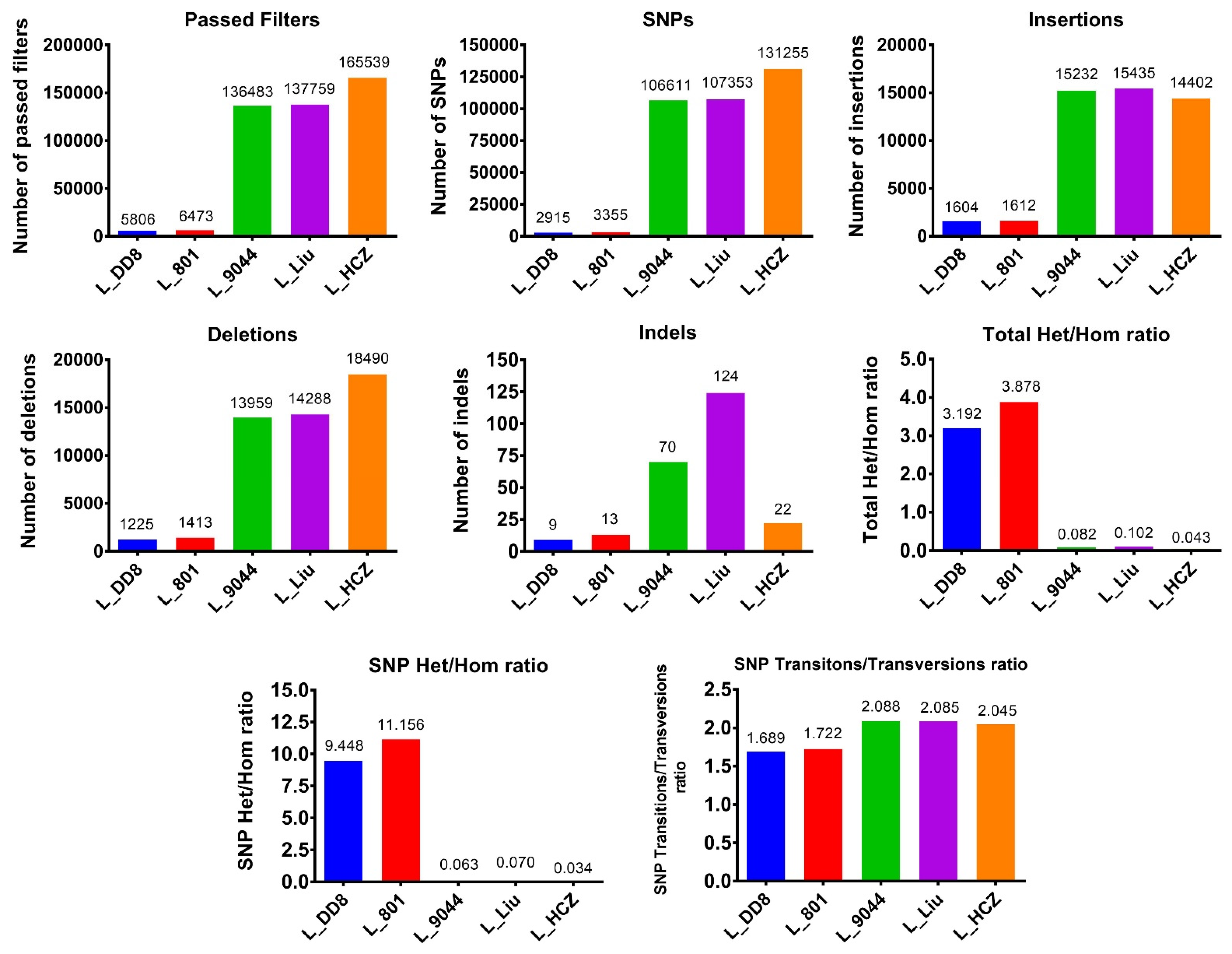
3.1.2. Detection Results of SNPs and InDels

Some differences in the number of SNPs and InDels among the five Leishmania isolates were found (Figure 1). The number of SNPs, insertions, deletions and InDels of L_801 and L_DD8 were basically the same and small, suggesting that they were close to the reference genome. The total Het/Hom ratio and SNP Het/Hom ratio of L_801 and L_DD8 were also basically consistent and high, reflecting that their heterozygous variations were more than homozygous variations. Moreover, the SNP Transitions/Transversions ratios of L_801 and L_DD8 were also basically the same, but they were lower than those of other Leishmania isolates. The number of SNPs, insertions, deletions, total Het/Hom ratio, SNP Het/Hom ratio and SNP Transitions/Transversions of L_9044, L_Liu and L_HCZ was approximately the same, and their homozygous variations were more than heterozygous variations, which was different from the results of L_801 and L_DD8. The number of InDels of L_Liu and L_9044 was higher than that of other Leishmania isolates.
The distribution of SNPs and InDels of the five Leishmania isolates is shown in Figure 2. Similarly, the distribution of SNPs and InDels of L_801 and L_DD8 was different from that of the other three isolates. The SNPs and InDels of L_801 and L_DD8 were mainly distributed upstream and downstream of genes, followed by the exonic region. However, the SNPs and InDels of L_9044, L_Liu and L_HCZ were most distributed in the exonic region, followed by upstream and downstream of genes, and their distribution in the upstream and downstream of genes was roughly the same. Only a very small number of SNPs and InDels in the five Leishmania isolates was distributed in the exonic region of ncRNA and the 5′ UTR region. It is worth noting that none of the five Leishmania SNPs and InDels were distributed in intronic, ncRNA_intronic, 3′ UTR and splicing regions.
The number of different types of mutations caused by SNPs and InDels in the coding region of the five Leishmania isolates is shown in Figure 3. L_801 and L_DD8 were slightly less synonymous than nonsynonymous, and the number of frameshifts was significantly more than that of non-frameshifts, and the number of stopgains was significantly higher than that of stoplosses. Among the mutations caused by the SNPs and InDels of L_9044, L_Liu and L_HCZ, the number of synonymous ones was basically equivalent to that of nonsynonymous ones, and the number of frameshifts was less than that of non-frameshifts, and the number of stopgains was more than that of stoplosses.

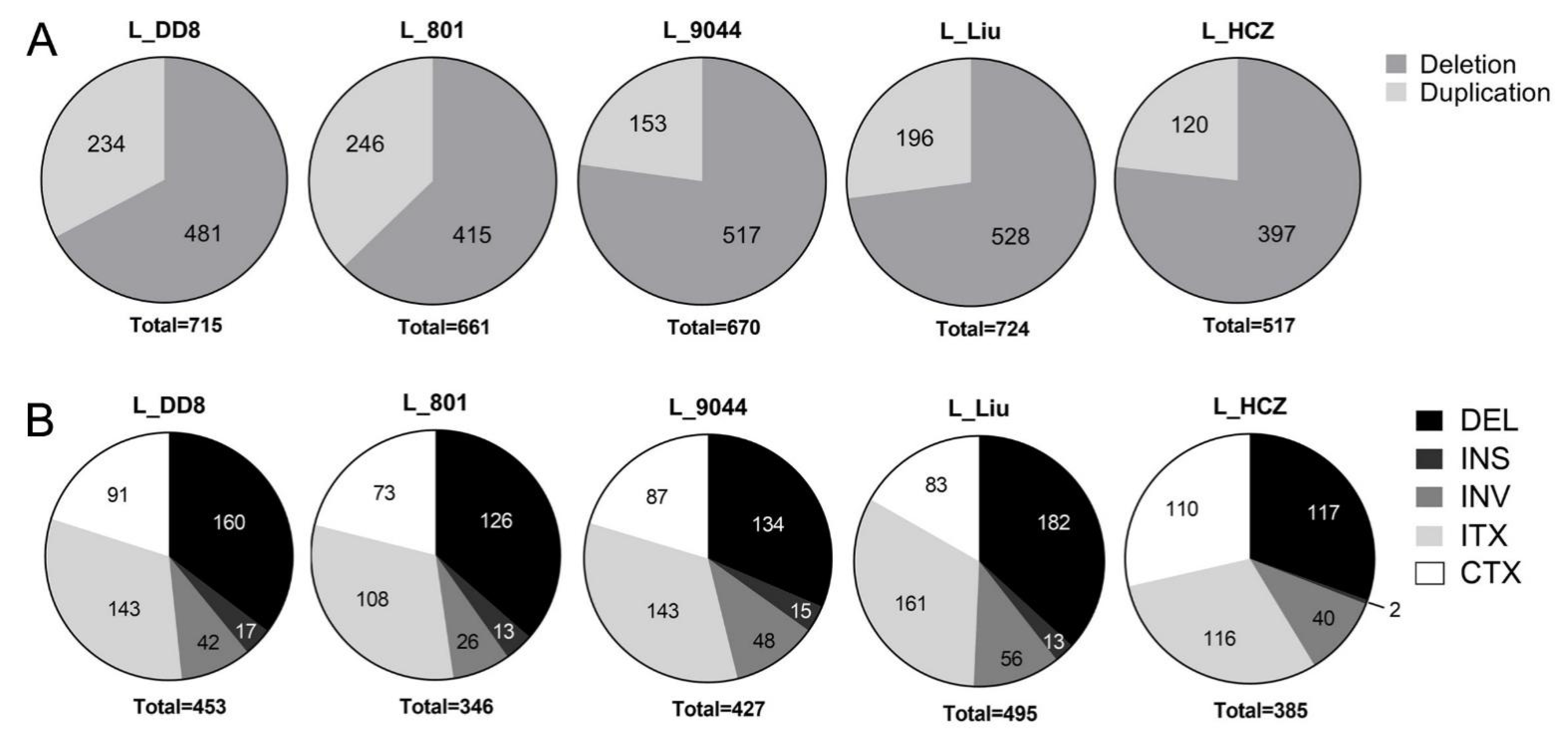
3.1.3. Detection Results of CNV and SV

As shown in Figure 4A, in CNV detection results, the number of duplications of all Leishmania isolates was greater than that of deletion, but their proportions of duplication and deletion were different. Similarly to the results of SNPs and InDels, the CNV results of L_801 and L_DD8 were also different from those of the other three Leishmania. The proportion of duplication of L_801 and L_DD8 was 62.8~67.2%, while that of L_9044, L_Liu and L_HCZ was 72.9~77.2%.
The SV results of L_801, L_DD8, L_9044 and L_Liu were similar, and their proportions of deletion (about 35%), insertion (about 3%), inversion (about 10%), intrachromosomal translocation (about 32%) and interchromosomal translocation (about 20%) were basically the same (Figure 4B). It is worth noting that the results of L_HCZ were different from those of the other four isolates, and its proportions of deletion, insertion, inversion, intrachromosomal translocation and interchromosomal translocation were 30.4%, 0.5%, 10.4%, 30.1% and 28.6%, respectively. Compared with the other four isolates, the proportions of deletion and insertion of L_HCZ decreased, and the proportion of interchromosomal translocation increased.

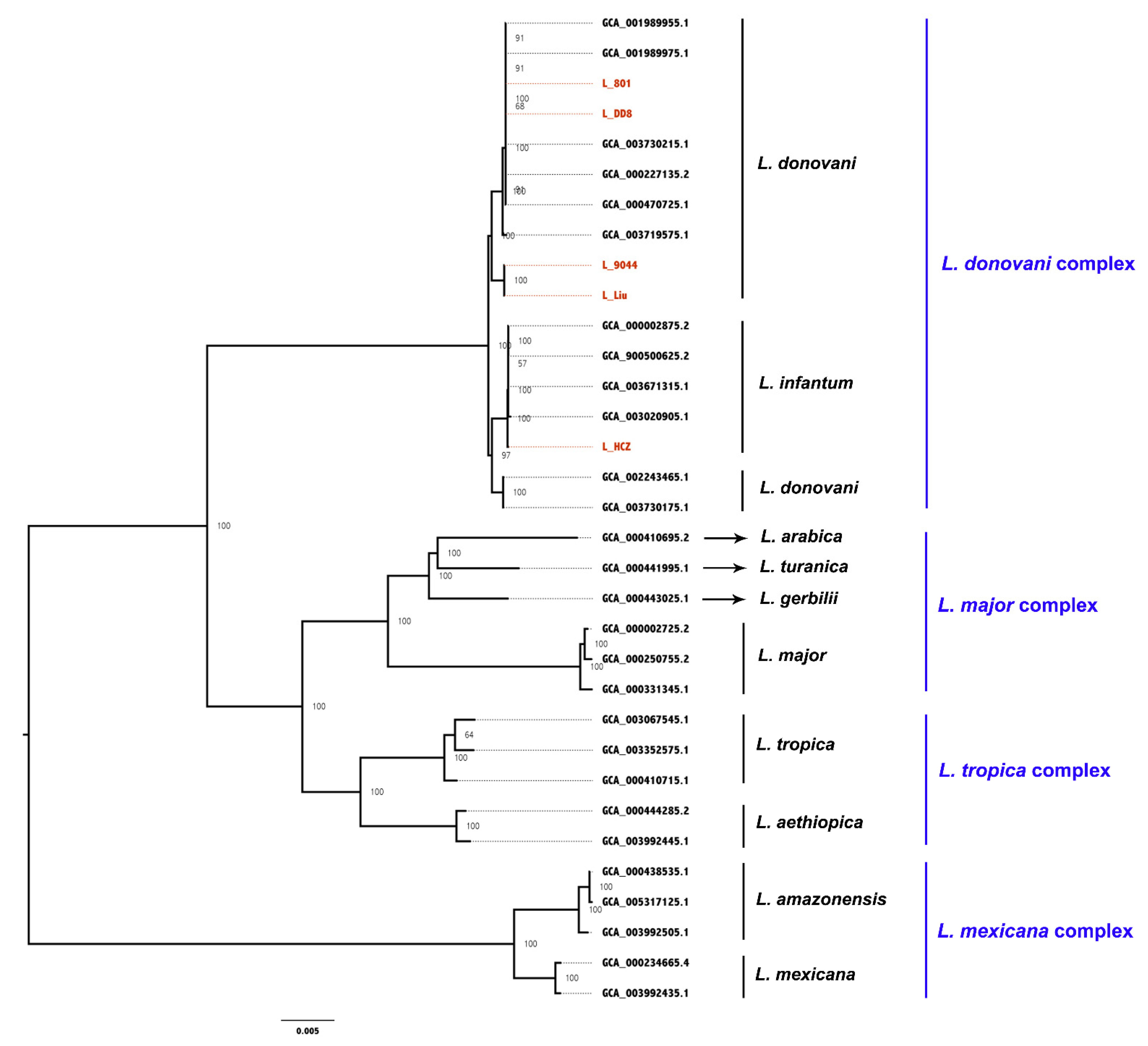
3.2. Whole-Gene Phylogenetic Analysis

The whole-genome sequences of 28 Leishmania strains were downloaded from NCBI and analyzed together with the whole-genome sequences of the five Leishmania clinical isolates. The pairwise comparison matrix of the whole genomic nucleic acids of the 33 Leishmania strains is shown in Figure 5. A core-genome alignment with a length of 7.3 Mbp was generated for phylogenetic analysis (Figure 6) based on the maximum-likelihood algorithm. These Leishmania strains were divided into four groups: L. donovani complex, L. major complex, L. tropica complex and L. mexcicana complex. The results of the phylogenetic tree show that L_801, L_DD8 and three L. donovani strains were clustered into a small branch, proving that they were closely related. The relationship between L_DD8 and L_801 also showed a high correlation on the matrix diagram. L_DD8 is the internationally common standard strain of L. donovani. Therefore, it could be determined that L_801 is L. donovani. L_9044 and L_Liu were clustered together in the tree, suggesting that their evolutionary relationship is close, but they were slightly distant from the rest of the L. donovani strains. L_9044 and L_Liu were temporarily incorporated into the Leishmania donovani complex. Due to the close evolutionary relationship between L_801 and L_DD8, the SNPs, InDels and CNV data of L_801 and L_DD8 were basically consistent and could be explained. Similarly, the consistent SNPs, InDels and CNV results of L_9044 and L_Liu may also be caused by their close evolutionary relationship. It should be noted that L_HCZ was clustered with two canine L. infantum strains and one human L. infantum strain in the tree. Consequently, the L_HCZ isolate could be identified as L. infantum. However, because of the close genetic relationship between L. donovani and L. infantum [40], the genome-wide phylogenetic tree in this study could not completely separate L. donovani and L. infantum.

3.3. Mutation Analysis of L_HCZ

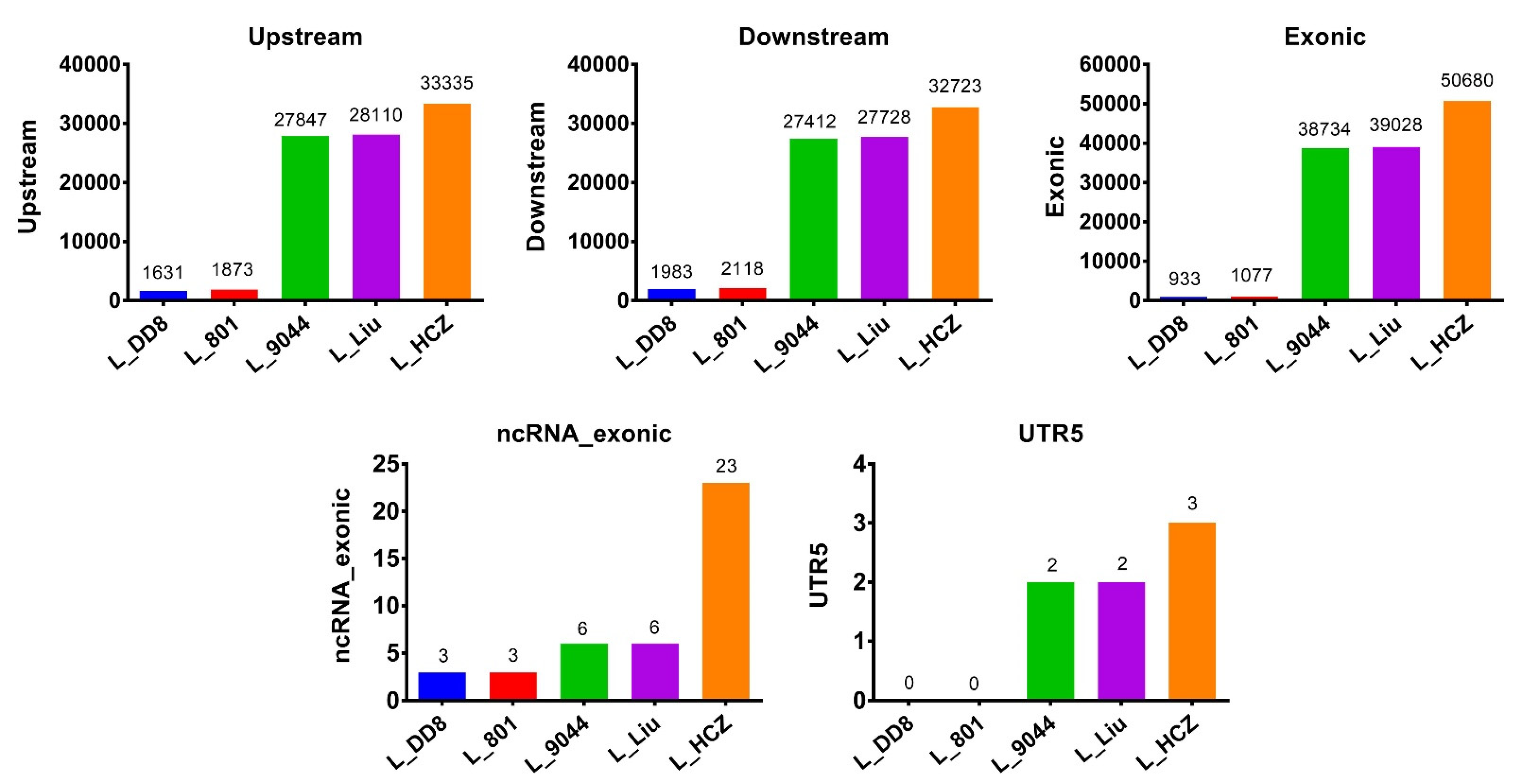
3.3.1. Exon Mutations in Five Leishmania Isolates

Compared with the reference genome, L_DD8 had 883 mutations in exons, 773 mutations were aneuploid, and the proportion of aneuploidy was 87.54%. L_801 had 1022 mutations in exons, 918 mutations were aneuploid, and the proportion of aneuploidy was 89.82%. L_9044 had 37,796 mutations in exons, 1648 mutations were aneuploid, and the proportion of aneuploidy was 4.36%. L_Liu had 38,078 mutations in exons, 2054 mutations were aneuploid, and the proportion of aneuploidy was 5.39%. L_HCZ had 49,352 mutations in exons, 1322 mutations were aneuploid, and the proportion of aneuploidy was 2.68%. We found that although L_HCZ, L_Liu and L_9044 had more exon mutations, most of them were euploid. However, although L_801 and L_DD8 had fewer exon mutations, most of them were aneuploid (Table 4).

3.3.2. Mutation Analysis in Nine Genes Associated with Antimony Resistance

The number of mutations in nine antimony resistance-related genes of five Leishmania isolates is shown in Figure 7. Most of the genes had only nonsynonymous and synonymous mutations, and only dihydrofolate reductase–thymidylate synthase (frameshift deletion), sodium stibogluconate resistance protein (frameshift deletion, frameshift insertion, non-frameshift deletion, non-frameshift insertion and stopgain) and trypanothione reductase (frameshift deletion and frameshift insertion) had other types of mutations. Among the nine genes, only sodium stibogluconate resistance protein and aquaglyceroporin had aneuploidy mutations. L_DD8 and L_801 only had mutations in the sodium stibogluconate resistance protein gene, and no mutations were found in the other eight genes. The nine genes in L_9044, L_Liu and L_HCZ all had varying degrees of mutation. The mutation types, numbers and sites of the nine genes in L_9044 and L_Liu were mostly the same, which was consistent with the results of previous resequencing and phylogenetic analysis. Except for dihydrofolate reductase–thymidylate synthase, mitogen-activated protein kinase and sodium stibogluconate resistance protein, the other genes had the most mutations in L_HCZ.

3.3.3. Mutation Analysis in Nine Genes Associated with Virulence

The number of mutations in nine virulence genes of five Leishmania isolates is shown in Figure 8. Only gp63 (nonframeshift deletion, nonframeshift insertion and stoploss), acid phosphatase (nonframeshift deletion) and kinesin k39 (frameshift insertion) genes had other types of mutations, while other genes had only nonsynonymous and synonymous mutations. Among the nine genes, only cysteine peptidase B, gp63, surface protein amastin and kinesin k39 had aneuploidy mutations. L_DD8 and L_801 only had synonymous mutations in kinesin k39 gene, and the numbers and gene sites of synonymous mutations in the two isolates were identical. Similar to the results of antimony resistance-related genes, the mutation types, numbers and sites of the nine virulence genes in L_9044 and L_Liu were mostly the same. Except for cyclophilin-40 and surface protein amastin, the other genes had the most mutations in L_HCZ.

3.3.4. Mutation Analysis of 18 Genes

We counted genes with more than 50 mutations in the L_HCZ isolate and finally obtained 18 genes. Genes involved in cell signaling and various biological functions: protein kinase [41], ubiquitin hydrolase [41], serine/threonine-protein kinase [42], phosphatidylinositol 3-kinase-like protein [43] and receptor-type adenylate cyclase a [44]. Genes related to the virulence of Leishmania: kinesin [45], calpain-like cysteine peptidase [46], phosphoglycan beta 1 [47], amastin-like surface protein [48] and amastin-like protein [48]. A gene related to cilia and flagella movement: dynein heavy chain [49]. A gene involved in protein synthesis and processing: RNA binding protein [50]. A gene related to cell metabolism: N-acyl-L-amino acid amidohydrolase [51]. Genes associated with drug resistance in Leishmania: ATP-binding cassette protein subfamily A [52], folate/biopterin transporter [53] and protein-like multidrug resistant [52]. Unknown functional gene: uncharacterized protein and kinetoplast-associated protein-like protein. The number of mutations in the 18 genes of five Leishmania is shown in Table 5. Most of these genes had the highest number of mutations in the L_HCZ isolate.

4. Discussion

Recently, the development of whole-genome sequencing and computational analysis has improved the resolution and accuracy of in-depth exploration and comparison of Leishmania genomes [54]. Although the various data of L_HCZ sequencing are close to the data of L_9044 and L_Liu, we still found some special features in the results of L_HCZ sequencing. Compared with the other four Leishmania isolates, L_HCZ had the largest number of SNPs and deletions, and had the largest number of variations in upstream, downstream, the exonic region of ncRNA and the 5′ UTR region. Furthermore, in coding regions, the number of SNV, insertion, deletion, synonymous, nonsynonymous, frameshift and non-frameshift mutations was also the highest in L_HCZ. It is interesting that L_HCZ had the lowest number of CNV and SV, especially the number of CNV duplication. The CNV duplication number of L_DD8 and L_801 was almost twice that of L_HCZ. Gene duplication would represent an important means of innovation by expanding the genomic information, and normal copies of the gene would compensate deleterious mutations, whereas successful mutations could be expanded [55]. In trypanosomatids, gene expression is regulated primarily at the post-transcriptional level rather than at the initial level and allows the up-regulation of expression through copy number amplification [55,56]. Therefore, whether these mutations have functional consequences needs to be further explored at the proteomic and metabonomic levels.
The results of the phylogenetic tree corresponded to the SNP/InDel results of five Leishmania isolates. In the phylogenetic tree, L_DD8 and L_801 were clustered together, while the data of the two isolates were similar in the SNP/InDel results. The same was true for the phylogenetic and SNP/InDel results of L_9044 and L_Liu. Genome-wide SNP variation provided new evolutionary insights into the ongoing diversification of the phenotypically variable set of Leishmania strains [57]. Based on genome-wide SNP/InDel variation analysis and phylogenetic analysis, it could be basically determined that the controversial L_801 in Genebank could be identified as L. donovani and had a close genetic relationship with L_DD8. L_Liu was isolated from a patient with cutaneous leishmaniasis in Karamay, Xinjiang, China [58]. An earlier study of cutaneous leishmaniasis in Karamay, Xinjiang found that the pathogen was L. infantum [59]. However, in our study, the results of both the phylogenetic tree and SNP/InDel show a close genetic relationship between L_9044 and L_Liu, which is basically consistent with the results of Zhang et al. [58]. In the previous studies, L_9044 and L_Liu were classified as L. donovani [58,60]. Therefore, in combination with our genome-wide phylogenetic tree, L_9044 and L_Liu should be identified as L. donovani. Moreover, L_HCZ was grouped with three L. infantum isolates in the phylogenetic tree, which basically confirmed L_HCZ as L. infantum. Whole-genome resequencing is economical, fast and convenient. It can be used for the identification of Leishmania isolates and searches for drug resistance targets. It has broad application prospects and may be put into clinical use with phylogenetic analysis in the future for identifying pathogens and epidemiological investigations [57,61].
SNPs in drug targets or transporters can cause drug resistance without changing gene expression, so single-nucleotide mutations constitute a strategy to avoid drug pressure [62]. Single-nucleotide mutations can lead to drug resistance by interfering with drug-target interactions or by changing drugs’ permeability to cells [37]. Another genomic mechanism for drug resistance in Leishmania is to regulate the expression of drug target, drug transporter or other drug resistance determinants by changing gene dose through copy number variations (aneuploidy, gene amplification or gene deletion) [62,63]. In this study, compared with the other four Leishmania isolates, the number of mutations in virulence genes and drug resistance-related genes of L_HCZ was larger. Except for L_HCZ, the other four Leishmania isolates were isolated from the 1980s and 1990s. As a newly isolated Leishmania strain, the mutations in virulence genes and drug resistance-related genes of L_HCZ might be caused by the variation accumulation during its evolution [64]. The mutations in L_HCZ might be one of the reasons for its antimony resistance and strong virulence in the clinical patient. Furthermore, in this study, L. infantum (L_HCZ) had more mutations in virulence genes than those of L. donovani (L_DD8, L_801, L_9044 and L_Liu), which might be one of the reasons why L. infantum is more virulent than L. donovani. In this study, we investigated the mutation characteristics of five Leishmania clinical isolates based on the results of whole-genome resequencing, and analyzed the phylogenetic relationship of these isolates by constructing a phylogenetic tree with the published whole-genome sequences of 28 Leishmania isolates. Genome-wide phylogenetic analysis would be helpful to improve the molecular identification of Leishmania species, and genome-wide mutation analysis revealed possible mechanisms of pathogenicity and drug resistance in different Leishmania isolates. The datasets of the five Leishmania clinical isolates will provide a molecular basis for future research on the pathogenic mechanism, drug resistance mechanism and vaccine development of Leishmania. In our previous study, we focused on verifying the virulence and antimony resistance of the Leishmania clinical isolate (L_HCZ) at molecular levels and proved that L_HCZ has high-level pathogenicity and evident antimony resistance in vitro, using an integrative approach of genome sequencing, proteomic profiling and phenotypic analysis to find the target spot of virulence and antimony resistance of L_HCZ [65]. According to these two papers, some interesting results attracted our attention. In this study, we found the genes of protein kinase, kinesin, amastin-like surface protein, N-acyl-L-amino acid amidohydrolase protein and sodium stibogluconate resistance protein in L_HCZ had more mutations, and in our previous study we found the expression of these genes at the protein level was up-regulated [65]. Similarly, the mutation of mitogen-activated protein kinase in L_HCZ was less than that in L_9044 and L_Liu, and the protein expressed by this gene was down-regulated. Therefore, these results suggest that mutations in these genes affected the expression levels of the corresponding proteins. The gene mutations of protein kinase, kinesin, amastin-like surface protein, N-acyl-L-amino acid amidohydrolase protein and sodium stibogluconate resistance protein in L_HCZ up-regulated the expression of the corresponding proteins and might play roles in the pathogenesis and drug resistance of L_HCZ. Virulence genes, drug resistance-related genes and genes with a high incidence of mutations selected from these Leishmania isolates may become targets for vaccines or drugs, as well as the molecular markers for screening of Leishmania drug-resistant strains and different virulence strains [38].

5. Conclusions

This study performed genome-wide resequencing and phylogenetic analysis of five isolates from the Leishmania donovani complex, focusing on finding mutations related to antimony resistance and virulence of the newly isolated Leishmania strain L_HCZ in 2016. By combining whole-genome sequencing and whole-genome phylogenetic analysis, Leishmania isolates L_801, L_9044 and L_Liu were identified as L. donovani, and L_HCZ was L. infantum. By discovering genome-wide single-nucleotide polymorphisms and structural variations, we identified mutations of drug resistance-related genes in the antimony-resistant Leishmania isolate L_HCZ. The new Leishmania isolate L_HCZ has strong virulence and strong drug resistance, which should be taken seriously by relevant health departments and scientific researchers.

Author Contributions

Z.Z., J.L. (Jiao Li) and J.C. designed and conceived the study. Z.Z. and J.H. wrote the original manuscript. Z.Z., J.Z., Q.Z., S.Y. and J.L. (Jie Luo) undertook the laboratory work. Z.Z., T.L. and J.H. collected and analysed the data. T.L. and D.C. revised the manuscript and polished the language. All authors have read and agreed to the published version of the manuscript.

Funding

This work was supported by the National Natural Science Foundation of China (No. 31802184 to Jiao Li, No. 81672048 to Jianping Chen, No. 31872959 to Dali Chen).

Institutional Review Board Statement

Not applicable.

Informed Consent Statement

Not applicable.

Data Availability Statement

The datasets supporting the conclusions of this article are included within the article. Genome sequencing data sets have been submitted to the National Center for Biotechnology Information (NCBI) sequence reads archive (SRA), which can be accessed under accession number PRJNA600762 (L_DD8, L_9044, L_HCZ, L_Liu and L_801). The datasets release on 30 December 2021 (L_DD8, L_9044 and L_HCZ) and on 28 February 2022 (L_Liu and L_801)).

Acknowledgments

The authors are very grateful to Junyun Wang from the National Institute of Parasitic Diseases, Chinese Center for Disease Control and Prevention, Shanghai, China, for providing the Leishmania isolates L_801, L_9044, L_DD8 and L_Liu.

Conflicts of Interest

The authors declare no conflict of interest.

References

  1. Guerin, P.J.; Olliaro, P.; Sundar, S.; Boelaert, M.; Croft, S.L.; Desjeux, P.; Wasunna, M.K.; Bryceson, A.D. Visceral leishmaniasis: Current status of control, diagnosis, and treatment, and a proposed research and development agenda. Lancet Infect. Dis. 2002, 2, 494–501. [Google Scholar] [CrossRef]
  2. Hide, M.; Bras-Gonçalves, R.; Bañuls, A.L. Specific cpb copies within the Leishmania donovani complex: Evolutionary interpretations and potential clinical implications in humans. Parasitology 2007, 134, 379–389. [Google Scholar] [CrossRef] [PubMed] [Green Version]
  3. Burza, S.; Croft, S.L.; Boelaert, M. Leishmaniasis. Lancet 2018, 392, 951–970. [Google Scholar] [CrossRef]
  4. Croft, S.L.; Sundar, S.; Fairlamb, A.H. Drug resistance in leishmaniasis. Clin. Microbiol. Rev. 2006, 19, 111–126. [Google Scholar] [CrossRef] [PubMed] [Green Version]
  5. Kostygov, A.Y.; Karnkowska, A.; Votýpka, J.; Tashyreva, D.; Maciszewski, K.; Yurchenko, V.; Lukeš, J. Euglenozoa: Taxonomy, diversity and ecology, symbioses and viruses. Open Biol. 2021, 11, 200407. [Google Scholar] [CrossRef] [PubMed]
  6. Maslov, D.A.; Opperdoes, F.R.; Kostygov, A.Y.; Hashimi, H.; Lukeš, J.; Yurchenko, V. Recent advances in trypanosomatid research: Genome organization, expression, metabolism, taxonomy and evolution. Parasitology 2019, 146, 1–27. [Google Scholar] [CrossRef] [PubMed] [Green Version]
  7. Akhoundi, M.; Kuhls, K.; Cannet, A.; Votýpka, J.; Marty, P.; Delaunay, P.; Sereno, D. A historical overview of the classification, evolution, and dispersion of Leishmania parasites and sandflies. PLoS Negl. Trop. Dis. 2016, 10, e0004349. [Google Scholar] [CrossRef]
  8. Fraga, J.; Montalvo, A.M.; De Doncker, S.; Dujardin, J.C.; Van der Auwera, G. Phylogeny of Leishmania species based on the heat-shock protein 70 gene. Infect. Genet. Evol. 2010, 10, 238–245. [Google Scholar] [CrossRef]
  9. Akhoundi, M.; Downing, T.; Votýpka, J.; Kuhls, K.; Lukeš, J.; Cannet, A.; Ravel, C.; Marty, P.; Delaunay, P.; Kasbari, M.; et al. Leishmania infections: Molecular targets and diagnosis. Mol. Asp. Med. 2017, 57, 1–29. [Google Scholar] [CrossRef]
  10. Cantacessi, C.; Dantas-Torres, F.; Nolan, M.J.; Otranto, D. The past, present, and future of Leishmania genomics and transcriptomics. Trends Parasitol. 2015, 31, 100–108. [Google Scholar] [CrossRef] [Green Version]
  11. Yurchenko, V.; Butenko, A.; Kostygov, A.Y. Genomics of trypanosomatidae: Where we stand and what needs to be done? Pathogens 2021, 10, 1124. [Google Scholar] [CrossRef] [PubMed]
  12. Real, F.; Vidal, R.O.; Carazzolle, M.F.; Mondego, J.M.; Costa, G.G.; Herai, R.H.; Würtele, M.; de Carvalho, L.M.; Carmona e Ferreira, R.; Mortara, R.A.; et al. The genome sequence of Leishmania (Leishmania) amazonensis: Functional annotation and extended analysis of gene models. DNA Res. 2013, 20, 567–581. [Google Scholar] [CrossRef] [PubMed] [Green Version]
  13. Peacock, C.S.; Seeger, K.; Harris, D.; Murphy, L.; Ruiz, J.C.; Quail, M.A.; Peters, N.; Adlem, E.; Tivey, A.; Aslett, M.; et al. Comparative genomic analysis of three Leishmania species that cause diverse human disease. Nat. Genet. 2007, 39, 839–847. [Google Scholar] [CrossRef] [PubMed] [Green Version]
  14. Wincker, P.; Ravel, C.; Blaineau, C.; Pages, M.; Jaufrett, Y.; Dedet, J.P.; Bastien, P. The Leishmania genome comprises 36 chromosomes conserved across widely divergent human pathogenic species. Nucleic Acids Res. 1998, 24, 1688–1694. [Google Scholar] [CrossRef] [PubMed] [Green Version]
  15. Ivens, A.C.; Peacock, C.S.; Worthey, E.A.; Murphy, L.; Aggarwal, G.; Berriman, M.; Sisk, E.; Rajandream, M.A.; Adlem, E.; Aert, R.; et al. The genome of the kinetoplastid parasite, Leishmania major. Science 2005, 309, 436–442. [Google Scholar] [CrossRef] [PubMed] [Green Version]
  16. Yardley, V.; Croft, S.L.; De Doncker, S.; Dujardin, J.C.; Koirala, S.; Rijal, S.; Miranda, C.; Llanos-Cuentas, A.; Chappuis, F. The sensitivity of clinical isolates of Leishmania from Peru and Nepal to miltefosine. Am. J. Trop. Med. Hyg. 2005, 73, 272–275. [Google Scholar] [CrossRef]
  17. Sánchez-Cañete, M.P.; Carvalho, L.; Pérez-Victoria, F.J.; Gamarro, F.; Castanys, S. Low plasma membrane expression of the miltefosine transport complex renders Leishmania braziliensis refractory to the drug. Antimicrob. Agents Chemother. 2009, 53, 1305–1313. [Google Scholar] [CrossRef] [Green Version]
  18. Horn, D.; Duraisingh, M.T. Antiparasitic chemotherapy: From genomes to mechanisms. Annu. Rev. Pharmacol. Toxicol. 2014, 54, 71–94. [Google Scholar] [CrossRef] [Green Version]
  19. Zhu, Q.; Kosoy, M.; Dittmar, K. HGTector: An automated method facilitating genome-wide discovery of putative horizontal gene transfers. BMC Genom. 2014, 15, 717. [Google Scholar] [CrossRef] [Green Version]
  20. Mardis, E.R. A decade’s perspective on DNA sequencing technology. Nature 2011, 470, 198–203. [Google Scholar] [CrossRef]
  21. He, J.; Huang, F.; Zhang, J.; Chen, H.; Chen, Q.; Zhang, J.; Li, J.; Zheng, Z.; Chen, D.; Chen, J. DNA prime-protein boost vaccine encoding HLA-A2, HLA-A24 and HLA-DR1 restricted epitopes of CaNA2 against visceral leishmaniasis. Immunology 2019, 156, 94–108. [Google Scholar] [CrossRef] [Green Version]
  22. Li, H.; Durbin, R. Fast and accurate long-read alignment with Burrows-Wheeler transform. Bioinformatics 2010, 26, 589–595. [Google Scholar] [CrossRef] [PubMed] [Green Version]
  23. McKenna, A.; Hanna, M.; Banks, E.; Sivachenko, A.; Cibulskis, K.; Kernytsky, A.; Garimella, K.; Altshuler, D.; Gabriel, S.; Daly, M.; et al. The genome analysis toolkit: A mapreduce framework for analyzing next-generation DNA sequencing data. Genome Res. 2010, 20, 1297–1303. [Google Scholar] [CrossRef] [PubMed] [Green Version]
  24. Wang, K.; Li, M.; Hakonarson, H. ANNOVAR: Functional annotation of genetic variants from high-throughput sequencing data. Nucleic Acids Res. 2010, 38, e164. [Google Scholar] [CrossRef] [PubMed]
  25. Abyzov, A.; Urban, A.E.; Snyder, M.; Gerstein, M. CNVnator: An approach to discover, genotype, and characterize typical and atypical CNVs from family and population genome sequencing. Genome Res. 2011, 21, 974–984. [Google Scholar] [CrossRef] [PubMed] [Green Version]
  26. Chen, K.; Wallis, J.W.; McLellan, M.D.; Larson, D.E.; Kalicki, J.M.; Pohl, C.S.; McGrath, S.D.; Wendl, M.C.; Zhang, Q.; Locke, D.P.; et al. BreakDancer: An algorithm for high-resolution mapping of genomic structural variation. Nat Methods. 2009, 6, 677–681. [Google Scholar] [CrossRef] [PubMed] [Green Version]
  27. Chen, S.; Zhou, Y.; Chen, Y.; Gu, J. fastp: An ultra-fast all-in-one FASTQ preprocessor. Bioinformatics. 2018, 34, i884–i890. [Google Scholar] [CrossRef] [PubMed]
  28. Jain, C.; Rodriguez-R, L.M.; Phillippy, A.M.; Konstantinidis, K.T.; Aluru, S. High throughput ANI analysis of 90K prokaryotic genomes reveals clear species boundaries. Nat. Commun. 2018, 9, 5114. [Google Scholar] [CrossRef] [Green Version]
  29. Fernández, A.; Gómez, S. Solving non-uniqueness in agglomerative hierarchical clustering using Multidendrograms. J. Classif. 2008, 25, 43–65. [Google Scholar] [CrossRef] [Green Version]
  30. Li, H. Minimap2: Pairwise alignment for nucleotide sequences. Bioinformatics 2018, 34, 3094–3100. [Google Scholar] [CrossRef]
  31. Li, H.; Handsaker, B.; Wysoker, A.; Fennell, T.; Ruan, J.; Homer, N.; Marth, G.; Abecasis, G.; Durbin, R. The sequence alignment/map format and SAMtools. Bioinformatics 2009, 25, 2078–2079. [Google Scholar] [CrossRef] [PubMed] [Green Version]
  32. Quinlan, A.R.; Hall, I.M. BEDTools: A flexible suite of utilities for comparing genomic features. Bioinformatics 2010, 26, 841–842. [Google Scholar] [CrossRef] [PubMed] [Green Version]
  33. Dozmorov, M.G. GitHub statistics as a measure of the impact of open-source bioinformatics software. Front. Bioeng. Biotechnol. 2018, 6, 198. [Google Scholar] [CrossRef]
  34. Nakamura, T.; Yamada, K.D.; Tomii, K.; Katoh, K. Parallelization of MAFFT for large-scale multiple sequence alignments. Bioinformatics 2018, 34, 2490–2492. [Google Scholar] [CrossRef] [PubMed] [Green Version]
  35. Capella-Gutiérrez, S.; Silla-Martínez, J.M.; Gabaldón, T. trimAl: A tool for automated alignment trimming in large-scale phylogenetic analyses. Bioinformatics 2009, 25, 1972–1973. [Google Scholar] [CrossRef]
  36. Minh, B.Q.; Schmidt, H.A.; Chernomor, O.; Schrempf, D.; Woodhams, M.D.; von Haeseler, A.; Lanfear, R. IQ-TREE 2: New Models and Efficient Methods for Phylogenetic Inference in the Genomic Era. Mol. Biol. Evol. 2020, 37, 1530–1534. [Google Scholar] [CrossRef] [Green Version]
  37. Leprohon, P.; Fernandez-Prada, C.; Gazanion, É.; Monte-Neto, R.; Ouellette, M. Drug resistance analysis by next generation sequencing in Leishmania. Int. J. Parasitol. Drugs Drug Resist. 2014, 5, 26–35. [Google Scholar] [CrossRef] [Green Version]
  38. Mohebali, M.; Kazemirad, E.; Hajjaran, H.; Kazemirad, E.; Oshaghi, M.A.; Raoofian, R.; Teimouri, A. Gene expression analysis of antimony resistance in Leishmania tropica using quantitative real-time PCR focused on genes involved in trypanothione metabolism and drug transport. Arch. Dermatol. Res. 2019, 311, 9–17. [Google Scholar] [CrossRef]
  39. Bifeld, E.; Clos, J. The genetics of Leishmania virulence. Med. Microbiol. Immunol. 2015, 204, 619–634. [Google Scholar] [CrossRef]
  40. Lukes, J.; Mauricio, I.L.; Schönian, G.; Dujardin, J.C.; Soteriadou, K.; Dedet, J.P.; Kuhls, K.; Tintaya, K.W.; Jirků, M.; Chocholová, E.; et al. Evolutionary and geographical history of the Leishmania donovani complex with a revision of current taxonomy. Proc. Natl. Acad. Sci. USA 2007, 104, 9375–9380. [Google Scholar] [CrossRef] [Green Version]
  41. Saxena, A.; Lahav, T.; Holland, N.; Aggarwal, G.; Anupama, A.; Huang, Y.; Volpin, H.; Myler, P.J.; Zilberstein, D. Analysis of the Leishmania donovani transcriptome reveals an ordered progression of transient and permanent changes in gene expression during differentiation. Mol. Biochem. Parasitol. 2007, 152, 53–65. [Google Scholar] [CrossRef] [PubMed] [Green Version]
  42. Manuse, S.; Fleurie, A.; Zucchini, L.; Lesterlin, C.; Grangeasse, C. Role of eukaryotic-like serine/threonine kinases in bacterial cell division and morphogenesis. FEMS Microbiol. Rev. 2016, 40, 41–56. [Google Scholar] [CrossRef] [PubMed] [Green Version]
  43. Quek, H.; Lim, Y.C.; Lavin, M.F.; Roberts, T.L. PIKKing a way to regulate inflammation. Immunol. Cell Biol. 2018, 96, 8–20. [Google Scholar] [CrossRef] [PubMed]
  44. Saada, E.A.; Kabututu, Z.P.; Lopez, M.; Shimogawa, M.M.; Langousis, G.; Oberholzer, M.; Riestra, A.; Jonsson, Z.O.; Wohlschlegel, J.A.; Hill, K.L. Insect stage-specific receptor adenylate cyclases are localized to distinct subdomains of the Trypanosoma brucei Flagellar membrane. Eukaryot. Cell 2014, 13, 1064–1076. [Google Scholar] [CrossRef] [PubMed] [Green Version]
  45. Dey, A.; Sharma, P.; Redhu, N.S.; Singh, S. Kinesin motor domain of Leishmania donovani as a future vaccine candidate. Clin. Vaccine Immunol. 2008, 15, 836–842. [Google Scholar] [CrossRef] [Green Version]
  46. Silva-Almeida, M.; Pereira, B.A.; Ribeiro-Guimarães, M.L.; Alves, C.R. Proteinases as virulence factors in Leishmania spp. infection in mammals. Parasites Vectors 2012, 5, 160. [Google Scholar] [CrossRef] [PubMed] [Green Version]
  47. Chang, K.P.; McGwire, B.S. Molecular determinants and regulation of Leishmania virulence. Kinetoplastid Biol. Dis. 2002, 1, 1. [Google Scholar] [CrossRef]
  48. Rochette, A.; McNicoll, F.; Girard, J.; Breton, M.; Leblanc, E.; Bergeron, M.G.; Papadopoulou, B. Characterization and developmental gene regulation of a large gene family encoding amastin surface proteins in Leishmania spp. Mol. Biochem. Parasitol. 2005, 140, 205–220. [Google Scholar] [CrossRef]
  49. Asai, D.J.; Wilkes, D.E. The dynein heavy chain family. J. Eukaryot. Microbiol. 2004, 51, 23–29. [Google Scholar] [CrossRef]
  50. Clayton, C.E. Gene expression in Kinetoplastids. Curr. Opin. Microbiol. 2016, 32, 46–51. [Google Scholar] [CrossRef]
  51. Sommer, A.; Christensen, E.; Schwenger, S.; Seul, R.; Haas, D.; Olbrich, H.; Omran, H.; Sass, J.O. The molecular basis of aminoacylase 1 deficiency. Biochim. Biophys. Acta. 2011, 1812, 685–690. [Google Scholar] [CrossRef] [PubMed] [Green Version]
  52. Sauvage, V.; Aubert, D.; Escotte-Binet, S.; Villena, I. The role of ATP-binding cassette (ABC) proteins in protozoan parasites. Mol. Biochem. Parasitol. 2009, 167, 81–94. [Google Scholar] [CrossRef] [PubMed]
  53. Ouameur, A.A.; Girard, I.; Légaré, D.; Ouellette, M. Functional analysis and complex gene rearrangements of the folate/biopterin transporter (FBT) gene family in the protozoan parasite Leishmania. Mol. Biochem. Parasitol. 2008, 162, 155–164. [Google Scholar] [CrossRef] [PubMed]
  54. Downing, T.; Imamura, H.; Decuypere, S.; Clark, T.G.; Coombs, G.H.; Cotton, J.A.; Hilley, J.D.; de Doncker, S.; Maes, L.; Mottram, J.C.; et al. Whole genome sequencing of multiple Leishmania donovani clinical isolates provides insights into population structure and mechanisms of drug resistance. Genome Res. 2011, 21, 2143–2156. [Google Scholar] [CrossRef] [Green Version]
  55. Victoir, K.; Dujardin, J.C. How to succeed in parasitic life without sex? Asking Leishmania. Trends Parasitol. 2002, 18, 81–85. [Google Scholar] [CrossRef]
  56. McDonagh, P.D.; Myler, P.J.; Stuart, K. The unusual gene organization of Leishmania major chromosome 1 may reflect novel transcription processes. Nucleic Acids Res. 2000, 28, 2800–2803. [Google Scholar] [CrossRef] [PubMed] [Green Version]
  57. Downing, T.; Stark, O.; Vanaerschot, M.; Imamura, H.; Sanders, M.; Decuypere, S.; de Doncker, S.; Maes, I.; Rijal, S.; Sunder, S.; et al. Genome-wide SNP and microsatellite variation illuminate population-level epidemiology in the Leishmania donovani species complex. Infect. Genet. Evol. 2012, 12, 149–159. [Google Scholar] [CrossRef] [Green Version]
  58. Zhang, C.Y.; Lu, X.J.; Du, X.Q.; Jian, J.; Shu, L.; Ma, Y. Phylogenetic and evolutionary analysis of Chinese Leishmania isolates based on multilocus sequence typing. PLoS ONE 2013, 8, e63124. [Google Scholar] [CrossRef] [Green Version]
  59. Guan, L.R.; Yang, Y.Q.; Qu, J.Q.; Ren, H.Y.; Chai, J.J. Discovery and study of cutaneous leishmaniasis in Karamay of Xinjiang, West China. Infect. Dis. Poverty 2013, 2, 20. [Google Scholar] [CrossRef] [Green Version]
  60. Yuan, D.; Qin, H.; Zhang, J.; Liao, L.; Chen, Q.; Chen, D.; Chen, J. Phylogenetic analysis of HSP70 and cyt b gene sequences for Chinese Leishmania isolates and ultrastructural characteristics of Chinese Leishmania sp. Parasitol. Res. 2017, 116, 693–702. [Google Scholar] [CrossRef]
  61. Mukherjee, I.; Chakraborty, A.; Chakrabarti, S. Identification of internalin-A-like virulent proteins in Leishmania donovani. Parasites Vectors 2016, 9, 557. [Google Scholar] [CrossRef] [PubMed] [Green Version]
  62. Laffitte, M.N.; Leprohon, P.; Papadopoulou, B.; Ouellette, M. Plasticity of the Leishmania genome leading to gene copy number variations and drug resistance. F1000Research 2016, 5, 2350. [Google Scholar] [CrossRef] [PubMed] [Green Version]
  63. Fernandez-Prada, C.; Sharma, M.; Plourde, M.; Bresson, E.; Roy, G.; Leprohon, P.; Ouellette, M. High-throughput Cos-Seq screen with intracellular Leishmania infantum for the discovery of novel drug-resistance mechanisms. Int. J. Parasitol. Drugs Drug Resist. 2018, 8, 165–173. [Google Scholar] [CrossRef] [PubMed]
  64. Vergnes, B.; Gazanion, E.; Mariac, C.; Du Manoir, M.; Sollelis, L.; Lopez-Rubio, J.J.; Sterkers, Y.; Bañuls, A.L. A single amino acid substitution (H451Y) in Leishmania calcium-dependent kinase SCAMK confers high tolerance and resistance to antimony. J. Antimicrob. Chemother. 2019, 74, 3231–3239. [Google Scholar] [CrossRef]
  65. Zheng, Z.; Chen, J.; Ma, G.; Satoskar, A.R.; Li, J. Integrative genomic, proteomic and phenotypic studies of Leishmania donovani strains revealed genetic features associated with virulence and antimony-resistance. Parasites Vectors 2020, 13, 510. [Google Scholar] [CrossRef]
Figure 1. SNPs/InDels detection results. Passed Filters: variations verified by the GATK quality control process. InDels: Insertions/Deletions. Total Het/Hom ratio: the ratio of total heterozygous variation to homozygous variation. SNP Het/Hom ratio: the ratio of heterozygous SNP to homozygous SNP. SNP Transitions/Transversions: the ratio of SNP transitions to transversions.
Animals 12 00321 g001
Figure 2. SNPs/InDels distributions. Upstream: variations located upstream of genes. Downstream: variations located downstream of genes. Exonic: variations located at the exonic region of genes. ncRNA_exonic: variations located at the exonic region of ncRNA. UTR5: variations located at the 5′ end of genes.
Animals 12 00321 g002
Figure 3. SNPs/InDels in the coding region. SNV: single-nucleotide variations. Stopgain: variations leading to early termination of codon in the coding region. Stoploss: variations leading to loss of normal termination codon in the coding region.
Animals 12 00321 g003
Figure 4. CNV and SV results. (A) Copy number variations of the five Leishmania isolates. (B) Structural variations of the five Leishmania isolates. DEL: Deletion. INS: Insertion. INV: Inversion. ITX: Intrachromosomal translocation. CTX: Interchromosomal translocation.
Animals 12 00321 g004
Figure 5. Whole-genome average nucleotide identity matrix of the 33 Leishmania strains. The heatmap was generated by the software HemI (Heatmap Illustrator, version 1.0.3.7) and shows the similarity value of genomic nucleic acids in pairwise comparison. Red represents the high similarity value of genomic nucleic acids, and blue represents the low similarity value of genomic nucleic acids.
Animals 12 00321 g005
Figure 6. The core-genome based maximum-likelihood phylogeny of the 33 Leishmania strains. Node label, bootstrap values. Tip label, GenBank accession or sample name of the 33 strains. The five strains sequenced in the current study are highlighted in red, L.: Leishmaina.
Animals 12 00321 g006
Figure 7. Mutation analysis of antimony resistance-related genes in five Leishmania isolates. * Means that in this type of mutation, the gene sites, numbers and bases of mutations in different Leishmania isolates were completely consistent.
Animals 12 00321 g007
Figure 8. Mutation analysis of virulence genes in five Leishmania isolates. * Means that in this type of mutation, the gene sites, numbers and bases of mutations in L_9044, L_Liu and L_HCZ were completely consistent. # Means that in this type of mutation, the gene sites, numbers and bases of mutations in L_DD8 and L_801 were completely consistent.
Animals 12 00321 g008
Table 1. Information of the five Leishmania clinical isolates in this study.
Leishmania SpeciesIsolateCountryHostAbbreviation in This Study
Leishmania donovaniMHOM/IN/80/DD8IndiaHomo L_DD8
Leishmania donovaniaMHOM/CN/80/801Kashgar, Xinjiang, ChinaHomoL_801
Leishmania donovaniMHOM/CN/90/9044Shandong, ChinaHomo L_9044
Leishmania infantumaMHOM/CN/94/KXG-LiuKaramay, Xinjiang, ChinaHomoL_Liu
Leishmania donovanibMHOM/CN/2016/SCHCZSichuan, ChinaHomoL_HCZ
a Identification of the species based on different techniques and genetic markers is controversial. b The preliminary identification of this isolate belongs to Leishmania donovani.
Table 2. Whole-genome sequence information of Leishmania strains downloaded from NCBI.
GenBank Assembly AccessionLeishmania SpeciesStrainCountryHostDate
GCA_003730215.1Leishmania donovaniFDAARGOS_361SudanHomo 2018/11/15
GCA_000227135.2Leishmania donovaniBPK282A1NepalHomo 2011/12/13
GCA_000470725.1Leishmania donovaniBHU 1220IndiaHomo 2013/09/30
GCA_003719575.1Leishmania donovaniLdCLCanadaHomo 2018/11/09
GCA_001989955.1Leishmania donovaniMHOM/IN/1983/AG83IndiaHomo 2017/02/07
GCA_001989975.1Leishmania donovaniMHOM/IN/1983/AG83IndiaHomo 2017/02/07
GCA_000002875.2Leishmania infantumJPCM5 (MCAN/ES/98/LLM-877)SpainCanis2011/11/08
GCA_900500625.1Leishmania infantumJPCM5SpainCanis2018/08/08
GCA_003671315.1Leishmania infantumHUUFS14UnknownHomo 2018/10/22
GCA_003020905.1Leishmania infantumTR01TurkeyHomo 2018/03/27
GCA_002243465.1Leishmania donovanipasteurFranceHomo 2017/08/08
GCA_003730175.1Leishmania donovaniFDAARGOS_360SudanHomo2018/11/15
GCA_000002725.2Leishmania majorFriedlin (MHOM/IL/81/Friedlin)IsraelHomo2011/02/14
GCA_000250755.2Leishmania majorSD 75.1UnknownUnknown2012/03/07
GCA_000331345.1Leishmania majorLV39c5UnknownUnknown2013/01/11
GCA_000441995.1 Leishmania turanicaLEM423 (MMEL/SU/79/MEL)Soviet unionMeles2013/07/29
GCA_000443025.1Leishmania gerbilliLEM452 (MRHO/CN/60/GERBILLI)ChinaRhombomys2013/08/25
GCA_000410695.2Leishmania arabicaLEM1108 (MPSA/SA/83/JISH220)Saudi ArabiaPsammomys2016/09/16
GCA_000410715.1Leishmania tropicaL590 (MHOM/IL/1990/P283)IsraelHomo2013/06/12
GCA_003352575.1Leishmania tropicaMHOM/LB/2015/IKLebanonHomo2018/08/01
GCA_003067545.1 Leishmania tropicaMHOM/LB/2017/IKLebanonHomo2018/04/23
GCA_000444285.2Leishmania aethiopicaL147 (MHOM/ET/1972/L100)EthiopiaHomo2016/09/30
GCA_003992445.1Leishmania aethiopica209-622EthiopiaHomo2019/01/04
GCA_000438535.1Leishmania amazonensisMHOM/BR/71973/M2269BrazilHomo2013/07/18
GCA_005317125.1 Leishmania amazonensisUA301ColombiaHomo2019/05/14
GCA_003992505.1Leishmania amazonensis210-660French GuianaHomo2019/01/04
GCA_000234665.4Leishmania mexicanaMHOM/GT/2001/U1103Guatemala Homo2011/11/08
GCA_003992435.1Leishmania mexicana215-49USAHomo2019/01/04
Table 3. Mapping results of the five Leishmania isolates.
SampleMap ReadsMap Rate (%)Average Depth (%)Coverage 1× (%)Coverage 4× (%)Coverage 10× (%)
L_DD810,781,47293.2338.4696.1595.7294.66
L_8019,059,36693.5032.5396.1095.5193.70
L_904411,278,92090.4138.0596.0995.6894.79
L_Liu11,870,39893.9342.8196.0995.6794.79
L_HCZ7,844,88290.0130.1296.0095.3290.54
Map rate: The number of reads matched to the reference genome divided by the number of reads for clean data. Average depth: average sequencing depth. The number of bases aligned to the reference genome divided by the length of the genome. Coverage 1×: the percentage of the genome covered by reads over 1×. Coverage 4×: the percentage of the genome covered by reads over 4×. Coverage 10×: the percentage of the genome covered by reads over 10×.
Table 4. Exonic mutation in five Leishmania isolates.
SampleExonic MutationAneuploidy MutationAneuploidy Mutation Rate (%)
L. DD888377387.54
L. 801102291889.28
L. 904437,79616484.36
L. Liu38,07820545.39
L. HCZ49,35213222.68
Table 5. Mutation analysis of 18 genes in five Leishmania isolates.
Mutated GeneNumber of Mutations
L_DD8L_801L_9044L_LiuL_HCZ
protein kinase549719801196
dynein heavy chain16353355416
ATP-binding cassette protein subfamily A6054110112344
kinesin66226231312
uncharacterized protein6983352345308
calpain-like cysteine peptidase014205193173
amastin-like surface protein6967132125136
ubiquitin hydrolase00111115132
phosphoglycan beta 11199492100
serine/threonine-protein kinase119910096
N-acyl-L-amino acid amidohydrolase007787
RNA binding protein02515184
folate/biopterin transporter1311809070
phosphatidylinositol 3-kinase-like protein01404069
protein-like multidrug resistant 00161665
amastin-like protein74433956
kinetoplast-associated protein-like protein00575655
receptor-type adenylate cyclase a2629303553
Genes are ranked according to the number of mutations in the L_HCZ isolate.
Publisher’s Note: MDPI stays neutral with regard to jurisdictional claims in published maps and institutional affiliations.

