Article

Prevalence and Antimicrobial Susceptibility of Campylobacter Species with Particular Focus on the Growth Promoting, Immunostimulant and Anti-Campylobacter jejuni Activities of Eugenol and Trans-Cinnamaldehyde Mixture in Broiler Chickens

by
Ahmed Aljazzar
1,
Marwa I. Abd El-Hamid
2,
Rania M. S. El-Malt
3,
Waleed Rizk El-Gharreb
4,5,*,
Sherief M. Abdel-Raheem
4,6,
Abdelazim M. Ibrahim
1,7,
Adel M. Abdelaziz
8,9 and
Doaa Ibrahim
10
1
Department of Pathology, College of Veterinary Medicine, King Faisal University, P.O. Box 400, Hofuf 31982, Saudi Arabia
2
Department of Microbiology, Faculty of Veterinary Medicine, Zagazig University, Zagazig 44519, Egypt
3
Department of Bacteriology, Zagazig Branch, Animal Health Research Institute, Agriculture Research Center, Zagazig 44516, Egypt
4
Department of Public Health, College of Veterinary Medicine, King Faisal University, P.O. Box 400, Hofuf 31982, Saudi Arabia
5
Food Control Department, Faculty of Veterinary Medicine, Zagazig University, Zagazig 44519, Egypt
6
Department of Animal Nutrition and Clinical Nutrition, Faculty of Veterinary Medicine, Assiut University, Assiut 71515, Egypt
7
Department of Pathology, College of Veterinary Medicine, Suez Canal University, Ismailia 41522, Egypt
8
Faculty of Veterinary Medicine, Zagazig University, Veterinary Educational Hospital, Zagazig 44511, Egypt
9
Department of Avian Diseases, El-Ahsaa Veterinary Laboratory, Ministry of Environment, Water and Agriculture, P.O. Box 400, El-Ahsaa 31982, Saudi Arabia
10
Department of Nutrition and Clinical Nutrition, Faculty of Veterinary Medicine, Zagazig University, Zagazig 44519, Egypt
*
Author to whom correspondence should be addressed.
Animals 2022, 12(7), 905; https://doi.org/10.3390/ani12070905
Submission received: 23 February 2022 / Revised: 24 March 2022 / Accepted: 30 March 2022 / Published: 1 April 2022

Abstract

:

Simple Summary

Campylobacter species are the leading cause of foodborne bacterial enteritis worldwide. Recently, extensively drug-resistant (XDR) and multi-drug-resistant (MDR) Campylobacter spp. have caused several global crises. Therefore, the present work aims to detect the prevalence and antimicrobial resistance patterns of Campylobacter spp. from various chicken sources in Egypt, and to investigate the efficacy of a mixture of eugenol and trans-cinnamaldehyde on the performance and immunity of challenged broilers and also to assess their effects on C. jejuni load and virulence gene expression in an in vivo model. Our results showed a high prevalence of campylobacter isolates (67.3%). Of note, 25.7 and 74.3% of campylobacter isolates were XDR and MDR, respectively. Interestingly, a mixture of eugenol and trans-cinnamaldehyde had significant enhancing and antimicrobial effects through improving the growth-performance variables, minimizing the C. jejuni fecal loads, and decreasing the C. jejuni virulence genes (flaA, virB11, and wlaN) expressions in broilers challenged with C. jejuni. Moreover, the mixture of eugenol and the trans-cinnamaldehyde had immunostimulant and anti-inflammatory activities. In conclusion, our findings suggest that the utilization of the mixture of eugenol and trans-cinnamaldehyde has a growth-promoting role and can be considered as a better replacement of the antimicrobial agents for the control and treatment of campylobacter infection in broiler chickens.

Abstract

Campylobacter species (spp.) are one of the most important causes of human bacterial gastroenteritis in foods of animal origin. Recently, with the spread of multi-drug-resistant (MDR) and extensively drug-resistant (XDR) Campylobacter spp., natural alternative therapeutic methods are urgently required. Phytogenic active principles have gained considerable attention due to their proficiency to enhance gut health and, thereby, performance of broiler chickens. Thus, the current study aims to determine the prevalence and antimicrobial resistance of Campylobacter spp. of different chicken sources in Sharkia Governorate, Egypt, and to assess the growth-promoting, immunostimulant and antimicrobial effects of a mixture of eugenol and trans-cinnamaldehyde in an in vivo approach. A total of 101 (67.3%) campylobacter isolates was identified, according to both phenotypic and genotypic techniques. Moreover, all of the campylobacter isolates were resistant to erythromycin, trimethoprim/sulfamethoxazole, and ampicillin (100% each). Of note, a dietary supplementation of the mixture of eugenol and trans-cinnamaldehyde led to a significant improvement of the feed conversion ratio and body weight gain and a decrease in the cecal C. jejuni loads in the broilers challenged with XDR C. jejuni. Additionally, eugenol and the trans-cinnamaldehyde mixture had protective activities via the down-regulation of XDR C. jejuni (flaA, virB11 and wlaN) virulence genes and proinflammatory cytokines (TNF-α, IL-2, IL-6, and IL-8), and the up-regulation of anti-inflammatory cytokine IL-10. Thus, we recommend the usage of a mixture of eugenol and trans-cinnamaldehyde as an alternative to antimicrobials for the control and treatment of campylobacter infections.

1. Introduction

For more than a century, there has been an evolution in the knowledge about the public health importance of human campylobacter infection. Campylobacteriosis is a global foodborne bacterial gastrointestinal infection and the majority of human campylobacter infection cases are caused by Campylobacter jejuni (C. jejuni) and, to a lesser extent, Campylobacter coli (C. coli) [1]. Since 2005, Campylobacter species (spp.) have been recognized as the most common cause of human foodborne enteritis in the European Union [2,3]. Furthermore, campylobacter infection is hyperendemic, mainly in young children and infants, in developing countries [4,5]. Campylobacter spp. are ubiquitous microorganisms and they are commensal bacteria in the gastrointestinal tract of poultry and domestic animals. Human campylobacteriosis cases usually occur by the consumption of contaminated water, raw or uncooked meat, mainly poultry and its products and unpasteurized milk [2,6].
The majority of campylobacter infection cases are self-limiting and antimicrobial therapy is not indicated; however, antimicrobial treatment may be necessary under specific clinical conditions, such as immunocompromised individuals and patients with persistent or severe symptoms, prolonged or intense enteritis, extraintestinal infections, and bacteremia [2,4]. In these specific circumstances, the treatment may be complicated because of the emergence of multi-drug-resistant (MDR) and extensively drug-resistant (XDR) Campylobacter spp., due to the excessive uncontrolled utilization of antimicrobials in the poultry industry, livestock breeding, and veterinary medicine [7,8]. The drugs of choice in the treatment of human campylobacter infections are erythromycin (macrolides) and ciprofloxacin (fluoroquinolones (FQs)) [9]. Recently, Campylobacter spp. has become resistant to multiple antimicrobial classes, such as macrolides, FQs, aminoglycosides, tetracyclines, and beta-lactams, which result in increasing the infections with MDR and XDR Campylobacter spp. [10].
For public health significance, it is important to identify the pathogenicity markers in campylobacter isolates, which are detected in food. Campylobacter spp. have several virulence factors, such as adhesions, acid resistance, heat and cold stress resistance [11], toxin production, hemolysin, capsule [12] and the flagella, and its secreted proteins [13]. The Flagellin A (flaA) gene is an important virulence marker in Campylobacter spp., which is responsible for the flagella formation and, consequently, bacterial adhesion, invasion, and motility [14]. Additionally, virB11, a plasmid-encoded gene, is correlated to the host cell invasion [5] and the wlaN gene is associated with the production of lipo-oligosaccharide and a β-1,3 galactosyltransferase. The wlaN gene products imitate the myelin sheath ganglioside structure on nerve cells, which result in the development of Guillain–Barré syndrome, an acute peripheral polyneuropathy, after campylobacter infection [15].
Chickens have been identified as the main reservoir of Campylobacter spp., and it is responsible for up to 80% of human campylobacteriosis cases. Human infection usually occurs through the handling and consumption of chicken meat and its products that are contaminated during the slaughtering and processing of carcasses [4,15]. Therefore, there is an important demand for effective protocols to control Campylobacter spp. at the farm level through minimizing the prevalence of Campylobacter spp. in poultry products, which will result in reducing the contamination of products and the prevalence of campylobacteriosis in humans [16]. Additionally, reducing campylobacter colonization, adhesion, and invasion of the intestinal epithelial cells through minimizing the production of their virulence genes and enhancing the immune response, might control the campylobacteriosis in humans [17].
Recent studies reported the capability of natural antimicrobials in controlling Campylobacter spp. in chickens, due to the consumers’ increasing demands for safe and natural products that use the least preservatives [18]. Herbal plant extracts, such as phytochemicals, have generally been used since ancient times as food flavoring agents, dietary supplements, and food preservatives to avoid the spoilage of food and for public health improvement. Furthermore, phytochemicals, such as essential oils (EOs), have antimicrobial characteristics [1,17] and are capable of modifying the pro- and anti-inflammatory cytokines and virulence genes’ expressions [19,20]; thus, they are used in herbal medicine for the treatment of various diseases [17]. Additionally, EOs can be utilized in poultry nutrition as feed additives because they enhance poultry growth performance, feed efficiency parameters, and meat quality by improving digestibility [19,20]. Eugenol is a polyphenol complex, which is considered as the primary antimicrobial active ingredient found in clove (Syzgium aromaticum) essential oil, whereas trans-cinnamaldehyde, an aldehyde found in the bark of the cinnamon (Cinnamomum zeylandicum) tree, is the principal component of cinnamon EO. The aforementioned EOs are classified by the Food and Drug Administration (FDA) under “Everything Added to Food in the United States” and as Generally Recognized as Safe (GRAS) with fast biodegradation and least cytotoxicity, and they can be used in food as good substitutes for the antimicrobial agents [17,21].
As the problem of resistant foodborne bacteria rises, infection with MDR pathogens will be substituted by XDR strains. Thus, we predict the worldwide spread of XDR foodborne bacteria, especially Campylobacter spp., soon. Eugenol and trans-cinnamaldehyde were reported as effective therapeutic options; however, to our best knowledge, no reports evaluated their activity against multi-virulent XDR Campylobacter spp. in vivo. Moreover, when a blend is utilized, the active principles might have synergistic effects influencing their modes of actions [22]. Hence, the present study aims to (i) detect the prevalence and antimicrobial resistance patterns of Campylobacter spp. from various chicken sources in Egypt; (ii) determine the virulence profiles of XDR campylobacter isolates; and (iii) assess, for the first time, the in vivo efficacy of a mixture of eugenol and trans-cinnamaldehyde, in comparison to the most susceptible examined antibiotics on the growth performance and expression of cytokines-related genes in broiler chickens experimentally infected with XDR and multi-virulent C. jejuni, in addition to investigating their activities on the viability and expression of the virulence genes of the infecting C. jejuni strain.

2. Materials and Methods

2.1. Ethical Statement

The experimental protocols were conducted following the regulations and approved guidelines of the Institutional Animal Care and Use Committee of Faculty of Veterinary Medicine, Zagazig University, Egypt under the reference number of ZU-IACUC/2/F/263/2021.

2.2. Sample Collection

A total of 150 different samples were collected from recently slaughtered broiler chickens (Ross 308) of 6 weeks of age during the period ranging from August 2019 to October 2020 (14 months) from different areas in Zagazig city, Sharkia Governorate, Egypt. A total of 11 samples were obtained per month from the 11 main chicken processing plants in Zagazig city, Egypt (n = 11, 14 samples from each outlet), including chicken luncheon meats, liver, breast meats, cecal parts, and cloacal swabs (n = 30 each), and 1 site was randomly tested per bird. Each sample represented a single bird (the samples were completely independent and randomly picked). The cloacal swabs and 25 g of each sample were transported directly into 225 mL of supplemented Bolton broth (Oxoid, Cambridge, UK), leaving a headspace of about 20 mm in the tightly capped tubes to generate microaerophilic conditions [23]. The obtained samples were aseptically transferred into an icebox and to the laboratory as soon as possible for the isolation and identification of Campylobacter spp.

2.3. Isolation and Identification of Campylobacter Species

For Campylobacter spp. isolation, the obtained specimen in Bolton broth was incubated at 42 °C/24–48 h in darkness in a microaerophilic atmosphere (85% N2, 10% CO2, and 5% O2) utilizing an anaerobic jar (Sigma-Aldrich, St. Louis, MO, USA) and CampyGen sachets (Oxoid, Cambridge, UK). Subsequently, 0.1 mL of the inoculated Bolton broth was inoculated onto the surface of supplemented modified charcoal cefoperazone deoxycholate agar (mCCDA) plates (Oxoid, Cambridge, UK), then the inoculated plates were incubated at 42 °C/48 h in a microaerophilic atmosphere. For further purification, the suspected campylobacter colonies were cultivated on 5% sheep blood agar plates (Oxoid, Cambridge, UK), then incubated at 42 °C/48 h in a microaerophilic atmosphere. Next, the suspected colonies were presumably identified through their culture characters on mCCDA and blood agar, Gram’s staining, motility test, susceptibility to nalidixic acid and cephalothin, and biochemical identification procedures, including oxidase, catalase, rapid sodium hippurate, and indoxyl acetate hydrolysis [23].

2.4. Antimicrobial Susceptibility Testing

The standard Kirby–Bauer disc diffusion method [24] was utilized to test the susceptibility of all the recovered campylobacter isolates to 18 antimicrobials. Briefly, a few (3–10) colonies from a single sample were suspended in sterile physiological saline and compared to the 0.5 McFarland standard solution. Then, Mueller–Hinton agar (Oxoid, Cambridge, UK) plates contained 5% of defibrinated sheep blood were cultivated with the prepared suspension. Next, the antimicrobial discs were placed onto the surface of dried plates and incubated at 42 °C/48 h in a microaerophilic atmosphere. A total of 18 antimicrobial discs (Oxoid, UK) that fall into 10 various antimicrobial categories, which were usually utilized in veterinary and human medicine in Egypt, were used: chloramphenicol (C, 30 µg); clindamycin (DA, 2 µg); colistin (CT, 10 µg); linezolid (LNZ, 30 µg); tobramycin (TOB, 10 µg); gentamicin (CN, 10 µg); amikacin (AK, 30 µg); azithromycin (AZM, 30 µg); erythromycin (E, 15 µg); doxycycline (DO, 30 µg); trimethoprim-sulfamethoxazole (SXT, 1.25 + 23.75µg); nalidixic acid (NA, 30 µg); aztreonam (ATM, 30 µg); imipenem (IMP, 10 µg); cefoxitin (FOX, 30 µg); sulbactam-ampicillin (SAM, 10 + 10 µg); ampicillin (AM, 10 µg); and ciprofloxacin (CIP, 5 µg). The susceptibility of the examined isolates was measured via the determination of the diameter of the inhibition zone of the antimicrobial discs, and the results were explained following the guidelines of the Clinical and Laboratory Standards Institute (CLSI) to classify each antimicrobial agent as either susceptible, intermediate, or resistant [25]. The XDR was defined as the non-susceptibility of an isolate to all antimicrobial agents, except two or fewer antimicrobial classes; meanwhile, MDR was described as the non-susceptibility of campylobacter isolates to at least one antimicrobial agent in three or more unrelated antimicrobial classes [26]. The multiple antibiotic resistance (MAR) indices were determined for the campylobacter isolates through the following equation: MAR = a/b, where (a) represents the number of antimicrobials to which the examined isolates were resistant and (b) is the total number of antimicrobial agents utilized [27].

2.5. Conventional PCR Assay

The PCR assays conducted in the current work were carried out on the highly resistant campylobacter isolates. The DNA extraction was performed utilizing the QIAamp DNA Mini Kit (Qiagen, Germantown, MD, USA), following the manufacturer’s instructions. Conventional PCR amplification procedures were carried out for the detection of the 23S rRNA, ceuE, and mapA genes of genus Campylobacter, C. coli, and C. jejuni, respectively. Additionally, three significant virulence genes (wlaN, virB11, and flaA) were also detected via PCR assays. All PCR protocols were performed, in triplicate, using the Emerald Amp GT PCR Master Mix (Takara, Mountain View, CA, USA), following the instructions of the manufacturer. The primer sequences for the tested genes in all the PCR procedures are presented in Table 1. All protocols of the PCR amplification were carried out as previously described [28,29,30]. Subsequently, ethidium bromide staining (Sigma-Aldrich, St. Louis, MO, USA) and agarose gel electrophoresis for the PCR products’ visualization were performed [5,31]. In all the PCR procedures, negative controls (no template DNA) were PCR grade water and positive controls were the reference strains of C. coli (NCTC11366) and C. jejuni (NCTC11322).

2.6. In Vivo Assessment of the Efficacy of the Eugenol and Trans-Cinnamaldehyde Mixture

One XDR and multi-virulent C. jejuni strain was selected for a further challenge experiment to assess the efficacy of the mixture of eugenol and trans-cinnamaldehyde, in comparison to the most effective examined antibiotic on the growth performance and the expression of the cytokines-related genes of broilers, and to also investigate their activities on the viability and expression of the virulence genes of the infecting strain.

2.6.1. Plant-Derived Antimicrobials

The eugenol and trans-cinnamaldehyde used in the current experiment were purchased from Sigma-Aldrich (St. Louis, MO, USA). Eugenol is a polyphenol complex that is considered as the primary antimicrobial compound found in clove EO, whereas trans-cinnamaldehyde is an aldehyde found in the bark of the cinnamon tree and it is the main component of cinnamon EO.

2.6.2. Experimental Infection by XDR and the Multi-Virulent C. jejuni Strain

An XDR and multi-virulent C. jejuni strain were used in the current experimental trial, in which the challenge was conducted orally at 23 days of age via the crop gavage with 1 mL of 108 CFU/mL of the bacterial inoculum. The bacterial infection was checked via the re-isolation and identification of the infecting strain. Additionally, the re-examination of its antimicrobial resistance and virulence genes profiles were carried out to make sure that the isolated strain corresponded to the infecting one.

2.6.3. Experimental Broiler Chickens, Design, and Feeding Regime

This experimental trial was conducted on 400 1-day-old broiler chicks (Ross 308) that were purchased from a local commercial poultry hatchery. On arrival, the chicks were weighed separately and randomly assigned into 4 groups in floor pens (100 birds in each pen, with 5 replicates/group and 20 chicks/replicate). The chicks in the negative control (NC) group were kept unchallenged, while the chicks in the positive control (PC) group were challenged. The chicks in the NC and PC groups were fed the basal diet without any supplementation. The PC and the other 2 treatment groups were challenged with XDR and a multi-virulent C. jejuni strain at 23 days of age. The EOs treatment group was fed a basal diet supplemented with a mixture of eugenol and trans-cinnamaldehyde (1:1) at a concentration of 400 mg/kg each from the first day of life as a prophylactic. Moreover, in the antibiotic treated group, all the birds were treated intramuscularly with 50 mg/kg of cefoxitin after the appearance of clinical signs (lethargy, depression, and decreased body weight) and re-isolation of the bacterium 3 days post infection. All chicks were kept under completely hygienic environments, according to the Ross Broiler Management Guide [33]. All the chicks were permitted free access to feed and drinking water throughout the 37-day experimental period. All the birds were fed coccidiostat- and antibiotic-free diets in the mash form for the starter (days 1–10), grower (days 11–20), and finisher (days 21–37) periods, according to the Ross broiler nutrition specifications [33], as shown in Table 2. All the feed constituents and diets were examined chemically for the determination of crude fiber, crude protein, moisture, and ether extract, and these tests were in accordance with the Association of Official Analytical Chemists [34].

2.6.4. Monitoring the Growth Performance of the Broilers

The body weight (BW) and feed intake (FI) were recorded for calculating the cumulative body weight gain (BWG) and feed conversion ratio (FCR) over the entire experimental period (1–37 days), as previously described [35,36,37,38].

2.6.5. Sampling

A total of 5 chicks from each replicate were slaughtered and sacrificed, and the cecal contents from each chick were aseptically removed and kept frozen at −80 °C in sterile tubes for the re-isolation and identification of the infecting C. jejuni strain and further quantification of the campylobacter populations through a quantitative real-time PCR (qPCR) assay and the analysis of the mRNA expression of campylobacter virulence genes via the reverse transcription quantitative polymerase chain reaction (RT-qPCR) assay at 30 (7 days post-infection, dpi) and 37 (14 dpi) days of age. Additionally, the spleen was rinsed with sterile phosphate-buffered saline and used for investigating the expression of cytokine-related genes using RT-qPCR assay 7 and 14 dpi.

2.6.6. Gene Expression Analysis by the Reverse Transcription Quantitative PCR Assay

Splenic tissues were utilized for detecting the mRNA expression levels of cytokines-related genes (tumor necrosis factor-alpha (TNF-α), interleukin (IL)-2, IL-6, IL-8, and IL-10), and the cecal contents were utilized for the subsequent investigation of the expression levels of campylobacter virulence genes (flaA, virB11, and wlaN). The total RNA was extracted utilizing the QIAamp RNeasy Mini kit (Qiagen, Hilden, Germany), according to the instructions of the manufacturer. The concentration of the extracted RNA was determined at 260 nm and the RNA clarity was determined utilizing a Spectrostar NanoDropTM 2000 spectrophotometer (Thermo Fisher, Sunnyvale, CA, USA). A one-step RT-qPCR assay was conducted, in triplicates, utilizing a QuantiTect SYBR Green RT-PCR Kit (Qiagen, Hilden, Germany) via the Strata-gene MX3005P real-time PCR identification system (Thermo Fisher, Sunnyvale, CA, USA), following the guidelines of the manufacturer. A melting curve analysis was used for the verification of all PCR amplifications. The transcript’s expression levels were normalized using glyceraldehyde 3-phosphate dehydrogenase (GAPDH) and campylobacter 23S rRNA genes as endogenous controls. The primer sequences of the virulence and cytokines-related genes used in the RT-qPCR assays are shown in Table 1 and Table 3, respectively. The relative mRNA expression data of the tested genes were assessed using the 2−∆∆Ct method [39].

2.6.7. Quantification of the Campylobacter DNA Copies by Quantitative Real-Time PCR Assay

DNA was extracted from the cecal contents using a QIAamp DNA stool Mini Kit (Qiagen, Hilden, Germany), according to the recommendations of the manufacturer. The absolute quantification of the campylobacter populations was conducted via qPCR assays, in triplicate, utilizing the Stratagene MX3005P RT-PCR machine and QuantiTect SYBR Green PCR Master Mix (Qiagen, Hilden, Germany), following the guidelines of the manufacturer. The primer sequence of the 23S rRNA gene is shown in Table 1. Ten-fold serial dilutions of the extracted DNA from pure campylobacter cultures were conducted to generate the standard calibration curves for the qPCR. The number of target genomic DNA copies were measured and the campylobacter concentration was expressed as the log10 CFU/gram of the cecal content.

2.7. Statistical Analysis

The results were examined statistically via the SPSS Inc. software version 26 (IBM Corp., Armonk, NY, USA). The chi-squared test was used to analyze the variations in the incidence of Campylobacter spp. from various chicken sources, and to assess the variations in the antimicrobial resistance profiles and the prevalence of virulence genes in the recovered isolates from different sources and between C. jejuni and C. coli isolates. Additionally, the homogeneity and normality between the experimental groups were conducted using Levene’s and Shapiro–Wilk’s tests, respectively. The differences between the data of the experimental trials were expressed as the standard error of the mean (SEM), and the ANOVA and Tukey’s test were used to assess the significant differences between the mean values. If the p-value was less than 0.05, it was considered statistically significant. All graphs were produced utilizing the GraphPad Prism software Version 8 (San Diego, CA, USA).

3. Results

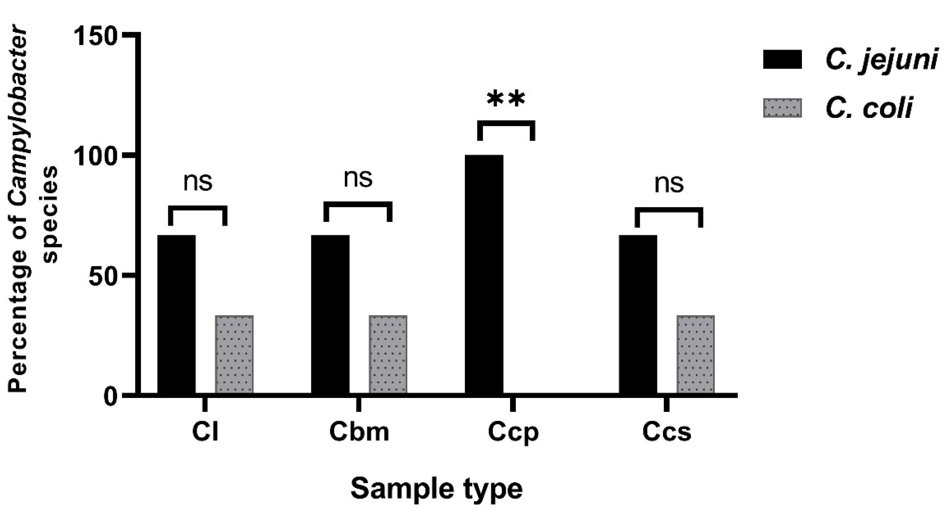
3.1. Prevalence of the Campylobacter Species in the Broiler Chickens in Sharkia Governorate, Egypt

According to the phenotypic identification results, a total of 101 (67.3%) campylobacter isolates were recovered from 150 various samples obtained from broiler chickens in Sharkia Governorate, Egypt. The prevalence of campylobacter isolates in the recovered specimens is presented in Table 4. Campylobacter spp. were highly distributed between chicken cloacal swabs (90%) and cecal parts (86.7%), while chicken luncheon meats were campylobacter negative (Table 4). Furthermore, C. jejuni was the most common Campylobacter spp. (54%), followed by C. coli (13.3%). Campylobacter jejuni was highly distributed among chicken cecal parts and cloacal swabs (70% each), followed by breast meats (66.7%). Meanwhile, C. coli isolates were more prevalent in the chicken livers and cloacal swabs (20% each), followed by cecal parts (16.7%) (Table 4). Additionally, there were statistically significant variations (p ˂ 0.001) in the incidences of Campylobacter spp., C. coli, and C. jejuni between the different sample origins.

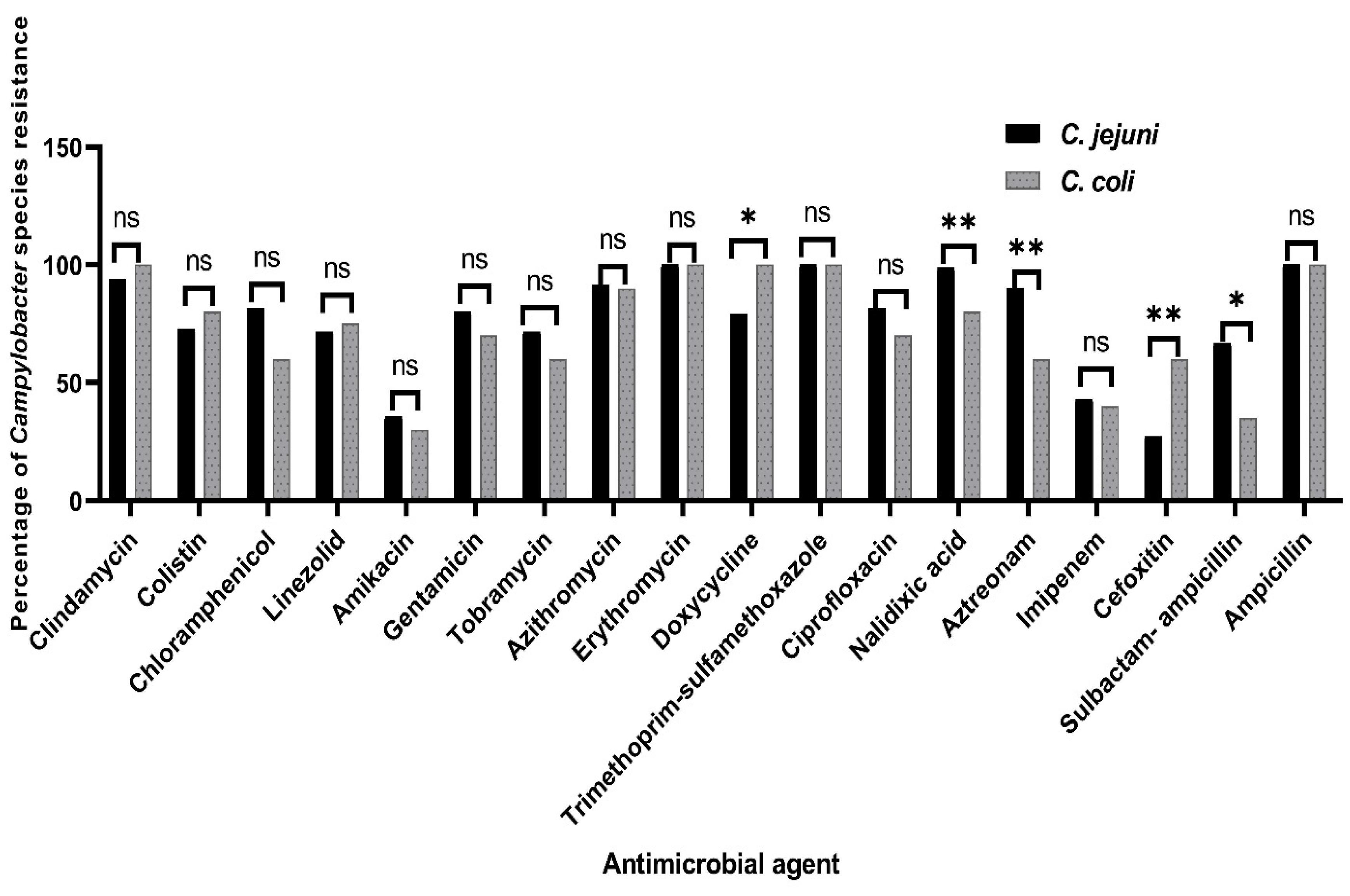
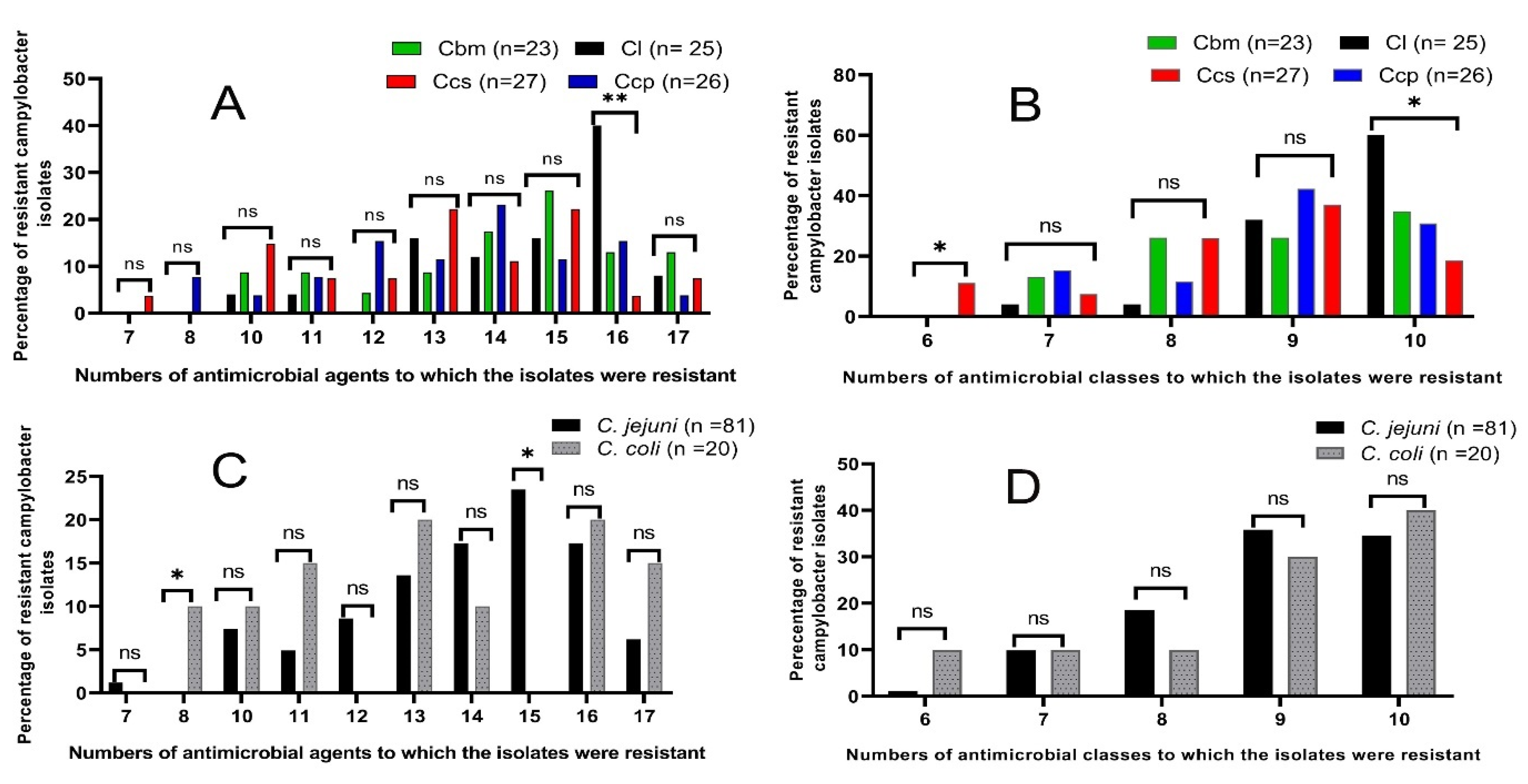
3.2. Antimicrobial Susceptibility Testing of the Campylobacter Species

All 101 recovered campylobacter isolates were examined for their susceptibility against the examined 18 antimicrobials, as presented in Table 5. Interestingly, the examined campylobacter isolates were completely resistant to trimethoprim/sulfamethoxazole erythromycin and ampicillin (100% each). Additionally, the majority of campylobacter isolates were resistant to clindamycin and nalidixic acid (95% each), followed by azithromycin (91.1%), aztreonam (84.2%), and doxycycline (83.2%). Meanwhile, the lowest resistance rates were detected against cefoxitin (33.7%), amikacin (34.7%), and imipenem (42.6%) (Table 5).
Regarding the isolates’ sources, the antimicrobial resistance patterns of the campylobacter isolates from various chicken sources were different (Table 5). It was observed that all the campylobacter isolates from the chicken liver and breast meat samples were 100% resistant to nalidixic acid, while the campylobacter isolates from the chicken cloacal swabs were 100% resistant to clindamycin. Additionally, statistical significance variations were observed in the resistance patterns between the campylobacter isolates from the different chicken sources to nalidixic acid, aztreonam, and cefoxitin (p = 0.03, 0.018, and 0.048, respectively). Additionally, there was a higher statistically significant difference in the linezolid resistance rates among the campylobacter isolates from the different chicken sources (p = 0.001) (Table 5). Meanwhile, there were no statistically significant differences (p ˃ 0.05) in the resistance percentages among the campylobacter isolates from the different chicken sources to the other examined antimicrobial agents.
According to the species level, our findings present that C. jejuni isolates are more resistant than C. coli isolates to the tested antimicrobial agents, except for clindamycin, colistin, linezolid, doxycycline, and cefoxitin (Figure 1). Moreover, statistical significance variations were detected in the resistance rates among the C. coli and C. jejuni isolates to doxycycline and sulbactam-ampicillin (p = 0.021 and 0.012, respectively). Additionally, higher statistical significance differences were observed in the resistance rates between the C. jejuni and C. coli isolates to cefoxitin, aztreonam, and nalidixic acid, (p = 0.008, 0.003, and 0.005, respectively). However, no statistical significance variations (p ˃ 0.05) were observed in the resistance patterns among the C. coli and C. jejuni isolates to the other investigated antimicrobials (Figure 1).
Interestingly, it was found that the C. jejuni isolates were more resistant to 8 and 9 antimicrobial categories (18.5 and 35.8%, respectively) than the C. coli isolates (10 and 30%, respectively). Meanwhile, the resistances to 6, 7, and 10 antimicrobial classes were higher in C. coli isolates (10, 10, and 40%, respectively) than the C. jejuni isolates (1.2%, 9.9%, and 34.6%, respectively). In total, the resistance rates to 10 antimicrobial classes were more prevalent among the campylobacter isolates from the chicken liver samples (60%). Meanwhile, the resistance patterns to 7 and 9 antimicrobials categories were more prevalent among the campylobacter isolates from the chicken cecal parts (15.3 and 42.3%, respectively). Moreover, the resistance proportions to the 8 antimicrobial classes were the highest among the campylobacter isolates from the chicken breast meats (26.1%) and those to the 6 classes were the highest among the campylobacter isolates from the chicken cloacal swabs (11.1%) (Figure 2).
Of note, our results reveal that 26 campylobacter isolates (25.7%) were XDR: 12 (48%), 6 (26.1%), 5 (19.2%), and 3 (11.1%) were obtained from chicken liver, breast meats, cecal parts, and cloacal swabs, respectively; meanwhile, 75 campylobacter isolates (74.3%) were recognized as MDR. Interestingly, our results present that all the tested campylobacter isolates showed an MAR index ≥0.39 (Table 6), which represent high-risk contamination sources, where the antimicrobials are frequently utilized. A total of 8 campylobacter isolates had MAR indices >0.9 (resistance to 17 antimicrobial agents); 2 (8%), 3 (13%), 1 (3.8%), and 2 (7.4%) were isolated from chicken liver, breast meats, cecal parts, and cloacal swabs, respectively (Table 6). Moreover, statistical significance variations were observed in the resistance profiles between the campylobacter isolates from various chicken sources to 16 antimicrobial agents (p = 0.006) and to 6 and 10 antimicrobial classes (p = 0.037 and 0.017, respectively) (Figure 2A). Additionally, statistical significance variations were observed in the resistance patterns among the C. jejuni and C. coli isolates to 8 and 15 antimicrobial agents (p = 0.038 and 0.024, respectively) (Figure 2B).

3.3. Molecular Grouping of the Campylobacter Species from Various Chicken Sources

A total of 26 XDR campylobacter isolates (resistant to 16 and 17 antimicrobials) were submitted for conventional PCR assays. Those isolates were recovered from the chicken liver (12), breast meats (6), cecal parts (5), and cloacal swabs (3). All the 26 screened campylobacter isolates (100%) were identified as genus Campylobacter by the PCR detection of the 23S rRNA gene. Additionally, 19 isolates (73.1%) and 7 isolates (26.9%) were positive for mapA and ceuE genes, respectively, and confirmed to be C. jejuni and C. coli, respectively. Of the 19 C. jejuni isolates, 8 (42.1%), 4 (21.1%), 5 (26.3%), and 2 (10.5%) were recovered from chicken liver, breast meats, cecal parts, and cloacal swabs, respectively. Moreover, 7 C. coli isolates were recovered from 4 (57.1%) chicken liver, 2 (28.6%) breast meat, and 1 (14.3%) cloacal swab samples. These findings were consistent with those of the phenotypic identification techniques. Additionally, statistically significant variations (p = 0.008) were observed in the incidences of the C. coli and C. jejuni isolates between the samples of the chicken cecal parts. Meanwhile, no statistical significance differences were observed in the incidence of the C. coli and C. jejuni isolates between the cloacal swabs, breast meat, and chicken liver samples (p = 1, 0.567, and 0.22, respectively) (Figure 3).

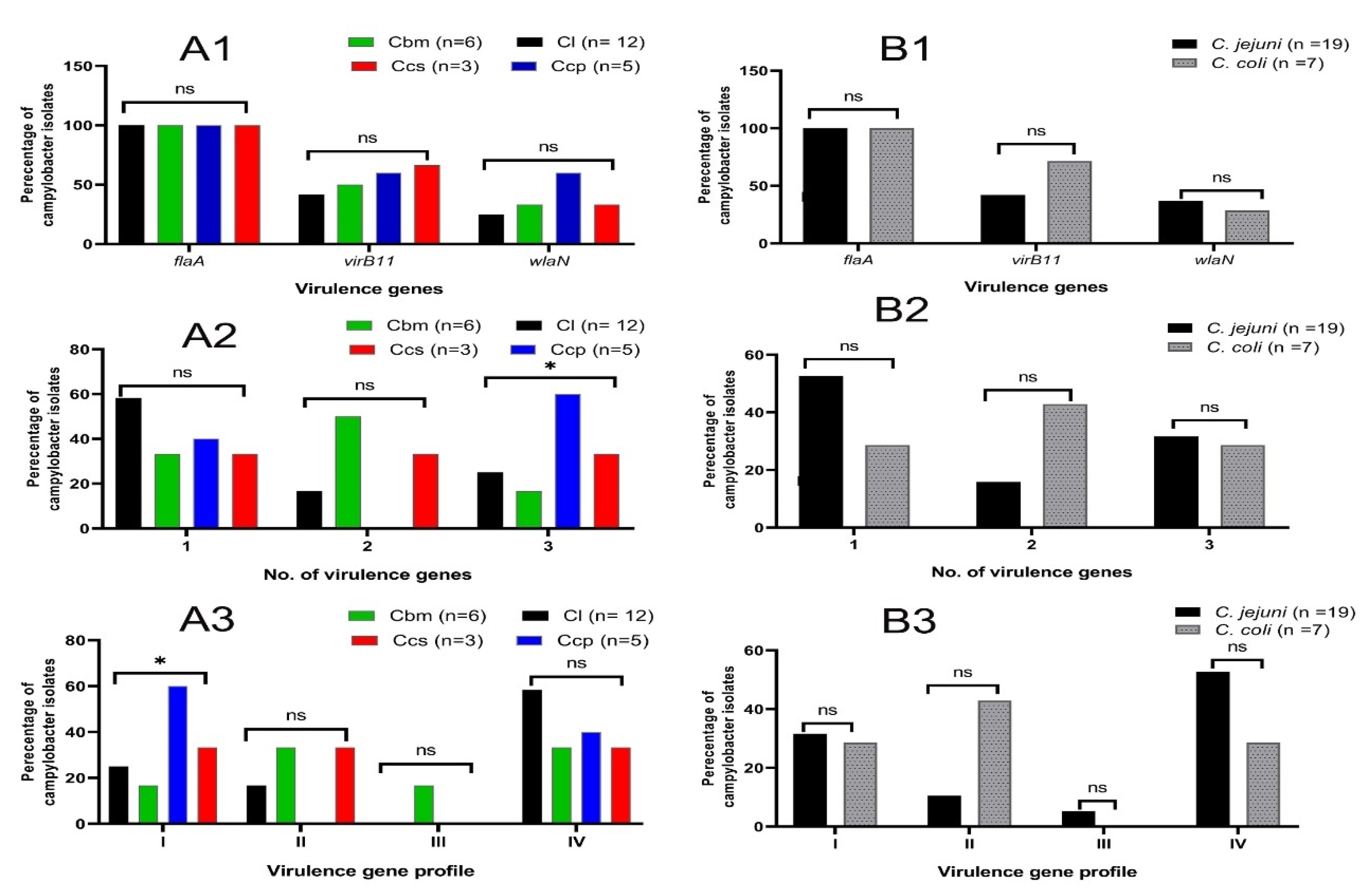
3.4. Molecular Investigation of the Virulence-Related Genes among the Investigated Isolates

All 26 molecularly confirmed campylobacter isolates were examined for the presence of three significant virulence genes, which have fundamental roles in the pathogenesis of Campylobacter spp. (flaA, virB11, and wlaN). Of the 26 examined isolates, 26 (100%) were positive for the flaA gene, 13 (50%) were positive for the virB11 gene, and 9 (34.6%) were positive for the wlaN gene. Of note, four virulence gene profiles were detected among the tested campylobacter isolates (Figure 4). A total of 12 XDR campylobacter isolates (46.2%) revealed the most frequent virulence gene profile (flaA).
Regarding the isolates’ sources, it was found that the virB11 gene was more prevalent among the campylobacter isolates from the chicken cloacal swabs (66.7%) and cecal parts (60%). Meanwhile, the wlaN gene was more prevalent among the campylobacter isolates from the chicken cecal parts (60%), followed by the cloacal swabs and breast meats (33.3% each). Moreover, 14 (53.8%) campylobacter isolates contained at least 2 virulence genes; 5 (41.7%), 4 (66.7%), 3 (60%), and 2 (66.7%) were recovered from the chicken liver, breast meats, cecal parts, and cloacal swabs, respectively. Additionally, 8 (30.8%) campylobacter isolates harbored 3 investigated virulence genes; 3 (25%), 1 (16.7%), 3 (60%), and 1 (33.3%) were recovered from the chicken liver, breast meats, cecal parts, and cloacal swabs, respectively. There was a statistically significant difference (p = 0.048) in the virulence profile I of the campylobacter isolates among the different chicken sources (Figure 4).
According to the spp. level, it was found that the virB11 gene was more prevalent among the C. coli isolates (71.4%), while the wlaN gene was more prevalent among the C. jejuni isolates (36.8%). Moreover, 5 C. coli isolates (71.4%) contained at least 2 virulence genes. Meanwhile, 6 C. jejuni isolates (31.6%) harbored 3 investigated virulence genes. Additionally, no statistical significance variations (p ˃ 0.05) were observed in the virulence gene profiles between the C. coli and C. jejuni isolates (Figure 4).

3.5. In Vivo Experimental Study

An identified XDR and multi-virulent C. jejuni strain, which was resistant to 17 tested antimicrobials, sensitive only to cefoxitin antibiotic and exhibited the virulence profile I (flaA, virB11, and wlaN), was used as a challenge strain in our in vivo experiment to assess the growth promoting, immunostimulant, antibacterial, anti-virulence, and activities of the eugenol and trans-cinnamaldehyde mixture.

3.5.1. Growth Performance

The growth performance variables throughout the experimental period are presented in Table 7. Significant differences were observed between the various experimental groups over the entire rearing period. The C. jejuni challenge significantly (p < 0.05) reduced the BW and BWG and increased the FI and FCR in the PC group, compared to the challenged groups treated with the mixture of eugenol and trans-cinnamaldehyde and cefoxitin. Interestingly, the FCR was most significantly improved (p < 0.05) in the NC and the mixture of eugenol and trans-cinnamaldehyde-treated groups, followed by the cefoxitin-treated group, with no significant differences between the NC and the mixture of eugenol and trans-cinnamaldehyde-treated groups. Meanwhile, chicks in the NC, cefoxitin, and the mixture of eugenol and trans-cinnamaldehyde-treated groups exhibited a significant (p < 0.05) increased BWG over the entire rearing period, unlike the PC group.

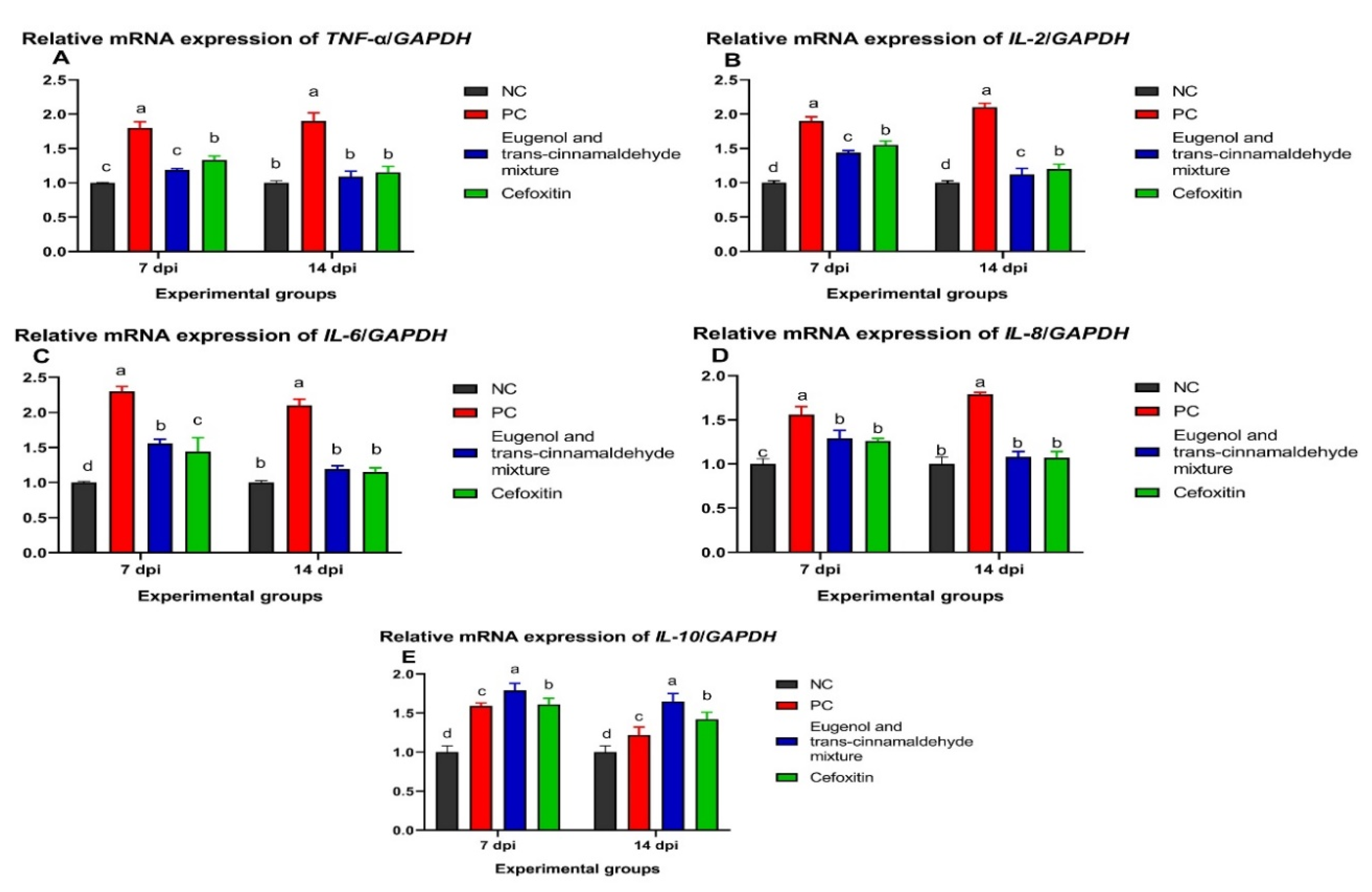
3.5.2. Analysis of Cytokines Genes Expression

The data of the cytokines genes expression analysis are shown in Figure 5. At 7 and 14 dpi, the relative expression levels of the TNF-α gene were significantly decreased (p < 0.05) in the mixture of the eugenol and trans-cinnamaldehyde-treated group, followed by the cefoxitin-treated group, in comparison to the PC group, and there was no significant difference between the NC and mixture of the eugenol and trans-cinnamaldehyde-treated groups. Moreover, the most marked reduction in the IL-2 expression level was observed in the mixture of the eugenol and trans-cinnamaldehyde-treated group, in respect to the PC. Meanwhile, the relative expression levels of the IL-6 and IL-8 genes were decreased in the cefoxitin-treated group, followed by the mixture of the eugenol and trans-cinnamaldehyde-treated group. Interestingly, no statistical significance variations were observed among the expression levels of the IL-6 and IL-8 genes among the NC, cefoxitin, and eugenol and trans-cinnamaldehyde mixture-treated groups at 14 dpi. Additionally, the IL-10 gene expression level was significantly upregulated (p < 0.05) in the group treated with the mixture of eugenol and trans-cinnamaldehyde, compared to the PC group, at both time points.

3.5.3. Expression Analysis of the C. jejuni Virulence Genes via the RT-qPCR Assay

The expression levels of the C. jejuni flaA, virB11, and wlaN virulence genes after treatment with the mixture of eugenol and trans-cinnamaldehyde and cefoxitin at 7 and 14 dpi are shown in Figure 6. The flaA, virB11, and wlaN mRNA expression levels were significantly (p < 0.05) downregulated in the cefoxitin, followed by the mixture of eugenol and trans-cinnamaldehyde-treated groups, compared to the PC group at both time points. Interestingly, no statistical significance variations were observed among the expression levels of C. jejuni flaA, virB11, the and wlaN genes between the cefoxitin- and mixture of eugenol and trans-cinnamaldehyde-treated groups at 14 dpi. Additionally, the flaA relative expression level was significantly (p < 0.05) decreased in the chicks treated with cefoxitin and the mixture of eugenol and trans-cinnamaldehyde at 7 dpi, with respect to the PC group, with no statistically significant differences between the latter groups.

3.5.4. Quantification of the C. jejuni DNA Copies

The quantification data of C. jejuni in the cecal contents of chicks are presented in Figure 7. The most significant (p < 0.05) reduction in C. jejuni loads was detected in the chicks treated with cefoxitin, followed by the mixture of eugenol and trans-cinnamaldehyde, compared to the PC group at 7 days post-challenge with XDR C. jejuni. Interestingly, no statistical significance variations were observed among the log10 copies of the C. jejuni populations in the cecal contents of the chicks treated with the mixture of eugenol and trans-cinnamaldehyde and cefoxitin at 14 dpi.

4. Discussion

It was stated that multiple worldwide crises were established as a result of the extensive spread of antimicrobial-resistant bacteria, including methicillin-resistant Staphylococcus aureus (MRSA), vancomycin-resistant Staphylococcus aureus (VRSA), Klebsiella spp., Mycoplasma spp., as well as zoonotic foodborne bacteria, such as Campylobacter spp., Salmonella Enteritidis, Salmonella Typhimurium, and E. coli [40,41,42,43,44,45,46,47,48]. Campylobacter spp., as commensal bacteria in the intestinal tract of domestic animals and chickens, are recognized as the primary sources of campylobacter infections in humans. Recently, Campylobacter spp. have become resistant to several antimicrobials, especially macrolides, FQ, and tetracyclines being important public health problems around the world. In the current work, we determined the high incidence of campylobacter isolates (67.3%) between different chicken specimens in Sharkia Governorate, Egypt. Our results are higher than those observed in previous studies carried out in Egypt, (7.6%) [49], (20.3%) [50], (26.9%) [51], and (32.8%) [7], meanwhile, these findings are consistent with the results of a recent study carried out in Kenya (69%) [52]. In the current work, Campylobacter spp. were more prevalent between the cloacal swabs (90%). These results are higher than those observed in India (71%) [53], Kenya (69%) [52], and Egypt (54.3%) [7]. In the present study, Campylobacter spp. were not detected in the chicken luncheon meats (0%), which is similar to the results of a previous study conducted in Egypt [54]. Additionally, C. jejuni was the most commonly recognized spp. (54%), which is consistent with the findings of previous studies carried out in Tunisia (68.9%) [55], and China; (20.3%) [56] and (12.3%) [12], respectively. Generally, the differences in the prevalence of Campylobacter spp. among the different studies might be correlated to the contamination and health conditions, climate factors, the geographical locations, the sources of tested samples, and the conventional identification methods [57].
Unsurprisingly, there are differences in the antibiotic resistance profiles between different countries, as a result of the variations in the prescribed antibiotics. Of note, our examined isolates showed complete resistance to ampicillin and erythromycin (100% each). These results are higher than those observed in other studies conducted in South Korea (57.4 and 14.9%, respectively) [58] and Pakistan (15 and 10%, respectively) [59]. Herein, high resistance levels to doxycycline and ciprofloxacin were observed among our examined isolates (83.2 and 79.2%, respectively); these results were lower than those reported in a previous study conducted in Tunisia (100 and 99.2%, respectively) [55]. Alarmingly, all global warnings are related to the emergence of MDR strains, but, recently, the XDR spread is taking concrete steps. In the current study, 74.3 and 25.7% of the examined campylobacter isolates were identified as MDR and XDR, respectively. These results are consistent with those detected in a recent study carried out in Egypt, where 28.5 and 69% of the tested isolates were MDR and XDR [54]. Herein, our examined isolates had MAR indices ≥ 0.39. These results were higher than those observed in another study conducted in South Korea, where all the tested isolates had MAR indices ≤ 0.2 [58]. In developing countries, the high resistance rates of campylobacters could result from the excessive uncontrolled utilization of antimicrobial agents in animal and human treatments without any prescription and as growth promoters in veterinary medicine. Moreover, our findings show alarmingly high resistance prevalence of doxycycline, erythromycin, and ciprofloxacin because they are the antibiotics of choice for the treatment of human campylobacter infection resulting in significant problems, where antimicrobial treatment becomes limited. Thus, the utilization of antimicrobials should be controlled in humans and animals [4]. Thus, it is significant to utilize medicinal plants, such as phytochemicals, as an alternative to the antimicrobials [60,61,62].
Out of the 26 molecularly investigated campylobacter isolates, 19 (73.1%) and 7 (26.9%) were positive for mapA and ceuE genes, respectively, being identified as C. jejuni and C. coli, respectively. Interestingly, there was a 100% correlation between the molecular and conventional identification results. These results were in agreement with a previous study conducted in Egypt, where 89.5% of the tested isolates were identified as C. jejuni, while the remaining isolates (10.5%) were identified as C. coli [63].
Herein, 50% of the examined campylobacter isolates were positive for the virB11 gene. These results are in contrast with the results of another study conducted in South Korea, where all the examined isolates were negative to the virB11 gene [64]. These differences in the prevalence of the virB11 gene may be attributed to the isolates’ sources and samples’ types.
Interestingly, our results reveal that all the examined campylobacter isolates (100%) are positive for the flaA gene. These findings are in complete agreement with a previous study conducted in Egypt [49], suggesting that the flaA gene has a significant virulence role in campylobacter isolates due to its significant part in the invasion, adhesion, and motility Campylobacter spp.
In the current work, the dietary supplementation of a mixture of eugenol and trans-cinnamaldehyde was able to modulate the immune responses of broiler chickens to overcome experimental C. jejuni infection and enhance their growth performance. In the present study, the mixture of eugenol and trans-cinnamaldehyde enhanced the overall growth rate and restored the decreased body weight and impaired the FCR post-infection with XDR C. jejuni, in comparison to the PC group. In agreement with our findings, a recent study reported the growth-promoting effect of carvacrol in broiler chickens challenged with C. jejuni [65]; however, the efficacy of the mixture of eugenol and trans-cinnamaldehyde on the broiler chickens’ performance after the experimental infection with the XDR C. jejuni strain has not been studied until now. Additionally, another study reported that broiler chicks fed with various levels of EOs exhibited an improved FCR and BWG after experimental infection, when compared to the PC group [66]. Moreover, the dietary supplementation of plant-active principles had better growth performance variables and C. jejuni load in both intestinal content and excreta [67]. According to previous studies, the positive effects of EOs on broiler chickens’ growth performance might result from the capability of their bioactive compounds to enhance their immune functions [68], stimulate their appetite and antimicrobial characteristics [69]. However, the EO extracts’ mode of actions are not yet defined and could vary as a result of the different forms, sources, and structures of the active components being utilized in diets [66].
Cytokines have significant regulatory roles in the inflammatory response of the intestinal tract. When the pathogens invade the gastrointestinal epithelial cells, the immune cells of the intestinal tract are triggered to produce cytokines that have fundamental roles in the host immune responses against microorganisms [70]. The TNF-α is a significant proinflammatory cytokine that regulates the host immune response against microorganisms via differentiating and proliferating the immune cells [71], but the excessive and prolonged secretion of proinflammatory cytokines might result in gastrointestinal damage [72]. Moreover, TNF-α, IL-2, IL-6, and IL-8 trigger an inflammatory response through recruiting the antimicrobial cells, including macrophages and neutrophils [71]. In the current study, the splenic proinflammatory cytokine (IL-2, IL-6, IL-8, and TNF-α) gene expression levels were upregulated in chicks that received a control basal diet and challenged experimentally with C. jejuni; however, the relative expression levels of IL-6 and IL-8 genes were significantly decreased in the cefoxitin-treated group, followed by the mixture of the eugenol and trans-cinnamaldehyde-supplemented group concerning the PC group. Additionally, supplementing our challenged birds with a mixture of eugenol and trans-cinnamaldehyde had regulatory effects on the proinflammatory cytokine genes expressions through a significant downregulation of TNF-α and IL-2 expression levels, which may counteract the inflammatory effects caused by C. jejuni, indicating their immunostimulant activities. This may result from the role of dietary EOs in improving the host non-specific immune response via the nonspecific killing of parasites, tumor cells, bacteria, and fungi, which lead to minimizing the microorganism loads [73]. Meanwhile, IL-10 has mainly opposed and complex effects on the immune and inflammatory responses via suppressing their actions [74]. In the current study, the highest expression level of the IL-10 gene was observed in the mixture of the eugenol and trans-cinnamaldehyde-supplemented group, which may minimize the excessive inflammatory response indicating their significant anti-inflammatory characteristics. In agreement with our results, recent studies showed the immunostimulant and anti-inflammatory effects of Eos via minimizing the expression levels of TNF-α [19], IL-2, and IL-6 [66] genes and decreasing that of the IL-10 gene [66] in broiler chicks challenged with Salmonella spp. due to their regulatory effects; however, the anti-inflammatory and immunostimulant effects of the mixture of eugenol and trans-cinnamaldehyde in broiler chickens challenged with the XDR C. jejuni strain has not yet been investigated.
Targeting the virulence factors of microorganisms through utilizing novel anti-virulence treatments is a promising alternative method, which could be utilized to disarm the bacterial microorganisms [60]. Following the previous statement, our data show that the mixture of eugenol and trans-cinnamaldehyde downregulated the expression levels of C. jejuni flaA, wlaN and virB11 virulence genes in challenged broilers concerning the PC group. In agreement with our results, recent studies reported the in vivo anti-virulence effects of EOs via suppressing the virulence genes’ expressions of Salmonella spp. in challenged broiler chicks [19]. Previous studies were continuously reporting the modulatory effects of EOs on C. jejuni virulence genes’ expressions in vitro [7,17,18]; however, to the best of our knowledge, there are no studies reporting the in vivo efficacy of the mixture of eugenol and trans-cinnamaldehyde on C. jejuni virulence in challenged broiler chickens.
Campylobacter spp. are commensal bacteria in the gastrointestinal tract of poultry and chickens, which considered the main reservoir for human campylobacter infections [1]. Herein, the quantitative analysis of cecal C. jejuni in challenged broilers showed that the dietary supplementation of a mixture of eugenol and trans-cinnamaldehyde significantly (p < 0.05) minimized C. jejuni populations 7 and 14 dpi, in comparison to the PC group. Our findings are in agreement with the results of previous studies, which demonstrate that the dietary supplementation of carvacrol significantly (p < 0.05) decreases C. jejuni counts in ceca [65], cloacal swabs, and the colon [75] of challenged broiler chickens, in comparison to the PC group. Additionally, a previous study reported a reduction in the cecal C. jejuni counts in challenged broilers after dietary supplementation with an EO mixture containing eugenol, indicating their inhibitory effects against C. jejuni [76]; however, the efficacy of the mixture of eugenol and trans-cinnamaldehyde on cecal C. jejuni loads in broiler chickens challenged with the XDR C. jejuni strain has not been explored to date.

5. Conclusions

The current work shows an alarming prevalence of avian XDR Campylobacter spp. in Egypt. The wlaN and virB11 genes were more prevalent among the examined campylobacter isolates from chicken cecal parts and cloacal swabs. Interestingly, our findings clearly prove the beneficial effects of a mixture of eugenol and trans-cinnamaldehyde on broiler chickens’ growth performance through decreasing the fecal load of C. jejuni and modulating the expression of proinflammatory and anti-inflammatory cytokines and decreasing the excessive inflammatory response. Furthermore, the administration of the mixture of eugenol and trans-cinnamaldehyde for broilers challenged with XDR C. jejuni had immunostimulant, anti-inflammatory, antimicrobial and anti-virulence effects. Therefore, our findings recommend the application of the mixture of eugenol and trans-cinnamaldehyde as an alternative to antimicrobials for the treatment and control of Campylobacter spp. infection in the veterinary fields, as well as for immunocompromised individuals.

Author Contributions

Conceptualization, A.A., M.I.A.E.-H., R.M.S.E.-M., W.R.E.-G., S.M.A.-R., A.M.I., A.M.A. and D.I.; methodology, A.A., M.I.A.E.-H., R.M.S.E.-M., W.R.E.-G., S.M.A.-R., A.M.I., A.M.A. and D.I.; software, A.A., M.I.A.E.-H., R.M.S.E.-M., W.R.E.-G., S.M.A.-R., A.M.I., A.M.A. and D.I.; validation, A.A., M.I.A.E.-H., R.M.S.E.-M., W.R.E.-G., S.M.A.-R., A.M.I., A.M.A. and D.I.; formal analysis, A.A., M.I.A.E.-H., R.M.S.E.-M., W.R.E.-G., S.M.A.-R., A.M.I., A.M.A. and D.I.; investigation, A.A., M.I.A.E.-H., R.M.S.E.-M., W.R.E.-G., S.M.A.-R., A.M.I., A.M.A. and D.I.; resources, A.A., M.I.A.E.-H., R.M.S.E.-M., W.R.E.-G., S.M.A.-R., A.M.I., A.M.A. and D.I.; data curation, A.A., M.I.A.E.-H., R.M.S.E.-M., W.R.E.-G., S.M.A.-R., A.M.I., A.M.A. and D.I.; writing—original draft preparation, A.A., M.I.A.E.-H., R.M.S.E.-M., W.R.E.-G., S.M.A.-R., A.M.I., A.M.A. and D.I.; writing—review and editing, A.A., M.I.A.E.-H., R.M.S.E.-M., W.R.E.-G., S.M.A.-R., A.M.I., A.M.A. and D.I.; visualization, A.A., M.I.A.E.-H., R.M.S.E.-M., W.R.E.-G., S.M.A.-R., A.M.I., A.M.A. and D.I.; supervision, A.A., M.I.A.E.-H., R.M.S.E.-M., W.R.E.-G., S.M.A.-R., A.M.I., A.M.A. and D.I.; project administration, A.A., M.I.A.E.-H., R.M.S.E.-M., W.R.E.-G., S.M.A.-R., A.M.I., A.M.A. and D.I.; funding acquisition, A.A., M.I.A.E.-H., R.M.S.E.-M., W.R.E.-G., S.M.A.-R., A.M.I., A.M.A. and D.I. All authors have read and agreed to the published version of the manuscript.

Funding

This study was supported by King Faisal University for Researchers Supporting under the research group support track (Grant No.1811022), Saudi Arabia.

Institutional Review Board Statement

The experimental protocols were conducted following the regulations and approved guidelines of the Institutional Animal Care and Use Committee of Faculty of Veterinary Medicine, Zagazig University, Egypt under the reference number of ZU-IACUC/2/F/263/2021.

Informed Consent Statement

The chicks in the experiment were brought by the authors, so informed consent is not needed.

Data Availability Statement

The data presented in this study are available on request from the corresponding author.

Acknowledgments

The authors acknowledge the Deanship of scientific research at King Faisal University for the financial support under the research group support track (grant No. 1811022), Saudi Arabia.

Conflicts of Interest

The authors declare no conflict of interest.

References

  1. Ammar, A.M.; El-Naenaeey, E.-S.Y.; Abd El-Hamid, M.I.; El-Gedawy, A.A.; El-Malt, R.M.S. Campylobacter as a Major Foodborne Pathogen: A Review of Its Characteristics, Pathogenesis, Antimicrobial Resistance and Control. J. Microbiol. Biotechnol. Food Sci. 2021, 10, 609–619. [Google Scholar]
  2. EFSA (European Food Safety Authority). Analysis of the baseline survey on the prevalence of Campylobacter in broiler batches and of Campylobacter and Salmonella on broiler carcasses, in the EU, 2008—Part B: Analysis of factors associated with Salmonella contamination of broiler carcasses. EFSA J. 2011, 9, 2017. [Google Scholar]
  3. EFSA (European Food Safety Authority). The European Union summary report on trends and sources of zoonoses, zoonotic agents and food-borne outbreaks in 2017. EFSA J. 2018, 16, e05500. [Google Scholar]
  4. Ammar, A.M.; El-Hamid, A.; Marwa, I.; El-Malt, R.M.S.; Azab, D.S.; Albogami, S.; Al-Sanea, M.M.; Soliman, W.E.; Ghoneim, M.M.; Bendary, M.M. Molecular detection of fluoroquinolone resistance among multidrug-, extensively drug-, and pan-drug-resistant Campylobacter species in Egypt. Antibiotics 2021, 10, 1342. [Google Scholar] [CrossRef]
  5. Same, R.G.; Tamma, P.D. Campylobacter infections in children. Pediatr. Rev. 2018, 39, 533–541. [Google Scholar] [CrossRef]
  6. Ugarte-Ruiz, M.; Domínguez, L.; Corcionivoschi, N.; Wren, B.W.; Dorrell, N.; Gundogdu, O. Exploring the oxidative, antimicrobial and genomic properties of Campylobacter jejuni strains isolated from poultry. Res. Vet. Sci. 2018, 119, 170–175. [Google Scholar] [CrossRef]
  7. Ammar, A.M.; El-Naenaeey, E.-S.Y.; El-Malt, R.M.S.; El-Gedawy, A.A.; Khalifa, E.; Elnahriry, S.S.; Abd El-Hamid, M.I. Prevalence, antimicrobial susceptibility, virulence and genotyping of Campylobacter jejuni with a special reference to the anti-virulence potential of Eugenol and beta-resorcylic acid on some multi-drug resistant isolates in Egypt. Animals 2021, 11, 3. [Google Scholar] [CrossRef]
  8. Silva, W.C.; Targino, B.N.; Gonçalves, A.G.; Silva, M.R.; Hungaro, H.M. Campylobacter: An important food safety issue. In Food Safety and Preservation; Elsevier: Amsterdam, The Netherlands, 2018; pp. 391–430. [Google Scholar]
  9. Luangtongkum, T.; Jeon, B.; Han, J.; Plummer, P.; Logue, C.M.; Zhang, Q. Antibiotic resistance in Campylobacter: Emergence, transmission and persistence. Future Med. 2009, 4, 189–200. [Google Scholar] [CrossRef] [Green Version]
  10. Wieczorek, K.; Denis, E.; Osek, J. Comparative analysis of antimicrobial resistance and genetic diversity of Campylobacter from broilers slaughtered in Poland. Int. J. Food Microbiol. 2015, 210, 24–32. [Google Scholar] [CrossRef]
  11. Bolton, D.J. Campylobacter virulence and survival factors. Food Microbiol. 2015, 48, 99–108. [Google Scholar] [CrossRef]
  12. Zhang, T.; Luo, Q.; Chen, Y.; Li, T.; Wen, G.; Zhang, R.; Luo, L.; Lu, Q.; Ai, D.; Wang, H. Molecular epidemiology, virulence determinants and antimicrobial resistance of Campylobacter spreading in retail chicken meat in Central China. Gut Pathog. 2016, 8, 48. [Google Scholar] [CrossRef] [PubMed] [Green Version]
  13. García-Sánchez, L.; Melero, B.; Rovira, J. Campylobacter in the food chain. Adv. Food Nutr. Res. 2018, 86, 215–252. [Google Scholar] [PubMed]
  14. Bhunia, A.K. Foodborne Microbial Pathogens: Mechanisms and Pathogenesis; Springer: Cham, Switzerland, 2018. [Google Scholar]
  15. García-Sánchez, L.; Melero, B.; Jaime, I.; Hänninen, M.-L.; Rossi, M.; Rovira, J. Campylobacter jejuni survival in a poultry processing plant environment. Food Microbiol. 2017, 65, 185–192. [Google Scholar] [CrossRef] [PubMed]
  16. Georgiev, M.; Beauvais, W.; Guitian, J. Effect of enhanced biosecurity and selected on-farm factors on Campylobacter colonization of chicken broilers. Epidemiol. Infect. 2017, 145, 553–567. [Google Scholar] [CrossRef] [Green Version]
  17. Upadhyay, A.; Arsi, K.; Wagle, B.R.; Upadhyaya, I.; Shrestha, S.; Donoghue, A.M.; Donoghue, D.J. Trans-cinnamaldehyde, carvacrol, and eugenol reduce Campylobacter jejuni colonization factors and expression of virulence genes in vitro. Front. Microbiol. 2017, 8, 713. [Google Scholar] [CrossRef] [Green Version]
  18. Wagle, B.; Shrestha, S.; Arsi, K.; Upadhyaya, I.; Donoghue, A.; Donoghue, D. Pectin or chitosan coating fortified with eugenol reduces Campylobacter jejuni on chicken wingettes and modulates expression of critical survival genes. Poult. Sci. 2019, 98, 1461–1471. [Google Scholar] [CrossRef]
  19. Bendary, M.M.; Ibrahim, D.; Mosbah, R.A.; Mosallam, F.; Hegazy, W.A.; Awad, N.F.; Alshareef, W.A.; Alomar, S.Y.; Zaitone, S.A.; Abd El-Hamid, M.I. Thymol Nanoemulsion: A New Therapeutic Option for Extensively Drug Resistant Foodborne Pathogens. Antibiotics 2021, 10, 25. [Google Scholar] [CrossRef]
  20. Ibrahim, D.; Ismail, T.A.; Khalifa, E.; El-Kader, A.; Shaimaa, A.; Mohamed, D.I.; Mohamed, D.T.; Shahin, S.E.; Abd El-Hamid, M.I. Supplementing Garlic Nanohydrogel Optimized Growth, Gastrointestinal Integrity and Economics and Ameliorated Necrotic Enteritis in Broiler Chickens Using a Clostridium perfringens Challenge Model. Animals 2021, 11, 2027. [Google Scholar] [CrossRef]
  21. FDA (Food and Drug Administration). Code of Federal Regulations Title 21 Part 172. Available online: https://www.accessdata.fda.gov/scripts/cdrh/cfdocs/cfCFR/CFRSearch.cfm?.CFRPart1/4172 (accessed on 22 July 2020).
  22. Yen, T.B.; Chang, S.T. Synergistic effects of cinnamaldehyde in combination with eugenol against wood decay fungi. Bioresour. Technol. 2008, 99, 232–236. [Google Scholar] [CrossRef]
  23. On, S. Identification methods for campylobacters, helicobacters, and related organisms. Clin. Microbiol. Rev. 1996, 9, 405–422. [Google Scholar] [CrossRef]
  24. Bauer, A. Antibiotic susceptibility testing by a standardized single disc method. Am. J. Clin. Pathol. 1966, 45, 149–158. [Google Scholar] [CrossRef]
  25. CLSI (Clinical and Laboratory Standards Institute). Performance Standards for Antimicrobial Susceptibility Testing, 27th ed.; CLSI supplement M100; CLSI: Wayne, PA, USA, 2017. [Google Scholar]
  26. Magiorakos, A.-P.; Srinivasan, A.; Carey, R.B.; Carmeli, Y.; Falagas, M.; Giske, C.; Harbarth, S.; Hindler, J.; Kahlmeter, G.; Olsson-Liljequist, B. Multidrug-resistant, extensively drug-resistant and pandrug-resistant bacteria: An international expert proposal for interim standard definitions for acquired resistance. Clin. Microbiol. Infect. 2012, 18, 268–281. [Google Scholar] [CrossRef] [PubMed] [Green Version]
  27. Sandhu, R.; Dahiya, S.; Sayal, P. Evaluation of multiple antibiotic resistance (MAR) index and Doxycycline susceptibility of Acinetobacter species among inpatients. Indian J. Microbiol. Res. 2016, 3, 299. [Google Scholar] [CrossRef]
  28. Wang, G.; Clark, C.G.; Taylor, T.M.; Pucknell, C.; Barton, C.; Price, L.; Woodward, D.L.; Rodgers, F.G. Colony multiplex PCR assay for identification and differentiation of Campylobacter jejuni, C. coli, C. lari, C. upsaliensis, and C. fetus subsp. fetus. J. Clin. Microbiol. 2002, 40, 4744–4747. [Google Scholar] [CrossRef] [Green Version]
  29. Shin, E.-J.; Lee, Y.-H. Comparison of three different methods for Campylobacter isolation from porcine intestines. J. Microbiol. Biotechnol. 2009, 19, 647–650. [Google Scholar] [PubMed]
  30. Datta, S.; Niwa, H.; Itoh, K. Prevalence of 11 pathogenic genes of Campylobacter jejuni by PCR in strains isolated from humans, poultry meat and broiler and bovine faeces. J. Med. Microbiol. 2003, 52, 345–348. [Google Scholar] [CrossRef] [PubMed]
  31. Sambrook, J.; Fritsch, E.; Maniatis, T. Molecular Cloning. A Laboratory Manual, 2nd ed.; Cold Spring Harbor Laboratory Press: New York, NY, USA, 1989. [Google Scholar]
  32. Talukder, K.A.; Aslam, M.; Islam, Z.; Azmi, I.J.; Dutta, D.K.; Hossain, S.; Nur-E-Kamal, A.; Nair, G.B.; Cravioto, A.; Sack, D.A. Prevalence of virulence genes and cytolethal distending toxin production in Campylobacter jejuni isolates from diarrheal patients in Bangladesh. J. Clin. Microbiol. 2008, 46, 1485–1488. [Google Scholar] [CrossRef] [Green Version]
  33. Aviagen, W. Ross 308: Broiler’s Management and Nutrition Specification; AOAC: Rockville, ML, USA, 2018. [Google Scholar]
  34. AOAC. Official Methods of Analysis of AOAC International; Association of Official Analytical Chemists: Rockville, ML, USA, 2012. [Google Scholar]
  35. Ibrahim, D.; Abdelfattah-Hassan, A.; Arisha, A.H.; Abd El-Aziz, R.M.; Sherief, W.R.; Adil, S.H.; El Sayed, R.; Metwally, A.E. Impact of feeding anaerobically fermented feed supplemented with acidifiers on its quality and growth performance, intestinal villi and enteric pathogens of mulard ducks. Livest. Sci. 2020, 242, 104299. [Google Scholar] [CrossRef]
  36. Al-Khalaifah, H.S.; Shahin, S.E.; Omar, A.E.; Mohammed, H.A.; Mahmoud, H.I.; Ibrahim, D. Effects of graded levels of microbial fermented or enzymatically treated dried brewer’s grains on growth, digestive and nutrient transporter genes expression and cost effectiveness in broiler chickens. BMC Vet. Res. 2020, 16, 424. [Google Scholar] [CrossRef]
  37. Ibrahim, D.; Moustafa, A.; Metwally, A.S.; Nassan, M.A.; Abdallah, K.; Eldemery, F.; Tufarelli, V.; Laudadio, V.; Kishawy, A.T. Potential Application of Cornelian Cherry Extract on Broiler Chickens: Growth, Expression of Antioxidant Biomarker and Glucose Transport Genes, and Oxidative Stability of Frozen Meat. Animals 2021, 11, 1038. [Google Scholar] [CrossRef]
  38. Ibrahim, D.; Moustafa, A.; Shahin, S.; Sherief, W.; Farag, M.; Nassan, M. Impact of fermented or enzymatically fermented dried olive pomace on growth, expression of digestive enzymes and glucose transporters genes, oxidative stability of frozen meat and economic efficiency of broiler chickens. Front. Vet. Sci. 2021, 8, 442. [Google Scholar] [CrossRef] [PubMed]
  39. Livak, K.J.; Schmittgen, T.D. Analysis of relative gene expression data using real-time quantitative PCR and the 2−ΔΔCT method. Methods 2001, 25, 402–408. [Google Scholar] [CrossRef] [PubMed]
  40. Bendary, M.; Solyman, S.; Azab, M.; Mahmoud, N.; Hanora, A. Genetic diversity of multidrug resistant Staphylococcus aureus isolated from clinical and non clinical samples in Egypt. Cell. Mol. Biol. 2016, 62, 55–61. [Google Scholar] [PubMed]
  41. Abd El-Hamid, M.I.; Bendary, M. Comparative phenotypic and genotypic discrimination of methicillin resistant and susceptible Staphylococcus aureus in Egypt. Cell. Mol. Biol. 2015, 61, 101–112. [Google Scholar]
  42. Abd El-Hamid, M.I.; Awad, N.F.; Hashem, Y.M.; Abdel-Rahman, M.A.; Abdelaziz, A.M.; Mohammed, I.A.; Abo-Shama, U.H. In vitro evaluation of various antimicrobials against field Mycoplasma gallisepticum and Mycoplasma synoviae isolates in Egypt. Poult. Sci. 2019, 98, 6281–6288. [Google Scholar] [CrossRef]
  43. Abd El-Aziz, N.K.; Abd El-Hamid, M.I.; Bendary, M.M.; El-Azazy, A.A.; Ammar, A.M.; Abd El-Aziz, N.K.; Abd El-Hamid, M.I.; Bendary, M.M.; El-Azazy, A.A.; Ammar, A.M. Existence of vancomycin resistance among methicillin resistant S. aureus recovered from animal and human sources in Egypt. Slov. Vet. Res. 2018, 55, 221–230. [Google Scholar]
  44. Ammar, A.M.; Attia, A.M.; Abd El-Hamid, M.I.; El-Shorbagy, I.M.; Abd El-Kader, S.A. Genetic basis of resistance waves among methicillin resistant Staphylococcus aureus isolates recovered from milk and meat products in Egypt. Cell. Mol. Biol. 2016, 62, 7–15. [Google Scholar]
  45. Abd El-Hamid, M.I.; Bendary, M.M.; Merwad, A.M.A.; Elsohaby, I.; Mohammad Ghaith, D.; Alshareef, W.A. What is behind phylogenetic analysis of hospital-, community- and livestock-associated methicillin-resistant Staphylococcus aureus? Transbound. Emerg. Dis. 2019, 66, 1506–1517. [Google Scholar]
  46. Awad, N.F.S.; Abd El-Hamid, M.I.; Hashem, Y.M.; Erfan, A.M.; Abdelrahman, B.A.; Mahmoud, H.I. Impact of single and mixed infections with Escherichia coli and Mycoplasma gallisepticum on Newcastle disease virus vaccine performance in broiler chickens: An in vivo perspective. J. Appl. Microbiol. 2019, 127, 396–405. [Google Scholar] [CrossRef]
  47. Ammar, A.M.; Attia, A.M.; Abd El-Aziz, N.K.; Abd El Hamid, M.I.; El-Demerdash, A.S. Class 1 integron and associated gene cassettes mediating multiple-drug resistance in some food borne pathogens. Int. Food Res. J. 2016, 23, 332–339. [Google Scholar]
  48. Ammar, A.M.; Abd El-Hamid, M.I.; Eid, S.E.A.; El Oksh, A.S. Insights into antimicrobial resistance and virulence genes of emergent multidrug resistant avian pathogenic Escherichia coli in Egypt: How closely related are they? Rev. Med. Vet. 2015, 166, 304–314. [Google Scholar]
  49. Abd El-Hamid, M.I.; Abd El-Aziz, N.K.; Samir, M.; El-Naenaeey, E.-S.Y.; Abo Remela, E.M.; Mosbah, R.A.; Bendary, M.M. Genetic diversity of Campylobacter jejuni isolated from avian and human sources in Egypt. Front. Microbiol. 2019, 10, 2353. [Google Scholar] [CrossRef] [PubMed]
  50. Awadallah, M.; Ahmed, H.; El-Gedawy, A.; Saad, A. Molecular identification of C. jejuni and C. coli in chicken and humans, at Zagazig, Egypt, with reference to the survival of C. jejuni in chicken meat at refrigeration and freezing temperatures. Int. Food Res. J. 2014, 21, 1801. [Google Scholar]
  51. Khalifa, N.O.; Afify, J.S.; Rabie, N.S. Zoonotic and molecular characterizations of Campylobacter jejuni and Campylobacter coli isolated from beef cattle and children. Glob. Vet. 2013, 11, 585–591. [Google Scholar]
  52. Nguyen, T.N.M.; Hotzel, H.; Njeru, J.; Mwituria, J.; El-Adawy, H.; Tomaso, H.; Neubauer, H.; Hafez, H.M. Antimicrobial resistance of Campylobacter isolates from small scale and backyard chicken in Kenya. Gut Pathog. 2016, 8, 39. [Google Scholar] [CrossRef] [Green Version]
  53. Begum, S.; Sekar, M.; Gunaseelan, L.; Gawande, M.; Suganya, G.; Malar, P.A.S.; Karthikeyan, A. Molecular identification of Campylobacter jejuni and coli from chicken, calves and dogs to determine its potential threat on human being. Vet. World. 2015, 8, 1420–1423. [Google Scholar] [CrossRef] [Green Version]
  54. El-Aziz, A.; Norhan, K.; Ammar, A.M.; Hamdy, M.M.; Gobouri, A.A.; Azab, E.; Sewid, A.H. First report of aacC5-aadA7Δ4 Gene Cassette array and phage tail tape measure protein on Class 1 Integrons of Campylobacter Species isolated from animal and human sources in Egypt. Animals 2020, 10, 2067. [Google Scholar] [CrossRef]
  55. Gharbi, M.; Béjaoui, A.; Ben Hamda, C.; Jouini, A.; Ghedira, K.; Zrelli, C.; Hamrouni, S.; Aouadhi, C.; Bessoussa, G.; Ghram, A. Prevalence and antibiotic resistance patterns of Campylobacter spp. isolated from broiler chickens in the North of Tunisia. BioMed Res. Int. 2018, 2018, 7943786. [Google Scholar] [CrossRef] [Green Version]
  56. Zhang, T.; Dong, J.; Cheng, Y.; Lu, Q.; Luo, Q.; Wen, G.; Liu, G.; Shao, H. Genotypic diversity, antimicrobial resistance and biofilm-forming abilities of Campylobacter isolated from chicken in Central China. Gut Pathog. 2017, 9, 62. [Google Scholar] [CrossRef] [Green Version]
  57. Chatur, Y.A.; Brahmbhatt, M.N.; Modi, S.; Nayak, J.B. Fluoroquinolone resistance and detection of topoisomerase gene mutation in Campylobacter jejuni isolated from animal and human sources. Int. J. Curr. Microbiol. App. Sci 2014, 3, 773–783. [Google Scholar]
  58. Wei, B.; Cha, S.-Y.; Yoon, R.-H.; Kang, M.; Roh, J.-H.; Seo, H.-S.; Lee, J.-A.; Jang, H.-K. Prevalence and antimicrobial resistance of Campylobacter spp. isolated from retail chicken and duck meat in South Korea. Food Control 2016, 62, 63–68. [Google Scholar] [CrossRef]
  59. Samad, A.; Abbas, F.; Ahmed, Z.; Akbar, A.; Naeem, M.; Sadiq, M.B.; Ali, I.; Bugti, F.S.; Achakzai, S.K. Prevalence, antimicrobial susceptibility, and virulence of Campylobacter jejuni isolated from chicken meat. J. Food Saf. 2019, 39, e12600. [Google Scholar] [CrossRef]
  60. Abd El-Hamid, M.I.; El-Sayed, M.; Ali, A.R.; Abdallah, H.; Arnaout, M.I.; El-Mowalid, G.A. Marjoram extract down-regulates the expression of Pasteurella multocida adhesion, colonization and toxin genes: A potential mechanism for its antimicrobial activity. Comp. Immunol. Microbiol. Infect. Dis. 2019, 62, 101–108. [Google Scholar] [CrossRef] [PubMed]
  61. El-Hamid, A.; Marwa, I.; El-Naenaeey, E.-S.Y.; Hegazy, W.A.; Mosbah, R.A.; Nassar, M.S.; Bakhrebah, M.A.; Abdulaal, W.H.; Alhakamy, N.A.; Bendary, M.M. Promising antibiofilm agents: Recent breakthrough against biofilm producing methicillin-resistant Staphylococcus aureus. Antibiotics 2020, 9, 667. [Google Scholar] [CrossRef]
  62. Elmowalid, G.A.; Abd El-Hamid, M.I.; Abd El-Wahab, A.M.; Atta, M.; Abd El-Naser, G.; Attia, A.M. Garlic and ginger extracts modulated broiler chicks innate immune responses and enhanced multidrug resistant Escherichia coli O78 clearance. Comp. Immunol. Microbiol. Infect. Dis. 2019, 66, 101334. [Google Scholar] [CrossRef]
  63. Girgis, S.A.; Rashad, S.S.; Othman, H.B.; Bassim, H.H.; Kassem, N.N.; El-Sayed, F.M. Multiplex PCR for identification and differentiation of Campylobacter species and their antimicrobial susceptibility pattern in Egyptian patients. Int. J. Curr. Microbiol. Appl. Sci. 2014, 3, 861–875. [Google Scholar]
  64. Oh, J.-Y.; Kwon, Y.-K.; Wei, B.; Jang, H.-K.; Lim, S.-K.; Kim, C.-H.; Jung, S.-C.; Kang, M.-S. Epidemiological relationships of Campylobacter jejuni strains isolated from humans and chickens in South Korea. J. Microbiol. 2017, 55, 13–20. [Google Scholar] [CrossRef]
  65. Kelly, C.; Gundogdu, O.; Pircalabioru, G.; Cean, A.; Scates, P.; Linton, M.; Pinkerton, L.; Magowan, E.; Stef, L.; Simiz, E. The in vitro and in vivo effect of carvacrol in preventing Campylobacter infection, colonization and in improving productivity of chicken broilers. Foodborne Pathog. Dis. 2017, 14, 341–349. [Google Scholar] [CrossRef]
  66. Ibrahim, D.; Abdelfattah-Hassan, A.; Badawi, M.; Ismail, T.A.; Bendary, M.M.; Abdelaziz, A.M.; Mosbah, R.A.; Mohamed, D.I.; Arisha, A.H.; Abd El-Hamid, M.I. Thymol nanoemulsion promoted broiler chicken’s growth, gastrointestinal barrier and bacterial community and conferred protection against Salmonella Typhimurium. Sci. Rep. 2021, 11, 7742. [Google Scholar] [CrossRef]
  67. Ibrahim, D.; Sewid, A.H.; Arisha, A.H.; Abd El-Fattah, A.H.; Abdelaziz, A.M.; Al-Jabr, O.A.; Kishawy, A.T. Influence of Glycyrrhiza glabra Extract on Growth, Gene Expression of Gut Integrity, and Campylobacter jejuni Colonization in Broiler Chickens. Front. Vet. Sci. 2020, 7, 612063. [Google Scholar] [CrossRef]
  68. Chowdhury, S.; Mandal, G.P.; Patra, A.K.; Kumar, P.; Samanta, I.; Pradhan, S.; Samanta, A.K. Different essential oils in diets of broiler chickens: 2. Gut microbes and morphology, immune response, and some blood profile and antioxidant enzymes. Anim. Feed Sci. Technol. 2018, 236, 39–47. [Google Scholar] [CrossRef]
  69. Windisch, W.; Schedle, K.; Plitzner, C.; Kroismayr, A. Use of phytogenic products as feed additives for swine and poultry. J. Anim. Sci. 2008, 86, E140–E148. [Google Scholar] [CrossRef] [PubMed]
  70. Kayamuro, H.; Yoshioka, Y.; Abe, Y.; Arita, S.; Katayama, K.; Nomura, T.; Yoshikawa, T.; Kubota-Koketsu, R.; Ikuta, K.; Okamoto, S. Interleukin-1 family cytokines as mucosal vaccine adjuvants for induction of protective immunity against influenza virus. J. Virol. 2010, 84, 12703–12712. [Google Scholar] [CrossRef] [PubMed] [Green Version]
  71. Wang, Y.-G.; Fang, W.-L.; Wei, J.; Wang, T.; Wang, N.; Ma, J.-L.; Shi, M. The involvement of NLRX1 and NLRP3 in the development of nonalcoholic steatohepatitis in mice. J. Chin. Med. Assoc. 2013, 76, 686–692. [Google Scholar] [CrossRef] [Green Version]
  72. Lee, S.H.; Lillehoj, H.S.; Jang, S.I.; Lillehoj, E.P.; Min, W.; Bravo, D.M. Dietary supplementation of young broiler chickens with Capsicum and turmeric oleoresins increases resistance to necrotic enteritis. Br. J. Nutr. 2013, 110, 840–847. [Google Scholar] [CrossRef] [PubMed] [Green Version]
  73. Khatun, J.; Loh, T.C.; Foo, H.L.; Akit, H.; Khan, K.I. Growth performance, cytokine expression, and immune responses of broiler chickens fed a dietary palm oil and sunflower oil blend supplemented with L-Arginine and varying concentrations of vitamin E. Front. Vet. Sci. 2020, 7, 619. [Google Scholar] [CrossRef]
  74. Wei, X.; Zhang, Y.; Zhou, H.; Tian, F.; Ni, Y. Antimicrobial activities and in vitro properties of cold-adapted Lactobacillus strains isolated from the intestinal tract of cold water fishes of high latitude water areas in Xinjiang, China. BMC Microbiol. 2019, 19, 247. [Google Scholar] [CrossRef] [Green Version]
  75. Szott, V.; Reichelt, B.; Alter, T.; Friese, A.; Roesler, U. In vivo efficacy of carvacrol on Campylobacter jejuni prevalence in broiler chickens during an entire fattening period. Eur. J. Microbiol. Immunol. 2020, 10, 131–138. [Google Scholar] [CrossRef]
  76. Grilli, E.; Vitari, F.; Domeneghini, C.; Palmonari, A.; Tosi, G.; Fantinati, P.; Massi, P.; Piva, A. Development of a feed additive to reduce caecal Campylobacter jejuni in broilers at slaughter age: From in vitro to in vivo, a proof of concept. J. Appl. Microbiol. 2013, 114, 308–317. [Google Scholar] [CrossRef]
Figure 1. Prevalence of Campylobacter jejuni and C. coli resistance against 18 antimicrobial agents. ns: non-significant, * p < 0.05, ** p < 0.01.
Animals 12 00905 g001
Figure 2. Antimicrobial resistance patterns of the total campylobacter isolates from different chicken sources (A,B) and C. jejuni and C. coli (C,D). Cl: chicken liver; Cbm: chicken breast meats; Ccp: chicken cecal parts; Ccs: chicken cloacal swabs, n: number, ns: non-significant, * p < 0.05, ** p < 0.01.
Animals 12 00905 g002
Figure 3. Prevalence of molecularly identified Campylobacter species in various chicken samples. Cl: chicken liver; Cbm: chicken breast meats; Ccp: chicken cecal parts; Ccs: chicken cloacal swabs; ns: non-significant; ** p < 0.01. The prevalence rate for each species was calculated regarding the total number of the examined samples.
Animals 12 00905 g003
Figure 4. Distribution of the virulence genes among the total campylobacter isolates from the different chicken sources (A1A3) and C. jejuni and C. coli (B1B3). Cl: chicken liver; Cbm: chicken breast meats; Ccp: chicken cecal parts; Ccs: chicken cloacal swabs, n: number; ns: non-significant; * p < 0.05; virulence gene profiles; I: flaA, virB11, and wlaN, II: flaA and virB11, III: flaA and wlaN, and IV: flaA.
Animals 12 00905 g004
Figure 5. Relative mRNA expression levels of tumor necrosis factor-alpha (TNF-α; (A)); interleukin-2 (IL-2; (B)); IL-6 (C), IL-8 (D), and IL-10 (E) in the spleen of chicks supplemented with the mixture of eugenol and trans-cinnamaldehyde and treated with cefoxitin antibiotic at 7 and 14 days post-infection (dpi) with XDR C. jejuni. Error bars represent the standard error of the mean (SEM). NC (negative control): chicks fed a basal diet and not challenged; PC (positive control): chicks fed a basal diet and challenged with XDR C. jejuni; eugenol and trans-cinnamaldehyde mixture: chicks fed the basal diet supplemented with a mixture of eugenol and trans-cinnamaldehyde; and cefoxitin: chicks fed a basal diet and treated with a cefoxitin antibiotic. All groups, except the NC group, were challenged with XDR C. jejuni at 23 days of age. a–d Means of the columns with different letters indicate a statistically significant difference (p < 0.05).
Animals 12 00905 g005
Figure 6. Relative mRNA expression levels of the C. jejuni flaA (A), virB11 (B), and wlaN (C) genes in response to supplementing the mixture of eugenol and trans-cinnamaldehyde and treatment with cefoxitin antibiotic at 7 and 14 days post-infection (dpi) with XDR C. jejuni, as were determined via the RT-qPCR assay. The results are expressed as the means ± standard error of the mean (SEM, error bars). PC (positive control): chicks fed a basal diet and challenged with XDR C. jejuni; eugenol and trans-cinnamaldehyde mixture: chicks fed a basal diet supplemented with a mixture of eugenol and trans-cinnamaldehyde; and cefoxitin: chicks fed a basal diet and treated with cefoxitin antibiotic. All the groups, except the NC group, were challenged with XDR C. jejuni at 23 days of age. a–c Means of the columns with different letters indicate a statistically significant difference (p < 0.05).
Animals 12 00905 g006
Figure 7. Quantification of the fecal Campylobacter jejuni loads (log10 CFU) in response to supplementing the mixture of eugenol and trans-cinnamaldehyde and treatment with cefoxitin antibiotic at 7 days (7 dpi) and 14 days post-infection (14 dpi) with XDR C. jejuni, as were determined via the qPCR assay. The results are expressed as the means ± standard error of the mean (SEM, error bars). PC (positive control): chicks fed a basal diet and challenged with XDR C. jejuni; eugenol and trans-cinnamaldehyde mixture: chicks fed a basal diet supplemented with a mixture of eugenol and trans-cinnamaldehyde; and cefoxitin: chicks fed a basal diet supplemented with a cefoxitin antibiotic. All groups, except the NC group, were challenged with XDR C. jejuni at 23 days of age. a–c Means of the columns with different letters indicate a statistically significant difference (p < 0.05).
Animals 12 00905 g007
Table 1. Sequences of the oligonucleotide primers targeting six genes of the Campylobacter species and their respective amplified PCR products.
Specificity (Target Gene)Primer Sequence (5’-3’)Amplified Product (bp)Denaturation TemperatureReferences
Genus Campylobacter (23S rRNA)F: TATACCGGTAAGGAGTGCTGGAG65095 °C[28,32]
R: ATCAATTAACCTTCGAGCACCG
Campylobacter jejuni (mapA)F: CTATTTTATTTTTGAGTGCTTGTG58995 °C[29,33]
R: GCTTTATTTGCCATTTGTTTTATTA
Campylobacter coli (ceuE)F: AATTGA AAATTG CTCCAACTATG46295 °C[29,33]
R: TGATTT TATTATTTGTAGCAGCG
FlaA (flaA)F: AATAAAAATGCTGATAAAACAGGTG85594 °C[30,34]
R: TACCGAACCAATGTCTGCTCTGATT
VirB (virB11)F: TCTTGTGAGTTGCCTTACCCCTTTT49494 °C[30,34]
R: CCTGCGTGTCCTGTGTTATTTACCC
WlaN (wlaN)F: TTAAGAGCAAGATATGAAGGTG67294 °C[32,35]
R: CCATTTGAATTGATATTTTTG
Table 2. Ingredients and nutrient composition of the basal diet.
Ingredient (%)Starter
(Days 1–10)
Grower
(Days 11–20)
Finisher
(Days 21–37)
Yellow corn60.1064.0065.70
Soybean oil1.572.504.20
Soybean meal34.529.325.90
Common salt0.30.30.3
Calcium diphasic phosphate1.21.21.2
Calcium carbonate1.11.11.1
Premix *0.80.80.8
Choline chloride0.20.20.2
DL-methionine 99%0.170.170.17
L-lysine HCL 78%0.330.330.33
Anti-mycotoxin0.10.10.1
Calculated composition
ME (kcal/kg)300131003222
CF (%)2.642.502.46
EE (%)4.075.146.83
CP (%)23.0121.0219.57
Available P (%)0.490.440.42
Ca (%)1.021.041.03
Methionine (%)0.5000.440.46
Lysine (%)1.411.281.18
* Vitamin premix supplied per kilogram of diet: Mn (sulphate and oxide), 100 mg; Zn (sulphate and oxide), 120 mg; I (iodide), 1.2 mg; Se (selenate), 0.28 mg; Cu (sulphate), 14 mg; Fe (sulphate), 30 mg; cyanocobalamin, 15 μg; biotin, 300 μg; pyridoxine, 6 mg; folate, 3 mg; niacin, 50 mg; pantothenate, 12 mg; thiamine, 4 mg; riboflavin, 7 mg; menadione, 3.5 mg; cholecalciferol, 6500 IU; tocopherol acetate, 75 mg; retinol, 12.000 IU; ME: metabolizable energy; CF: crude fiber; EE: ether extract; CP: crude protein; P: phosphorus; and Ca: calcium.
Table 3. Primer sequences of the cytokine-related genes used in the reverse transcription quantitative real-time PCR assay.
Target GenePrimer Sequence (5’-3’)Accession No.
TNF-αF: CTGCACTTCAGGGTGATCGXM_008262537.2
R: CTACGTGGGCTAGAGGCTTG
IL-2F: GCTTATGGAGCATCTCTATCATCAXM_015276098.2
R: GGTGCACTCCTGGGTCTC
IL-6F: GCCAACCCTACAACAAGANC_013678
R: AGAGCCACAACGACTGAC
IL-8F: CTCTCTTGGCAACCTTCCTGKT216053.1
R: TTGCACAGTGAGGTCCACTC
IL-10F: AAAAGCTAAAAGCCCCAGGANM001082045.1
R: CGGGAGCTGAGGTATCAGAG
GAPDHF: TGTTTGTGATGGGCGTGAANC_013676.1
R: CCTCCACAATGCCGAAGT
TNF-α: tumor necrosis factor-alpha; IL: interleukin; and GAPDH: glyceraldehyde 3-phosphate dehydrogenase.
Table 4. Prevalence of Campylobacter species in various chicken samples in Sharkia Governorate, Egypt.
Sample Type (Symbol, No.)Total No. of Campylobacter Isolates (%) *No. of Campylobacter spp. (%) *
C. jejuniC. coli
Chicken luncheon meats (Clm, 30)000
Chicken livers (Cl, 30)25 (83.3)19 (63.3)6 (20)
Chicken breast meats (Cbm, 30) 23 (76.7)20 (66.7)3 (10)
Chicken cecal parts (Ccp, 30)26 (86.7)21 (70)5 (16.7)
Chicken cloacal swabs (Ccs, 30)27 (90)21 (70)6 (20)
Total (150)101 (67.3)81 (54)20 (13.3)
* The isolation rates were calculated regarding the total number of the examined samples.
Table 5. Antimicrobial resistance patterns of the Campylobacter species from various chicken sources.
Antimicrobial
Class
Antimicrobial AgentNo. of Resistant Campylobacter Isolates from Various Chicken Sources (%)Total No. of Campylobacter Isolates (%)
(n = 101)
p-Value
Liver
(n = 25)
Breast Meats (n = 23)Cecal Parts (n = 26)Cloacal Swabs (n = 27)
LincosamideClindamycin24 (96)20 (87)25 (96.2)27 (100)96 (95)0.18
PolypeptidesColistin19 (76)17 (73.9)22 (84.6)17 (63)75 (74.3)0.348
PhenicolsChloramphenicol24 (96)16 (69.6)18 (69.2)20 (74.1)78 (77.2)0.076
OxazolidonesLinezolid23 (92)17 (73.9)21 (80.8)12 (44.4)73 (72.3)0.001 **
AminoglycosidesAmikacin9 (36)9 (39.1)7 (26.9)10 (37)35 (34.7)0.821
Gentamicin21 (84)21 (91.3)16 (61.5)21 (77.8)79 (78.2)0.07
Tobramycin19 (76)15 (65.2)20 (76.9)16 (59.3)70 (69.3)0.458
MacrolidesAzithromycin23 (92)22 (95.7)23 (88.5)24 (88.9)92 (91.1)0.871
Erythromycin25 (100)23 (100)26 (100)27 (100)101 (100)NA
TetracyclinesDoxycycline23 (92)18 (78.3)20 (76.9)23 (85.2)84 (83.2)0.472
SulphonamidesTrimethoprim-sulfamethoxazole25 (100)23 (100)26 (100)27 (100)101 (100)NA
QuinolonesCiprofloxacin20 (80)19 (82.6)21 (80.8)20 (74.1)80 (79.2)0.903
Nalidixic acid25 (100)23 (100)23 (88.5)25 (92.6)96 (95)0.04 *
Beta-lactamsAztreonam18 (72)21 (91.3)21 (80.8)25 (92.6)85 (84.2)0.015 *
Imipenem14 (56)11 (47.8)7 (26.9)11 (40.7)43 (42.6)0.195
Cefoxitin13 (52)8 (34.8)8 (30.8)5 (18.5)34 (33.7)0.047 *
Sulbactam-ampicillin18 (72)17 (73.9)13 (50)13 (48.1)61 (60.4)0.114
Ampicillin25 (100)23 (100)26 (100)27 (100)101 (100)NA
n: number, * and ** represent the statistically significant differences in the resistance profiles among the campylobacter isolates from different chicken sources to the indicated antimicrobial agent using a chi-squared test; * p < 0.05, ** p < 0.01, NA: non-applicable.
Table 6. Multiple antibiotic resistance indices and the frequency of resistance of the campylobacter isolates from different chicken sources.
MAR IndexNo. of Antimicrobials to Which the Isolates Were Resistant *No. of AMCNo. of Resistant Campylobacter Isolates from Different Chicken Sources (%)Total No. of Campylobacter Isolates (%)
(n = 101)
The Character of Resistant Strains
Liver
(n = 25)
Breast Meats (n = 23)Cecal
Parts
(n = 26)
Cloacal Swabs (n = 27)
0.3977---1 (3.7)1 (1)MDR
0.4487--2 (7.7)-2 (2)
0.56106---3 (11.1)3 (3)
71 (4)2 (8.7)1 (3.8)1 (3.7)5 (5)
0.611181 (4)2 (8.7)1 (3.8)1 (3.7)5 (5)
9--1 (3.8)1 (3.7)2 (2)
0.67127-1 (4.3)1 (3.8)-2 (2)
8--1 (3.8)2 (7.4)3 (3)
9--2 (7.7)-2 (2)
0.72138---2 (7.4)2 (2)
92 (8)2 (8.7)3 (11.5)2 (7.4)9 (8.9)
102 (8)--2 (7.4)4 (4)
0.78148--1 (3.8)-1 (1)
91 (4)-3 (11.5)2 (7.4)6 (5.9)
102 (8)4 (17.4)2 (7.7)1 (3.7)9 (8.9)
0.83158-1 (4.3)-2 (7.4)3 (3)
92 (8)4 (17.4)2 (7.7)4 (14.8)12 (11.9)
102 (8)1 (4.3)1 (3.8)-4 (4)
0.89168-3 (13)--3 (3)XDR
93 (12)--1 (3.7)4 (4)
107 (28)-4 (15.4)-11 (10.9)
0.9417102 (8)3 (13)1 (3.8)2 (7.4)8 (7.9)
MAR: multiple antibiotic resistance; AMC: antimicrobial classes; n: number; MDR: multi-drug resistant; XDR: extensively drug resistant; * all the tested isolates were resistant to at least 7 antimicrobials.
Table 7. Effects of supplementing the mixture of eugenol and trans-cinnamaldehyde and cefoxitin treatment on the growth performance variables of the C. jejuni-challenged broilers over a 37-day experimental period.
Experimental Groupp-ValueSEM
NCPCEugenol and Trans-Cinnamaldehyde MixtureCefoxitin
BW, g/bird2372 a1873 c2355 ab2363 a0.035.44
BWG, g/bird2327 a1830 c2311 ab2319 a0.046.74
FI, g/bird3843 b3998 a3884 b4075 a0.0017.60
FCR1.65 c2.18 a1.68 bc1.76 b0.0010.05
BW: body weight; BWG: body weight gain; FI: feed intake; FCR: feed conversion ratio; PC (positive control): chicks fed a basal diet and challenged with C. jejuni at 23 days of age; NC (negative control): chicks fed a basal diet; and SEM: standard error of the mean. Means with no common superscripts within the same row differ significantly (p < 0.05) using Tukey’s test and the a, ab, and b indexes from Tukey’s test as corresponding values.
Publisher’s Note: MDPI stays neutral with regard to jurisdictional claims in published maps and institutional affiliations.

