Article

Enterocytozoon hepatopenaei (EHP) Infection Alters the Metabolic Processes and Induces Oxidative Stress in Penaeus vannamei

Laboratory of Disease Research of Aquatic Animal, School of Agriculture, Ludong University, Yantai 264025, China
*
Authors to whom correspondence should be addressed.
Animals 2023, 13(23), 3661; https://doi.org/10.3390/ani13233661
Submission received: 7 November 2023 / Revised: 20 November 2023 / Accepted: 24 November 2023 / Published: 27 November 2023
(This article belongs to the Section Aquatic Animals)

Abstract

:

Simple Summary

Enterocytozoon hepatopenaei (EHP) is highly contagious and has been detected in almost all major shrimp farms in our country. The main symptom of EHP infection is the retarded growth of shrimp and it causes huge economic losses to the aquaculture industry. However, the mechanism of growth retardation remains unclear, so we explored the physiological response mechanisms under EHP experimental infection, including the metabolic processes and oxidative stress. The results showed that EHP infection changed the substance metabolism and growth process. In addition, EHP induced oxidative stress and led to the accumulation of lipid peroxidation products, which may also be an important cause of hepatopancreas tissue damage. It has been observed that severe EHP infection may lead to white feces. This study provides new insights into the subsequent physiological study of host infection with EHP and contributes to the disease prevention and treatment of EHP.

Abstract

Enterocytozoon hepatopenaei (EHP) is highly contagious and can cause hepatopancreatic microsporidiosis (HPM), which is typically characterized by the slow growth of shrimp. In this study, the differences in histology, metabolism, oxidative stress and growth between healthy and EHP-infected Penaeus vannamei were analyzed using an EHP challenge experiment. Histology showed that EHP caused lesions in the hepatic tubules of P. vannamei, such as hepatic tubular atrophy and epithelial cell shedding, with mature spores. Meanwhile, white feces may appear when the infection is severe. Furthermore, the content of total protein, glycogen, ATP and glucose in the EHP challenge group was significantly reduced. The qPCR results showed that EHP infection changed the expression of key genes in glucose metabolism, among which hexokinase (HK), phosphofructokinase (PFK), pyruvatekinase (PK), citrate synthase (CS) and isocitric dehydrogenase (IDH) were significantly down-regulated, while phosphoenolpyruvate carboxykinase (PEPCK), fructose bisphosphatase (FBP) and glucose-6-phosphatase (G6P) were significantly up-regulated. Obviously, the expression of growth-related genes was disordered. Simultaneously, the antioxidant genes manganese superoxide dismutase (MnSOD), catalase (CAT), glutathione peroxidase (GPX), glutathione-S-transferases (GST) and nuclear factor E2-related factor2 (Nrf2) were up-regulated to varying degrees in the EHP challenge group, and EHP infection induced significant increases in the oxidative damage products lipid peroxide (LPO) and malondialdehyde (MDA). Ultimately, the shrimp weight of the challenge group was 6.85 ± 0.86 g, which was significantly lower than that of the control group (8.95 ± 0.75 g). Taken together, we speculate that EHP changes the substance metabolism and growth process by causing oxidative damage to the hepatopancreas, which may lead to the growth retardation of P. vannamei.

1. Introduction

Penaeus vannamei (Boone, 1931) is the major shrimp culture species in the world, accounting for the highest proportion of crustacean culture production, and has important economic value [1]. As a major producer of shrimp culture, our country has made an important contribution to the global total shrimp production [2]. However, shrimp are threatened by many diseases in intensive cultivation [3]. Among them, Enterocytozoon hepatopenaei (EHP) (Tourtip et al., 2009) is one of the important diseases that reduces production in the process of shrimp culture [4]. It is reported that EHP has been detected in many shrimp culture areas in our country, and the positive rates of EHP in Shandong and Jiangsu have reached 51.2% and 54.40%, respectively, which led to serious economic losses [5,6,7,8]. Internationally, the economic losses caused by EHP in India were about US$ 567.62 million [9], and Shinn et al. estimated that the annual EHP-related losses in Thailand have reached US$ 232 million [10].
Tourtip et al. (2009) described the morphology of EHP in detail for the first time and classified it as a new species of microsporidia [4]. EHP can cause hepatopancreatic microsporidiosis (HPM), resulting in different body sizes and growth retardation of shrimp [11]. At the late stage of EHP infection, shrimp usually exhibit soft shells, reduced food intake, drowsiness and other physiological phenomena. In addition, it has been suggested that there is a relationship between EHP and white feces syndrome, which is characterized by white feces and the detection of EHP spores in the feces of shrimp, and can lead to shrimp death in severe cases [12,13,14]. It is well known that normal material metabolism is a necessary condition to ensure the growth and development of the body. Coincidentally, EHP is mainly parasitic in the hepatopancreas of shrimp. As the infection worsens, the epithelial cells of the hepatic tubular will shed and atrophy [4]. Importantly, as the core metabolic organ of shrimp, the hepatopancreas is responsible for the storage, transportation, absorption and digestion of nutrients [15,16]; its damage affects the metabolic processes of total protein, glucose and glycogen [17,18]. In addition, microspores have lost a large number of metabolism-related genes during their evolution, which means that they are not able to produce enough energy to meet their own needs. Therefore, they are highly dependent on intercepting ATP from the host [19]. Our group and other related omics studies have found that EHP can disrupt the normal glucose metabolism and lipid metabolism of shrimp, change the expression of growth-related genes, and affect the growth and development of shrimp [15,18,20].
As we all know, pathogen invasion can disrupt cellular homeostasis, and then induce abnormal physiological metabolism, mainly by affecting the cellular antioxidant defense system, and even lead to tissue damage. Antioxidant enzymes mainly include superoxide dismutase (SOD), catalase (CAT), glutathione peroxidase (GPX) and glutathione-S-transferase (GST), which play an important role in antioxidant stress and repair of oxidative damage. Moreover, lipid peroxidation is an important marker of oxidative stress. Typically, the content of lipid peroxide (LPO) and malondialdehyde (MDA) can reflect the oxidation state of the body [21]. Importantly, when shrimp were infected with Vibrio parahaemolyticus (Fujino, 1953) and white spot syndrome virus (WSSV) (Lightner et al. 1996) [22], the expression levels of antioxidant genes in the hepatopancreas were significantly increased [23,24]. It has also been reported that EHP infection can lead to severe oxidative stress in shrimp. However, the pathogen is co-infected with vibrio, so the effect of single EHP infection on the oxidative stress of shrimp remains unclear [25].
In recent years, the research on EHP has mainly focused on infection under natural conditions, and related research has mainly focused on improving the detection methods of EHP [26,27,28]. Hitherto, there has been no research on the effects of EHP infection on the glucose metabolism and oxidative stress of shrimp. Therefore, this study analyzed the changes in key genes of glucose metabolism and antioxidation after P. vannamei were infected with EHP via experimental challenge. Simultaneously, related physiological indicators, such as total protein, ATP, glucose and glycogen content, as well as the influence of EHP on shrimp growth performance, were measured. The purpose of this study is to provide evidence for further research on the mechanism of growth retardation of P. vannamei under EHP infection by exploring the effects of EHP infection on growth, metabolism and oxidative damage.

2. Materials and Methods

2.1. Experimental Design and Sample Collection

The P. vannamei (3.7 ± 0.3 g) were obtained from a shrimp farm in Weifang City, Shandong Province, China. In order to acclimate the shrimp to the laboratory environment, the shrimp were domesticated in an aquarium (L: 60 cm, W: 50 cm, H: 50 cm) for 10 days and fed twice a day with commercial pellet feed (Tongwei, Chengdu, China). During this period, the shrimp used in the experiments were ensured to be healthy, and randomly selected shrimp were tested for WSSV [29], EHP [5] and infectious hypodermal and hematopoietic necrosis virus (IHHNV) (Lightner, 1983) [30] using PCR. The experimental environment simulated aquaculture ponds with aerated sand-filtered seawater at 25 ± 0.2 °C, salinity 20‰ and pH 7.5 ± 0.2, and half of the seawater was changed twice daily. Healthy shrimp were divided into a challenge group and control group. In the challenge group, 30 shrimp in triplicate were fed with the fresh hepatopancreas of EHP-infected shrimp (EHP copies 105 copies/ng DNA) for 3 d, and the shrimp were fed multiple times a day to keep the shrimp satiated. In the control group, 30 shrimp in triplicate were fed with the fresh hepatopancreas of healthy shrimp for 3 d, and fed multiple times a day to keep the shrimp satiated. After 3 d, both groups of shrimp were fed with commercial feed (Tongwei, China). The shrimp were randomly sampled at the 0th, 5th, 10th and 20th days post-challenge (dpc), and the control group were also collected concurrently. All procedures were in accordance with the guidelines of the respective Animal Research and Ethics Committees of Ludong University and did not involve endangered or protected species.
The sampled tissues were hepatopancreas, muscle and hemolymph. The hepatopancreas and muscle samples were immediately frozen in liquid nitrogen and stored in an ultra-low-temperature refrigerator (−80 °C). The hemolymph was collected with a 1 mL sterile syringe containing anticoagulant (450 mmol/L NaCl, 10 mmol/L KCl, 10 mmol/L EDTA-Na2 and 10 mmol/L HEPES, pH 7.45, 780 mOsm/kg) at a ratio of 1:1 (hemolymph:anticoagulant), which was located in the abdominal sinus of the shrimp [31]. After sample collection, direct centrifugation (4 °C) at 800× g for 10 min was performed and the supernatant was collected for subsequent experiments.

2.2. DNA Extraction and EHP Detection

The total DNA of the shrimp hepatopancreas was extracted using a marine animal tissue genomic DNA extraction kit (DP324, TIANGEN, Beijing, China). EHP-specific primers were used for PCR amplification to identify whether the shrimp were infected with EHP, and the primer sequences were EHP-358F (5′-ATTAGACACCGCTGTAGTTC-3′) and EHP-358R (5′-GTTATTGCCTTCTCCCTC-3′) [5]. The total volume of the PCR reaction was 20 μL, including 10 μL of 2 × Pro Taq Master Mix (AgBio, Changsha, China), 1.0 μL each of the EHP-358 forward and reverse primers, 1.0 μL of DNA template and 7 μL of ddH2O. The PCR procedure began at 94 °C for 5 min, followed by 35 cycles of 94 °C for 30 s, 54 °C for 40 s and 72 °C for 40 s and was finally extended for 10 min at 72 °C. To determine whether the shrimp were positive or negative for EHP infection, the PCR products were electrophoresed on 1.0% agarose gels. The EHP load were detected using qPCR using the specific primers EHP185 (F: 5′-GTAGCGGAACGGATAGGG-3′) and EHP185 (R: 5′-CCAGCATTGTCGGCATAG-3′) [32]. The qPCR reaction system consisted of 10 μL of 2 × SYBR qPCR Master Mix (Vazyme, Nanjing, China), 0.4 μL each of the EHP-185 forward and reverse primers (10 μM), 2.0 μL of DNA template and ddH2O at a total volume of 20 μL. A total of 40 cycles of the qPCR reaction program were set at 95 °C for 30 s, 95 °C for 10 s and 60 °C for 45 s. A 96-well plate was used for the experiment, and each sample was subjected to three technical and three biological replicates. Gradient dilution was performed according to the constructed plasmid concentration, and a standard curve was established to calculate the EHP load.

2.3. Microstructure and Histology

The hepatopancreas at different sampling times was fixed using Davidson fixative [33]. After transparency, embedding, hematoxylin and eosin staining and sealing were carried out, it was placed under an optical microscope (Olympus, IX73SC) for observation.
The white feces were stained using calcofluor white (CFW) dye (Sigma-Aldrich, St. Louis, MO, USA). First, the feces were collected in a 1.5 mL EP tube, homogenized using a grinding rod and centrifuged, and a drop was placed on a glass slide. After removing the excess water, the CFW working solution was added and mixed for dyeing at room temperature, and then the size of spores was observed and calculated using a fluorescence microscope (Olympus, IX73SC) (Image J 2. software) [34].

2.4. RNA Extraction and Gene Expression Analysis

Trizol (Invitrogen, Carlsbad, CA, USA) was used to extract the total hepatopancreas. After chloroform extraction, isopropanol precipitation, multiple centrifugation and other steps, the RNA was collected in an enzyme-free EP tube and stored at −80 °C. The RNA quality was evaluated using DeNovix DS-11 (DeNovix, Wilmington, DE, USA) and 1% agarose gel electrophoresis. The cDNA was synthesized using a Fast King RT Kit (with gDNase, TIANGEN, China), according to the commercial instructions and stored at −20 °C for the experiments.
The differences in glucose metabolism, growth-related genes and antioxidant genes between the control group and the challenge group were analyzed using qPCR experiments. They mainly included three key genes, hexokinase (HK), phosphofructokinase (PFK) and pyruvatekinase (PK), of the glycolyic pathway; two key genes, citrate synthase (CS) and isocitric dehydrogenase (IDH), of the tricarboxylic acid cycle (TCA) and three key enzymes, phosphoenolpyruvate carboxykinase (PEPCK), fructose bisphosphatase (FBP) and glucose-6-phosphatase (G6P), of the gluconeogenic pathway; as well as three growth-related genes, chitinase (CHI), ecdysteroid regulated-like protein (ERP) and juvenile hormone esterase-like carboxylesterase (JHEC). In addition, five antioxidant genes, including MnSOD, CAT, GPX, GST and nuclear factor E2-related factor 2 (Nrf2), were detected. The Primers were designed using the Primer 5.0 software (Table 1) and synthesized by Sangon (Shanghai, China). The β-actin and ribosome 18s genes were selected as candidate internal reference genes. Since β-actin was evaluated for higher stability with lower variation using the BestKeeper method, it was selected as the housekeeping gene for subsequent analysis. qPCR was performed using an ABI 7500 Fast Real-Time thermal cycler machine (Applied Biosystems, Foster City, CA, USA). The total reaction system was 20 μL, including 10 μL of 2 × qPCR Master Mix (Vazyme, Nanjing, China), 2 μL of cDNA template, 0.4 μL of each primer (10 μM) and 7.2 μL of ddH2O. The qPCR procedure was performed as follows: 95 °C for 2 min, followed by 40 cycles of 95 °C for 10 s and 60 °C for 35 s and melting curve analysis at 60–95 °C. All reactions were carried out in triplicate using separate templates. The comparative CT method (2−ΔΔCT) was used to analyze the relative expression levels of the genes [35].

2.5. Determination of Total Protein, Glycogen, Glucose and ATP Content

The total protein content was determined using a total protein assay kit (Jiancheng, Nanjing, China, No. A045-4). 100 mg of the muscle samples was weighed. The ratio of the sample to PBS was mixed at 1:9, and then the total protein content of the muscle was determined according to the instructions. The measurement process required incubation at 37 °C for 30 min, and the OD value was calculated at 562 nm.
The determination of the hepatopancreas and muscle glycogen content was carried out using a liver/muscle glycogen assay kit (Jiancheng, Nanjing, China, No. A043-1-1). 100 mg of the hepatopancreas and muscle samples was weighed separately. The glycogen detection solution was prepared with a ratio of sample to alkali solution of 1:3, and boiled for 5 min. The OD value was measured and calculated using a wavelength of 620 nm. The above experiments were performed using an Infinite 200 PRO multifunctional enzyme marker (TECAN, Männedorf, Switzerland).
A glucose kit (glucose oxidase method) (Jiancheng, Nanjing, China, No. A154-1-1) was used to analyze the changes in the hemolymph glucose levels. The content of hemolymph glucose was measured directly in accordance with the instructions, and incubated at 37 °C for 10 min. Ultimately, the OD value at 505 nm was evaluated.
The ATP content was measured using an ATP assay kit (Jiancheng, Nanjing, China, No. A095-1-1). The hepatopancreas samples (100 mg) and ddH2O were homogenized at a ratio of 1:9, diluted to a concentration of 10%, bathed in water for 10 min and the supernatant was collected for determination after centrifugation. 100 μL of the hemolymph sample was mixed with ddH2O at a ratio of 1:4, and the supernatant was also collected using centrifugation for detection. The OD value was measured and calculated using a wavelength of 636 nm.

2.6. Detection of Oxidation Products

A lipid peroxidation assay kit (Jiancheng, Nanjing, China, No. A160-1) was used to determine the LPO content. The proportion of hepatopancreas to normal saline was homogenized at 1:9. Incubation was required at 45 °C for 60 min, and the OD value was measured and calculated using a wavelength of 586 nm.
The MDA content was determined using a Malondialdehyde (MDA) Assay Kit (Jiancheng, Nanjing, China, No. A003-1). The tissue homogenate was prepared according to the instructions, incubated at 95 °C for 40 min and the OD value was measured and calculated using a wavelength of 532 nm.

2.7. Growth Performance

The growth of the shrimp in the control group and EHP challenge group was recorded, and the relevant growth indicators were calculated. They comprised initial weight, final weight, average daily weight gain (ADG), weight gain, feed conversion ratio (FCR) and survival rate.
The above indicators referred to the method of Zarain-Herzberg et al. [36], where FCR = feed provided/weight gain.

2.8. Data Analysis

The data statistics were carried out using the SPSS 17.0 software. One-way analysis of variance and the t-test was used to analyze the statistical significance at p < 0.05. All numerical data shown in the figures and tables are mean ± SE. Asterisks and double asterisks represented the statistically significant difference and the extremely significant difference between the challenge group and the control group, respectively.

3. Results

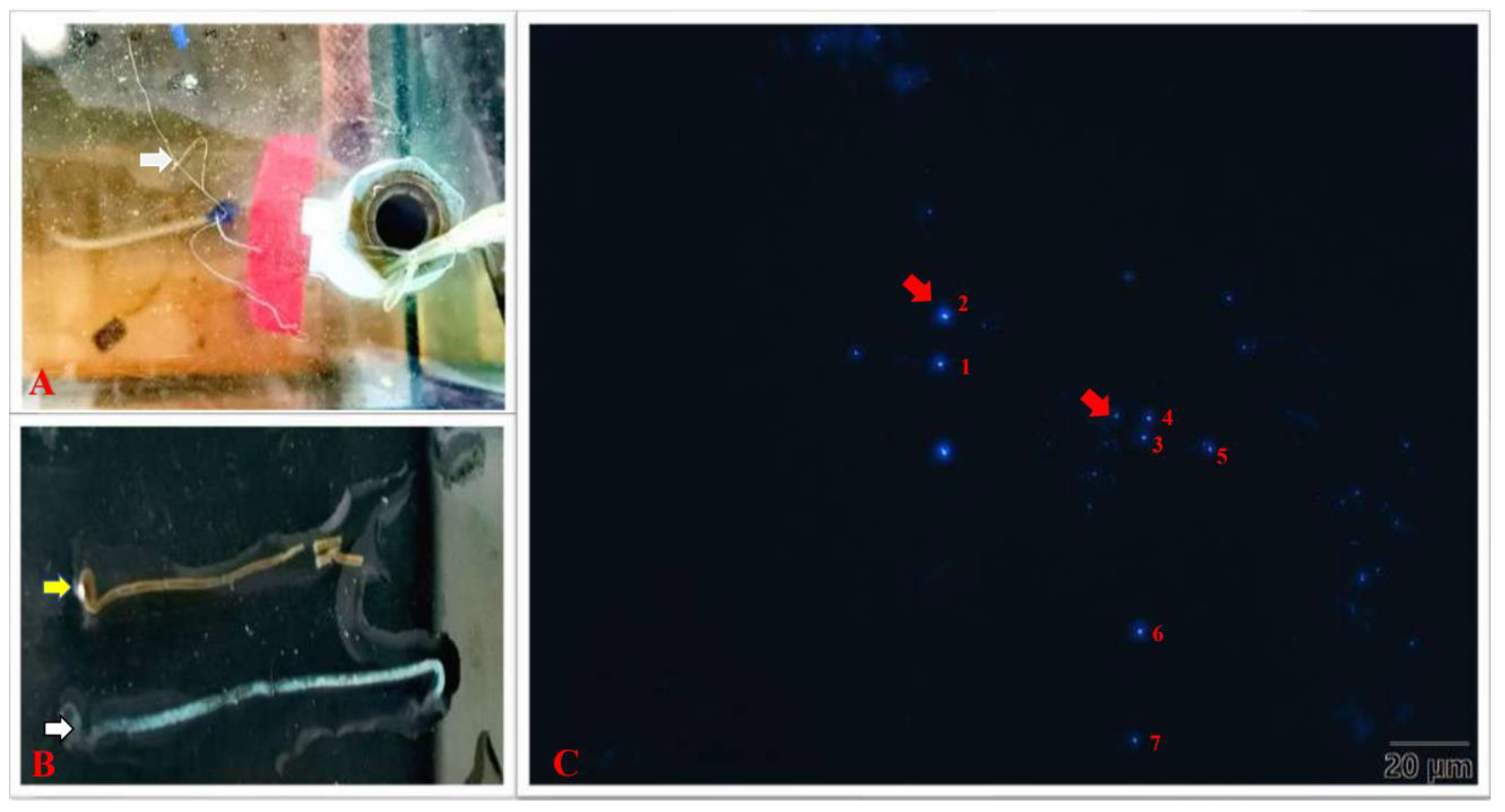
3.1. Detection of EHP Infection

The infection of EHP in the control group and the challenge group was detected using PCR. The shrimp in the challenge group were EHP-positive after the 5th dpc, while the control group remained EHP-negative throughout the experiment. The EHP load was detected using qPCR, and the results showed that the maximum load appeared on the 20th dpc (5.42 × 10⁵ ± 1.51 × 10⁵ EHP copies−1 ng HP DNA), followed by the 10th dpc (2.34 × 10⁵ ± 1.25 × 10⁵ EHP copies−1 ng HP DNA) and 5th dpc (3.30 × 10⁴ ± 6.19 × 10³ EHP copies−1 ng HP DNA).

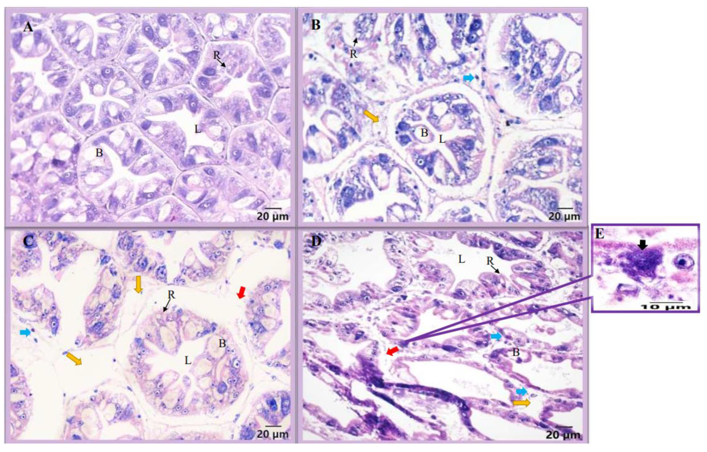
3.2. Microscopic Observation and Histology

Histological analysis of the hepatopancreas in the control group and the challenge group showed that the hepatic tubules of the healthy hepatopancreas were tightly arranged, with a full structure, a star-shaped lumen and an intact basement membrane (Figure 1A). At the 5th dpc, the hepatopancreas presented with mild atrophy of the hepatic tubules, with exfoliated cells between the tubules and vacuoles between the hepatic tubules (Figure 1B). At the 10th dpc, there were large vacuoles between the hepatic tubules of the hepatopancreas, exfoliated cells between the hepatic tubules and the basement membrane was damaged (Figure 1C). At the 20th dpc, the hepatic tubules showed more serious damage with a looser structure, the basement membrane was severely damaged and the epithelial cells had spore clusters (Figure 1D,E).
In addition, when the white feces were stained using CFW working solution, a large number of spores could be observed in the feces (Figure 2C). The size of EHP spores detected in the feces is shown in Table 2.

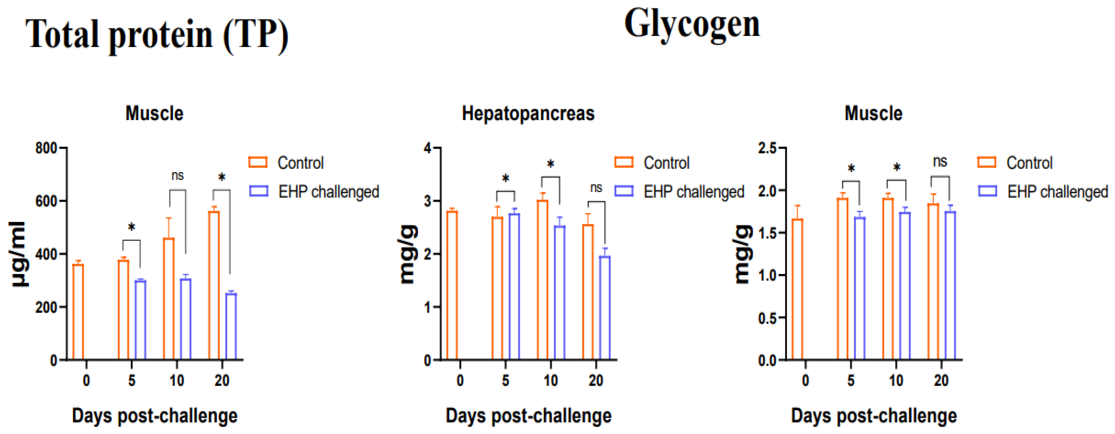
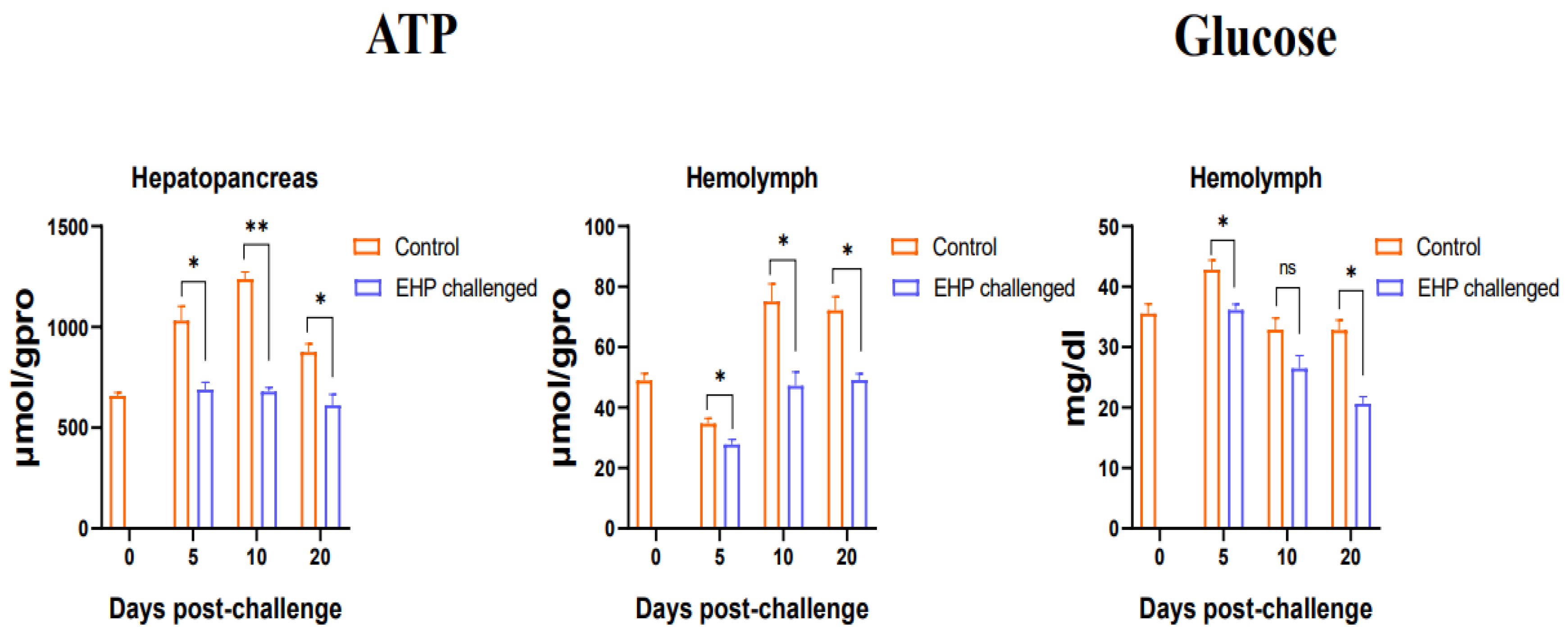
3.3. Total Protein, Glycogen, Glucose and ATP Content Assay

Compared with the healthy shrimp, the muscle total protein level of the EHP-infected shrimp decreased significantly at the 5th dpc and 20th dpc (p < 0.05). At the same time, the results of the determination of the hepatopancreas glycogen and muscle glycogen showed that the hepatopancreas and muscle glycogen of the shrimp in the challenge group decreased significantly at the 5th dpc and 10th dpc (p < 0.05), and recovered to the control level at the 20th dpc (Figure 3). Additionally, the results on ATP content showed that the ATP content of the hepatopancreas in the challenge group decreased significantly during the entire experiment (p < 0.05), and similarly, the ATP content of the hemolymph also decreased significantly at the 5th dpc, 10th dpc and 20th dpc (p < 0.05). Moreover, the hemolymph glucose content of the EHP-infected shrimp decreased significantly at the 5th dpc and 20th dpc (p < 0.05) (Figure 4).

3.4. Analysis of Glucose Metabolism and Growth-Related Genes

The expression of key genes in the glucose metabolism pathway was analyzed using qPCR. The qPCR results showed that the key genes of glycolysis in the challenge group were all down-regulated compared to the control group. Among them, HK and PK were significantly down-regulated at the 5th dpc and 20th dpc (p < 0.05), and PFK was significantly down-regulated at the 5th dpc, 10th dpc and 20th dpc (p < 0.05). The key genes CS and IDH in the TCA cycle were significantly down-regulated at the 5th dpc, 10th dpc and 20th dpc compared with the control group (p < 0.05). However, the key genes in the gluconeogenesis pathway were up-regulated to a certain extent after EHP infection. The expression level of PEPCK was increased at the 5th dpc and 20th dpc, of which it was significantly up-regulated at the 5th dpc (p < 0.05). The expression of FBP increased significantly at the 10th dpc and 20th dpc (p < 0.05). G6P was significantly up-regulated at the 5th dpc and 10th dpc (p < 0.05) (Figure 5). In addition, compared with the control group, the growth-related gene CHI was significantly down-regulated during the experiment (p < 0.05). JHEC was significantly down-regulated at the 5th dpc and 20th dpc (p < 0.05). However, ERP was significantly up-regulated at the 5th dpc and 20th dpc (p < 0.05), but the difference was not significant at the 10th dpc (Figure 6).

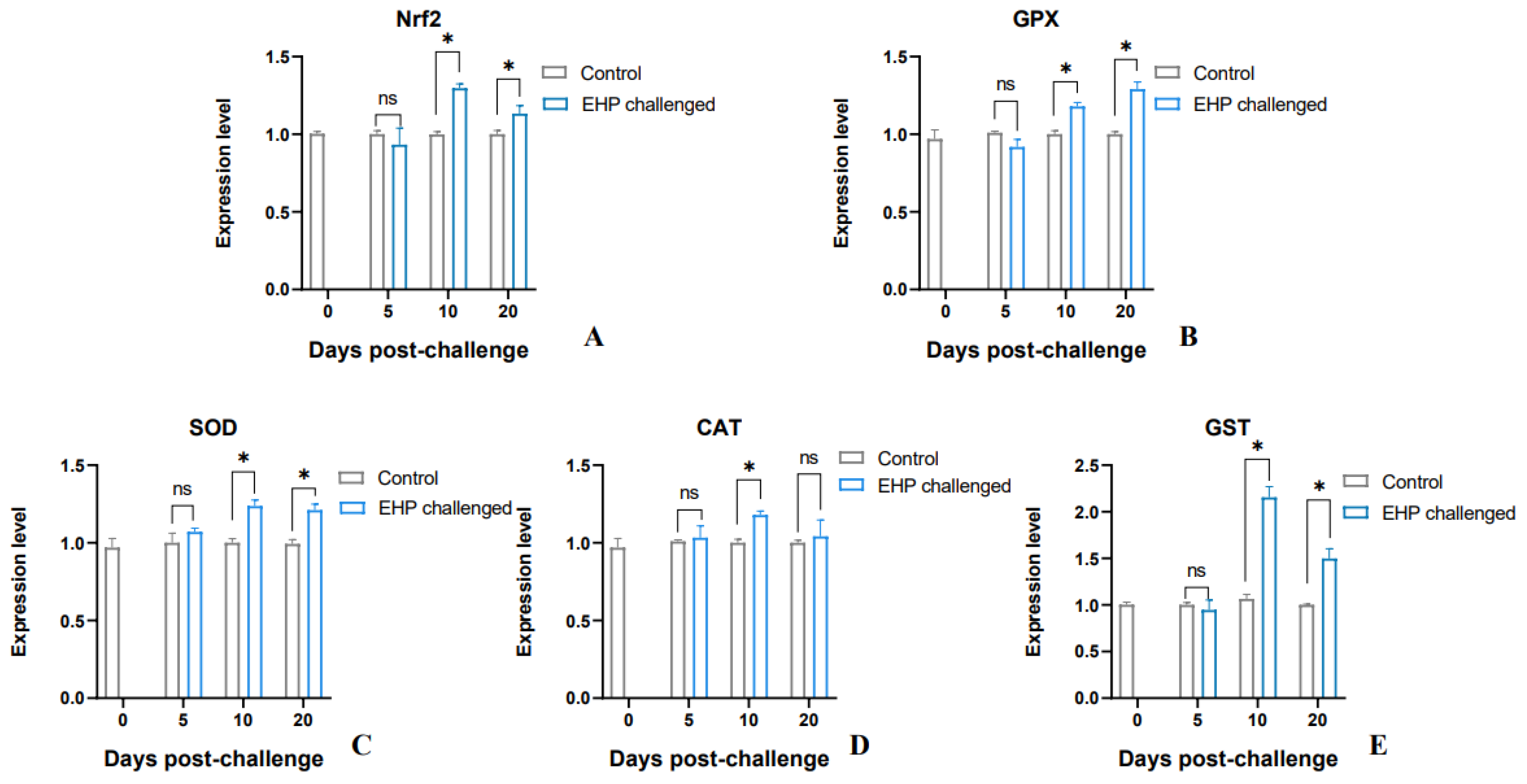
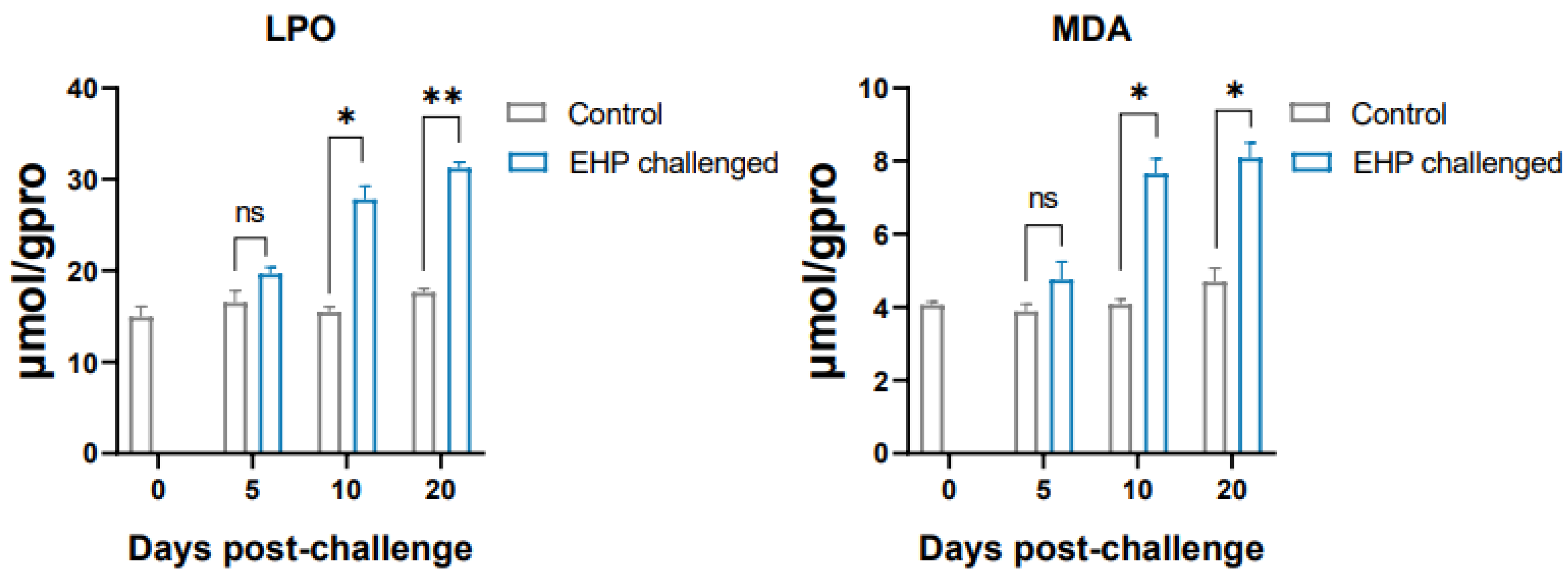
3.5. Analysis of Antioxidant Genes and Lipid Peroxidation Products

The results of qPCR showed that the antioxidant enzyme genes SOD, GPX and GST and the antioxidant pathway factor Nrf2 of the shrimp in the EHP challenge group were significantly up-regulated at the 10th dpc and 20th dpc (p < 0.05). However, CAT was only significantly up-regulated at the 10th dpc (p < 0.05) (Figure 7). Overall, compared with the healthy shrimp, the expression level of antioxidant genes in the EHP-infected shrimp showed an increasing trend. Moreover, the lipid peroxidation products LPO and MDA accumulated significantly at the 10th dpc and 20th dpc (p < 0.05) (Figure 8).

3.6. Growth Performance

After 20 days of challenge, the weight of the EHP-infected shrimp was 6.85 ± 0.86 g, which was distinctly lower than that of the control group (8.95 ± 0.75 g). At the same time, the weight gain of the shrimp in the challenge group (2.36 ± 0.34 g) was significantly lower than that in the control group (4.46 ± 0.23 g) (p < 0.05). Moreover, the ADG of the shrimp in the challenge group (118.06 ± 17.0 mg) was significantly lower than that in the control group (222.50 ± 11.5 mg) (p < 0.05), while the FCR of the challenge group (2.82 ± 0.21) was significantly higher than that in the control group (1.49 ± 0.06) (p < 0.05). However, the survival rate of the challenge group (76.67%) was lower than that of the control group (93.34%) (Figure 9). Equally, the growth changes in the shrimp were observed every day during the experiment. At the 7th dpc, shrimp in the challenge group showed a decrease in food intake and activity. At the 16th dpc, it was observed that shrimp in the challenge group showed fecal dragging and slow movement, as well as floating white feces in the breeding tank (Figure 2A,B).

4. Discussion

The hepatopancreas of crustaceans integrates the functions of the mammalian liver and pancreas, and is an important organ for nutrient storage, synthesis and digestion. The hepatopancreas, as the main target organ of EHP, even plays an important role in regulating the secretion of growth-related hormones in crustaceans [17,37], which is closely related to the growth retardation caused by EHP. Histology analysis observed that the hepatic tubules of EHP-infected shrimp showed varying degrees of atrophy with the duration of infection, and there were vacuoles between the hepatic tubules, accompanied by the shedding of epithelial cells, which is similar to the results of a previous study [38]. At the 20th dpc, the basement membrane of the hepatic tubules in the challenge group was severely disrupted and EHP spores were observed. At the same time, the EHP load also reached the maximum at the 20th dpc (5.42 × 10⁵ ± 1.51 × 10⁵). This suggests that there is a positive correlation between the degree of hepatopancreas damage and the severity of infection. Compared with a healthy hepatopancreas, these symptoms indicate that the normal absorption and digestion functions of the hepatopancreas are impaired, which may result in the inability of the hepatopancreas to provide nutritional support for the normal growth and development of the shrimp.
As an important nutrient, protein is considered the main source for animal growth and development. This study found that EHP infection caused a decrease in the total protein content of muscle, which may be closely related to the hepatopancreas damage directly caused by EHP parasitism. This further indicates that EHP interfered with the normal muscle development and affected the molting process of the shrimp, resulting in growth retardation [39]. Similarly, infection with WSSV resulted in a decrease in the total protein content in the muscle of shrimp [40], meaning that increasing protein consumption in the body is a self-protection strategy initiated by shrimp against viruses and microsporidia infection. In addition, the significant decrease in ATP content in the hepatopancreas also indicates that the function of the hepatopancreas is damaged and the metabolic process is disordered. Hemolymph is an important component in maintaining a normal metabolism and immune response in shrimp. The content of ATP and glucose in the hemolymph decreased, indicating that EHP infection disrupted the normal metabolic process and increased energy consumption, which is consistent with the findings of previous research [37]. Analogously, it has been reported that after shrimp are infected with WSSV, the ATP content in the hemolymph decreased significantly as the infection time was prolonged [41,42]. Meanwhile, the glucose content in the hemolymph can be regarded an important physiological indicator of shrimp health. As an important energy storage substance, glycogen can rapidly provide energy to the body under stress and starvation. It has been suggested that microsporidia act as a “carbohydrate collector”, absorbing glucose from the host and forcing the hepatopancreas to mobilize stored carbohydrates [43,44]. In the present study, it was found that the glycogen levels in the hepatopancreas and muscle of shrimp decreased significantly after EHP infection. Likewise, a significant reduction in glycogen content in the hepatopancreas has also been reported in the Norway lobster Nephrops norvegicus (Linnaeus, 1758) infected with parasitic dinoflagellates [44]. It is possible that EHP starves the shrimp during infection and consumes the glycogen of the host, as if EHP is issuing instructions to the shrimp that require large amounts of energy. Moreover, impaired hepatopancreatic function is associated with severe infection of glycogen storage R cells and secretory B cells [45,46].
Whether carbohydrate metabolism is normal or not can affect the molting and growth process of crustaceans, as well as the ability to cope with hypoxia stress and the invasion of pathogenic microorganisms [47,48,49]. Critically, changes in key enzymes in carbohydrate metabolism are the decisive factor in determining whether a certain metabolic pathway is able to proceed normally. Among them, glycolysis, the TCA cycle and gluconeogenesis are the basic pathways of glucose metabolism in eukaryotes. Overall, the present study showed that EHP infection suppressed the gene expression levels of key regulatory enzymes in glycolysis and the TCA cycle, possibly reducing the metabolic rate, which is consistent with previous studies [15,18,20,50]. However, the gene expression of key regulatory enzymes in the gluconeogenesis pathway was significantly up-regulated. HK, PFK and PK are important key regulatory enzymes in the glycolytic pathway, and changes in their expression affect the entire pathway [51]. The expression of key enzymes in the glycolysis pathway decreased significantly after EHP infection, indicating that the metabolic pathway was inhibited. Among these enzymes, PFK is considered to play a restrictive role in the pathway, determining the rate and direction of the reaction [52,53]. EHP significantly inhibited the gene expression of PFK, which means that the capacity of anaerobic metabolism decreased, the metabolic rate slowed down and the metabolic process changed. Meanwhile, the key regulatory enzymes in the TCA cycle, CS and IDH, were significantly down-regulated. As the primary key regulatory enzyme of the TCA cycle, CS controls the speed of acetyl-CoA entering the TCA cycle and releasing ATP via oxidative phosphorylation [54]. IDH, another key regulatory enzyme in the TCA cycle, catalyzes the oxidative decarboxylation of isocitrate into 2-oxoglutarate and releases ATP via the electron transport chain [55]. The decrease in the expression level of key enzymes in the TCA cycle may be due to the inhibition of the upstream glycolysis reaction by EHP, resulting in fewer products entering the TCA cycle, precisely because the parasitism of EHP disrupts the glucose metabolic process of the host. Glycolysis is an important way for crustaceans to obtain energy. As the core of carbohydrate, protein and lipid metabolism, the TCA cycle is the main source of cellular energy [56]. If the activity of its key enzyme is inhibited, it will directly cause a decrease in ATP production, resulting in a shortage of energy necessary for cell development. This may be an important reason for the slow growth of shrimp. Therefore, host cells may adopt strategies to reduce enzyme activity and decrease the metabolic intensity to maintain the host’s survival for as long as possible in the presence of prolonged infection. In addition, the glycolysis and TCA cycle metabolic processes were impaired by EHP, while the key regulatory enzymes in the gluconeogenesis pathway tended to increase. FBP is a rate-limiting enzyme for gluconeogenesis, which controls the rate of gluconeogenesis and catalyzes the conversion of fructose-1,6-diphosphate into fructose-6-phosphate [51]. After EHP infection, FBP expression was significantly increased at the 10th dpc and 20th dpc, and the expression of key enzymes PEPCK and G6P was also up-regulated, indicating that the gluconeogenesis process was enhanced. Since the parasitism of EHP needs to continuously absorb the energy from the host to maintain its own proliferation, the host is in a state of starvation [11]. This situation has also been observed in bees infected with parasites [57]. It is possible that the organism can maintain a normal hemolymph glucose level in shrimp by enhancing gluconeogenesis, but the degree of enhancement is not enough to compensate for the glucose consumption caused by EHP parasitism. Therefore, the hemolymph glucose content was still lower than that of the control group. Meanwhile, the consumption of carbohydrates was greater than the intake, which may be another reason for the decrease in glucose content. Interestingly, when shrimp were infected with WSSV [58] or Vibrio parahaemolyticus, the glucose metabolism process in vivo was significantly enhanced [59]. WSSV infection may be similar to the Warburg effect by enhancing glucose metabolism in response to viral replication [40,60]. Perhaps the study of changes in key metabolites can be regarded as a way to distinguish between viral infections and pathogenic microbial infections.
Oxidative stress is induced by excessive accumulation of free radicals and reactive oxygen species (ROS) in the body, which is an important factor leading to apoptosis and disease. It can be caused by stressors in vivo or in vitro. In order to prevent the damage caused by ROS, organisms have evolved an antioxidant defense system, with enzymatic responses to oxidative stress as the main strategy [61]. Therefore, a series of antioxidant enzymes play an important role, such as SOD, CAT, GPX and GST [62]. MnSOD is mainly distributed in the mitochondria and can catalyze the decomposition of superoxide into oxygen and hydrogen peroxide. Previous results found that the transcription level of MnSOD increased significantly after shrimp were injected with the pathogen [63]. Similarly, in this study, significant increases in the expression levels of MnSOD and GPX were detected at the 10th dpc and 20th dpc, suggesting that EHP activated the antioxidant defense system of the shrimp. In addition, the body produces a large amount of ROS, which can induce the expression of antioxidant genes, and in turn protect host cells to reduce oxidative damage [64]. Usually, the change in CAT is consistent with that in SOD, which is responsible for the decomposition of hydrogen peroxide into oxygen and water. However, CAT was significantly elevated only at the 10th dpc. It may be because both CAT and GPX perform the function of decomposing peroxides, but GPX is more sensitive to peroxides in the body and can play a role even at low concentrations. This was verified using nitrite stress experiments [65,66]. Meanwhile, GPX has the function of detoxification metabolism, which is considered to be more important than CAT. Importantly, GPX in shrimp can be induced by Vibrio alginolyticus [67]. The expression of GST also increased significantly in this experiment, suggesting that it may participate in the transport of endogenous substances and play an irreplaceable role in detoxification and cell protection [68]. The Nrf2-Keap1 signaling pathway is the most important antioxidant pathway in the body, and can regulate the activity of antioxidant enzymes [69]. Unfortunately, Keap1 has not been identified in P. vannamei. It has been reported that inhibiting the expression of Nrf2 resulted in a significant decrease in the expression of related antioxidant genes (SOD, GPX, CAT) in shrimp [70]. In this study, Nrf2 was significantly up-regulated after EHP infection, which was consistent with the expression of antioxidant enzymes, indicating that this gene may regulate the expression of antioxidant genes. Nrf2 has also been reported to be significantly activated in the response of P. vannamei to Vibrio harveyi [71]. In addition, LPO is a lipid peroxidation product generated by the reaction of ROS with polyunsaturated fatty acids, which causes oxidative damage to cells. MDA is also the result of lipid peroxidation and is considered to be a marker of oxidative damage [72]. In organisms, polyunsaturated fatty acids are the main components of the membrane structure that make up various organelles. The increase in LPO and MDA content detected in this experiment after EHP infection suggests that the hepatopancreas cells may be damaged and interfere with normal physiological functions. Corresponding to the histology, it was clearly observed that the hepatopancreas was significantly damaged, with vacuoles and membrane damage between the hepatic tubules, which may be tissue damage caused by a large number of peroxide products. These results reveal that the activation of the antioxidant pathway and the up-regulated expression of antioxidant oxidase genes induced by EHP are not sufficient to clear excess lipid peroxidation products, and the tissue damage caused by oxidative stress may be the key reason that affects the metabolic function of the hepatopancreas.
The present study also demonstrated that EHP can lead to the abnormal expression of growth-related genes, among which JHEC, ERP and CHI play important regulatory roles in molting, growth and reproduction in crustaceans. In this experiment, JHEC (at the 5th dpc and 20th dpc) was significantly down-regulated, whereas ERP (at the 5th dpc and 20th dpc) was significantly up-regulated. JHEC is a key enzyme in the degradation of methyl farnesoate (MF) [73]. The reduction of the MF titer is beneficial to the growth and development of shrimp. On the contrary, the down-regulation of JHEC reduced the degradation rate of MF, resulting in stunted growth and development. The increase in ecdysterone is conducive to molting, but there is a negative regulatory relationship between ERP and ecdysterone. When the level of ecdysterone is high, the expression of ERP is inhibited, and the up-regulation of ERP in the present study was manifested as a delayed molting process [74,75]. Concurrently, CHI can digest chitin, and the enhanced activity of this enzyme before molting can ensure a smooth molting process by decomposing the old shell [76,77]. However, in this study, EHP invasion resulted in a significant decrease in CHI, indicating that shrimp molting was blocked. Therefore, the parasitism of EHP destroys the normal substance metabolism in shrimp, resulting in insufficient energy for cell growth and development. Furthermore, EHP invasion breaks the balance between growth and molting-related hormones, which may be an important cause of retarded development of shrimp.
EHP can significantly damage the hepatopancreas of shrimp, disrupt the normal intestinal flora and cause nutrient absorption and transport disorders. Therefore, the final body weight and ADG of shrimp in the EHP challenge group were significantly lower than those in the control group. Similarly, this phenomenon of different individual sizes is more obvious in aquaculture ponds [78]. In addition, the FCR of shrimp in the EHP challenge group was significantly higher than that in the control group (20th dpc) (p < 0.05) due to the disruption of nutrient absorption and transport, indicating that the shrimp infected with EHP could not adequately absorb the nutrients in the feed. Fortunately, the administration of 5-aminolaevulinic acid and linolenic acid to EHP-infected shrimp could improve the function of the hepatopancreas to a certain extent [75,79]. In addition, EHP infection did not cause a large number of deaths in shrimp, but increased the risk of infection with other diseases such as acute hepatopancreatic necrosis disease (AHPND) (Lightner et al. 2012) [12], taura syndrome [80] and white feces syndrome [13]. Previous studies have shown that white feces can be present with the aggravation of EHP infection, but white feces are not real feces and may be composed of EHP spores, intestinal mucus and necrotic tubular epithelial cells. Many studies have shown that EHP spores have been detected in white feces [13,81]. Consistent with previous reports, floating white feces were also found in the tanks of EHP-challenged shrimp (at the 16th dpc). A large number of spores were observed after the suspended feces were stained, indicating that the phenomenon of white feces has a certain relationship with EHP infection.

5. Conclusions

In this study, the changes in hepatopancreas histology and the effects of EHP on the metabolism, oxidative stress and growth-related genes of healthy shrimp and EHP-infected shrimp were analyzed using EHP challenge experiment. Our studies have shown that the hepatopancreas became damaged with an increasing duration of EHP infection, and the degree of damage was positively correlated with the intensity of infection. At the same time, EHP infection affected the normal metabolism of substances and growth, which was characterized by the inhibition of glycolysis and the TCA cycle, activation of the gluconeogenesis pathway, and interference with the expression of growth-related genes. Furthermore, EHP induced oxidative stress in shrimp and resulted in the accumulation of a large amount of the lipid peroxidation products LPO and MDA, which was also an important cause of oxidative damage to the hepatopancreas. Additionally, severe EHP infection may result in white feces. In summary, this study concludes that EHP infection can disrupt the metabolic process of shrimp and cause oxidative stress, both of which will lead to the slow growth of shrimp and even death in severe cases.

Author Contributions

Conceptualization, Z.C. and L.S.; methodology, Z.C. and L.C.; software, C.W. and T.L.; validation, Z.C. and C.C.; formal analysis, Z.C. and C.C.; investigation, Z.C., C.C. and C.W.; data curation, T.L. and L.C.; writing—original draft preparation, Z.C.; writing—review and editing, Z.C. and L.S.; visualization, Z.C., L.S. and D.Y.; supervision, L.S. and D.Y.; project administration, L.S. All authors have read and agreed to the published version of the manuscript.

Funding

This work was supported by the Natural Science Foundation of Shandong Province (ZR2023QC050) and start-up funding for scientific research from Ludong University (20220022).

Institutional Review Board Statement

This study was approved by the Institutional Review Board of Ludong University (protocol code: LDU-IRB20221127NXY-1; approval date: 27 November 2022). All procedures involving animals were performed in accordance with the Guide for the Care and Use of Laboratory Animals in China.

Informed Consent Statement

Not applicable.

Data Availability Statement

The datasets used in this study are available from the corresponding authors upon reasonable request.

Conflicts of Interest

The authors declare no conflict of interest.

References

  1. Food and Agriculture Organization of the United Nations. Food and Agriculture Organization of the United Nations TUNA. GLOBEFISH Highlights April 2019, Statistics a Quarterly Update on World Seafood Markets FAO. Roma. December 2018. Available online: https://www.fao.org/in-action/globefish/publications/details-publication/es/c/1201730/ (accessed on 7 June 2022).
  2. National modern agricultural industry technology system white shrimp industry development report. China Fish. 2021, 5, 27–36.
  3. Chang, Z.Q.; Neori, A.; He, Y.Y.; Li, J.T.; Qiao, L.; Preston, S.I.; Liu, P.; Li, J. Development and current state of seawater shrimp farming, with an emphasis on integrated multi-trophic pond aquaculture farms, in China a review. Rev. Aquacult. 2020, 12, 2544–2558. [Google Scholar] [CrossRef]
  4. Tourtip, S.; Wongtripop, S.; Stentiford, G.D.; Bateman, K.S.; Sriurairatana, S.; Chavadej, J.; Sritunyalucksana, K.; Withyachumnarnkul, B. Enterocytozoon hepatopenaei sp. nov. (microsporida: Enterocytozoonidae), a parasite of the black tiger shrimp Penaeus monodon (decapoda: Penaeidae): Fine structure and phylogenetic relationships. J. Invertebr. Pathol. 2009, 102, 21–29. [Google Scholar] [CrossRef] [PubMed]
  5. Hou, Z.H.; Yu, J.Y.; Wang, J.J.; Li, T.; Chang, L.R.; Fang, T.; Yan, D.C. Development of a PCR assay for the effective detection of Enterocytozoon hepatopenaei (EHP) and investigation of EHP prevalence in Shandong Province, China. J. Invertebr. Pathol. 2021, 184, 107653. [Google Scholar] [CrossRef] [PubMed]
  6. Shen, H.; Jiang, G.; Wan, X.H.; Fan, X.P.; Qiao, Y.; Shi, W.J.; Wang, L.B. Multiple pathogens prevalent in shrimp Penaeus vannamei cultured from greenhouse ponds in Jiang Su province of China. J. Aquac. Res. Dev. 2017, 27, 675–683. [Google Scholar] [CrossRef]
  7. Ma, B.; Yu, H.; Fang, J.; Sun, C.; Zhang, M. Employing DNA binding dye to improve detection of Enterocytozoon hepatopenaei in real-time LAMP. Sci. Rep. 2019, 9, 15860. [Google Scholar] [CrossRef]
  8. Shi, H.; Xu, W.J.; Xie, J.J.; Wang, G.S. Pathogenicity of Penaeus vannamei slow growth syndrome in intensively cultured penaeid shrimp in Zhoushan. J. Fish. Sci. China 2017, 24, 387–394. [Google Scholar] [CrossRef]
  9. Patil, P.K.; Geetha, R.; Ravisankar, T.; Avunje, S.; Solanki, H.G.; Abraham, T.J.; Vinoth, S.P.; Jithendran, K.P.; Alavandi, S.V.; Vijayan, K.K. Economic loss due to diseases in Indian shrimp farming with special reference to Enterocytozoon hepatopenaei (EHP) and white spot syndrome virus (WSSV). Aquaculture 2021, 533, 9736231. [Google Scholar] [CrossRef]
  10. Shinn, A.P.; Pratoomyot, J.; Griffiths, D.; Trong, T.Q.; Vu, N.T.; Jiravanichpaisal, P.; Briggs, M. Asian shrimp production and the economic costs of disease. Special Acute Hepatopancreatic Necrosis Disease (AHPND). Asian Fish. Sci. 2018, 31, 29–58. [Google Scholar] [CrossRef]
  11. Chaijarasphong, T.; Munkongwongsiri, N.; Stentiford, G.D.; Aldama-Cano, D.J.; Thansa, K.; Flegel, T.W.; Sritunyalucksana, K.; Itsathitphaisarn, O. The shrimp microsporidian Enterocytozoon hepatopenaei (EHP): Biology, pathology, diagnostics and control. J. Invertebr. Pathol. 2021, 186, 107458. [Google Scholar] [CrossRef]
  12. Aranguren, L.F.; Han, J.E.; Tang, K.F.J. Enterocytozoon hepatopenaei (EHP) is a risk factor for acute hepatopancreatic necrosis disease (AHPND) and septic hepatopancreatic necrosis (SHPN) in the Pacific white shrimp Penaeus vannamei. Aquaculture 2017, 471, 37–42. [Google Scholar] [CrossRef]
  13. Munkongwongsiri, N.; Prachumwat, A.; Eamsaard, W.; Lertsiri, K.; Flegel, T.W.; Stentiford, G.D.; Sritunyalucksana, K. Propionigenium and Vibrio species identified as possible component causes of shrimp white feces syndrome (WFS) associated with the microsporidian Enterocytozoon hepatopenaei. J. Invertebr. Pathol. 2022, 192, 107784. [Google Scholar] [CrossRef] [PubMed]
  14. Sathish Kumar, T.; Makesh, M.; Alavandi, S.V.; Vijayan, K.K. Clinical manifestations of White feces syndrome (WFS), and its association with Enterocytozoon hepatopenaei in Penaeus vannamei grow-out farms: A pathobiological investigation. Aquaculture 2022, 547, 737463. [Google Scholar] [CrossRef]
  15. Duan, Y.F.; Chen, H.G.; Wang, J.L.; Zeng, S.M.; Wang, Y.; Mo, Z.Q.; Dan, X.M.; Li, Y.W. Response signatures of Litopenaeus vannamei to natural Enterocytozoon hepatopenaei infection revealed by the integration of the microbiome and transcriptome. Aquaculture 2021, 542, 736885. [Google Scholar] [CrossRef]
  16. Vogt, G. Life-cycle and functional cytology of the hepatopancreatic cells of Astacus astacus (Crustacea, Decapoda). Zoomorphology 1994, 114, 83–101. [Google Scholar] [CrossRef]
  17. Wang, W.; Wu, X.; Liu, Z.; Zheng, H.; Cheng, Y. Insights into hepatopancreatic functions for nutrition metabolism and ovarian development in the crab Portunus trituberculatus: Gene discovery in the comparative transcriptome of different hepatopancreas stages. PLoS ONE 2014, 9, e84921. [Google Scholar] [CrossRef]
  18. Ning, M.; Wei, P.; Shen, H.; Wan, X.; Jin, M.; Li, X.; Shi, H.; Qiao, Y.; Jiang, G.; Gu, W.; et al. Proteomic and metabolomic responses in hepatopancreas of whiteleg shrimp Litopenaeus vannamei infected by microsporidian Enterocytozoon hepatopenaei. Fish Shellfish Immun. 2019, 87, 534–545. [Google Scholar] [CrossRef]
  19. Wiredu Boakye, D.; Jaroenlak, P.; Prachumwat, A.; Williams, T.A.; Bateman, K.S.; Itsathitphaisarn, O.; Sritunyalucksana, K.; Paszkiewicz, K.H.; Moore, K.A.; Stentiford, G.D.; et al. Decay of the glycolytic pathway and adaptation to intranuclear parasitism within Enterocytozoonidae microsporidia. Environ. Microbiol. 2017, 19, 2077–2089. [Google Scholar] [CrossRef]
  20. Cao, Z.; Gao, Y.; Xu, J.H.; Yang, N.; Li, T.; Chang, L.R.; Si, L.J.; Yan, D.C. Transcriptome analysis of the hepatopancreas in Penaeus vannamei under experimental infection with Enterocytozoon hepatopenaei (EHP). Fish Shellfish Immun. 2023, 134, 108605. [Google Scholar] [CrossRef]
  21. Oakes, K.D.; Van Der Kraak, G.J. Utility of the TBARS assay in detecting oxidative stress in white sucker (Catostomus commersoni) populations exposed to pulp mill effluent. Aquat. Toxicol. 2003, 29, 447–463. [Google Scholar] [CrossRef]
  22. Lightner, D.V. A Handbook of Shrimp Pathology and Diagnostic Procedures for Diseases of Cultured Penaeid Shrimp; World Aquatic Society: Baton Rouge, LA, USA, 1996. [Google Scholar]
  23. Ji, P.F.; Yao, C.L.; Wang, Z.Y. Reactive oxygen system plays an important role in shrimp Litopenaeus vannamei defense against Vibrio parahaemolyticus and WSSV infection. Dis. Aquat. Organ. 2011, 29, 9–20. [Google Scholar] [CrossRef] [PubMed]
  24. Bianchini, A.; Monserrat, J.M. Effects of methyl parathion on Chasmagnathus granulatus hepatopancreas: Protective role of sesamol. Ecotoxicol. Environ. Saf. 2007, 67, 100–108. [Google Scholar] [CrossRef] [PubMed]
  25. Subash, P.; Chrisolite, B.; Sivasankar, P.; Rosalind George, M.; Vijay Amirtharaj, K.S.; Padmavathy, P.; Rani, V.; Sankar Sri Balaje, R.; Gowtham, S.; Mageshkumar, P. White feces syndrome in Penaeus vannamei is potentially an Enterocytozoon hepatopenaei (EHP) associated pathobiome origin of Vibrio spp. J. Invertebr. Pathol. 2023, 198, 107932. [Google Scholar] [CrossRef] [PubMed]
  26. Gao, W.; Guo, X.M.; Wang, H.L.; Zhao, R.H.; Xie, G.S.; Li, C.; Huang, J. A double staining method using calcofluor white and acridine orange to differentiate life stages of Enterocytozoon hepatopenaei (EHP) on hepatopancreatic sections. Aquaculture 2020, 528, 735628. [Google Scholar] [CrossRef]
  27. Li, G.; Cong, F.; Cai, W.Y.; Li, J.H.; Wu, M.L.; Xiao, L.; Hu, X.L.; Zeng, W.W.; He, D.S. Development of a recombinase polymerase amplification (RPA) fluorescence assay for the detection of Enterocytozoon hepatopenaei (EHP). Aquacult. Rep. 2021, 19, 100584. [Google Scholar] [CrossRef]
  28. Tang, K.F.J.; Pantoja, C.R.; Redman, R.M.; Han, J.E.; Tran, L.H.; Lightner, D.V. Development of in situ hybridization and PCR assays for the detection of Enterocytozoon hepatopenaei (EHP), a microsporidian parasite infecting penaeid shrimp. J. Invertebr. Pathol. 2015, 130, 37–41. [Google Scholar] [CrossRef]
  29. Otta, S.K.; Karunasagar, I.; Karunasagar, I. Detection of monodon baculovirus and white spot syndrome virus in apparently healthy Penaeus monodon postlarvae from India by polymerase chain reaction. Aquaculture 2003, 220, 59–67. [Google Scholar] [CrossRef]
  30. OIE. Manual of Diagnostic Tests for Aquatic Animals. World Organisation for Animal Health. 2019. Available online: http://www.oie.int/en/international-standard-setting/aquatic-manual (accessed on 13 June 2022).
  31. Söderhäll, K.; Smith, V.J. Separation of the haemocyte populations of Carcinus maenas and othermarine decapods, and prophenoloxidase distribution. Dev. Comp. Immunol. 1983, 7, 229–233. [Google Scholar] [CrossRef]
  32. Liu, Z.; Zhang, Q.L.; Wan, X.Y.; Ma, F.; Huang, J. Development of real-time PCR assay for detecting microsporidian Enterocytozoon hepatorenal and the application in shrimp samples with different growth rates. Progress. Fish. Sci. 2016, 37, 119–126. [Google Scholar] [CrossRef]
  33. Bell, T.A.; Lightner, D.V. A Handbook of Normal Penaeid Shrimp Histology; World Aquaculture Society: Sorrento, LA, USA, 1988. [Google Scholar]
  34. Zhao, R.H.; Gao, W.; Qiu, L.; Chen, X.; Dong, X.; Li, C.; Huang, J. A staining method for detection of Enterocytozoon hepatopenaei (EHP) spores with calcofluor white. Aquaculture 2020, 172, 107347. [Google Scholar] [CrossRef]
  35. Livak, K.J.; Schmittgen, T.D. Analysis of Relative Gene Expression Data Using Real-Time Quantitative PCR and the 2−ΔΔCT Method. Methods 2001, 25, 402–408. [Google Scholar] [CrossRef] [PubMed]
  36. Zarain-Herzberg, M.; Fraga, I.; Hernandez-Llamas, A. Advances in intensifying the cultivation of the shrimp Litopenaeus vannamei in floating cages. Aquaculture 2010, 300, 87–92. [Google Scholar] [CrossRef]
  37. Sathish Kumar, T.; Ezhil Praveena, P.; Sivaramakrishnan, T.; Joseph Sahaya Rajan, J.; Makesh, M.; Jithendran, K.P. Effect of Enterocytozoon hepatopenaei (EHP) infection on physiology, metabolism, immunity, and growth of Penaeus vannamei. Aquaculture 2022, 553, 738105. [Google Scholar] [CrossRef]
  38. Zhu, B.; Lu, X.D.; Liu, Y.H.; Wu, Z.N.; Cai, H.F.; Jin, S.; Li, Z.; Xie, J.S.; Li, X.B.; Sun, F.Y.; et al. Effects of Enterocytozoon hepatopenaei single-infection or co-infection with Vibrio parahaemolyticus on the hepatopancreas of Penaeus vannamei. Aquaculture 2022, 549, 737726. [Google Scholar] [CrossRef]
  39. de Oliveira Cesar, J.R.; Zhao, P.B.; Malecha, S.; Ako, H.; Yang, J.Z. Morphological and biochemical changes in the muscle of the marine shrimp Litopenaeus vannamei during the molt cycle. Aquaculture 2006, 261, 688–694. [Google Scholar] [CrossRef]
  40. Yoganandhan, K.; Thirupathi, S.; Sahul Hameed, A.S. Biochemical, physiological and hematological changes in white spot syndrome virus-infected shrimp, Penaeus indicus. Aquaculture 2003, 221, 1–11. [Google Scholar] [CrossRef]
  41. Chen, I.T.; Aoki, T.; Huang, Y.T.; Hirono, I.; Chen, T.C.; Huang, J.Y.; Chang, G.D.; Lo, C.F.; Wang, H.C. White spot syndrome virus induces metabolic changes resembling the warburg effect in shrimp hemocytes in the early stage of infection. J. Virol. 2011, 85, 19–28. [Google Scholar] [CrossRef]
  42. Zhang, Y.S.; Li, F.X.; Yao, C.L. Glycogen phosphorylase of shrimp (Litopenaeus vannamei): Structure, expression and anti-WSSV function. Fish Shellfish Immun. 2019, 91, 275–283. [Google Scholar] [CrossRef]
  43. Lin, L.J.; Chen, Y.J.; Chang, Y.S.; Lee, C.Y. Neuroendocrine responses of a crustacean host to viral infection: Effects of infection of white spot syndrome virus on the expression and release of crustacean hyperglycemic hormone in the crayfish Procambarus clarkii. Comp. Biochem. Physiol. A Mol. Integr. Physiol. 2013, 164, 327–332. [Google Scholar] [CrossRef]
  44. Stentiford, G.D.; Chang, E.S.; Chang, S.A.; Neil, D.M. Carbohydrate dynamics and the crustacean hyperglycemic hormone (CHH): Effects of parasitic infection in Norway lobsters (Nephrops norvegicus). Gen. Comp. Endocrinol. 2001, 121, 13–22. [Google Scholar] [CrossRef]
  45. Aranguren Caro, L.F.; Mai, H.N.; Cruz-Florez, R.; Marcos, F.L.A.; Alenton, R.R.R.; Dhar, A.K. Experimental reproduction of White Feces Syndrome in whiteleg shrimp, Penaeus vannamei. PLoS ONE 2021, 16, e0261289. [Google Scholar] [CrossRef] [PubMed]
  46. Cuomo, C.A.; Desjardins, C.A.; Bakowski, M.A.; Goldberg, J.; Ma, A.T.; Becnel, J.J.; Didier, E.S.; Fan, L.; Heiman, D.I.; Levin, J.Z.; et al. Microsporidian genome analysis reveals evolutionary strategies for obligate intracellular growth. Genome. Res. 2012, 22, 2478–2488. [Google Scholar] [CrossRef]
  47. Wang, Z.Y.; Zhang, Y.L.; Yao, D.F.; Zhao, Y.Z.; Tuan, T.N.; Li, S.K.; Ma, H.Y. Aweya Jude Juventus, Metabolic reprogramming in crustaceans: A vital immune and environmental response strategy. Rev. Aquacult. 2022, 14, 1094–1119. [Google Scholar] [CrossRef]
  48. Xu, C.; Li, E.; Liu, Y.; Wang, S.; Wang, X.; Chen, K.; Qin, G.J.; Chen, L. Effect of dietary lipid level on growth, lipid metabolism and health status of the Pacific white shrimp Litopenaeus vannamei at two salinities. Aquacult. Nutr. 2018, 24, 204–214. [Google Scholar] [CrossRef]
  49. Zhan, Q.Y.; Han, T.; Li, X.Y.; Wang, J.T.; Yang, Y.X.; Yu, X.J.; Zheng, P.Q.; Liu, T.; Xu, X.Y.; Wang, C.L. Effects of dietary carbohydrate levels on growth, body composition, and gene expression of key enzymes involved in hepatopancreas metabolism in mud crab Scylla paramamosain. Aquaculture 2020, 529, 735638. [Google Scholar] [CrossRef]
  50. Wu, Y.J.; Chen, J.; Liao, G.L.; Hu, M.J.; Zhang, Q.; Meng, X.Z.; Li, T.; Long, M.X.; Fan, X.D.; Yu, Q.; et al. Down-Regulation of Lipid Metabolism in the Hepatopancreas of Shrimp Litopenaeus vannamei upon Light and Heavy Infection of Enterocytozoon hepatopenaei: A Comparative Proteomic Study. Int. J. Mol. Sci. 2022, 23, 11574. [Google Scholar] [CrossRef]
  51. Bian, X.L.; Jiang, H.F.; Meng, Y.; Li, Y.P.; Fang, J.; Lu, Z.M. Regulation of gene expression by glycolytic and gluconeogenic enzymes Trends. Cell. Biol. 2022, 32, 786–799. [Google Scholar] [CrossRef]
  52. Cota-Ruiz, K.; Peregrino-Uriarte, A.B.; Felix-Portillo, M.; Martínez-Quintana, J.A.; Yepiz-Plascencia, G. Expression of fructose 1,6-bisphosphatase and phosphofructokinase is induced in hepatopancreas of the white shrimp Litopenaeus vannamei by hypoxia. Mar. Environ. Res. 2015, 106, 1–9. [Google Scholar] [CrossRef]
  53. Jenkins, C.M.; Yang, J.Y.; Sims, H.F.; Gross Reversible, R.W. high affinity inhibition of phosphofructokinase-1 by acyl-CoA: A mechanism integrating glycolytic flux with lipid metabolism. J. Biol. Chem. 2011, 286, 11937–11950. [Google Scholar] [CrossRef]
  54. Ulaje, S.A.; Rojo-Arreola, L.; Lluch-Cota, S.E.; Ascencio, F.; Cruz-Hernández, P.; Sicard, M.T. Gene expression and energetic metabolism changes in the whiteleg shrimp (Litopenaeus vannamei) in response to short-term hypoxia. Aquac. Res. 2019, 50, 994–1004. [Google Scholar] [CrossRef]
  55. Zhu, F.; Zhang, X.B. The Wnt signaling pathway is involved in the regulation of phagocytosis of virus in Drosophila. Sci. Rep. 2013, 3, 2069. [Google Scholar] [CrossRef] [PubMed]
  56. Reyes, M.; Chandel, N.S. Mitochondrial TCA cycle metabolites control physiology and disease. Nat. Commun. 2020, 11, 102. [Google Scholar] [CrossRef] [PubMed]
  57. Mayack, C.; Naug, D. Energetic stress in the honeybee Apis mellifera from Nosema ceranae infection. J. Invertebr. Pathol. 2009, 100, 185–188. [Google Scholar] [CrossRef] [PubMed]
  58. Fan, W.W.; Ye, Y.F.; Chen, Z.; Shao, Y.N.; Xie, X.L.; Zhang, W.W.; Liu, H.P.; Li, C.H. Metabolic product response profiles of Cherax quadricarinatus towards white spot syndrome virus infection. Dev. Comp. Immunol. 2016, 61, 236–241. [Google Scholar] [CrossRef] [PubMed]
  59. Nguyen, T.V.; Alfaro, A.; Arroyo, B.B.; Leon, J.A.R.; Sonnenholzner, S. Metabolic responses of penaeid shrimp to acute hepatopancreatic necrosis disease caused by Vibrio parahaemolyticus. Aquaculture 2021, 533, 736174. [Google Scholar] [CrossRef]
  60. Chen, I.T.; Lee, D.Y.; Huang, Y.T.; Kou, G.H.; Wang, H.C.; Chang, G.D.; Lo, C.F. Six Hours after Infection, the Metabolic Changes Induced by WSSV Neutralize the Host's Oxidative Stress Defenses. Sci. Rep. 2016, 6, 27732. [Google Scholar] [CrossRef]
  61. Wang, H.; Pan, L.; Xu, R.; Si, L.; Zhang, X. The molecular mechanism of Nrf2-Keap1 signaling pathway in the antioxidant defense response induced by BaP in the scallop Chlamys farreri. Fish Shellfish Immun. 2019, 92, 489–499. [Google Scholar] [CrossRef]
  62. Parrilla Taylor, D.P.; Zenteno Savín, V.T.; Magallón Barajas, F.J. Antioxidant enzyme activity in pacific white leg shrimp (Litopenaeus vannamei) in response to infection with white spot syndrome virus. Aquaculture 2013, 380, 41–46. [Google Scholar] [CrossRef]
  63. Sookruksawong, S.; Pongsomboon, S.; Tassanakajon, A. Genomic organization of the cytosolic manganese superoxide dismutase gene from the Pacific white shrimp, Litopenaeus vannamei, and its response to thermal stress. Fish Shellfish Immun. 2013, 35, 1395–1405. [Google Scholar] [CrossRef]
  64. Zelko, I.N.; Mariani, T.J.; Folz, R.J. Superoxide dismutase multigene family: A comparison of the CuZn-SOD (SOD1), Mn-SOD (SOD2), and EC-SOD (SOD3) gene structures, evolution, and expression. Free Radic. Biol. Med. 2002, 33, 337–349. [Google Scholar] [CrossRef]
  65. Kang, S.W.; Rhee, S.G.; Chang, T.S.; Jeong, W.; Choi, M.H. 2-Cys peroxiredoxin function in intracellular signal transduction: Therapeutic implications. Trends Mol. Med. 2005, 11, 571–578. [Google Scholar] [CrossRef]
  66. Guo, H.; Xian, J.A.; Li, B.; Ye, C.X.; Wang, A.L.; Miao, Y.T.; Liao, S.A. Gene expression of apoptosis-related genes, stress protein and antioxidant enzymes in hemocytes of white shrimp Litopenaeus vannamei under nitrite stress. Comp. Biochem. Physiol. C Toxicol. Pharmacol. 2013, 157, 366–371. [Google Scholar] [CrossRef] [PubMed]
  67. Liu, C.H.; Tseng, M.C.; Cheng, W. Identification and cloning of the antioxidant enzyme, glutathione peroxidase, of white shrimp, Litopenaeus vannamei, and its expression following Vibrio alginolyticus infection. Fish Shellfish Immun. 2007, 23, 34–45. [Google Scholar] [CrossRef] [PubMed]
  68. Zhou, W.N.; Wang, A.L.; Wang, W.Y.; He, Q.T.; Zhou, Y.; Xu, J. Glutathione S-transferase in the white shrimp Litopenaeus vannamei: Characterization and regulation under pH stress. Comp. Biochem. Physiol. C Toxicol. Pharmacol. 2009, 150, 224–230. [Google Scholar] [CrossRef] [PubMed]
  69. Wang, H.; Pan, L.; Si, L.; Ji, R.; Cao, Y. Effects of Nrf2-Keap1 signaling pathway on antioxidant defense system and oxidative damage in the clams Ruditapes philippinarum exposure to PAHs. Environ. Sci. Pollut. Res. 2021, 26, 33060–33071. [Google Scholar] [CrossRef]
  70. Huang, Y.; Li, Q.; Yuan, Y.; Zhang, Z.; Jiang, B.; Yang, S.; Jian, J. Silencing of Nrf2 in Litopenaeus vannamei, decreased the antioxidant capacity, and increased apoptosis and autophagy. Fish Shellfish Immun. 2022, 122, 257–267. [Google Scholar] [CrossRef] [PubMed]
  71. Yuan, Y.; Guan, H.; Huang, Y.; Luo, J.; Jian, J.; Cai, S.; Yang, S. Involvement of Nrf2 in the immune regulation of Litopenaeus vannamei against Vibrio harveyi infection. Fish Shellfish Immun. 2023, 133, 108547. [Google Scholar] [CrossRef]
  72. Duan, Y.; Wang, Y.; Zhang, J.; Liu, Q.; Ding, X. Morphologic, digestive enzymes and immunological responses of intestine from Litopenaeus vannamei after lipopolysaccharide injection. J. Invertebr. Pathol. 2018, 153, 186–194. [Google Scholar] [CrossRef]
  73. Lee, S.O.; Jeon, J.M.; Oh, C.W.; Kim, Y.M.; Kang, C.K.; Lee, D.S.; Mykles, D.L.; Kim, H.W. Two juvenile hormone esterase-like carboxylesterase cDNAs from a Pandalus shrimp (Pandalopsis japonica): Cloning, tissue expression, and effects of eyestalk ablation. Comp. Biochem. Physiol. B Biochem. Mol. Biol. 2011, 159, 148–156. [Google Scholar] [CrossRef]
  74. He, C.B.; Chen, P.H.; Gao, X.G.; Gao, L.; Li, L. Expression and purification of ecdysteroid-regulated protein from Chinese mitten crab Eriocheir sinensis in E. coli. Mol. Biol. Rep. 2013, 40, 6987–6995. [Google Scholar] [CrossRef]
  75. Ning, N.X.; Bi, J.X.; Sun, W.; Xie, X.J.; Huang, Y.L.; Gu, W.; Wang, W.; Qiao, Y.; Jiang, G.; Shen, H.; et al. Linolenic acid improves growth performance and immune status of Penaeus vannamei infected by Enterocytozoon hepatopenaei. Aquaculture 2021, 535, 736397. [Google Scholar] [CrossRef]
  76. Priya, T.A.; Li, F.H.; Zhang, J.Q.; Wang, B.; Zhao, C.; Xiang, J.H. Molecular characterization and effect of RNA interference of retinoid X receptor (RXR) on E75 and chitinase gene expression in Chinese shrimp Fenneropenaeus chinensis. Comp. Biochem. Physiol. B Biochem. Mol. Biol. 2009, 153, 121–129. [Google Scholar] [CrossRef] [PubMed]
  77. Proespraiwong, P.; Tassanakajon, A.; Rimphanitchayakit, V. Chitinases from the black tiger shrimp Penaeus monodon: Phylogenetics, expression and activities. Comp. Biochem. Physiol. B Biochem. Mol. Biol. 2010, 156, 86–96. [Google Scholar] [CrossRef] [PubMed]
  78. Liu, Y.M.; Qiu, L.; Cheng, D.Y.; Zhang, Q.L.; Wan, X.Y.; Huang, J. The Relationship of Body Length and Weight in the Litopenaeus vannamei Populations Detected Enterocytozoon hepatopenaei. Progress. Fish. Sci. 2017, 38, 96–103. [Google Scholar] [CrossRef]
  79. Kongplong, S.; Kanjanasopa, D.; Pongtippatee, P.; Vanichviriyakit, R.; Withyachumnarnkul, B. 5-Aminolaevulinic acid reduced the mortality of the Pacific white shrimp Litopenaeus vannamei infected with Enterocytozoon hepatopenaei. Aquaculture 2023, 568, 739322. [Google Scholar] [CrossRef]
  80. Tang, K.F.J.; Aranguren, L.F.; Piamsomboon, P.; Han, J.E.; Maskaykina, I.Y.; Schmidt, M.M. Detection of the microsporidian Enterocytozoon hepatopenaei (EHP) and Taura syndrome virus in Penaeus vannamei cultured in Venezuela. Aquaculture 2017, 480, 17–21. [Google Scholar] [CrossRef]
  81. Wang, H.L.; Wan, X.Y.; Xie, G.S.; Dong, X.; Wang, X.H.; Huang, J. Insights into the histopathology and microbiome of Pacific white shrimp, Penaeus vannamei, suffering from white feces syndrome. Aquaculture 2020, 527, 735447. [Google Scholar] [CrossRef]
Figure 1. Histology of shrimp hepatopancreas under EHP infection. (A) Hepatopancreas of the control shrimp; hepatopancreas of EHP-challenged shrimp at 5th dpc (B), 10th dpc (C) and 20th dpc (D). The blue arrow indicates scattered cells, the yellow arrow indicates vacuoles between hepatic tubules, the red arrow indicates broken basement membrane and the black arrow indicates EHP spores (E). B, B cells; L, lumen; R, R cells.
Animals 13 03661 g001
Figure 2. White feces of EHP-infected shrimp and fluorescence microscopic observation of EHP spores in the white feces. (A) White feces floating in the aquaculture tank (white arrow); (B) the yellow arrow indicates healthy feces, and the white arrow indicates white feces; (C) the red arrow indicates EHP spores in white feces.
Animals 13 03661 g002
Figure 3. Total protein content in the muscle and glycogen content in the hepatopancreas and muscle of shrimp after EHP infection (mean ± SE, n = 6). The superscript symbol * depicts the statistical significance at p < 0.05. ns: not significant.
Animals 13 03661 g003
Figure 4. ATP content in the hepatopancreas and hemolymph, and glucose content in the hemolymph of shrimp after EHP infection (mean ± SE, n = 6). The superscript symbol * depicts the statistical significance at p < 0.05, and ** depicts the statistical significance at p < 0.01. ns: not significant.
Animals 13 03661 g004
Figure 5. Relative expression levels of glucose metabolism-related genes in the hepatopancreas of P. vannamei after EHP infection (mean ± SE, n = 6). Glycolysis-related genes: HK, PFK, PK (AC); Gluconeogenesis-related genes: PEPCK, FBP, G6P (DF); TCA cycle-related genes: CS, IDH (G,H). The superscript symbol * depicts the statistical significance at p < 0.05, and ** depicts the statistical significance at p < 0.01. ns: not significant.
Animals 13 03661 g005
Figure 6. Relative expression levels of growth-related genes CHI (A), ERP (B) and JHEC (C) in the hepatopancreas of P. vannamei after EHP infection (mean ± SE, n = 6). The superscript symbol * depicts the statistical significance at p < 0.05, and ** depicts the statistical significance at p < 0.01. ns: not significant.
Animals 13 03661 g006
Figure 7. Relative expression levels of antioxidant genes Nrf2 (A), GPX (B), SOD (C), CAT (D)and GST (E) in the hepatopancreas of P. vannamei after EHP infection (mean ± SE, n = 6). The superscript symbol * depicts the statistical significance at p < 0.05. ns: not significant.
Animals 13 03661 g007
Figure 8. LPO and MDA content in the hepatopancreas of shrimp after EHP infection (mean ± SE, n = 6). The superscript symbol * depicts the statistical significance at p < 0.05, and ** depicts the statistical significance at p < 0.01. ns: not significant.
Animals 13 03661 g008
Figure 9. The growth performance of the control group and the EHP challenge group. It mainly includes final weight (A), weight gain (B), ADG (C), FCR (D) and survival rate (E). The superscript symbol * depicts the statistical significance at p < 0.05, and ** depicts the statistical significance at p < 0.01.
Animals 13 03661 g009
Table 1. Primers used for qPCR in the experiment.
GenePrimers (5′-3′)GenBank Accession No.
Growth and metabolic genes
ERPF: CCTACAGCGTCAACATCCA
R: GCATCCGTCGGTGTCTATT
JQ009182.1
JHEC1F: GGCGGAGCAGAGGACTAT
R: CGAGGTCACGGATGTTGTC
APO14259.1
CHIF: GAAGGCGTCGTATATTGTGTCAC
R: AGCACGGTCCTGATGGTAGT
ACR23315
G6PF: CTTATGAATCGTGGGTGGTGC
R: CAATAGGGTCGGTCTCCTCTGA
PRJNA660490
FBPF: TGTGTCGGAAGAAAACAAAACT
R: CTATGGAGACGAGGCAATCAAT
KP057246
PEPCKF: AAGACCAGTGATGGAGGAGTG
R: GGGAGTTGGGATGAGCAG
FJ441189
PKF: ATCCTTGATGGTGCTGAC
R: TGAAGAGTTGCTTGTGCC
EF102105
PFKF: AGAGGACGGGGAAGTTTTACAG
R: GTTCTTGCCTGGGTTCAAATAG
PRJNA660490
HKF: ATCGGCAAGTTAGATACGC
R: AGGACACCACGGTAGGAA
EF102106
CSF: AAAGAATACGGTAACACCAAAGT
R: ATGGAGTAACCTCGGAACCTGAT
XM027367291
IDHF: GCCTCTGCCTCAAGGGTAT
R: CCTCAGTCTGCTCACGGAT
XM027354101.1
Antioxidant genes
CATF: TCAGCGTTTGGTGGAGAA
R: GCCTGGCTCATCTTTATC
AY518322
MnSODF: CGTAGAGGGTATTGTCGT
R: TTGAAATCATACTTGAGGG
DQ005531
GPXF: AGGGACTTCCACCAGATG
R: CAACAACTCCCCTTCGGTA
AY973252
GSTF: AAGATAACGCAGAGCAAGG
R: TCGTAGGTGACGGTAAAGA
AY573381
Nrf2F: GATGAAGCGAGCCAGAGCG
R: GCCGTCGGATGTCTCGGATAA
XM_027367068.1
β-actinF: TGGACTTCGAGCAGGAGATG
R: GGAATGAGGGCTGGAACAGG
AF300705
18sF: TATACGCTAGTGGAGCTGGAA
R: GGGGAGGTAGTGACGAAAAAT
EU920969
Table 2. The size of EHP spores in the experiment.
Number1234567
Size (μm)1.1021.1930.9280.7171.0091.0550.820
Disclaimer/Publisher’s Note: The statements, opinions and data contained in all publications are solely those of the individual author(s) and contributor(s) and not of MDPI and/or the editor(s). MDPI and/or the editor(s) disclaim responsibility for any injury to people or property resulting from any ideas, methods, instructions or products referred to in the content.

