Article

Zoonotic Bacteria in Anolis sp., an Invasive Species Introduced to the Canary Islands (Spain)

by
Néstor Abreu-Acosta
1,2,
Román Pino-Vera
2,3,
Elena Izquierdo-Rodríguez
2,3,
Oscar Afonso
4 and
Pilar Foronda
2,3,*
1
Nertalab S.L.U. Santa Cruz de Tenerife, Tenerife, 38001 Canary Islands, Spain
2
Instituto Universitario de Enfermedades Tropicales y Salud Pública de Canarias, Universidad de La Laguna, La Laguna, Tenerife, 38200 Canary Islands, Spain
3
Department Obstetricia y Ginecología, Pediatría, Medicina Preventiva y Salud Pública, Toxicología, Medicina Legal y Forense y Parasitología, Universidad de La Laguna, La Laguna, Tenerife, 38200 Canary Islands, Spain
4
Área de Medio Ambiente, Gestión y Planeamiento Territorial y Ambiental (Gesplan), Santa Cruz de Tenerife, Tenerife, 38200 Canary Islands, Spain
*
Author to whom correspondence should be addressed.
Animals 2023, 13(3), 414; https://doi.org/10.3390/ani13030414
Submission received: 28 October 2022 / Revised: 11 January 2023 / Accepted: 20 January 2023 / Published: 26 January 2023

Abstract

:

Simple Summary

The anoles are a group of lizards native to America that have been introduced to other regions, such as the Canary Islands (Spain). With this study we aimed to analyze the presence of pathogenic bacteria in a population of this invasive lizard in the Canary Islands. The results highlight the presence of a variety of pathogens of relevance to health, most of them related to gastrointestinal diseases. This archipelago is considered a hotspot of biodiversity, where some species are considered endangered, so the presence of anoles can also be a risk for the biodiversity conservation, by the spread and/or transmission of pathogenic bacteria to the native fauna. In conclusion, the invasive anoles population in the Canary Islands should be considered as a potential risk factor for public health and biodiversity conservation.

Abstract

Lizards belonging to the genus Anolis are native to America and have been introduced in many parts of the world. In this work, a gastrointestinal microbiological analysis from Anolis sp. introduced to Tenerife, Canary Island, was carried out. A total of 74 individuals were analyzed by culture and molecular tools. Pseudomonas spp. was the most prevalent bacteria isolated (64.3%), followed by enteropathogenic Escherichia coli with at least one of the investigated virulent genes (stx1, stx2, and eae) (44.6%). The stx2 gene was more prevalent which differs to that reported in other reptiles, probably due to wastewater transmission. Campylobacter spp. was detected in 32.4% of the animals, highlighting the detection of C. jejuni and C. fetus by their relevance to public health. The zoonotic Staphylococcus lugdunensis, found in 14.9% of the animals, was firstly detected in reptiles. Vibrio sp. which is more associated with aquatic environments was found in 10.8% of the lizards in this study, with Vibrio cholerae being found in two of the animals. The prevalence of Salmonella sp. (5.4%) was low, compared with other studies carried out in reptiles. These results indicate that Anolis sp. in Tenerife could be playing a role in the maintenance and spread of the pathogens detected, being a possible risk factor for public health and biodiversity conservation.

1. Introduction

Following the description of the Convention on Biological Diversity (CBD, 2010), invasive non-native species (INNS) are organisms introduced outside their natural distribution area due to human activity, or are species that can threaten biodiversity, being one of the largest global threats to biodiversity (IPBES global assessment, 2019). Invasions are not the result of single species introduction events, but units of biological organization that include the host and all its symbionts, including pathogenic species [1]. Therefore, invasive species can constitute a health risk for humans, animals, and plants, acting as reservoirs for pathogens [2]. Island communities are more vulnerable to introduced diseases because of their geographical isolation [3,4]. While the impact of foreign diseases on the human communities of the islands is well-documented [5], the effect of introduced diseases on biodiversity is less reported [6]. However, there are evident examples of introduced diseases that are seriously affecting the survival of island species [7].
The Canary Archipelago (Spain), located in NW Africa (13°23′–18°8′ W and 27°37′–29°24′ N), is a particularly suitable place for the settlement of invasive species due to its climatic conditions, its wealth of resources, and the lack of large predators that control the populations [8]. Tenerife, the largest island in the Archipelago, is also the island with the largest number of exotic species. According to the latest available data, around 1200 of the species present are not native to the island, and a significant percentage of these could become naturalized and behave in an invasive manner, affecting the island biodiversity [9]. Among the invasive species, lizards belonging to the genus Anolis Daudin, 1802 (Squamata, Dactyloidae) were introduced in the south of the island through the ornamental plant trade since 1995 [10]. These small tree lizards are native to North America, but they have been introduced to many parts of the world [11,12,13], harming the ecosystems of some regions, as in the case of Chichi Island, Japan [14].
Due to the lack of data on the presence of pathogenic micro-organisms in this invasive Anolis species in the Canary Islands, the main purpose of this study was to determine the presence of pathogenic bacteria in these lizards and to evaluate the possible health risks for public health and native fauna.

2. Materials and Methods

A total of 74 animals, 70 adults (25 males and 45 females) and 4 young individuals were captured manually and by adhesive traps in the South of Tenerife (Canary Islands, Spain). The captured anoles were euthanized after authorization of “Dirección General de Lucha Contra el Cambio Climático y Medio Ambiente” (Gobierno de Canarias) (Expte. 31280/2021). The animals were measured and weighed, and then necropsied and their sex determined. Fecal samples were obtained during the dissection for culture.

2.1. Bacterial Strains

The bacterial strains used as positive amplification controls were obtained from American Type Culture Collection (ATCC). All the bacterial strains were stored at −70 °C and were grown on Tryptic Soy Broth (TSB; Labkem, Barcelona, Spain) at 37 °C for 18 to 24 h under aerobic or microarophilic conditions, in the case of Campylobacter spp. Sebald and Veron, 1963; Vandamme et al., 2010.

2.2. Isolation of Pathogenic Bacteria

For the investigated bacteria, except for Campylobacter sp. and Vibrio spp. Pacini, 1854, 0.1 g of intestinal content was extracted and incubated (37 °C) in 5 mL of Buffered Peptone Water (BPW) (Labkem, Barcelona, Spain) for 24 h thus achieving enriched liquid culture. In the case of Campylobacter sp., 0.1 g of the intestinal content was taken in a 2 mL tube with BPW. The tubes were incubated at 42 °C for 18 h in microaerophilic condition. For Vibrio spp., 0.1 g of intestinal content was transferred into 5 mL of Alkaline Peptone Water (APW) containing 1% NaCl as a pre-enrichment, incubated for 8 h at 37 °C.
After the incubation, for the isolation of Salmonella spp. Lignieres, 1900, 0.5 mL of each culture of BPW was transferred into 4.5 mL of Rappaport Vassiliadis Broth (VWR International, Leuven, Belgium) and incubated a 42 °C for 20 h. Then, these samples were cultured on Salmonella-Shigella agar (Merck, Darmstadt, Germany) at 37 °C as a secondary enrichment culture. For the isolation of the other pathogenic bacteria considered in this study, 100 µL of each culture of BPW were inoculated in selective media for Staphylococcus sp. Rosenbach, 1884 (Baird Parker agar; Labkem, Barcelona, Spain), Pseudomonas sp. Migula, 1894; Yang et al., 2013 (Cetrimide agar; VWR International, Leuven, Belgium), and Listeria monocytogenes (Murray et al., 1926) Pirie, 1940 (Oxford agar; Labkem, Barcelona, Spain). In the case of Vibrio spp., 100 µL of each culture of APW were inoculated onto Thiosulfate–Citrate–Bile salts Sucrose agar (TCBS; VWR International, Leuven, Belgium) and incubated for 24 h at 37 °C.

2.3. Molecular Identification of Isolates and Pathogenicity Genes

2.3.1. DNA Isolation

In the case of Campylobacter spp., 1 mL of BPW incubated at 42 °C, and in the case of Escherichia. coli (Migula, 1895) Castellani and Chalmers, 1919, Mycobacterium spp. Lehmann and Neumann, 1896; Gupta et al., 2018, and Yersinia enterocolitica (Schleifstein and Coleman, 1939) Frederiksen, 1964, 1 mL of BPW incubated at 37 °C were taken, respectively. They were washed twice with PBS and the pellet was subjected to DNA extraction according to López et al. [15].
For the rest of the bacteria analyzed, 5 colonies of each culture were dissolved in 1 mL of PBS and centrifuged at 12,000× g. The supernatant was removed and centrifuged again under the same conditions. The pellet was subjected to DNA extraction following López et al. [15].

2.3.2. PCR Assays

A multiplex-PCR (m-PCR) assay was used for the identification of the most important zoonotic Campylobacter species, C. jejuni (Jones et al., 1931) Veron and Chatelain, 1973, C. coli (Doyle 1948) Veron and Chatelain, 1973, C. lari Benjamin et al., 1984; Debruyne et al., 2009, C. upsaliensis Sandstedt and Ursing, 1991, and y C. fetus (Smith and Taylor, 1919) Sebald and Veron, 1963, according to the specifications of Wang et al. [16].
Enteropathogenic E. coli (producing shigatoxins, STEC) were analyzed by the detection of the genes that encode for Shiga toxins (stx1 and stx2) and the gene that codes for the intimin protein, which is another factor associated with virulence in E. coli, following the protocol described by Blanco et al. [17].
Other m-PCR assay was carried out for the identification of Mycobacterium sp. and, in addition, for the differentiation of the species belonging to M. tuberculosis complex, according to Kim et al. [18].
For the identification of Yersinia enterocolitica, a duplex PCR assay described by Wannet et al. [19] was developed. With this protocol, non-virulent and pathogenic strains are detected.
Other PCR assay was used for the detection and identification of Vibrio spp. according to Liu et al. [20]. The samples that were positive were subjected to an m-PCR assay according to Neogi et al. [21], for the identification of Vibrio cholerae Pacini, 1854, V. parahaemolyticus (Fujino et al., 1951) Sakazaki et al., 1963; West et al., 1986, and V. vulnificus (Reichelt et al., 1979) Farmer, 1980; West et al., 1986.
For the identification of the most prevalent zoonotic Salmonella serotypes, two m-PCR assays were used, according to Guimarães de Freitas et al. [22]. In a first test, all Salmonella species are detected, in addition to Salmonella ser. Enteritidis and Salmonella ser. Typhi. In the second trial, Salmonella ser. Typhimurium is identified.
One m-PCR was used for the detection of clinically relevant antibiotic resistance genes harbored by some Staphylococcus aureus Rosenbach, 1884 isolates, and for the simultaneous identification of these isolates to the species level, according to Pérez-Roth et al. [23].
An m-PCR assay was used for the detection of fluorescent pseudomonads and the identification of Pseudomonas aeruginosa (Schroeter, 1872) Migula, 1900 based on the simultaneous amplification of the oprI and oprL genes, as described by De Vos et al. [24].
To confirm suspicious colonies grown on Oxford agar as Listeria monocytogenes, a region of the iap gene was amplified by PCR following the protocol described by Jaton et al. [25].
Positive and negative controls were included in all the PCRs performed.
After PCR amplification, 5 µL was analyzed in an agarose gel (Fisher Bioreagents, Madrid, Spain) electrophoresis to estimate the sizes of the amplification products by comparison with a molecular size standard ladder (GeneRuler 50 bp DNA Ladder, Thermo Scientific, Vilnius, Lithuania and HyperLadder 50 bp, Bioline, London, England). The gel was stained with Real-Safe (Durviz SL, Valencia, Spain), and the amplicons were visualized with ChemiDoc™ XRS+ (Bio-Rad, Hercules, CA, USA) system. Some amplicons were purified using the UltraClean PCR Clean-up (MO BIO Laboratories, Inc., Carlsbad, CA, USA) kit, following manufacturer recommendations. Sequencing in both directions was performed in Macrogen (Spain) and then analyzed with software MEGA X (Molecular Evolutionary Genetic Analysis) [26]. Sequences were compared and similarity values obtained with available published sequences in GenBank by using the nucleotide–nucleotide BLAST (blastn) program [27].

2.4. Index Co-Infection (Ic)

Ginsberg, H.S. [28] developed an index of co-infection (Ic) which quantifies the degree of departure of the number of mixed infections from independence. This is defined as the difference of the number of co-infections from the number expected due to chance alone, as a percentage of the total number of infected lizards.
Ic = [(O − E)/N] × 100,
where O = number of observed co-infections, E = expected number of co-infected Anolis sp. due to chance alone, and N = total number of Anolis sp. infected by either or both micro-organisms.
E = (a + b) (a + c)/(a + b + c + d),
N = a + b + c,
where a = number of lizards infected with both bacteria (equals O), b = number of Anolis sp. infected only with micro-organism 1, c = number of Anolis sp. infected only with micro-organism 2, and d = number of lizards not infected with either micro-organism. Ic is positive when the number of co-infections is greater than expected, and negative when there are fewer co-infections than would be expected due to chance alone.
Significance of the index was calculated by a chi-square test.

2.5. Statistical Analysis

A chi-square test, setting the p value in 0.05, was conducted to compare prevalence between age and sex using SPSS for Windows statistical software (IBM Corporation, Armonk, NY, USA).

3. Results

Seventy-four individuals were analyzed. Table 1 shows the percentages of isolation of the investigated bacteria. Pseudomonas spp. was the most prevalent bacteria (64.3%), followed by STEC with at least one of the investigated genes (45.6%), and Campylobacter spp. (32.4%).

3.1. Campylobacter spp.

In 24 individuals out of 74, Campylobacter species were detected by PCR, which represents a prevalence of 32.4%. No significant differences were found with the Chi-square test, between the number of positive male individuals (6/25; 24.0% of males) and females (16/45; 35.6% of females), or between positive adults and positive juveniles (2/4; 50.0% of positives) (Table 2). The length and weight of the animals that were infected with Campylobacter spp. were similar to those individuals that were free of these bacteria.
Four species were identified, with C. fetus being the most prevalent species, and the only one that was isolated in juvenile individuals. The second most prevalent species was C. coli, isolated only in females. The presence of C. jejuni and C. upsaliensis was also detected (Table 2). Moreover, non-identified Campylobacter species were detected in seven animals.

3.2. Vibrio spp.

Eight of the reptiles (10.8%) were positive for Vibrio sp., six of them females (13.3% of the females) and two males (8.0% of the males). The juveniles analyzed were free of Vibrio spp. The number of positives was statistically (p > 0.05) similar between males and females. The length and weight of the animals that presented Vibrio spp. were similar to those individuals that were free of these bacteria. Due to the infrequent detection of Vibrio spp. in terrestrial reptiles, the amplicons obtained in the PCR assay were sequenced. In two individuals, one male and one female, Vibrio cholerae was identified. Two of the obtained sequences had 100% similarity with Vibrio cholerae and were uploaded to the GenBank database (accession number OP688106 and OP688107).

3.3. Enteropathogenic Shiga Toxin Producing Escherichia coli (STEC)

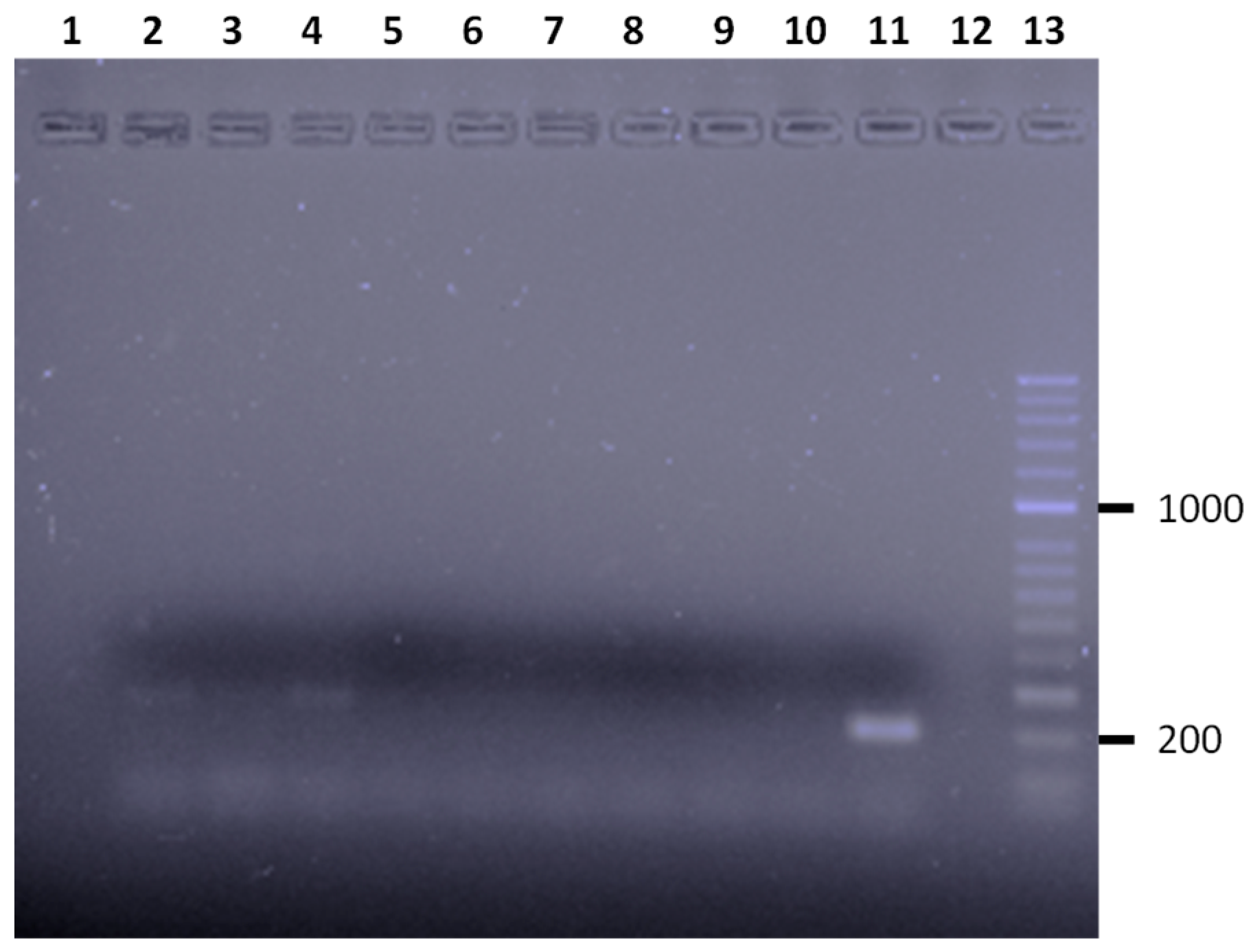
The presence of Shiga toxin (stx1, stx2) and intimin (eae) genes, related to the ability of enteropathogenic Escherichia coli to produce disease, was analyzed (Figure 1). E. coli carrying these genes were found in 33 individuals (44.6%). The prevalence of STEC that carried the stx2 gene (28.4%) was higher than that of those that carried the stx1 gene (10.8%) and the eae gene (5.4%) (Table 3) (p < 0.05). There were no differences between the number of positive males and females or between adults and juveniles for the different virulence genes (Table 3). Finally, the coexistence of two virulence genes (stx1 and stx2) was detected in two individuals, one female and one male.

3.4. Salmonella spp.

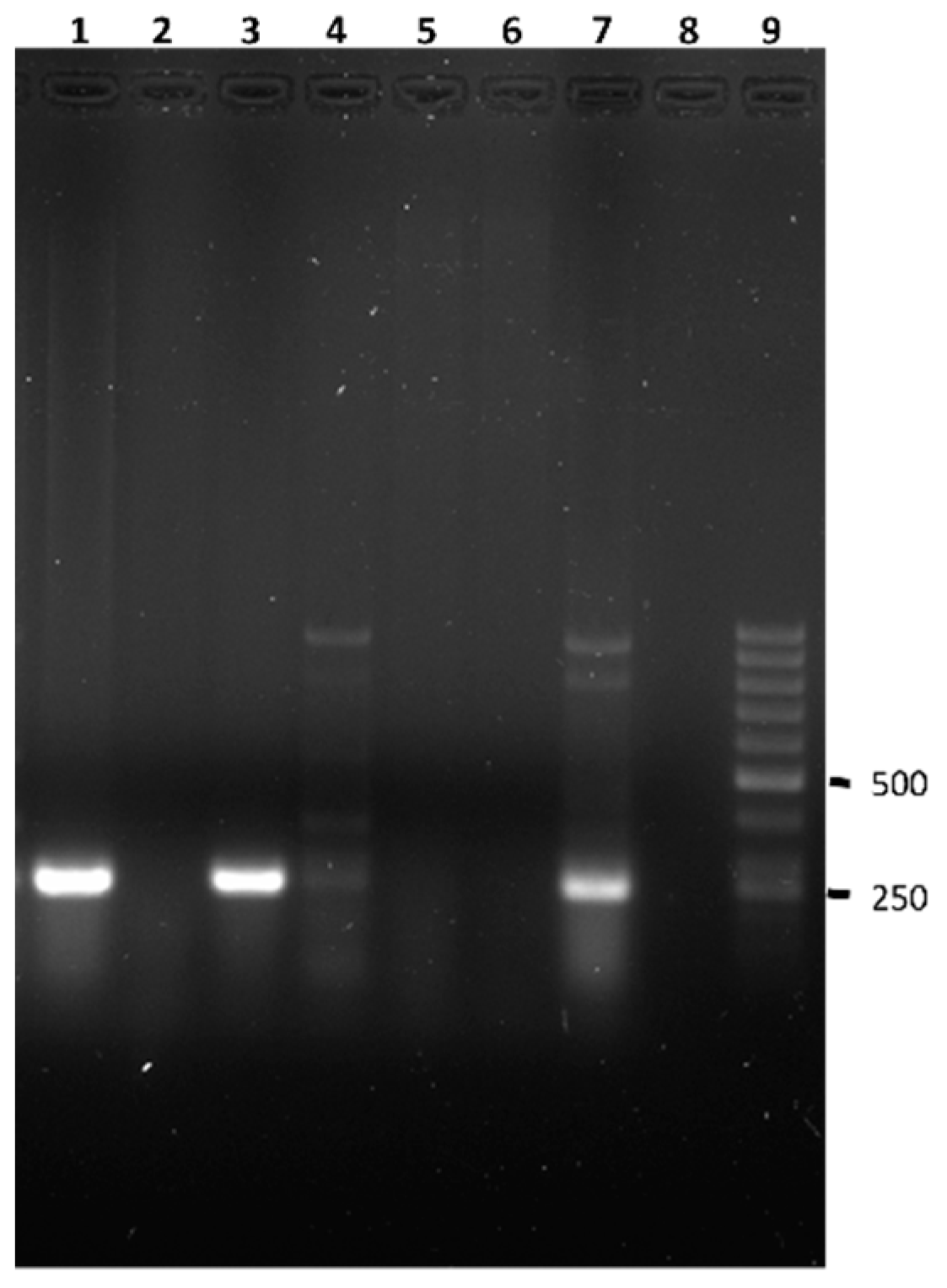
The presence of Salmonella spp. was revealed by obtaining a 204 bp amplification fragment corresponding to the ompC gene, responsible for the C protein involved in the invasion of the epithelial cells of bacteria of the Salmonella genus, in four individuals (5.4%) of the total analyzed (Figure 2).
Two females were positive for Salmonella spp. (4.4%; n = 45), one juvenile (25.0%; n = 4), and one male (4.0%; n = 25). The investigated zoonotic serotypes (Salmonella enteritidis, S. typhimurium, and S. typhi) were not detected, since the expected amplification fragments of the sdfI, spy, and viab genes, respectively, were not obtained.

3.5. Staphylococcus sp.

Of the six species of this genus investigated, only Staphylococcus lugdunensis Freney et al. 1988 was detected, with a prevalence of 14.9% (11 positive/74 individuals).
The size of Anolis sp. individuals positive for this bacteria genus was similar to that of those in which these bacteria were not detected. However, the anoles that contained Staphylococcus lugdunensis had significantly lower weight ( X ¯ = 4.36 g; Des. St = 0.57) than those individuals that were free of these bacteria ( X ¯ = 5.76 g; Des. St = 2.25) (p < 0.05).

3.6. Pseudomonas spp.

The presence of Pseudomonas spp. was investigated in 28 anoles, of which 12 were males, 12 females, and four juveniles. Pseudomonas spp. was detected in 18 animals (64.3%) based on the presence of the amplification of the expected 249 bp fragment of the oprI gene, present in all members of the genus Pseudomonas (Figure 3). Of these positive Anolis sp., eight were males (66.7% of the males studied), six females (50.0% of the females studied), and four juveniles (100%). In none of the cases was the amplification fragment of 504 bp of the oprL gene obtained and, therefore, none of the isolates corresponded to P. aeruginosa. Both weight and length were similar among the individuals with and without Pseudomonas sp.

3.7. Listeria monocytogenes, Mycobacterium sp., and Yersinia enterocolitica

All individuals investigated were negative for Listeria monocytogenes, Mycobacterium sp., and Yersinia enterocolitica.

3.8. Co-Infection and Index of Co-Infection (Ic)

Because Pseudomonas sp. were not analyzed in all animals, they were not taken into account in the study of co-infections.
Of the 74 animals analyzed, in 48 individuals (64.9%), at least one of the investigated pathogenic bacteria was detected. Regarding the infection rate, 26 Anolis sp. were hosting just one pathogen (35.1%), 19 hosted two pathogens (25.7%), five hosted three (6.8%), and one hosted four pathogens (1.4%) (C. upsaliensis, E. coli (stx1), Vibrio spp. y S. lugdunensis). The most common combination (Table 4) was STEC with the gen stx2 and some species of the Campylobacter genus (30.4%), followed by the combination of Campylobacter spp. and S. lugdunensis (26.1%), STEC that carried the stx1 gen and some Campylobacter species (21.7%), and lastly, S. lugdunensis and STEC that carried the stx2 gen (17.4%). Finally, in 81.1% of the co-infection cases detected, some of the genes involved in the virulence of STEC were found, while in 64.9%, some Campylobacter species was found.
The co-infection index (Ic) for interactions between the different pathogenic bacteria taken into account is presented in Table 4. All the co-infections that presented positive Ic and also the differences between the number of co-infected Anolis sp. and the expected numbers were significant (values in bold), suggesting that there is significant association between the bacteria.
According to the data obtained, there is a close association between Campylobacter spp. with S. lugdunensis, stx1 gene, and Salmonella spp., while the presence of the stx2 gene is significantly associated with Vibrio spp. (Table 5). Some of the co-infections presented negative Ic, which suggests non-significant associations between the pathogens studied.

4. Discussion

4.1. Salmonella spp.

The Salmonella genus is widely distributed in nature, being found in the gastrointestinal tract of domestic and wild mammals, birds, insects, and reptiles, causing significant disease in both humans and animals [29]. Several studies have shown that reptiles are often infected by different Salmonella spp. serovars [30]. The transport of Salmonella spp. in the intestinal contents of cold-blooded animals and its importance as a natural reservoir in the epidemiology of Salmonella has been recognized in several reports from different geographical areas [31,32,33]. A large number of species, including wild animals found in zoos or laboratories, have been investigated as potential reservoirs for Salmonella. Prevalence is variable, although some groups of reptiles, particularly lizards, show higher rate of infection [34,35,36]. Salmonella infections in these animals can entail a risk for public health, since they can be a source of infection for the human population living in their vicinity [37,38].
The prevalence of Salmonella spp. found in Anolis sp. from Tenerife (5.40%) was similar to that detected in the natural populations from Florida (USA) with a prevalence of 7.5% [39], and to the prevalence obtained in other invasive populations of these reptiles established on the main island of Okinawa (2.1%) [40], but lower than that detected in other parts of the world such as Guam (Polynesia, USA) (76.2%) [12] or Chichi Island (Japan) (34.2%) [41]. The low prevalence of these bacteria found in the invasive population of the Canary Islands stands out with the high prevalence found in some endemic lizards, such as Gallotia stehlini, with values of 100% [42]. Reptiles do not continually excrete Salmonella in their feces, which can present a barrier to identifying infected reptiles if they are subjected to a single bacteriological examination [43]. This fact may have contributed to the lower prevalence found in the population of Anolis sp. studied in Tenerife. Another reason could be that not enough time has elapsed since the arrival of these reptiles on the island for the cycle of Salmonella infection, that originates in the soil and/or in the feces of other wildlife [12]. The interactions that have to occur between anoles and other infected animals and/or the environment have not occurred frequently enough in the environment where they are found [44].
The absence of the zoonotic species of Salmonella sp. investigated in this study does not rule out the potential transmission of these reptiles from other species or serotypes that can potentially be transmitted to humans, which is demonstrated by the detection of other species of the genus in the animals analyzed.

4.2. Campylobacter spp.

Bacteria included in the genus Campylobacter are common pathogens of great important to public and veterinary health worldwide [45,46], due to their zoonotic potential, wide host range, ability to colonize diverse habitats, and emerging resistance to some of the commonly used antimicrobials [47].
Some Campylobacter species can be found in many different host species, such as the C. jejuni and C. coli species found in this work. Other species are more associated with, or even restricted to, certain hosts such as C. upsaliensis in dogs, a species found also in the anoles studied in this work [48].
In reptiles, the most common Campylobacter species are C. iguaniorum Gilbert et al., 2015 and C. hyointestinalis Gebhart et al., 1985; On et al., 1995, and the subspecies C. fetus subsp. fetus and C. fetus subsp. testudinum [49]. Other species apart from these have been detected in reptiles that lived near birds in zoos or fed on poultry and most likely were exposed to thermotolerant species of Campylobacter, such as C. coli and C. jejuni [50].
In this work, C. fetus was identified and, although the PCR technique used was not able to discriminate at the subspecies level, the C. fetus isolates obtained probably correspond to the most common cited in reptiles, fetus and/or testudinum.
Campylobacter jejuni is typically found in mammals and birds, although it has also been isolated from lizards, as in this work. This differential presence could be explained by the optimal temperatures for growth in endotherms and ectotherms. Mammals and birds have a constant body temperature of 37° and 41–42 °C, respectively; these temperatures would favor the growth of C. jejuni, whose optimum growth temperature is 37–42 °C [51].
The temperature range of ectothermic vertebrates is 5–46 °C, while the optimum temperature for growth in C. iguaniorum and C. fetus subsp. testudinum is from 20 to 37 °C [52]. This temperature range may be an adaptation that favors the growth of these pathogens in the reptilian host, since the average voluntary temperature for reptiles ranges between 20 and 35 °C [50].
The presence of C. jejuni, C. coli, and C. upsaliensis in the analyzed anoles may be due to these animals sharing the habitat with other animals in which these thermotolerant species are usually found, such as birds and dogs. In fact, it has been shown that, for example, C. jejuni can alter some gene expression pathways and adapt to new growth temperatures when exposed to a sudden change in temperature [53], a fact that may explain the development of these thermotolerant species in anoles.
Apart from C. jejuni, C. coli, and C. upsaliensis, species implicated in causing infections in humans, the reptile-associated C. fetus species has been also reported, causing infection in humans with underlying disease [54]. Campylobacter fetus subsp. fetus and C. fetus subsp. testudinum have been identified as the predominant causes of human campylobacteriosis linked to reptiles. Additionally, Campylobacter jejuni has been frequently isolated from squamates and reported as a potential health risk to humans [55].
Reptiles are potentially involved in the horizontal transmission of Campylobacter spp. either by cross-contamination through their feces, handling of pets, or, in general, as a result of close interaction with human habitats [56].
For all these reasons, the presence of the Campylobacter species identified in the anoles analyzed implies that these animals can be a potential source of infection for people who interact with these animals.

4.3. Vibrio spp.

Vibrio species are highly related to aquatic ecosystems. This genus contains 66 species [57], of which 12 are pathogenic for humans or have been isolated from clinical samples. Of these species, the ones that stand out for their pathogenicity are V. cholerae, V. parahaemolyticus, and V. vulnificus [58].
Although water constitutes the main reservoir of this etiological agent, Vibrio species have been isolated in numerous species of mammals, both domestic and wild, and in birds [59,60]. In non-cholera-endemic areas, strains of choleric and non-cholera (non-cholera-producing) vibrios have been isolated from domestic animals (goats, cows, dogs, and birds) [61].
Although the agent has been isolated from many animal species, their role as reservoirs is unclear [62], although some researchers have considered them to be reservoirs or possible sources of infection for humans [59,61]. The presence of V. cholerae in these reptiles makes them a potential source of this bacteria for people who come into contact with them.

4.4. E. coli STEC

There are numerous studies on the virulence of E. coli isolated from mammals, including humans and birds; however, data on the presence of these bacteria in reptiles are scarce. What has been shown is that the prevalence of E. coli in reptiles is low compared to that detected in warm blooded animals [63,64,65,66]. In addition, the existing works have only been focused on investigating the presence of virulence factors in reptiles in captivity [67,68].
Bautista-Trujillo et al. [68] found the presence of virulent strains of E. coli in captive green iguanas, with a higher prevalence for the stx1 gene (38.7%) than for stx2 (1.6%) or eae genes (3.2%). However, in this study the highest prevalence was detected for the stx2 gene (28.4%), followed by the stx1 (10.8%) and the eae genes (5.4%). On the contrary, Dec et al. [69] did not detect any of the genes investigated in this study in pet lizards, as in the case of the captive ocellated lizard (Timon Lepidus, Daudin, 1802) in a research center in Spain [67]. The population of Anolis studied in this work is located in a landscaped area irrigated with treated wastewater with fecal coliform values between 3.0·103 and 1.0·105 CFU/mL [70], so the presence of E. coli is guaranteed. It has been shown that wastewater of human origin, as is the case, and of animal origin, are potential reservoirs of bacteria carrying the stx2 and stx1 genes [71,72], and that the stx2 gene is present with a higher proportion than the stx1 gene [72]. The continuous contact of anoles with purified water may justify the presence of the virulence genes in this population, and the fact that the most prevalent gene is stx2 instead of stx1, contrary to that which occurs in other reptile populations.
Stx toxins represent the key virulence factor for diarrheal disease caused by STEC strains [73]. Both stx1 and stx2 are related to infections in humans, although stx2 toxin is more virulent as it is responsible for causing hemolytic uremic syndrome in humans [73,74]. The intimin adhesin, encoded by the eae gene, is involved in the adherence of bacteria to enterocytes [75]. However, STEC strains that lack the latter gene can also cause disease in humans. Although it is not established which combination of markers define a STEC strain to be pathogenic, the presence of the stx/eae genes is associated with a risk of more serious human disease [76]. Therefore, the high prevalence found for the virulence genes analyzed in this work, especially for the stx2 gene, confirms the role of these reptiles as reservoirs of E. coli STEC, and a risk of transmission both for human and the surrounding wildlife.

4.5. Staphylococcus sp.

There are few studies on the presence of Staphylococcus sp. in wild populations of scaled reptiles, most focusing on the study of skin infections by S. aureus [77] or on the detection of species in oropharyngeal and cloacal isolates [78]. There are few works that describe the prevalence of this bacterial genus in the feces of this type of reptiles. The prevalence of Staphylococcus sp. found in the Anolis population from Tenerife (14.9%) was among those described in feces from populations of other scaly animals with similar eating habits in other parts of the world, such as Gecko gecko Linnaeus in Singapore (7.9%) or Hemidactylus turcicus Linnaeus, 1758 in Iraq (19.0%) [79]. In the Canary Islands, Staphylococcus sp. has been detected in lizards belonging to Gallotia Boulenger, 1916 species kept in captivity, with 100% prevalence for G. intermedia Barbadillo, Lacoba, Pérez-Mellado, Sancho and Lόpez-Jurado, 1999, and 88.9% for G. bravoana Hutterer, 1985 he [80]. The difference between the prevalence found in Anolis and in these Canary endemic lizards may be due to the captive condition of the latter, which favors the spread of this and other bacteria.
The detection of Staphylococcus lugdunensis as the only species of this genus in the anoles was surprising, since it has not been previously detected in reptiles. This coagulase-negative staphylococci is a commensal organism, found on the skin and mucous membranes of humans and companion animals [81], such as dogs, cats, goats, chinchillas, and guinea pigs [82]. Anoles may be acquiring these bacteria from their closeness to people and pets.
The relationship found between the presence of S. lugdunensis with a lower weight of the anoles may be due to the fact that there are species of this bacterial genus that can cause stomatitis in lizards [83]. Early symptoms may be subtle and often overlooked: slight petechia; inappetence, a reluctance to feed or change in food selection; and increased, thickened, ropey, or sheeting saliva [83]. This can lead to weight loss by the animal. The human infections caused by coagulase-negative staphylococci, unlike those caused by S. aureus, manifest as less severe or subacute illnesses. The exception to this rule is the species S. lugdunensis. The special virulence, microbiological, clinical, and antimicrobial susceptibility characteristics of this species make it unique and different from another negative coagulase. S. lugdunensis behaves more like S. aureus in many respects, especially its high virulence and its ability to produce suppurative infections [84]. Due to these characteristics, the presence of S. lugdunensis in the anoles studied poses a potential risk to the human population that comes into contact with these animals.

4.6. Pseudomonas sp.

About 120 species are grouped within Pseudomonas genus, all related to humid environments such as water and soil ecosystems, and are infectious for plants, animals, and humans. Within the species of this genus, P. aeruginosa is most often associated with human infection, although it is found naturally in the environment [85,86].
Pseudomonas spp. can present high pathogenicity under certain conditions and have been associated with high morbidity and mortality in infected animals due to their ability to develop resistance to antibiotics and express multiple virulence factors [87]. In the case of reptiles, the isolation of Pseudomonas sp. is frequent in the oral cavity and intestinal tract [78], since they are part of the normal microbiota. However, and mainly in the case of P. aeruginosa, they can be opportunistic pathogens with various clinical manifestations in susceptible animals [88]. In the anoles analyzed, apparently, there was no negative effect due to the presence of these bacteria, since there were no differences in weight and length between individuals with and without Pseudomonas sp., possibly due to the fact that P. aeruginosa was not detected in this study.
It has been found that the prevalence of bacteria of the genus Pseudomonas sp. in different types of lizards is highly variable, with values of 6.2% in the case of the wall gecko (Hemydactilus turcius) in Iraq [80], 33.3% in Agama agama Linnaeus 1758 in Nigeria [89], 25.0% in Gallotia intermedia, and 11.1% in Gallotia bravoana in the Canary Islands, Spain [80], or 41.7% in Podarcis sp. Wagler, 1830 in Italy [90]. In the case of the population of Anolis sp. studied, the prevalence of Pseudomonas sp. was higher than those in the cited studies, with a value of 64.29%. As in these other works, this bacterial genus was one of the most prevalent with respect to other pathogenic bacteria.
Several studies describe that, in some gecko species not often associated with the soil, where Pseudomonas sp. is frequent, hardly any bacterium of this genus is isolated from the intestinal content of these animals [91], or in a small proportion [79]. On the contrary, in the Anolis sp. population of Tenerife, the prevalence of Pseudomonas sp. is elevated, despite being arboreal animals rarely found on the ground. This could be due to the exposure of these lizards to these bacteria, since the area where they are located is irrigated with treated wastewater with an abundant presence of Pseudomonas sp. [92].
As for the other pathogens analyzed, improper handling of these lizards can be a risk to human health, a fact that has been demonstrated by cases of cross-contamination between captive reptiles, such as snakes, and their owners [93]. In the case of the surrounding fauna, especially lizards and geckos, they are not supposed to pose a risk since all reptiles are normal carriers of this type of bacteria.

4.7. Listeria monocytogenes

In this study, L. monocytogenes was not detected. This is not surprising, since although it has been detected in a wide variety of mammals, few reports of infections in reptiles are available. The first reported case of reptiles was from alligators that had been fed infected pork [94]. Two other cases involved bearded dragons (Pogona vitticeps Ahl, 1926) [95,96], being confirmed that one of them was infected by feeding frozen mice. Our study indicates that Anolis sp. in Tenerife are not an important reservoir for this bacterium.

4.8. Mycobacterium spp.

Many cases of spontaneous mycobacterial infection have been described in a wide variety of reptiles, including lizards, snakes, crocodiles, and turtles [97,98]. Transmission is still poorly understood, but skin lesions or ingestion are generally believed to be the most likely form of infection [99]. Although they are bacteria that are frequently isolated from soils, waters, and plants [100], and are frequent in lizards [101], however, in the population of Anolis sp. studied, no bacteria of this genus were detected.

4.9. Yersinia enterocolitica

Yersinia enterocolitica is widely distributed in the environment. As one of the few intestinal bacteria that can grow at low temperatures, it has a wide host range, including livestock, poultry, rodents, reptiles, and aquatic animals [102]. However, as in the case of some species of giant lizards in the Canary Islands [80], in the Anolis sp. studied in this work, this bacterium was not detected, so it seems that it is not frequent in these animals.

4.10. General

In the Anolis sp. population investigated, the most prevalent pathogenic bacteria were Pseudomonas sp., followed by bacteria containing genes stx1, stx2, or eae, and Campylobacter spp. These results are surprising due to the fact that Salmonella sp., one of the most prevalent bacteria in reptiles, was one of the least isolated genera.
Most wild animals are usually affected by multiple simultaneous infections caused by various infectious organisms [103], but the relationships between these micro-organisms can vary. As observed in this study, some of them show antagonistic interactions, others positive, and others do not interact. Interpretation of the results can be difficult because many factors, in addition to simple interactions between micro-organisms, can influence the number of co-infections. In addition, the presence of these pathogens in nature can be influenced by a series of factors, such as the presence of microclimates, vegetation, and the density of reptiles. Likewise, the immune responses caused by some micro-organisms can increase the susceptibility to other infectious agents, or vice versa [104] and, therefore, it can be an important factor in co-infection.
The major mechanism of acquiring several bacteria by lizards occurs by a close contact with mothers just after hatching; studies indicated that mothers may transfer microbes to their progeny vertically through different ways depending on their biology [105]. This generation-to-generation transmission may also be occurring in the anole population studied.
Environment is the second source of bacteria; lizards might acquire them horizontally from air, water, and food [106]. Irrigation with purified wastewater from the existing vegetation in the area where the Anolis sp. are located may be an important factor that contributes to the transmission of the pathogenic bacteria to these reptiles..
Anoles are arboreal insectivores, actively consuming large numbers of invertebrates and sometimes small vertebrates, such as geckos [107,108,109]. Most of the insects captured by these reptiles serve as a major source of pathogens as these insects often come into extensive contact with human and animal feces and waste [110].
The presence of pathogenic bacteria detected in the anole population studied poses a risk of zoonosis for the population that comes into contact with them, if proper handling is not performed.

5. Conclusions

The Anolis sp. population introduced to the Canary Islands (Spain) harbors a variety of pathogens of relevance to public health. Considering the zoonotic importance of the majority of the pathogens detected, handling Anolis sp. from Tenerife can pose a risk for human health. Among the bacteria detected, enteropathogenic Escherichia coli, Campylobacter jejuni, C. fetus, Staphylococcus lugdunensis, Vibrio cholera, and Salmonella sp. are all recognized species with zoonotic potential; moreover, this is the first reported isolation of S. lugdunensis in reptiles.
The anoles can also transmit the pathogenic bacteria to native animals, including endemic species, and/or spread them to the environment, so the possible role in threatening biodiversity should be taking into account.
Considering the public health and veterinary importance of the present results, even by analyzing a limited number of samples, more studies are required, increasing the sample size to better understand the epidemiology of the pathogenic bacteria present in the invasive Anolis species in the Canary Islands.

Author Contributions

Conceptualization, P.F. and N.A.-A.; methodology, N.A.-A. and P.F.; formal analysis, N.A.-A.; investigation, E.I.-R., N.A.-A. and R.P.-V.; resources, P.F. and O.A.; data curation, N.A.-A.; writing—original draft preparation, N.A.-A. and P.F.; writing—review and editing, all the authors; visualization, N.A.-A.; supervision, P.F.; project administration, N.A.-A. and P.F.; funding acquisition, P.F. All authors have read and agreed to the published version of the manuscript.

Funding

This research was funded by “Consejería de Transición Ecológica, Lucha contra el Cambio Climático y Planificación Territorial” (Gobierno de Canarias)-Universidad de La Laguna agreement “Estudio de patógenos en aves migratorias y en especies exóticas en un escenario de cambio climático”. R.P.-V. was granted a predoctoral scholarship of the training program of the Department of Economy, Knowledge, and Employment of the Canary Government, co-funded by the European Social Fund (ESF) with a co-financing rate of 85% within the framework of the Canary Islands ESF Operational Program 2014–2020, priority axis 3, investment priority 10.2, specific object 10.2.1. E.I.-R. was granted a scholarship by Ministerio de Ciencia, Innovación y Universidades de España and Universidad de La Laguna (Becas M-ULL, convocatoria 2019).

Institutional Review Board Statement

The animal study protocol was approved by the “Dirección General de Lucha Contra el Cambio Climático y Medio Ambiente” (Gobierno de Canarias) (Expte. 31280/2021).

Informed Consent Statement

Not applicable.

Data Availability Statement

Data is contained within the article.

Acknowledgments

We want to thank the REDEXOS staff for helping to supply the lizards.

Conflicts of Interest

The authors declare no conflict of interest. The funders had no role in the design of the study; in the collection, analyses, or interpretation of data; in the writing of the manuscript; or in the decision to publish the results.

References

  1. Skillings, D. Holobionts and the ecology of organisms: Multi-species communities or integrated individuals? Biol. Philos. 2016, 31, 875–892. [Google Scholar] [CrossRef]
  2. Capdevila-Argüelles, L.; Zilletti, B.; Suárez Álvarez, V.A. Causas de la pérdida de biodiversidad: Especies invasoras. Memorias R. Soc. Esp. Hist. Nat. 2013, 2, 10. [Google Scholar]
  3. Daszak, P.; Cunningham, A.A.; Hyatt, A.D. Emerging infectious diseases of wildlife—Threats to biodiversity and human health. Science 2000, 287, 443–449. [Google Scholar] [PubMed]
  4. Crump, J.A.; Murdoch, D.R.; Baker, M.G. Emerging infectious diseases in an island ecosystem: The New Zealand perspective. Emerg. Infect. Dis. 2001, 7, 767–772. [Google Scholar] [CrossRef]
  5. Mazza, G.; Tricarico, E.; Genovesi, P.; Gherardi, F. Biological invaders are threats to human health: An overview. Ethol. Ecol. Evol. 2014, 26, 112–129. [Google Scholar] [CrossRef]
  6. Young, H.S.; Parker, I.M.; Gilbert, G.S.; Guerra, A.S.; Nunn, C.L. Introduced species, disease ecology, and biodiversity–Disease relationships. Trends Ecol. Evol. 2017, 32, 41–54. [Google Scholar] [PubMed]
  7. Beever, R.; Waipara, N.; Ramsfield, T.D.; Dick, M.; Horner, I. Kauri (Agathis australis) under threat from Phytophthora. In Phytophthoras in Forests and Natural Ecosystems; Goheen, E., Frankel, S., Eds.; USDA/Forest Service, Pacific Southwest Research Station: Albany, CA, USA, 2009; pp. 74–85. [Google Scholar]
  8. Gobierno de Canarias. Available online: https://www.cienciacanaria.es/secciones/a-fondo/1174-invasoras-las-especies-que-acechan-en-las-islas (accessed on 19 September 2022).
  9. Medio Ambiente de Tenerife—Especies Exóticas Invasoras—Cabildo de Tenerife. Available online: https://www.tenerife.es/portalcabtfe/es/temas/medio-ambiente-de-tenerife/biodiversidad/especies-exoticas-invasoras (accessed on 19 September 2022).
  10. Gobierno de Canarias. Available online: https://www.biodiversidadcanarias.es/exos/especie/E09272 (accessed on 19 September 2022).
  11. Fritts, T.; Rodda, G. The role of introduced species in the degradation island ecosystems: A case history of Guam. Annu. Rev. Ecol. Syst. 1998, 29, 113–140. [Google Scholar] [CrossRef] [Green Version]
  12. Haddock, R.L.; Toda Nocon, F.A.; Santos, E.A.; Taylor, T.G. Reservoirs and vehicles of Salmonella infection on Guam. Environ. Int. 1990, 16, 11–16. [Google Scholar] [CrossRef]
  13. Beletsky, L. Hawaii: The Ecotravellers Wildlife Guide; Academic Press: London, UK, 2000; p. 416. [Google Scholar]
  14. Toda, M.; Takahashi, H.; Nakagawa, N.; Sukigara, N. Ecology and control of the green anole (Anolis carolinensis), an invasive alien species on the Ogasawara Islands. In Restoring the Oceanic Island Ecosystem; Kawakami, K., Okochi, I., Eds.; Springer: Tokyo, Japan, 2010; pp. 145–152. [Google Scholar]
  15. López, C.; Clemente, S.; Almeida, C.; Brito, A.; Hernández, M. A genetic approach to the origin of Millepora sp. in the eastern Atlantic. Coral. Reefs 2015, 34, 631–638. [Google Scholar]
  16. Wang, G.; Clark, C.G.; Taylor, T.M.; Pucknell, C.; Barton, C.; Price, L.; Woodward, D.L.; Rodgers, F.G. Colony Multiplex PCR Assay for Identification and Differentiation of Campylobacter jejuni, C. coli, C. lari, C. upsaliensis, and C. fetus subsp. Fetus. J. Clin. Microbiol. 2002, 40, 4744–4747. [Google Scholar] [CrossRef] [PubMed] [Green Version]
  17. Blanco, M.; Blanco, J.E.; Mora, A.; Dahbi, G.; Alonso, M.P.; González, E.A.; Bernárdez, M.I.; Blanco, J. Serotypes, virulence genes, and intimin types of shiga toxin (verotoxin)-producing Escherichia coli isolates from cattle in Spain and identification of a new intimin variant gene (eae-ξ). J. Clin. Microbiol. 2004, 42, 645–651. [Google Scholar] [CrossRef] [PubMed] [Green Version]
  18. Kim, Y.; Choi, Y.; Jeon, B.Y.; Jin, H.; Cho, S.N.; Lee, H. A simple and efficient Multiplex PCR assay for the identification of Mycobacterium genus and Mycobacterium tuberculosis complex to the species level. Yonsei Med. J. 2013, 54, 1220–1226. [Google Scholar] [CrossRef] [PubMed]
  19. Wannet, W.J.B.; Reessink, M.; Brunings, H.A.; Maas, H.M.E. Detection of pathogenic Yersinia enterocolitica by rapid and sensitive duplex PCR assay. J. Clin. Microbiol. 2001, 39, 4483–4486. [Google Scholar] [CrossRef] [Green Version]
  20. Liu, Y.; Yang, G.; Wang, H.; Chen, J.; Shi, X.; Zou, G.; Wei, Q.; Sun, X. Design of Vibrio 16S rRNA gene specific primers and their application in the analysis of seawater Vibrio community. J. Ocean Univ. China 2006, 5, 157–164. [Google Scholar]
  21. Neogi, S.B.; Chowdhury, N.; Asakura, M.; Hinenoya., A.; Haldar., S.; Saidi, S.M.; Kogure, K.; Lara, R.J.; Yamasaki, S. A highly sensitive and specific multiplex PCR assay for simultaneous detection of Vibrio cholerae, Vibrio parahaemolyticus and Vibrio vulnificus. Lett. Appl. Microbiol. 2010, 51, 293–300. [Google Scholar] [CrossRef]
  22. Guimarães de Freitas, C.; Patrícia Santana, A.; Helena Caldeira da Silva, P. Salvador Picão Gonçalves, V.; Ferreira Barros, M.A.; Gonçalves Torres, F.A.; Sayori Murata, L.; Perecmanis, S. PCR multiplex for detection of Salmonella Enteritidis, Typhi and Typhimurium and occurrence in poultry meat. Int. J. Food Microbiol. 2010, 139, 15–22. [Google Scholar] [CrossRef]
  23. Pérez-Roth, E.; Claverie-Martín, F.; Villar, J.; Méndez-Álvarez, S. Multiplex PCR for simultaneous identification of Staphylococcus aureus and detection of methicillin and mupirocin resistance. J. Clin. Microbiol. 2001, 39, 4037–4041. [Google Scholar] [CrossRef] [PubMed] [Green Version]
  24. De Vos, D.; Lim, A., Jr.; Pirnay, J.P.; Struelens, M.; Vandenvelde, C.; Duinslaeger, L.; Vanderkelen, A.; Cornelis, P. Direct detection and identification of Pseudomonas aeruginosa in clinical samples such as skin biopsy specimens and expectorations by Multiplex PCR Based on two outer membrane lipoprotein genes, oprI and oprL. J. Clin. Microbiol. 1997, 35, 1295–1299. [Google Scholar]
  25. Jaton, K.; Sahli, R.; Bille, J. Development of Polymerase Chain Reaction assays for detection of Listeria monocytogenes in clinical cerebrospinal fluid samples. J. Clin. Microbiol. 1992, 30, 1931–1936. [Google Scholar] [CrossRef] [Green Version]
  26. Kumar, S.; Stecher, G.; Li, M.; Knyaz, C.; Tamura, K. MEGA X: Molecular Evolutionary Genetics Analysis across Computing Platforms. Mol. Biol. Evol. 2018, 35, 1547–1549. [Google Scholar] [CrossRef]
  27. Altschul, S.F.; Gish, W.; Miller, W.; Myers, E.W.; Lipman, D.J. Basic local alignment search tool. J. Mol. Biol. 1990, 215, 403–410. [Google Scholar] [CrossRef] [PubMed]
  28. Ginsberg, H.S. Potential Effects of Mixed Infections in Ticks on Transmission Dynamics of Pathogens: Comparative Analysis of Published Records. Exp. Appl. Acarol. 2008, 46, 29–41. [Google Scholar] [CrossRef] [PubMed]
  29. Home | Typhoid Fever | CDC. Available online: https://www.cdc.gov/typhoid-fever/ (accessed on 2 September 2021).
  30. Pedersen, K.; Lassen-Nielsen, A.M.; Nordentoft, S.; Hammer, A.S. Serovars of Salmonella from captive reptiles. Zoonoses Public Health 2009, 56, 238–242. [Google Scholar] [CrossRef] [PubMed]
  31. Collard, P.; Montefiore, C. Agama as a reservoir of Salmonella infection in Ibadan. West Afr. Med. J. 1954, 5, 154–156. [Google Scholar]
  32. Kourany, M.; Telford, S.R. Lizards in the ecology of salmonelosis in Panama. Appl. Environ. Microbiol. 1981, 41, 1248–1253. [Google Scholar] [CrossRef]
  33. Gugnani, H.C.; Oguike, J.U.; Sakazaki, R. Salmonellae and other enteropathogenic bacteria in the intestines of wall geckos in Nigeria. Anton. Leeuw. 1986, 52, 117–120. [Google Scholar] [CrossRef]
  34. Cambre, R.C.; Green, D.E.; Smith, E.E.; Montali, R.J.; Bush, M. Salmonellosis and arizonosis in the reptile collection at the National Zoological Park. J. Am. Vet. Med. Assoc. 1980, 177, 800–803. [Google Scholar]
  35. Onderka, D.K.; Finlayson, M.C. Salmonella and salmonelosis in captive reptiles. Can. J. Comp. Med. 1985, 49, 268–270. [Google Scholar]
  36. Kalvig, B.A.; Maggio-Price, L.; Tsuji, J.; Giddens, W.E. Salmonellosis in laboratory-housed iguanid lizards (Sceloporus spp.). J. Wil. Dis. 1991, 2, 551–556. [Google Scholar] [CrossRef] [PubMed] [Green Version]
  37. Baylet, R.; Diop, S.; Baylet, J.F. Simultaneous study of Salmonella infection in the population of eleven villages in Senegal and among lizards (species Agama). Pathol. Biol. 1981, 29, 617–619. [Google Scholar]
  38. Tauxe, R.V.; Rigau-Pérez, J.G.; Wells, J.G.; Blake, P.A. Turtle-associated salmonellosis in Puerto Rico: Hazards of the global turtle trade. J. Am. Med. Assoc. 1985, 254, 237–239. [Google Scholar] [CrossRef]
  39. Hoff, G.L.; White, F.H. Salmonella in reptiles: Isolation from free-ranging lizards (Reptilia, Lacertilia) in Florida. J. Herpetol. 1977, 11, 123–129. [Google Scholar] [CrossRef]
  40. Sumiyama, D.; Shimizu, A.; Kanazawa, T.; Anzai, H.; Murta, K. Prevalence of Salmonella in green anoles (Anolis carolinensis), an invasive alien species in Naha and Tomigusuku Cities, Okinawa Main Island, Japan. J. Vet. Med. Sci. 2020, 82, 678–680. [Google Scholar] [CrossRef] [PubMed] [Green Version]
  41. Sumiyama, D.; Izumiya, H.; Kanazawa, T.; Murata, K. Salmonella infection in green anoles (Anolis carolinensis), an invasive alien species on Chichi Island of the Ogasawara archipelago in Japan. J. Vet. Med. Sci. 2014, 76, 461–465. [Google Scholar] [CrossRef] [PubMed] [Green Version]
  42. Monzón Moreno, C.; Ojeda-Vargas, M.M.; Echeita, A.; Usera, M.A. Ocurrence of Salmonella in cold-blooded animals in Gran Canaria, Canary Islands. Ant. Leeuw. 1995, 68, 191–194. [Google Scholar] [CrossRef] [PubMed]
  43. Ebani, V.V. Domestic reptiles as source of zoonotic bacteria: A mini review. Asian Pac. J. Trop. Med. 2017, 10, 723–728. [Google Scholar] [CrossRef] [PubMed]
  44. Sumiyama, D.; Hayashida, I.; Kanazawa, T.; Anzai, H.; Murata, K. Prevalence and antimicrobial-resistance profiles of Salmonella spp. isolated from green anoles (Anolis carolinensis) collected on the Haha-jima of the Ogasawara archipelago, Japan. J. Vet. Med. Sci. 2020, 82, 1558–1561. [Google Scholar] [CrossRef]
  45. Kaakoush, N.O.; Castaño-Rodríguez, N.; Mitchell, H.M.; Man, S.M. Global epidemiology of Campylobacter infection. Clin. Microbiol. Rev. 2015, 28, 687–720. [Google Scholar] [CrossRef]
  46. Hsieh, Y.H.; Sulaiman, I.M. Campylobacteriosis: An emerging infectious foodborne disease. In Foodborne Diseases; Academic Press: Cambridge, MA, USA, 2018; pp. 119–155. [Google Scholar]
  47. Epps, S.V.; Harvey, R.B.; Hume, M.E.; Phillips, T.D.; Anderson, R.C.; Nisbet, D.J. Foodborne Campylobacter: Infections, metabolism, pathogenesis, and reservoirs. Int. J. Environ. Res. Public Health 2013, 10, 6292–6304. [Google Scholar] [CrossRef] [Green Version]
  48. Gilbert, M.; Duim, B.; Zomer, A.L.; Wagenaar, A. Living in cold blood: Arcobacter, Campylobacter, and Helicobacter in reptiles. Front. Microbiol. 2019, 10, 1086. [Google Scholar] [CrossRef]
  49. Masila, N.M.; Ross, K.E.; Gardner, M.G.; Whiley, H. Zoonotic and public health implications of Campylobacter species and squamates (lizards, snakes and amphisbaenians). Pathogens 2020, 9, 799. [Google Scholar] [CrossRef] [PubMed]
  50. Gilbert, M.J.; Miller, W.G.; Yee, E.; Kik, M.; Wagenaar, J.A.; Duim, B. Complete genome sequence of Campylobacter iguaniorum strain 1485ET, isolated from a bearded dragon (Pogona vitticeps). Genome Announc. 2014, 2, e00844-14. [Google Scholar] [CrossRef] [PubMed] [Green Version]
  51. Mihaljevic, R.R.; Sikic, M.; Klancnik, A.; Brumini, G.; Mozina, S.S.; Abram, M. Environmental stress factors affecting survival and virulence of Campylobacter jejuni. Microb. Pathog. 2007, 43, 120–125. [Google Scholar] [CrossRef] [PubMed]
  52. Vitt, L.J.; Zug, G.R.; Caldwell, J.P. Herpetology: An Introductory Biology of Amphibians and Reptiles, 2nd ed.; Academic press: Waltham, MA, USA, 2001. [Google Scholar]
  53. Stintzi, A. Gene expression profile of Campylobacter jejuni in response to growth temperature variation. J. Bacteriol. 2003, 185, 2009–2016. [Google Scholar] [CrossRef] [Green Version]
  54. Tu, Z.C.; Zeitlin, G.; Gagner, J.P.; Keo, T.; Hanna, B.A.; Blaser, M.J. Campylobacter fetus of reptile origin as a human pathogen. J. Clin. Microbiol. 2004, 42, 4405–4407. [Google Scholar] [CrossRef] [Green Version]
  55. Gilbert, M.J.; Miller, W.G.; Yee, E.; Kik, M.; Zomer, A.L.; Wagenaar, J.A.; Duim, B. Comparative genomics of Campylobacter iguaniorum to unravel genetic regions associated with reptilian hosts. Genome Biol. Evol. 2016, 8, 3022–3029. [Google Scholar] [CrossRef] [Green Version]
  56. Whiley, H.; McLean, R.; Ross, K. Detection of Campylobacter jejuni in lizard faeces from central Australia using quantitative PCR. Pathogens 2017, 6, 1. [Google Scholar] [CrossRef] [Green Version]
  57. Garrity, G.; Bell, J.; Lilburn, T. Taxonomic Outline of the Prokaryotes. Bergey’s Manual of Systematic Bacteriology, 2nd ed.; Springer: New York, NY, USA, 2004; pp. 112–113. [Google Scholar]
  58. Austin, B.; Austin, D.A. Bacterial Fish Pathogens: Disease of Farmed and Wild Fish, 4th ed.; Springer: Berlin, Germany, 2007. [Google Scholar]
  59. Sack, R.B. A search for canine carriers of Vibrio. J. Infect. Dis. 1973, 127, 709–712. [Google Scholar] [CrossRef]
  60. Izquierdo-Rodríguez, E.; Martín-Carrillo, N.; Valladares, B.; Foronda, P. Study of zoonotic enteric pathogens of Atelerix algirus in Tenerife, Canary Islands, Spain. Front. Vet. Sci. 2020, 7, 579602. [Google Scholar] [CrossRef]
  61. Sanyal, S.C.; Singh, S.J.; Tiwari, P.C.; Sen, I.C.; Marwah, S.M.; Hazarika, U.R.; Singh, H.; Shimada, T.; Sakazaki, R. Role of household animals in maintenance of cholerae infection in a community. J. Infect. Dis. 1974, 130, 575–579. [Google Scholar] [CrossRef] [PubMed]
  62. Morris, J.G. Non-O group 1 Vibrio cholerae: A look at the epidemiology of an occasional pathogen. Epidemiol. Rev. 1990, 12, 179–191. [Google Scholar] [CrossRef] [PubMed]
  63. Day, M.J.; Rodríguez, I.; van Essen-Zandbergen, A. Diversity of STs, plasmids and ESBL genes among Escherichia coli from humans, animals and food in Germany, the Netherlands and the UK. J. Antimicrob. Chemother. 2016, 71, 1178–1182. [Google Scholar] [CrossRef] [Green Version]
  64. Schmidt, V.; Mock, R.; Burgkhardt, E.; Junghanns, A.; Ortlieb, F.; Szabo, I. Cloacal aerobic bacterial flora and absence of viruses in free-living slow worms (Anguis fragilis), grass snakes (Natrix natrix) and European Adders (Vipera berus) from Germany. EcoHealth 2014, 11, 571–580. [Google Scholar] [CrossRef] [PubMed]
  65. Gordon, D.M.; Cowling, A. The distribution and genetic structure of Escherichia coli in Australian vertebrates: Host and geographic effects. Microbiol. Read. Engl. 2003, 149, 3575–3586. [Google Scholar] [CrossRef] [PubMed] [Green Version]
  66. Gopee, N.V.; Adesiyun, A.A.; Caesar, K. A longitudinal study of Escherichia coli strains isolated from captive mammals, birds, and reptiles in Trinidad. J. Zoo Wildl. Med. Off. Publ. Am. Assoc. Zoo Vet. 2000, 31, 353–360. [Google Scholar]
  67. Martínez, R.; Sánchez, S.; Alonso, J.M.; Herrera-León, S.; Rey, J.; Echeita, M.A.; Morán, J.M.; García-Sánchez, A. Salmonella spp. and Shiga toxin-producing Escherichia coli prevalence in an ocellated lizard (Timon lepidus) research center in Spain. Foodborne Pathog. Dis. 2011, 8, 1309–1311. [Google Scholar] [CrossRef]
  68. Bautista-Trujillo, G.U.; Gutiérrez-Micele, F.A.; Mandujano-García, L.; Oliva-Llaven, M.A.; Ibarra-Martínez, C.; Mendoza-Nazar, P.; Ruiz-Sesma, B.; Tejeda-Cruz, C.; Pérez-Vázquez, L.C.; Pérez-Batrez, E.; et al. Captive green iguana is a reservoir of diarrheogenic Escherichia coli pathotypes. bioRixv 2019, 624544. [Google Scholar] [CrossRef] [Green Version]
  69. Dec, M.; Stepien-Pysniak, D.; Szczepaniak, K.; Turchi, B.; Urban-Chmiel, R. Virulence Profiles and Antibiotic Susceptibility of Escherichia coli Strains from Pet Reptiles. Pathogens 2022, 11, 127. [Google Scholar] [CrossRef]
  70. Abreu Acosta, N.; Martín Delgado, M.; Ortega Rivas, A.; del Castillo Remiro, A.; Aguiar González, E.; Valladares Hernández, B. Giardia lamblia and Cryptosporidium spp. presence in treated wastewater reutilised for irrigation in Tenerife Island, Spain. Long-distance transport effects in the reutilised water quality. Rev. Salud Ambient. 2002, 2, 2–7. [Google Scholar]
  71. Blanch, A.R.; García-Aljaro, C.; Muniesa, M.; Jofre, J. Detection, enumeration and isolation of strains carrying the stx2 gene from urban sewage. Water Sci. Technol. 2003, 47, 109–116. [Google Scholar] [CrossRef]
  72. García-Aljaro, C.; Muniesa, M.; Jofre, J.; Blanch, A. Prevalence of the stx2 gene in coliform populations from aquatic environments. Appl. Environ. Microbiol. 2004, 70, 3535–3540. [Google Scholar] [CrossRef] [PubMed] [Green Version]
  73. Taghadosi, R.; Shakibaie, M.R.; Alizade, H.; Hosseini-Nave, H.; Askari, A.; Ghanbarpour, R. Serogroups, subtypes and virulence factors of shiga toxin-producing Escherichia coli isolated from human, calves and goats in Kerman, Iran. Gastroenterol. Hepatol. Bed. Bench 2018, 11, 60–67. [Google Scholar] [PubMed]
  74. Orth, D.; Grif, K.; Khan, A.B.; Naim, A.; Dierich, M.P.; Würzner, R. The Shiga toxin genotype rather than the amount of Shiga toxin or the cytotoxicity of Shiga toxin in vitro correlates with the appearance of the hemolytic uremic syndrome. Diagn. Microbiol. Infect. Dis. 2007, 59, 235–242. [Google Scholar] [CrossRef]
  75. Louie, M.; de Azavedo, J.C.; Handelsman, M.Y.; Clark, C.G.; Ally, B.; Dytoc, M.; Sherman, P.; Brunton, J. Expression and characterization of the eaeA gene product of Escherichia coli serotype O157:H7. Infect Immun. 1993, 61, 4085–4092. [Google Scholar] [CrossRef] [PubMed] [Green Version]
  76. EFSA Panel on Biological Harzards (BIOHAZ). Scientific Opinion on VTEC-seropathotype and scientific criterio regarding pathogenicity assessment. EFSA J. 2013, 11, 3138. [Google Scholar] [CrossRef]
  77. Lazić, M.M.; Carretero, M.A.; Mihailov-Krstev, T.; Lazarecvić-Macanović, M.; Krstić, N.; Crnobrnja-Isailović, J. Incidence patterns of ectodermic lesions in wild populations of common wall lizard (Podarcis muralis). Ambiphia-Reptil. 2012, 33, 327–336. [Google Scholar] [CrossRef] [Green Version]
  78. Naldo, J.L.; Libanan, N.L.; Samour, J.H. Health assessment of a spiny-tailed lizard (Uromastyx spp.) population in Abu Dhabi, United Arab Emirates. J. Zoo Wildl. Med. 2009, 40, 445–452. [Google Scholar] [CrossRef]
  79. Al-Taii, N.A.; Khalil Hoff, N.K.; Abd Al-Rudha, A.M.H. Pathogenic bacteria isolated from Hemidactylus turcicus in Baghdad Province, Iraq. J. Entomol. Zool. Stud. 2017, 5, 1348–1350. [Google Scholar]
  80. Martínez-Silvestre, A.; Silveira, L.; Mateo, J.A.; Urioste, J.; Rodríguez-Domínguez, M.A.; Pether, J. Microbiología cloacal en lagartos gigantes amenazados de las Islas Canarias (género Gallotia) en cautividad. Rev. Esp. Herp. 2003, 17, 29–37. [Google Scholar]
  81. Piette, A.; Verschraegen, G. Role of coagulase-negative staphylococci in human disease. Vet. Microbiol. 2009, 134, 45–54. [Google Scholar] [CrossRef] [PubMed] [Green Version]
  82. Becker, K.; Heilmann, C.; Peters, G. Coagulase-negative staphylococci. Clin. Microbiol. Rev. 2014, 27, 870–926. [Google Scholar] [CrossRef] [PubMed] [Green Version]
  83. Kaplan, M.; Jereb, R. Ulcerative stomatitis (Mouthrot) in reptiles. J. Wildl. Rehabil. 1995, 18, 13. [Google Scholar]
  84. Cercenado, E. Staphylococcus lugdunensis: Un estafilococo coagulasa negativo diferente de los demás. Enferm. Infecc. 2009, 27, 139–142. [Google Scholar] [CrossRef]
  85. Peix, A.; Ramirez-Bahena, M.H.; Velazquez, E. Historical evolution and current status of the taxonomy of genus Pseudomonas. Infect. Genet. Evol. 2009, 9, 1132–1147. [Google Scholar] [CrossRef] [PubMed]
  86. Spiers, A.J.; Buckling, A.; Rainey, P.B. The causes of Pseudomonas diversity. Microbiology 2000, 146, 2345–2350. [Google Scholar] [CrossRef] [Green Version]
  87. Palamthodi, S.M.; Gaikwad, V.J.; Ghasghase, N.V.; Patil, S.S. Antibacterial targets in Pseudomonas aeruginosa. Int. J. Pharmacol. 2011, 2, 159–164. [Google Scholar]
  88. Cushing, A.; Pinborough, M.; Stanford, M. Review of bacterial and fungal culture and sensitivity results from reptilian samples submitted to a UK laboratory. Vet. Rec. 2011, 169, 390. [Google Scholar] [CrossRef]
  89. Ajayi, J.O.; Ogunleye, A.O.; Happi, A.N.; Okunlade, A.O. Bacteria Isolated from the Oral and Cloaca Swabs of Lizards co-habitating with Poultry in Some Poultry Farms in Ibadan, Oyo State, Nigeria. Afr. J. Biomed. Res. 2015, 18, 211–215. [Google Scholar]
  90. Dipineto, L.; Raia, P.; Varriale, L.; Borrelli, L.; Botta, V.; Serio, C.; Capasso, M.; Rinaldi, L. Bacteria and parasites in Podarcis sícula and P. sícula klemmerii. BMC Vet. Res. 2018, 14, 392–398. [Google Scholar] [CrossRef] [Green Version]
  91. Graves, S.R.; Rawlinson, P.A.; Kennelly-Merrit, S.A.; McLaren, D.A.; Harvey, K.J.; Thornton, W.B. Enteric bacteria of reptiles on Java and Krakatau Islands. Phil. Trans. R. Soc. Lond. B 1988, 322, 355–361. [Google Scholar]
  92. Abreu-Acosta, N. Protozoos Emergentes en Canarias. Ph. D. Thesis, Universidad de La Laguna, La Laguna, Spain, 2009. [Google Scholar]
  93. Colinon, C.; Jocktane, D.; Brothier, E.; Rossolini, G.M.; Cournoyer, B.; Nazaret, S. Genetic analyses of Pseudomonas aeruginosa isolated from healthy captive snakes: Evidence of high inter-and intrasite dissemination and occurrence of antibiotic resistance genes. Environ. Microbiol. 2010, 12, 716–729. [Google Scholar] [CrossRef] [PubMed]
  94. Frye, F.L. Biomedical and Surgical Aspects of Captive Reptile Husbandry; Krieger Publishing Co.: Malabar, FL, USA, 1991; Volume 1. [Google Scholar]
  95. Girling, S.J.; Fraser, M. Listeriosis in an Inland Bearded Dragon, Pogona vitticeps. J. Herpetol. Med. Surg. 2014, 14, 6–9. [Google Scholar] [CrossRef] [Green Version]
  96. Vancraeynest, D.; Pasmans, F.; De Graef, E.; Hermans, K.; Decostere, A. Listeria monocytogenes associated myocardial perforation in a bearded dragon (Pogona vitticeps). Vlaams Diergeneeskd. Tijdschr. 2006, 75, 232–234. [Google Scholar]
  97. Soldati, G.; Lu, Z.H.; Vaughan, L.; Polkinghorne, A.; Zimmermann, D.R.; Huder, J.B.; Pospischil, A. Detection of Mycobacteria and Chlamydiae in granulomatous inflammation of reptiles: A retrospective study. Vet. Pathol. 2004, 41, 388–397. [Google Scholar] [CrossRef] [PubMed] [Green Version]
  98. Slany, M.; Knotek, Z.; Skoric, M.; Knotkova, Z.; Svobodova, J.; Mrlik, V.; Moravkova, M.; Pavlik, I. Systemic mixed infection in a brown caiman (Caiman crocodilus fuscus) caused by Mycobacterium szulgai and M. chelonae: A case report. Vet. Med. 2010, 55, 91–96. [Google Scholar] [CrossRef] [Green Version]
  99. Reil, I.; Špičić, S.; Kompes, G.; Duvnjak, S.; Zdelar-Tuk, M.; Stojević, D.; Cvetnić, Ž. Nontuberculous mycobacteria in captive and pet reptiles. Acta Vet. BRNO 2017, 86, 101–107. [Google Scholar] [CrossRef] [Green Version]
  100. Ebani, V.V.; Fratini, F.; Bertelloni, F.; Cerri, D.; Tortoli, E. Isolation and identification of mycobacteria from captive reptiles. Res. Veter. Sci. 2012, 93, 1136–1138. [Google Scholar] [CrossRef]
  101. Girling, S.; Fraser, M. Systemic mycobacteriosis in an inland bearded dragon (Pogona vitticeps). Vet. Rec. 2007, 160, 526–528. [Google Scholar] [CrossRef]
  102. Yang, H.; Gu, W.; Qiu, H.; Sun, G.; Liang, J.; Li, K.; Xiao, Y.; Duan, R.; Jing, H.; Wang, X. Comparison of Growth and the Cytokines Induced by Pathogenic Yersinia enterocolitica Bio-Serotypes 3/O: 3 and 2/O: 9. Front. Cell. Infect. Microbiol. 2017, 7, 158. [Google Scholar] [CrossRef] [Green Version]
  103. Risco, D.; García, A.; Serrano, E.; Fernández-Llario, P.; Benítez, J.M.; Martínez, R.; García, W.L.; de Mendoza, J.H. High-density dependance but low impact of selected reproduction parameters of Brucella suis Biovar 3 in wild boar Hunting Estates from South-Western Spain. Transbound Emerg. Dis. 2013, 61, 555–6224. [Google Scholar] [CrossRef] [PubMed]
  104. Buendia, A.; Fallon, P.G.; Del Rio, L.; Ortega, N.; Caro, M.R.; Gallego, M.C.; Salinas, J. Previous infection with the nematode Nippostrongylus brasiliensis alters the immune specific response against Chlamydophila abortus infection. Microb. Pathog. 2002, 33, 7–15. [Google Scholar] [CrossRef] [PubMed]
  105. Dalla Valle, L.; Benato, F.; Maistro, S.; Quinzani, S.; Alibardi, L. Bioinformatic and molecular characterization of betadefensins-like peptides isolated from the green lizard Anolis carolinensis. Dev. Comp. Immunol. 2012, 36, 222–229. [Google Scholar] [CrossRef] [PubMed]
  106. Singh, B.R.; Singh, V.; Ebibeni, N.; Singh, R.K. Maternal transfer of bacteria to eggs of common house gecko (Hemidactylus frenatus). J. Micro. Res. 2014, 4, 78–85. [Google Scholar]
  107. Jenssen, T.A.; Greenberg, N.; Hovde, K.A. Behavioral profile of free-ranging male lizards, Anolis carolinensis, across breeding and post-breeding seasons. Herpetol. Monogr. 1995, 9, 41–62. [Google Scholar] [CrossRef]
  108. Nunez, S.C.; Jenssen, T.A.; Ersland, K. Female activity profile of a polygynous lizard (Anolis carolinensis): Evidence of intersexual asymmetry. Behaviour 1997, 134, 205–223. [Google Scholar] [CrossRef]
  109. Losos, J. Lizards in an Evolutionary Tree: Ecology and Adaptive Radiation of Anoles; University of California Press: Berkeley, CA, USA, 2009; p. 528. [Google Scholar]
  110. Otokunefor, T.V.; Kindzeka, B.I.; Ubitey, I.O.; Osuji, G.U.; Obi, F.O.; Jack, A.W.K. Salmonella in gut and droppings of three pest lizards in Nigeria. World J. Micro. Biotech. 2003, 19, 545–548. [Google Scholar] [CrossRef]
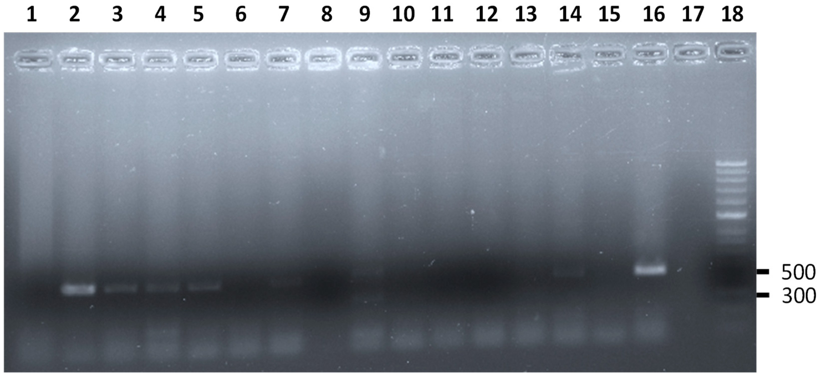
Figure 1. Results of the m-PCR assay for the simultaneous detection of stx1 and stx2 genes of E. coli STEC from Anolis sp. analyzed. Lane 2, 3, 4, 5, 7, and 14 amplification fragments of the stx2 gene characteristic of E. coli STEC obtained from Anolis sp. analyzed in this study (516 bp). Lane 9 amplification fragments of the stx1 (302 bp) and stx2 gene characteristic of E. coli STEC obtained from one Anolis sp. analyzed in this study. Lane 1, 6, 8, and 15 Anolis sp. negative for E. coli STEC. Lane 16, positive control stx2 gene. Lane 17, control negative. Lane 18, molecular size marker (HyperLadderTM 50 bp, Bioline).
Animals 13 00414 g001
Figure 2. Results of the m-PCR assay for the simultaneous detection of Salmonella spp. and S. enteritidis, S. typhimurium, and S. typhi from various Anolis sp. analyzed. Lane 2, 3, and 4, amplification fragment of the ompC gene characteristic of Salmonella spp. obtained from Anolis sp. analyzed in this study. Lane 5 to 10, Anolis sp. negative for Salmonella spp. Lane 11, positive control. Lane 12, control negative. Lane 9, molecular size marker (HyperLadderTM 50 bp, Bioline).
Animals 13 00414 g002
Figure 3. Results of the m-PCR assay for the simultaneous detection of Pseudomonas spp. and P. aeruginosa from the Anolis sp. analyzed. Lane 1 and 3, amplification fragment of the oprI gene characteristic of Pseudomonas sp. obtained from Anolis sp. analyzed in this study. Lane 2, 4, 5, and 6, Anolis sp. negative for Pseudomonas sp. Lane 7, positive control. Lane 8, control negative. Lane 9, molecular size marker (GeneRuler 50 bp DNA Ladder, Thermo Scientific).
Animals 13 00414 g003
Table 1. Percentage of pathogen bacteria isolated from Anolis sp. from Tenerife (Canary Islands).
Bacteria TypeNo. of Isolates (n)Prevalence (%)
(CI 95%)
Pseudomonas spp.18 (28)64.3 (45.5–82.1)
E. coli (stx1/stx2/eae genes)33 (74)44.6 (38.8–50.3)
Campylobacter spp.24 (74)32.4 (21.4–42.6)
Staphylococcus lugdunensis11 (74)14.9 (10.7–19.1)
Vibrio spp.8 (74)10.8 (3.7–17.8)
Salmonella spp.4 (74)5.4 (4.4–6.6)
Listeria monocytogenes0 (74)0.00
Yersinia enterocolitica0 (74)0.00
Mycobacterium spp.0 (74)0.00
Table 2. Prevalence of Campylobacter sp. detected in Anolis sp. from Tenerife, Canary Islands.
Campylobacter SpeciesPositive
Males
(Prevalence) (CI 95%)
n = 25
Positive
Females
(Prevalence) (CI 95%)
n = 45
Positive Juveniles
(Prevalence) (CI 95%)
n = 4
Total of Positive Individuals
(Prevalence) (CI 95%)
n = 74
Campylobacter fetus2 (8.0%)
(2.6–18.6)
2 (4.4%)
(1.6–10.4)
2 (50.00%)
(1.0–99.0)
6 (8.1%)
(1.9–14.3)
Campylobacter coli05 (11.1%)
(1.9–20.3)
05 (6.8%)
(1.0–12.5)
Campylobacter jejuni3 (12.0%)
(0.7–24.7)
1 (2.2%)
(0.0–6.5)
04 (5.4%)
(0.2–10.5)
Campylobacter upsaliensis2 (8.0%)
(2.6–18.6)
1 (2.2%)
(0.0–6.5)
03 (4.1%)
(0.0–8.5)
Campylobacter lari0000
Table 3. Prevalence of enteropathogenic E. coli detected in Anolis sp. from Tenerife, Canary Islands.
Virulence GenesPositive
Males
(Prevalence)
(CI 95%)
n = 25
Positive
Females
(Prevalence)
(CI 95%)
n = 45
Positive
Juveniles
(Prevalence)
(CI 95%)
n = 4
Total Positive
Individuals
(Prevalence)
(CI 95%)
n = 74
stx14 (16.0%)
(1.6–30.3)
4
(0.5–17.1)
08 (10.8%)
(3.7–17.9)
stx26 (24.0%)
(7.3–40.7)
14
(17.6–44.6)
1 (25.0%)
(0.0–67.4)
21 (28.8%)
(18.1–38.7)
eae1 (4.0%)
(3.0–11.7)
2 (4.4%)
(0.0–5.9)
1 (25.0%)
(0.0–67.4)
4 (5.4%)
(0.3–10.5)
Table 4. Percentage of co-infections of pathogen bacteria isolated from Anolis sp. from Tenerife (Canary Islands, Spain) (n = 74).
No. of IndividualsMixed Infection of Bacteria/Virulence GenPercentage %
7Campylobacter spp. + stx2 gene30.4
6Campylobacter spp. + Staphylococcus lugdunensis26.1
5Campylobacter spp. + stx1 gene21.7
3Campylobacter spp. + Salmonella spp.13.0
3stx2 gene + Vibrio spp.13.0
3stx2 gene + Staphylcoccus lugdunensis13.0
2Campylobacter spp. + Vibrio spp.8.7
2stx1 gene + Staphylcoccus lugdunensis8.7
2stx1 gene + stx2 gene8.7
1Campylobacter spp. + eae gene4.4
1stx1 gene + Salmonella spp.4.4
1stx1 gene + Vibrio spp.4.4
1Staphylcoccus lugdunensis + Vibrio spp.4.4
Table 5. Index of co-infection (Ic) for the different co-infections obtained from Anolis sp.
Mixed Infection of Bacteria/Virulence GenIcChi-Square
Campylobacter spp. + stx2 gen0.280.29
Campylobacter spp. + Staphylococcus lugdunensis4.806.32 *
Campylobacter spp. + stx1 gen16.9210.2 *
Campylobacter spp. + Salmonella spp.10.4817.6 *
stx2 gen + Vibrio spp.7.807.24 *
stx2 gen + Staphylcoccus lugdunensis2.863.98
Campylobacter spp. + Vibrio spp.−1.9610.2 *
stx1 gen + Staphylcoccus lugdunensis10.230.54
stx1 gen + stx2 gen−1.007.24 *
Campylobacter spp. + eae gen−0.9317.1 *
stx1 gen + Salmonella spp.5.181.44
stx1 gen + Vibrio spp.2.260.00
Staphylcoccus lugdunensis + Vibrio spp.−1.050.52
* and bold type: significant differences between the number of co-infections obtained and expected. * no bold type: non-significant associations between the pathogens studied.
Disclaimer/Publisher’s Note: The statements, opinions and data contained in all publications are solely those of the individual author(s) and contributor(s) and not of MDPI and/or the editor(s). MDPI and/or the editor(s) disclaim responsibility for any injury to people or property resulting from any ideas, methods, instructions or products referred to in the content.