Article

Metabolic Responses and Resilience to Environmental Challenges in the Sedentary Batrachoid Halobatrachus didactylus (Bloch & Schneider, 1801)

by
Juan Manuel Molina
1,2,3,*,
Andreas Kunzmann
2,
João Pena Reis
3 and
Pedro Miguel Guerreiro
3
1
Instituto Argentino de Oceanografía (CONICET), Bahía Blanca B8000, Argentina
2
Leibniz-Zentrum für Marine Tropenforschung (ZMT), 28359 Bremen, Germany
3
Centro de Ciências do Mar (CCMAR), Universidade do Algarve, 8005-139 Faro, Portugal
*
Author to whom correspondence should be addressed.
Animals 2023, 13(4), 632; https://doi.org/10.3390/ani13040632
Submission received: 26 December 2022 / Revised: 6 February 2023 / Accepted: 10 February 2023 / Published: 11 February 2023
(This article belongs to the Special Issue Metabolic Adaptation and Regulation in Aquatic Animals)

Abstract

:

Simple Summary

In the context of climate change, warming of the seas and expansion of hypoxic zones are challenges that most species of fish are, or will be subjected to. We provide first-time estimates on the metabolic rates, thermal, and oxygen-related limits for Halobatrachus didactylus, a coastal sedentary fish that lives in intertidal environments of the Northeast Atlantic. The metrics obtained in this study prove that H. didactylus is remarkably resilient to acute environmental variations in temperature and oxygen content, which might enable it to adapt to the extreme abiotic conditions forecasted for the world’s oceans in the near future.

Abstract

In the context of climate change, warming of the seas and expansion of hypoxic zones are challenges that most species of fish are, or will be subjected to. Understanding how different species cope with these changes in their environment at the individual level can shed light on how populations and ecosystems will be affected. We provide first-time estimates on the metabolic rates, thermal, and oxygen-related limits for Halobatrachus didactylus, a coastal sedentary fish that lives in intertidal environments of the Northeast Atlantic. Using respirometry in different experimental designs, we found that this species is highly resistant to acute thermal stress (CTmax: 34.82 ± 0.66 °C) and acute hypoxia (Pcrit: 0.59–1.97 mg O2 L−1). We found size-specific differences in this stress response, with smaller individuals being more sensitive. We also quantified its aerobic scope and daily activity patterns, finding this fish to be extremely sedentary, with one of the lowest standard metabolic rates found in temperate fish (SMR: 14.96 mg O2 kg−1h−1). H. didactylus activity increases at night, when its metabolic rate increases drastically (RMR: 36.01 mg O2 kg−1h−1). The maximum metabolic rate of H. didactylus was estimated to be 67.31 mg O2 kg−1h−1, producing an aerobic scope of 52.35 mg O2 kg−1h−1 (77.8% increase). The metrics obtained in this study prove that H. didactylus is remarkably resilient to acute environmental variations in temperature and oxygen content, which might enable it to adapt to the extreme abiotic conditions forecasted for the world’s oceans in the near future.

1. Introduction

Climate warming produces changes in water temperature and, consequently, in oxygen availability, which forces marine ectotherms to acclimate and, in the long-term, adapt by means of metabolic and behavioral changes [1,2,3]. According to the most favorable prediction of the Special Report on Emissions Scenarios, by the year 2100, the oceans’ temperature and oxygen minimum zones will increase [4]. These changes in the abiotic conditions of the oceans are expected to produce severe impacts on marine organisms [4,5]. Most of these changes will occur in coastal areas, often in systems already affected by important natural fluctuations in temperature and oxygen levels [1,3,4,5]. One example of such environments are coastal lagoons, which have a major ecological role, functioning as nurseries for pelagic species and habitats for many sedentary fish that also provide important ecosystem services [6,7].
In this context, increasing our understanding of thermal and hypoxia tolerance and the stress responses of fish on an organismal level is of high relevance. This is also important from an economic point of view, for fisheries and aquaculture [5]. Despite the relevance of this type of research, information on the thermal and oxygen-related limits is scarce or lacking for a large proportion of fish species [8]. Temperature and hypoxia tolerance have been traditionally approached by measuring the critical thermal maximum (CTmax) and critical oxygen partial pressure (Pcrit) [9]. CTmax is a measure of an animal’s upper thermal tolerance limit. Its determination consists of exposing groups of fish to a constant increase in temperature until some form of physiological failure is evident (e.g., loss of equilibrium, gill ventilation stops, inability to move or react) [8,10]. The biological threshold meaning of this metric is “the thermal point at which locomotory activity becomes disorganized and the animal loses its ability to escape from conditions that will promptly lead to its death” [10] (p. 1562). CTmax is considered to be ecologically relevant, with relatively high repeatability, and is methodologically straight-forward, making it relevant for continued use as a metric for acute thermal tolerance [8,9,11]. Aside from CTmax, which has been employed extensively and provides an important metric of thermal tolerance, other methods have also been employed to measure the effect of increased temperature on the physiology of fish [12]. Temperature ramps, for example, have been employed to measure the effect of acutely increasing the temperature on fish, and are considered to be good at replicating natural stressors like those affecting migrant fish or coastal and intertidal pool inhabitants [13,14,15,16,17].
Pcrit represents the oxygen partial pressure at which oxygen consumption switches from a regulator to conformer on oxygen partial pressure [18,19,20]. Pcrit has also been given the biological threshold meaning of a value below which activity can no longer be sustained aerobically [20,21,22]. Several authors have challenged this view of Pcrit as the sole metric to look into hypoxia, raising concerns about inconsistent or incorrectly applied methodologies as well as suggesting alternative metrics [23,24]. Methodologically, however, alternatives to Pcrit have not been fully explored, and the theoretical alternatives are not widespread enough to be validated by their use on different species [24]. Regardless of these discrepancies, the basic methodology is subjecting fish to an ever-decreasing oxygen tension while measuring their oxygen consumption. The tension at which the fish consumption of oxygen becomes dependent on the oxygen tension is considered the Pcrit [20].
The European toadfish Halobatrachus didactylus is a sedentary species, with mostly sit-and-wait predatory habits, and is a member of the order Batrachoidiformes. This fish is distributed all along the Western Mediterranean coast [25] and the Central and Northeast Atlantic [26,27]. It has low commercial value, but is commonly caught by artisanal fishermen of southern Portugal and is sold at local markets [28]. H. didactylus can reach lengths of 50 cm in total, being one of the largest Batrachoididae species [29]. It inhabits shallow waters near coasts, estuaries, and coastal lagoons [30]. It prefers muddy and sandy sediments where it can burrow, and remains covered by sediment, algae, or rocks for long periods of time, waiting for prey or resting [31]. The species has been used as an experimental model for studies on toxicology, cardiac function, behavior, and spatial ecology [32,33,34,35]. Physiological and life-history traits influence how species respond to abiotic factors, which means that lifestyle alone could be an important proxy for how a certain species will respond to environmental change [12,36,37,38,39]. The benthic-associated life history traits of H. didactylus make it an inevitable subject of the particular stressors of its habitat, and its coastal distribution and association with intertidal mudflats subjects them to diurnal changes in temperature as a result of the tidal regime. These changes in temperature are of quite notable magnitudes (10–30 °C) [40]. H. didactylus, other batrachoids, and even other fish inhabiting these highly variable ecosystems, with a wide range of temperatures and oxygen levels, have developed physiological adaptations that allow them to be able to survive and reproduce. Despite the relative wealth of knowledge on H. didactylus, many of its physiological traits remain unknown or understudied, which are needed to fully understand their physiological resilience. The work presented here is the first of a series of studies in which we explore the effects of man-made climate change stressors on the physiology of H. didactylus.
In this paper, we aimed to quantify and determine the aerobic energy budget, thermal limits (CTmax), and critical oxygen tension (Pcrit) of the Lusitanian toadfish H. didactylus.

2. Materials and Methods

2.1. Sourcing and Housing

Individuals of H. didactylus were sourced from the coastal lagoon Ria Formosa, located near Faro, in the Algarve region of Portugal, during December 2019 and January 2020. This lagoon is highly productive and suffers from considerable anthropogenic pressure and economic exploitation, but as a Natural Park, it has benefited from several protection measures against highly destructive commercial fishing methodologies [41]. H. didactylus individuals were captured using a beam trawl, at night, close to the shore or close to seagrass beds [42] at depths ranging from 1 to 2 m and for up to 15 min. The net was pulled by hand and when the catch was onboard, H. didactylus were immediately sorted by size and retained in containers with aerated seawater. Fish were then quickly transported to a common open circulation system fed with water pumped from the lagoon, located in the nearby Ramalhete Marine Station (University of Algarve), and allocated by size range in large acclimation tanks (volume 600 L, bottom area 1.3 m2). The fish were allowed to recover in these acclimation tanks for 1 month from the stress originated from the capture and transport. During this period, they were fed squid and mussels (every other day, 3% w/w tank biomass), and kept at natural temperature, salinity, and photoperiod (Jan 2020: 12.9 ± 0.28 °C; 35.1 ± 0.05 PSU; 10 light hours (L) and 14 no light hours (D); Feb 2020: 15.9 ± 0.11 °C; 35.6 ± 0.06 PSU; 11L:13D). As the species inhabits crevices and small caves, PVC tubes for shelter were provided, which were readily accepted. Only juvenile fish were employed in this study because of the confounding effects that gonadal development may produce on energy expenditure.
Each of the trials described in the following sections utilized independent samples, that is, once a fish was used for an experiment, it was returned to a different acclimation tank, and not used again for any other procedure. This protocol ensured independence of the results, albeit limiting the size range and number of individuals for each trial.
Fish were collected under special license from the Portuguese Agency for Nature Conservancy (ICNF; 7048/2020/DCBN/DAN) and the General Directorate for Marine Resources (DGRM; 1009/2019/DRI). Experiments were conducted following the guidelines established by the EU Directive 2010/63/EU and the Portuguese Decree Law No. 113/2013 on “The protection of animals used for scientific purposes”. The experimental design was previously approved by the CCMAR Ethical Committee for Managing Animal Welfare (ORBEA) and by the Portuguese Veterinary Authority (DGAV) under permit 009272. Fish manipulation was performed by accredited scientists in laboratory animal science by the Portuguese DGAV, following the FELASA category C recommendations.

2.2. Metabolic Rates Measurement Procedure

To estimate the metabolic rates (MRs) of H. didactylus, we measured the oxygen consumption by means of a respirometry array. In designing our respirometry system, we followed the guidelines outlined in [43]. This array consisted of four flow-through transparent respiration chambers made of acrylic (1.7 L and 2.5 L chambers, approximately between 20 and 55 respirometer:fish volume ratio depending on the fish size) that were outfitted with circulation and in/out flow control pumps, and placed inside 200 L tanks (measuring tanks). The tanks had top PVC covers to reduce the visual disturbance of the fish. The dissolved oxygen concentrations were measured inside each chamber with an optical oxygen sensor probe (Pyroscience GmbH, Germany) that directly fed data in 20 s intervals (computing an average of 20x 1 s data points) to a computer using the FirestingGo interface and dedicated Pyroscience software. Oxygen was measured in “respiration cycles” that consisted of two phases. In the first phase, the chamber was sealed, allowing the fish to consume the oxygen in the chamber (from 100% to a minimum of 85% O2 saturation). The second phase consisted of flushing the chamber with fresh sea water by means of an electrically activated pump. The system was configured to measure for 8 min and flush for 2 min. Animals measured for oxygen consumption were left unfed 48 h prior to the measurement to ensure a post absorptive state [9]. The number of cycles conducted (and therefore the total measuring time) varied depending on the experimental set-up performed, as described in the following sections. Similar set-ups are described in greater detail in [9,44,45].

2.3. Standard, Routine and Maximum Metabolic Rate

In this study, we estimated the standard, routine, and maximum metabolic rates (SMR, RMR, and MMR, respectively) of H. didactylus. Sixteen individuals (size range: 13.9–147.9 g) were selected to run the SMR and RMR determinations, while a different set of 12 animals (size range: 60.0–119.3 g) were selected for the MMR trial. For this work, and following the framework for respirometry research [9], SMR is considered to be the MR of a post-absorptive fish in a quiescent and calm state. The RMR is the consumption of oxygen of a fish that is calm, but performs at-will movements or other minor activities during the measurement [9]. For SMR and RMR, fish were simply transferred to the measuring tanks and into the respiration chambers at 9 am, and the oxygen measurement was started right away. These procedures lasted for 75 hs. For MMR determination, we followed the standard protocols outlined in the literature [9,43,46,47]. Briefly, fish were transferred to an annular pool (0.75 m in circumference) with water fed from the acclimation tanks, and chased around by gently touching the caudal region. The fish would swim in bursts for a short period and then remain motionless, a point at which further stimulus was provided. The fish were considered exhausted when they stopped reacting to tail pinching. Time until exhaustion (in minutes) and the number of laps completed were recorded. Bony fish have been shown to recuperate from exercise quickly, and such recovery can occur even when the fish is unresponsive, and when being moved from the exercise pool and inserted in the respirometry chamber [46,47,48,49]. For this reason, fish in this state were transferred to the measuring tanks and into the respiration chambers, and the oxygen measurement was started right away (less than 20 s). Respiration cycles in this case were inverted, with 2 min of measuring time and 8 min of flushing to allow the fish to recuperate properly, preventing fast depletion of oxygen within the sealed chamber [49]. This procedure lasted for 24 h.
We performed 24 h measurements (based on suggestions from the literature, e.g., [9,43,46]) to determine SMR, RMR, MMR and the time until the MR values stabilized after the initial handling and after exhausting exercise. These experiments were run at 15 °C with fish previously acclimated in the acclimation tanks (12–16 °C, see sourcing and housing section for more details on acclimation conditions).
After completing the respirometry trial, fish were measured for TL (mm), weight (g), and volume (L) before being returned to the acclimation tanks. The volume of each animal was measured using the water displacement technique, which consists of measuring the volume of water displaced by submerging the animal in a graded container.

2.4. Mass-Scaling Exponent

The dissolved oxygen (DO) data to calculate the mass-scaling exponent (ME) were taken from the measurements collected from the 16 individuals used in the SMR/RMR section. Data points were those corresponding to the stabilized and resting individuals. There are no external sexual dimorphisms in the juveniles of this species, and since the individuals were not euthanized, it was impossible to determine an exponent for females and males separately. All animals were at a length below sexual maturity, therefore, a separate exponent for adults was not calculated.

2.5. Critical Thermal Maximum (CTmax) Trial

CTmax was estimated by subjecting 16 individuals (26–82 g) to a rapid increase in temperature until they lost equilibrium by recording the time and temperature. Fish were placed in an aquarium connected to a tank, outfitted with a circulation pump and a heating element, and left undisturbed for 6 h (double of what was determined in the SMR/RMR trial) before the trial started. The water temperature started at 15 °C (the acclimation temperature) and was increased by the heating element in the tank to provide a gradual and smooth increase in the water temperature in the aquarium, at a rate of approximately 1 °C each 20 min [12,15]. Oxygen saturation was monitored and maintained at 85–100% throughout the trial [9] with compressed air delivered through several aeration stones. Water temperature in the aquarium was increased until all fish had lost equilibrium, which was the CTmax endpoint [9]. When a fish lost equilibrium, the time and temperature were noted, and it was removed from the container, weighed, and measured (TL (mm), weight (g), and volume (L)). After these procedures, they were placed in a container with the same water. When the temperature naturally dropped to 15 °C, the fish were returned to the acclimation tanks. All except one fish recuperated from this trial.

2.6. Temperature Ramp

To further explore the effects of acute temperature stress as a result of tidal regime, we employed a temperature ramp protocol. This protocol was suggested by Vinagre et al. [15], and further employed in Campos et al. and Rangel and Johnson [12,50]. Thirty-two fish (size range: 13.2–159.1 g) were taken, eight at a time, from the acclimation tanks, and transferred to the respiration chambers of the measuring tanks of the respirometry array at 16:00 in the afternoon. The water temperature of the measuring tanks was 12 °C. According to the results of the aerobic scope section, fish were left in the chambers undisturbed for 6 h (double of what was determined in the SMR/RMR trial), with the flushing pump always on, so that their MR values stabilized. After this time, the computer-controlled water temperature was reduced to 8 °C over 4 h in order to further reduce the oxygen consumption. After 4 h at 8 °C, the temperature was increased. The increases were programmed so that the temperature would rise 2 °C in an hour, remain stable for another hour, and then rise again in the same stepwise manner up to 32 °C in the course of 24 h, while measuring the oxygen consumption. Temperature was left to descend to 28 °C before removing the fish from the chambers, measuring their TL (mm), volume (L), and weight (g). After these procedures, they were placed in a container with the same water. When the temperature naturally dropped to 15 °C, the fish were returned to the acclimation tanks.

2.7. Critical Oxygen Tension Trial

Acute hypoxia tolerance was estimated using eight individuals (43.7–81 g), which were taken from the acclimation tanks and transferred to the respiration chambers of the respirometry array at 07:00 in the morning, and left undisturbed for 6 h (double of what was determined in the SMR/RMR trial). The respirometry array was started with 100% O2 saturated water (8.5 mg O2 L−1 at 15 °C), and thereafter, the saturation was lowered 10% every 30 min. We achieved this by bubbling N2 in the measuring tank, which was covered with plastic bubble wrap to minimize the loss of N2, until the desired saturation point was reached. Oxygen saturation in the measuring tank was monitored using a handheld ProSolo Optical Dissolved Oxygen and Conductivity Meter fitted with an optical, non-consumptive DO sensor (YSI, Yellow Springs, OH, USA, https://www.ysi.com/prosolo-odoct (accessed on 15 January 2020)). Saturation was lowered until 2% saturation (0.25 mg O2 L−1 at 15 °C) was reached, or the fish lost equilibrium [9,20,24]. Loss of equilibrium in this species was readily apparent, as the fish tilted to one side on the gentle water flow of the chamber. Immediately after the fish lost equilibrium, or by the end of the trial (at 2% DO), the fish were removed from the chambers, weighed, and measured for TL and volume, and then returned to their acclimation tanks.

2.8. Data Analysis

The normality of the error structure of the oxygen data was assessed with a quantile–comparison plot, and the kurtosis and skewness tests as well as mean–variance plots. Certain distributions were normal (i.e., SMR, RMR, MMR, CTmax data) and others were log-transformed (i.e., ME, Pcrit, temperature ramp), after which the data fitted well to a normal distribution; therefore, we employed linear statistics for further analysis [51].

2.8.1. Metabolic Rates

The dissolved oxygen level in the respiration chambers was plotted against time for each respirometry cycle of an individual fish and a regression slope was obtained. These regression slopes were used to calculate the metabolic rates, modified from Clark et al. [52] as:
MR = VO2 × VR × (MME)1 × t−1,
where MR is the metabolic rate of the fish, measured as mg O2 kg−1h−1; VO2 is the slope of the linear model fitted to the oxygen concentration (mg L−1) data from the start through to the end of each cycle; VR is the volume of the respirometer chamber (L) calculated as the total volume of the chamber minus the volume of each animal; M is the total wet body mass of the animal (kg); and t is the duration (hours) of the cycle. ME is the mass-scaling exponent. This exponent was estimated fitting a linear regression model using the logarithm of DO and fish weight (n = 28) and the slope (the ME exponent) was extracted from the model. This exponent was used to calculate MR in the SMR set of individuals to showcase the effectiveness of the calculated ME. An ANOVA, followed by a Tukey pairwise comparison, was used to test the possible differences in the MR between fish sizes [51].
To estimate the acclimation-to-respirometer times, we performed an ANCOVA test to detect the differences in the slopes between the cycles for each individual fish from the SMR/RMR trial during the 24 h of measurements [53]. The initial slopes were more pronounced, and gradually became smaller with the passage of time. Once the analysis found no significant differences between the slopes, the animal was assumed to be stable and resting. Time to acclimation was computed and averaged between all the fish that took part in the trial. This acclimation-to-respirometer time was estimated to be 3.2 h. This exact same analytical process was employed to determine how much time it took for the fish in the MMR experiment to recuperate from exhaustion and regain steady oxygen consumption.
In order to estimate the daily oscillations in the activity of this species, the MR data from the SMR/RMR trial were used to fit a sine wave function using non-linear regression (function nls of R base library). We used three error terms for harmonic oscillation in order to mechanistically characterize the daily fluctuations in activity, according to the formula:
Metabolic rate (mg O2 kg−1h−1) = amplitude × sine (2 × π × frequency × time of day + phase) + offset
Additionally, data were grouped obtaining a median, standard deviation, and the first and third quartiles per hour. SMR and RMR were determined following the recommendations of Remeyer and Rees, and Killen et al. [20,39] for standardized metabolic measurements and calculations. Briefly, for SMR, we opted for the median of the quantiles that placed SMR below the lowest 20% of the daylight (inactive phase) observations (q 0.2) for each fish. For RMR, the median of the quantiles that placed RMR above the highest 80% of the night (active phase) observations (q 0.8) for each fish were used [20].
To determine MMR, we employed the rolling regression method with a window width of 1 min [49]. Next, we used an ANCOVA test to detect the differences in the slopes between the cycles for each individual fish [53]. Oxygen consumption recorded for the highest significantly different period of measurements in the MMR trial was recorded per fish, and expressed as the median of the quantiles that placed MMR above the highest 80% of the measurements of all fish (q 0.8). We then plotted the MR vs. time using boxplots, with the significantly different time interval where MMR was found, followed by hourly intervals to visualize recovery. Finally, individual splines were fitted to each fish data points [51].
Aerobic scope and routine aerobic scope were calculated both as the differences and as the rate between MMR and SMR, and RMR and SMR, respectively [9,46,49,54].
The effect of size (as weight) in the MRs determined in this section was tested using linear regression [51].
Additionally, for MMR, the effect on the estimation of MR from the trainer and time being chased was also tested for significance. Stepwise deletion of non-significant terms in a linear model including all terms was the method employed [51]. Concrete examples and a more thorough description of this method are given elsewhere (e.g., [55,56]).

2.8.2. CTmax and Acute Thermal Tolerance

A linear regression model was used to test whether CTmax changed with weight, given the normal distribution of the data [51].
The acute temperature exposure data were analyzed using recursive partitioning and regression trees (RPRT) [57]. This method is used to split the predictor variable/s (in our case, temperature and fish size) into statistically significant ranges within which the response variable (in our case, the MR) remains significantly constant. Briefly, the algorithm starts with the complete dataset and tests if it should be split. If the null hypothesis is rejected, the variable is split into two new ranges. The steps will be repeated within both of the new ranges (hence the “recursive partitioning”). The tree stops growing, if in all partitions a global null hypothesis of independence cannot be rejected [57]. The procedure defines “nodes” or divergent values in the continuous variable from which the statistically different ranges are grouped [57], making it possible to discern where the changes in MR occur in the ranges employed. Using the R package rpart, with the “anova” method, we ran a RPRT using MR as the response variable with fish weight and temperature as the predictor variables to partition them into ranges [57]. The MR of each size class at each range of temperature was compared by ANOVA and Tukey pairwise comparisons [51].

2.8.3. Pcrit

Pcrit for each fish was calculated following the methods proposed by [20,24] (herein presented as the “α method” and the “LLO method”, respectively). The former study suggested the estimation of α as a species and temperature specific constant based on the metabolic rate dependence of Pcrit (Pcrit = MR/α). Oxygen consumption was monitored as the O2 partial pressure declined and was divided by the corresponding O2 partial pressure to obtain α0. The mean of the three highest values of α0 was designated as α. The later study defined Pcrit as the O2 partial pressure at which O2 consumption dropped below its SMR. A linear relationship between O2 consumption and O2 partial pressure was fitted (to data collected after O2 consumption fell below that individual’s SMR) and from this relationship, the O2 partial pressure where O2 consumption equals SMR is considered as Pcrit. We performed both calculations for each of the eight fish in our critical oxygen tension trial, and then averaged the results to obtain an estimate for the species’ Pcrit.
All tests used a significance level of α ≤ 0.05. Data were processed using R statistical software [58] using scripts specifically developed for this purpose and functions in the respirometry, FSA, and lme4 packages.

3. Results

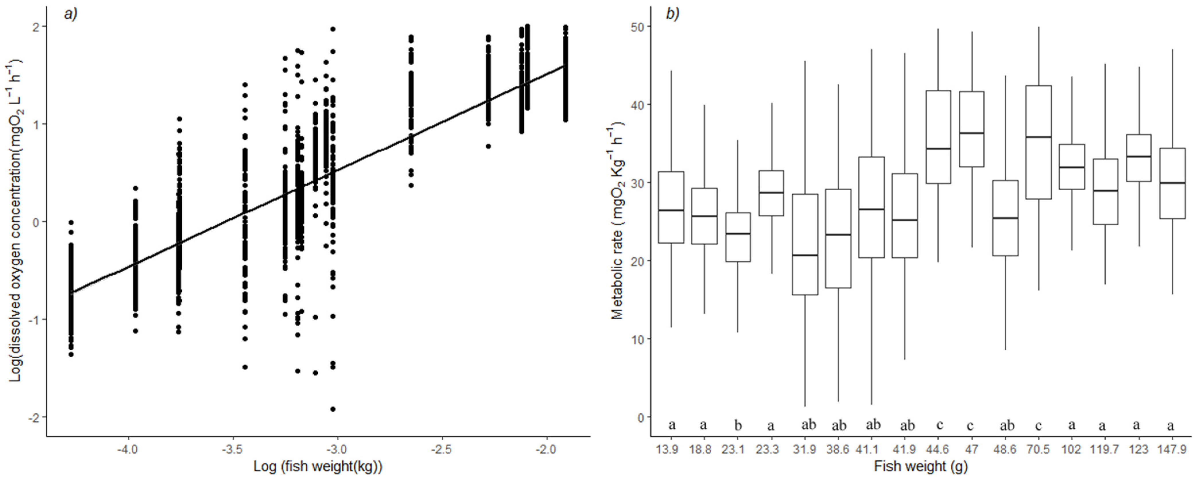
3.1. Mass-Scaling Exponent of Oxygen Consumption

The general mass-scaling exponent (ME) calculated for H. didactylus was 0.96 +/− 0.08 (Figure 1a) at 15 °C. For each of the 16 fish in the SMR/RMR trial, Figure 1b presents the individual variations of MR. This figure was built using ME. As expected, there was no observable pattern in the weight–MR relationship, meaning that the general ME fit well with the data. Mean MR was 26.44 mg O2 kg−1h−1. Three individuals presented a higher-than-average MR (p < 0.05) (Figure 1b) partly due to excessive spontaneous movement during measurement.

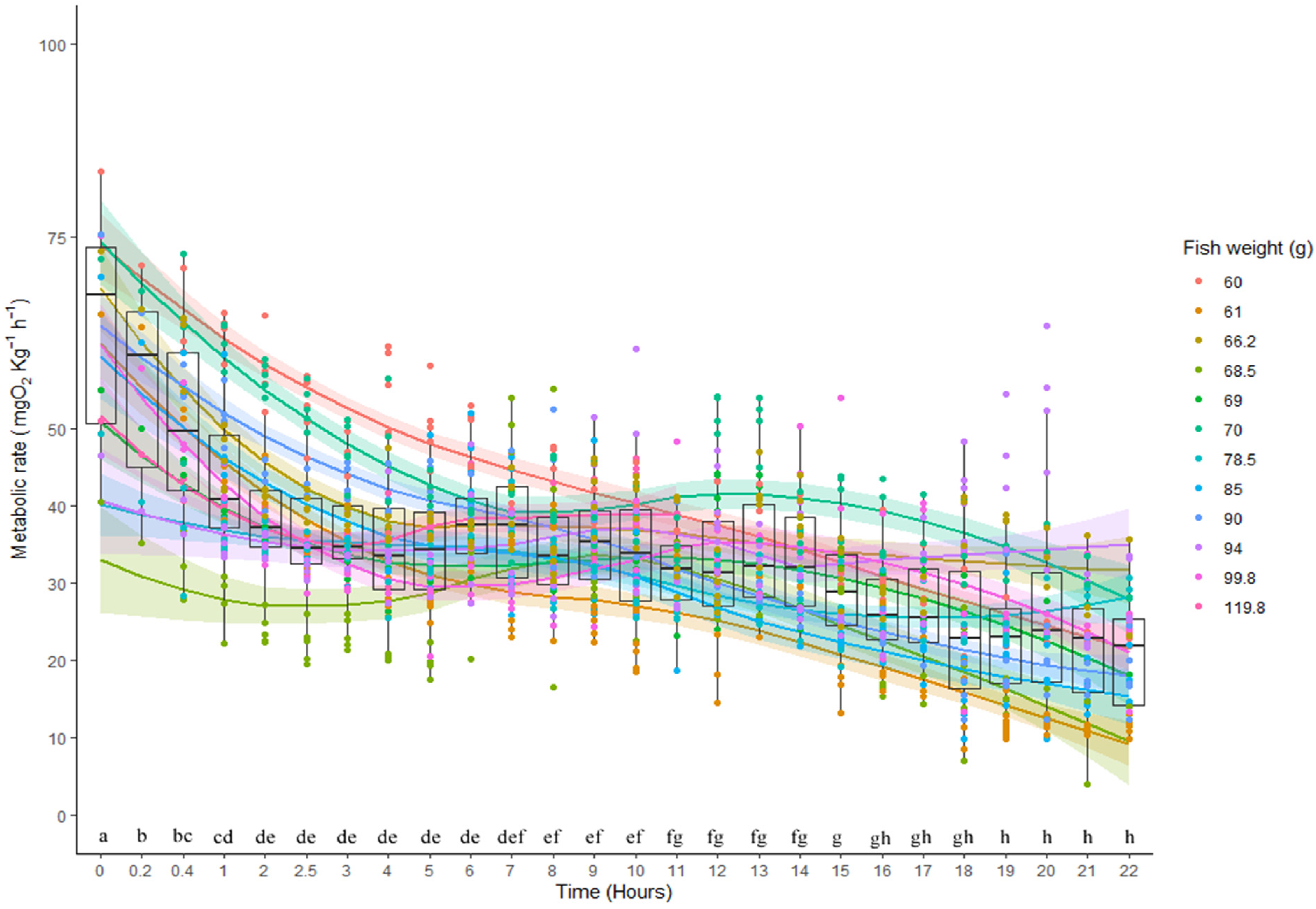
3.2. Metabolic Rates and Aerobic Scope

At 15 °C, the SMR of Halobatrachus didactylus was estimated at 14.96 mg O2 kg−1h−1 and the RMR at 36.01 mg O2 kg−1h−1. Based on these estimates, the routine aerobic scope for this species was calculated to be 21.05 mg O2 kg−1h−1 (58.5%). In Figure 2, the hourly variations in the MR of the 16 fish that took part in the SMR/RMR determination were plotted for the total duration of 75 h. The daily pattern of higher activity (RMR) and quiescence (mainly SMR) was evident for each 24 h period. The initial highly variable period of approximately 3 h was in accordance with the acclimation-to-respirometer estimation presented in the previous section. This activity pattern of H. didactylus can be described by the fitted sine function. Despite the high variability in the data, it was observed that the activity pattern was nocturnal, with peak oxygen consumption occurring at around 21:00, when the fish were actively moving and swimming, followed by a decrease in oxygen consumption as the day progressed, with minimum activity at around 09:00 (Figure 2).
The following function describes the sinusoidal oscillation of the MR of H. didactylus throughout the day:
Metabolic rate (mg O2 kg−1h−1)= 5 (+/−0.5) × sine (2 × π × 1 (+/−0.01) × time of day + 5.3 (+/−0.1)) + 29 (+/−3).
Exhaustive exercise produced a marked increase in the MR of H. didactylus. We estimated a MMR of 67.31 mg O2 kg−1h−1, producing an aerobic scope of 52.35 mg O2 kg−1h−1 (77.8%). Figure 3 showcases this high MR and the subsequent decrease in a 24 h period. It was evident that after 5 to 6 h, the MR seemed to have stabilized, only to fall to lower values after the 19 h mark. This second decrease in MR might be explained by the dial activity pattern of the species. The SMR estimated using the latter values (20.8 mg O2 kg−1h−1) was close to the SMR estimation produced by the set of individuals used in the SMR trial (Table 1). H. didactylus was able to tolerate exhaustive exercise for a median of 22 min before being completely exhausted.
Linear regressions fitted to SMR, RMR, and MMR showed no effect of size on these variables (t = 1.287, p = 0.22; t = 0.63, p = 0.53; t = −0.723, p = 0.48, respectively) as would be expected from employing an appropriate ME. Size and time until exhaustion were not found to have an independent statistical effect on MMR. The full model including the interactions between size and time was not found to be different from the null model (F = 0.4406, p = 0.7303).

3.3. CTmax

Halobatrachus didactylus presented a mean CTmax of 34.82 +/− 0.66 °C (n = 16). Results from the CTmax trial are summarized in Figure 4. The linear relationship of CTmax and weight is described by the function:
CTmax = 33.39(+/−0.43) °C + 0.02 (+/−0.008) * Weight.
The fit of the CTmax to weight showed a low positive correlation (R2: 0.380, n = 16, F: 3.094, p = 0.039).

3.4. Temperature Ramp

The regression tree employed on the temperature ramp data found significantly different MRs at 13 temperature ranges, with a total of 22 tree nodes (Supplementary Material Figure S1a) and at five size ranges, termed as follows: smallest: 13–22.9 g, small: 23–33.9 g, medium: 34–48.9 g, large: 49–140.9 g, largest: 141–159 g (Supplementary Material Figure S1b). More temperature ranges were found in the 9 degrees above 23 °C than in the 11 degrees below, showing a differential effect of temperature on the MR of this species at higher temperatures. In Figure 5, these differences are showcased for each fish size considered. As the temperature increased, the size effect of the fish became more variable and higher at a steady pace. Beyond 29 °C, the MR of H. didactylus became increasingly higher and variable. The large and largest animals were consistently similar in their responses to increased temperature, with smaller animals being more variable, and consistently differently affected by temperature increases, as can be observed by the statistical differences in Figure 5. Smaller fish seem to be more susceptible to increased temperature, showing increased metabolic rates and higher variability in their responses. The smallest fish had to be taken out of the chambers before the experiment could be concluded due to the loss of equilibrium at 26 °C.

3.5. Pcrit

Critical oxygen tension for H. didactylus was estimated at 1.61 +/− 0.36 mg O2 L−1 using the α method, and 1.27 +/− 0.68 mg O2 L−1 using the LLO method, for fish acclimated at 18 °C (Figure 6). Single regression lines (MR versus PO2) were plotted through the oxygen-limited portion of the curve and intersected with individual SMR established at high PO2. The results obtained using both methods showed Pcrit to range between 0.59 and 1.97 mg O2 L−1. The average SMR for these trials was 38.59 mg O2 L−1 Kg−1. Fish weights ranged from 43.7 to 81.0 g.

4. Discussion

In this article, we explore the responses of H. didactylus to two of the most typical climate change abiotic stressors. We profiled the metabolic performance of this species, providing the first estimates of SMR, RMR, MMR, aerobic scope, CTmax, Pcrit, and daily aerobic activity. Aside from these physiological parameters, we also provide reference methodological information such as the first ME estimation, acclimation times, and significant temperature ranges for use in future studies concerning this and other closely related species. Toadfishes have been used as models in several metabolism-related studies in the past 50 years (e.g., [59,60,61,62,63]), but there is little information on the active metabolic rates and responses to environmental changes of toadfishes. Indeed, this is, in fact, to our knowledge, the first study to show the whole organism metabolic rates in relation to environmental temperature and oxygen levels, in any member of the Batrachoididae family, since the seminal work of [64], later revisited by Ultsch et al. [18], evaluating the oxyconformity, and by Haschemeyer [65], who studied the impact of temperature in the oyster toadfish Opsanus tau. Work on the genus Opsanus has been more prolific, and studies dealing with nitrogen excretion and the metabolic costs of sound production have been published ([60,61], respectively). In the present study, the measured SMRs in H. didactylus were rather low, 14.96 mg O2 kg−1h−1 in fish maintained at 15 °C, a temperature that reflects the median of their natural thermal amplitude in the wild. The mass specific SMR value of H. didactylus is only about half of that measured earlier in the taxonomically related Opsanus tau at 21–22 °C [18,61,65], and is surprisingly close to the tropical marine stonefish Synanceia verrucosa [66], described as one of the most motionless fish species for which SMR has been estimated. While the reported value of 24 mg O2 kg−1h−1 for S. verrucosa is higher, it was measured at 24 °C; assuming a Q10 of 1.8 [46]; this value becomes 13.3 mg O2 kg−1h−1 at 15 °C. Other Batrachoidiformes such as Opsanus tau (24.5 mg O2 kg−1h−1 at 15 °C; [61]) and O. beta (53.3 mg O2 kg−1h−1 at 15 °C; [60]) were found to have higher MR than H. didactylus, which in the case of the former was much closer than the latter to the values calculated in this study. Table S1 compiles the SMR values from several papers dealing with intertidal sedentary fish from around the world, from Antarctic to tropical fish. Values were converted from the original units to mg O2 kg−1h−1 at 15 °C (using the suggested Q10 of 1.8 from Norim and Clarke [46] when possible; otherwise the study was not included in the table. More complete resources compiling SMR of several fish species exist (e.g., [39]), Table S1 is only intended to provide the general reference values.
Additionally of note is the relatively fast acclimation shown by this species to a respirometry chamber. General recommendations are around 24 h of acclimation; however, for H. didactylus, 3 h was enough (Figure 2), similar to the stonefish [66], making it extremely convenient for use in stress-related studies. This crepuscular species mainly remains still during the daylight hours, even in the respirometry chamber, which allowed us to record such low SMR values. It became more active during lowlight, consuming almost 30% (Table 1) more oxygen at night. Recently, Pereira et al. [35] used acceleration acoustic tags to explore the behavioral patterns of H. didactylus, finding that it was much more active during nighttime, a finding that is now supported by our results. Night activity would be compatible with foraging behavior as this species is not solely a sit-and-wait ambush predator, but also actively engages in prey searching [35,67]. The authors of Costa [68] suggest that H. didactylus performs daily movements according to temperature, light, and tide. In our study, the temperature was kept constant, and an artificial 11:13 h of day–night lighting (emulating that of the natural environment at Faro, Portugal for January–February) was employed, and the activity pattern inferred from the MR data shown in Figure 2 closely followed this light oscillation, providing evidence that daylight plays a major role in driving the activity patterns in H. didactylus. It would be interesting to evaluate whether these circadian oscillations would change in magnitude if the temperature fluctuates according to natural daily variations. It is likely that our estimation of RMR is at the lower end of the activity spectrum for this fish, considering that we only found an increase of about 30% (Table 1) from the basal energy expenditure during the active phase of the day. In natural conditions, where the individuals are able to freely roam and engage in more complex behavior, it is likely that their RMR would be much higher.
Benthic fish species present, in general, lower MMR when compared to species living in the pelagic zone, possibly due to different behaviors related to prey capture and predator avoidance [12,69]. Consistently, the MMR calculated for H. didactylus is similar in magnitude to that of other benthic ambush predators such as Solea solea and Esox lucius (a more complete list of values for many other species is provided in Norim and Clarke [46]). Several factors, both intrinsic and extrinsic, play different roles in determining the MMR of a given species, and even within a species, individual variations in MMR can be considerable, and they can often be measured consistently [70,71,72]. However, our fish showed no sign of significant individual differences in MMR, which might be due to two methodological causes, aside from a potential intraspecific homogeneity in the MMR of this species: the n might not have been large enough to capture differences, or the size range employed might not have been sufficient. Regardless of intraspecific differences, the MMR obtained positions H. didactylus on the less active end of the spectrum for bony fish [46], as was previously suggested based on the SMR obtained. Further telemetry studies (similar to the one performed by Pereira et al. [35], but with a different objective) would be helpful at providing insights on how often this species increases its MR by 70% to reach its MMR, which would clarify the ecological importance of this measurement for H. didactylus.
Temperature is a major driving variable for the metabolism of ectotherms, and the increase in global temperature due to climate change is a cause of concern for this very reason. Species that are able to tolerate wide ranges of temperatures in their natural distributions are usually “touching the edges” of their metabolic competence to maintain homeostasis [12,16,73,74,75,76]. In the case of H. didactylus, the effects of acute increases in the temperature past 29 °C had a marked effect on their MR (Figure 6). This effect was greater in smaller fish than in larger ones, due probably to the size-related threshold effects [39,77,78] that were not covered by the allometric exponent employed. This may also have relevant ecological consequences: temperature is an important reason for the distribution of the species in estuaries and coastal lagoons. A study by Cotter et al. [79] revealed that the conditions responsible for higher densities of small individuals including recruits, in the Tagus Estuary, were mainly high water temperature, low water flow, and the predominance of a muddy substrate. In Ria Formosa, smaller fish are more abundant in shallow, warm water areas of mud or seagrass beds, but are unlikely to be exposed to extreme elevated temperatures. Meanwhile, larger juveniles often find shelter in shell concretions, abandoned bricks, or other debris, while larger males, which like other batrachoidids build nests and guard the eggs [35], may be exposed to high temperatures in very shallow water during low tides. Indeed, the calculated CTmax values (34.61–35.39 °C) were not much higher than the extreme temperatures these fish may encounter in such conditions, but the virtual absence of mortalities after the experimental procedure suggests that at the moment, the species is able to cope well with the acute changes in temperature present in their environment. This situation may change in the near future, with the increase in water temperature levels [4], which makes a mechanistic understanding of the thermal responses of fish essential to predict the vulnerability of species and populations to climate change [80].
Describing the oxygen dependence in marine fish is relevant in the context of climate change, where the geographic extension and intensity of aquatic hypoxia has increased [20,81,82,83]. This phenomenon is even more relevant in shallow coastal lagoons, where elevated temperatures may contribute to low DO. Recently, there has been some discussion as to which methodological and statistical approach to employ in estimating Pcrit, and details about several possible methodologies are available and have been discussed extensively [20,24,83]. H. didactylus proved to be resilient to hypoxia, as evidenced by the low Pcrit estimated. The bibliographic search revealed only two papers describing hypoxia tolerance measured as oxygen consumption for other batrachoid fish: Ultsch et al. [18] presented similar low Pcrit results for Opsanus tau, 1.43 mg O2 L−1, and Craig et al. [84] provided no estimate, but rather categorized Porichthys notatus as a complete oxyconformer. LeMonie et al. [85], also working with P. notatus, described a high hypoxia tolerance when fish were subjected to 8% oxygen saturation at 11 °C; however, no estimate of Pcrit was supplied. In our trials, all individuals of H. didactylus fully recovered after the trials that exposed them to an oxygen concentration lower than 1 mg O2 L−1. Other species with similar Pcrit have shown substantially delayed mortality after similar procedures [86,87,88,89], which further showcases the resilience of H. didactylus to hypoxia. In fact, the species is known to survive for long periods exposed to air, either in shallow tide pools, sandbanks, or in market benches, which also raises questions about the ability of the species to perform aerial or cutaneous respiration. Studies have been done onthe taxonomically close P. notatus [90,91] and the findings indicate low, but ecological relevant, aerial respiration in males, collected while guarding egg nests exposed during extremely low tides [92]. In said study, P. notatus consumed O2 and released CO2 continuously into the air throughout emergence, but the fish remained quite inactive and showed the lowest mass-specific metabolic rates of all species tested [90]. The mechanisms for aerial respiration were not explored, and the possibility of cutaneous O2 exchange is unknown for H. didactylus, neither during air exposure nor when immersed. In the current case, as the decrease in O2 concentration was acute, it is unlikely that any structural reconfiguration of gill surface [93] or increase in hemoglobin–O2 binding [94] takes place, as shown in hypoxia-acclimated experiments on other species [95,96]. Instead, we believe that metabolic depression (e.g., [97]) would be the likely mechanism underlying H. didactylus’ resilience [98,99,100,101]. This phenomenon has been shown to occur in other related species of Osteichthyes [102,103,104] and Chondrichthyes [105,106,107], and may also help H. didactylus to survive when exposed to air. While further research employing biomarkers and blood physiology studies are needed to deepen our understanding of hypoxia tolerance in this species, Pcrit has scientific importance, as it allows for predictive statements [108].
The increase in water temperatures and oxygen minimum zones has caused changes in the distribution of many fish species [109,110] and may also induce a shift in this species distribution. H. didactylus has a complex reproduction process, with egg nests, parental care, and lack of a pelagic larval phase, and a seemingly sedentary, site-dependent life-style [111], which may suggest changes in its distribution to be unlikely. However, studies using acoustic transmitters have shown that particular individuals can actually cover quite some ground [112], and eventually colonize new areas along the coastline. Until recently, the Tagus estuary was considered the northern limit of the species distribution, with genetic data pointing to a very recent Holocenic (hundreds to thousand years) (re)colonization from the south (e.g., Algarve region, where the present study was conducted), with possible successive colonizations/extinctions driven by changes in temperature [113]. However, such a rate of dissemination could now be increased, as recent surveys have shown that the species can migrate north along the Portuguese and Spanish coast, and there is anecdotal evidence that H. didactylus is now captured more often at the Atlantic coast in the north of Portugal and Galicia [114]. Their remarkable tolerance to high temperatures and hypoxia found in the present study, certainly attest to their adaptability to new environments. Cases of isolated individuals captured as far as Greece have already been reported [115].
It has been suggested that a physiological trade-off between a fast and slow lifestyle exists in fishes [12,38]. Sedentary species would present low oxygen demands and be more resistant to thermal stress and hypoxia (as described for the plain midshipman [92]). From the metrics presented in this article, we can conclude that H. didactylus is highly resilient to acute environmental variations in temperature and oxygen content, which might translate to a good acclimation capacity when exposed to chronic, rather than acute, stressors. Studies employing an experimental setup that replicates chronic exposure to high temperature and hypoxia would be very useful to understand the acclimation potential of this and other resident species (good examples of such setups can be found in Di Santo [116] and McArley et al. [117]). Furthermore, studies combining respirometry with biomarkers such as oxidative stress enzymes, damaged DNA, and hematological parameters [45,118] would also provide a more physiologically complete panorama of how H. didactylus and other batrachoids cope with the unpredictable and variable world they thrive in.

5. Conclusions

Halobatrachus didactylus is an extremely sedentary fish, with one of the lowest standard metabolic rates found in temperate fish (SMR: 14.96 mg O2 kg−1h−1). H. didactylus activity increases at night, when its metabolic rate increases drastically (RMR: 36.01 mg O2 kg−1h−1). The maximum metabolic rate of this species was estimated to be 67.31 mg O2 kg−1h−1, producing an aerobic scope of 52.35 mg O2 kg−1h−1 (77.8% increase). This batrachoid is highly resistant to thermal and hypoxia stress, with a CTmax of 34.82 +/− 0.66 °C and Pcrit ranging from 0.59 to 1.97 mg O2 L−1. We found size-specific differences in this stress response, with smaller individuals being more sensitive. The metrics obtained in this study prove that H. didactylus is remarkably resilient to acute environmental variations in temperature and oxygen content, which might enable it to adapt to the extreme abiotic conditions forecasted for the world’s oceans in the near future. Future studies looking into the chronic effects of increased temperature and hypoxia would help complete our understanding on the remarkable resilience of this species.

Supplementary Materials

The following supporting information can be downloaded at: https://www.mdpi.com/article/10.3390/ani13040632/s1, Figure S1: Regression tree analysis applied to a) temperature ramp and b) weight data of Halobatrachus didactylus. Separation nodes (bubbles) present the p of the statistical test, between the ranges (numbers on the connections). End nodes present total data points and their distribution on an individual boxplot.; Table S1: Standard metabolic rates (SMR) of several sedentary fish. SMR at 15 °C was calculated using the suggested Q10 of 1.8 [46]). Refs. [119,120,121] are cited in Supplementary.

Author Contributions

Conceptualization, J.M.M., A.K. and P.M.G.; Methodology, J.M.M., A.K., J.P.R. and P.M.G.; Software, J.M.M.; Validation, J.M.M., A.K. and P.M.G.; Formal analysis, J.M.M.; Resources, A.K., J.P.R. and P.M.G.; Data curation, J.M.M. and P.M.G.; Writing—original draft preparation, J.M.M.; Writing—review and editing, J.M.M., A.K. and P.M.G.; Visualization, J.M.M.; Supervision, A.K. and P.M.G.; Project administration, P.M.G.; Funding acquisition, J.M.M., A.K. and P.M.G. All authors have read and agreed to the published version of the manuscript.

Funding

This study received Portuguese national funds from FCT—Foundation for Science and Technology through project UIDB/04326/2020, from the Portuguese node of EMBRC-ERIC, specifically EMBRC.PT ALG-01-0145-FEDER-022121 and funding from the European Union’s Horizon 2020 Research and Innovation Program through project Assemble Plus under grant agreement no. 730984. JMM was supported by the Alexander von Humboldt Foundation through a Georg Forster postdoctoral fellowship.

Institutional Review Board Statement

Fish were collected under special license from the Portuguese Agency for Nature Conservancy (ICNF; 7048/2020/DCBN/DAN) and the General Directorate for Marine Resources (DGRM; 1009/2019/DRI). Experiments were conducted following the guidelines established by the EU Directive 2010/63/EU and the Portuguese Decree Law No. 113/2013 on “The protection of animals used for scientific purposes”. The experimental design was previously approved by the CCMAR ethical committee for Managing Animal Welfare (ORBEA) and by the Portuguese Veterinary Authority (DGAV) under permit 009272. Fish manipulation was performed by accredited scientists in laboratory animal science by the Portuguese DGAV, following the FELASA category C recommendations.

Informed Consent Statement

Not applicable.

Data Availability Statement

Data used in this article are available on the VLIZ website and can be found here: “Molina, J.M.; Guerreiro, P.M.; Argentine Institute of Oceanography (IADO): Argentina; Centre of Marine Sciences (CCMAR): Portugal; (2021): Measurements of respiratory metabolism in Halobatrachus didactylus. Marine Data Archive. https://doi.org/10.14284/503“.

Acknowledgments

The authors greatly acknowledge the funding provided by the Assemble PLUS grant (Project code 8307) awarded to JMM and the Alexander von Humboldt Foundation, who supported JMM. This study received Portuguese national funds from the FCT—Foundation for Science and Technology through project UIDB/04326/2020 from the Portuguese node of EMBRC-ERIC, specifically EMBRC.PT ALG-01-0145-FEDER-022121 and funding from the European Union’s Horizon 2020 Research and Innovation Program through project Assemble Plus under grant agreement no. 730984. The authors wish to also express their gratitude to the fisherman Isidoro Costa for procuring the fish used in this study and the support of the CCMAR and ZMT technical and administrative staff (MAREE and BioLab) for the development of this project.

Conflicts of Interest

The authors declare no conflict of interest.

References

  1. Sunday, J.M.; Bates, A.E.; Dulvy, N.K. Thermal Tolerance and the Global Redistribution of Animals. Nat. Clim. Chang. 2012, 2, 686–690. [Google Scholar] [CrossRef]
  2. Pörtner, H.-O.; Karl, D.M.; Boyd, P.W.; Cheung, W.; Lluch−Cota, S.E.; Nojiri, Y.; Schmidt, D.N.; Zavialov, P.O.; Alheit, J.; Aristegui, J. Ocean Systems. In Climate Change 2014: Impacts, Adaptation, and Vulnerability. Part A: Global and Sectoral Aspects. Contribution of Working Group II to the Fifth Assessment Report of the Intergovernmental Panel on Climate Change; Cambridge University Press: Cambridge, UK, 2014; pp. 411–484. [Google Scholar]
  3. Ern, R.; Norin, T.; Gamperl, A.K.; Esbaugh, A.J. Oxygen Dependence of Upper Thermal Limits in Fishes. J. Exp. Biol. 2016, 219, 3376–3383. [Google Scholar] [CrossRef] [Green Version]
  4. Parmesan, C.; Pörtner, H.-O.; Roberts, D.C. IPCC 2022: Summary for Policymakers. In Climate Change 2022: Impacts, Adaptation and Vulnerability. Contribution of Working Group II to the Sixth Assessment Report of the Intergovernmental Panel on Climate Change; Cambridge University Press: Cambridge, UK, 2022. [Google Scholar]
  5. Koenigstein, S.; Mark, F.C.; Gößling−Reisemann, S.; Reuter, H.; Poertner, H.−O. Modelling Climate Change Impacts on Marine Fish Populations: Process−Based Integration of Ocean Warming, Acidification and Other Environmental Drivers. Fish Fish. 2016, 17, 972–1004. [Google Scholar] [CrossRef] [Green Version]
  6. Ribeiro, D.; Gkenas, C.; Gago, J.; Ribeiro, F. Variation in Diet Patterns of the Invasive Top Predator Sander lucioperca (Linnaeus, 1758) across Portuguese Basins. Water 2021, 13, 2053. [Google Scholar] [CrossRef]
  7. Pérez−Ruzafa, A.; Pérez−Ruzafa, I.M.; Newton, A.; Marcos, C. Coastal Lagoons: Environmental Variability, Ecosystem Complexity, and Goods and Services Uniformity. In Coasts and Estuaries; Elsevier: Amsterdam, Netherlands, 2019; pp. 253–276. [Google Scholar]
  8. Moyano, M.; Candebat, C.; Ruhbaum, Y.; Alvarez−Fernandez, S.; Claireaux, G.; Zambonino−Infante, J.-L.; Peck, M.A. Effects of Warming Rate, Acclimation Temperature and Ontogeny on the Critical Thermal Maximum of Temperate Marine Fish Larvae. PLoS ONE 2017, 12, e0179928. [Google Scholar] [CrossRef] [PubMed] [Green Version]
  9. Chabot, D.; Steffensen, J.F.; Farrell, A.P. The Determination of Standard Metabolic Rate in Fishes. J. Fish Biol. 2016, 88, 81–121. [Google Scholar] [CrossRef] [PubMed]
  10. Lutterschmidt, W.I.; Hutchison, V.H. The Critical Thermal Maximum: History and Critique. Can. J. Zool. 1997, 75, 1561–1574. [Google Scholar] [CrossRef]
  11. Morgan, R.; Finnøen, M.H.; Jutfelt, F. CTmax Is Repeatable and Doesn’t Reduce Growth in Zebrafish. Sci. Rep. 2018, 8, 7099. [Google Scholar] [CrossRef] [PubMed] [Green Version]
  12. Campos, D.F.D.; Val, A.L.; Almeida−Val, V.M.F. The Influence of Lifestyle and Swimming Behavior on Metabolic Rate and Thermal Tolerance of Twelve Amazon Forest Stream Fish Species. J. Therm. Biol. 2018, 72, 148–154. [Google Scholar] [CrossRef] [PubMed]
  13. Madeira, D.; Costa, P.M.; Vinagre, C.; Diniz, M.S. When Warming Hits Harder: Survival, Cellular Stress and Thermal Limits of Sparus aurata Larvae under Global Change. Marine Biology 2016, 163, 91. [Google Scholar] [CrossRef]
  14. Madeira, D.; Vinagre, C.; Diniz, M.S. Are Fish in Hot Water? Effects of Warming on Oxidative Stress Metabolism in the Commercial Species. Sparus Aurata. Ecol. Indic. 2016, 63, 324–331. [Google Scholar]
  15. Vinagre, C.; Leal, I.; Mendonça, V.; Flores, A.A. Effect of Warming Rate on the Critical Thermal Maxima of Crabs, Shrimp and Fish. J. Therm. Biol. 2015, 47, 19–25. [Google Scholar] [CrossRef] [PubMed]
  16. Bennett, W.A.; Beitinger, T.L. Temperature Tolerance of the Sheepshead Minnow, Cyprinodon variegatus. Copeia 1997, 77–87. [Google Scholar] [CrossRef]
  17. Bennett, W.A.; Judd, F.W. Comparison of Methods for Determining Low Temperature Tolerance: Experiments with Pinfish, Lagodon rhomboides. Copeia 1992, 1059–1065. [Google Scholar] [CrossRef]
  18. Ultsch, G.R.; Jackson, D.C.; Moalli, R. Metabolic Oxygen Conformity among Lower Vertebrates: The Toadfish Revisited. J. Comp. Physiol. 1981, 142, 439–443. [Google Scholar] [CrossRef]
  19. Rogers, N.J.; Urbina, M.A.; Reardon, E.E.; McKenzie, D.J.; Wilson, R.W. A New Analysis of Hypoxia Tolerance in Fishes Using a Database of Critical Oxygen Level (Pcrit). Conserv. Physiol. 2016, 4. [Google Scholar] [CrossRef]
  20. Reemeyer, J.E.; Rees, B.B. Standardizing the Determination and Interpretation of Pcrit in Fishes. J. Exp. Biol. 2019, 222, jeb210633. [Google Scholar] [CrossRef] [Green Version]
  21. Claireaux, G.; Chabot, D. Responses by Fishes to Environmental Hypoxia: Integration through Fry’s Concept of Aerobic Metabolic Scope. J. Fish Biol. 2016, 88, 232–251. [Google Scholar] [CrossRef]
  22. Snyder, S.; Nadler, L.E.; Bayley, J.S.; Svendsen, M.B.S.; Johansen, J.L.; Domenici, P.; Steffensen, J.F. Effect of Closed v. Intermittent−Flow Respirometry on Hypoxia Tolerance in the Shiner Perch Cymatogaster aggregata. J. Fish Biol. 2016, 88, 252–264. [Google Scholar] [CrossRef] [Green Version]
  23. Wood, C.M. The Fallacy of the P Crit—Are There More Useful Alternatives? J. Exp. Biol. 2018, 221, jeb163717. [Google Scholar] [CrossRef] [Green Version]
  24. Seibel, B.A.; Andres, A.; Birk, M.A.; Burns, A.L.; Shaw, C.T.; Timpe, A.W.; Welsh, C.J. Oxygen Supply Capacity Breathes New Life into Critical Oxygen Partial Pressure (Pcrit). J. Exp. Biol. 2021, 224. [Google Scholar] [CrossRef]
  25. Gamito, S.; Pires, A.; Pita, C.; Erzini, K. Food Availability and the Feeding Ecology of Ichthyofauna of a Ria Formosa (South Portugal) Water Reservoir. Estuaries 2003, 26, 938–948. [Google Scholar] [CrossRef]
  26. Costa, J.L.; Costa, M.J. Distribution and Abundance of the Lusitanian Toadfish Halobatrachus didactylus (Bloch & Schneider, 1801) in Portugal with Some Remarks on Its Population Fragmentation. Rev. Biol. 2002, 20, 156–167. [Google Scholar]
  27. Bauchot, M.L. Poissons Osseux. In Fiches FAO d’identification Pour Les Besoins de La Pêche. (Rev. 1). Méditerranée et Mer Noire. Zone de Pêche 37; Fischer, M.S., Bauchot, M.L., Schneider, M., Eds.; Commission des Communautés Européennes and FAO: Rome, Italy, 1987; Volume 2. [Google Scholar]
  28. Veiga, P.; Ribeiro, J.; Gonçalves, J.M.S.; Erzini, K. Quantifying Recreational Shore Angling Catch and Harvest in Southern Portugal (North−East Atlantic Ocean): Implications for Conservation and Integrated Fisheries Management. J. Fish Biol. 2010, 76, 2216–2237. [Google Scholar] [CrossRef]
  29. Greenfield, D.W.; Winterbottom, R.; Collette, B.B. Review of the Toadfish Genera (Teleostei: Batrachoididae); California Academy of Sciences: San Francisco, CA, USA, 2008. [Google Scholar]
  30. Roux, C. Fiches FAO d’identifcation Des Espèces Pour Les Besoins de La Pêche. In Fiches Fao D’Identifcation Des Espèces Pour Les Besoins de la Pêche.; Fischer, W., Bianchi, G., Scott, W.B., Eds.; Atlantique Centreest: Zones de Pêche; Canada Fonds de Dépôt: Ottawa, ON, Canada, 1987; Volume 1. [Google Scholar]
  31. Muzavor, S.; Arruda, L.M.; Andrade, J.P. Roteiro Ecológico Da Ria Formosa; Peixes; Foco Editora: Faro, Portugal, 1993; Volume 2. [Google Scholar]
  32. Palazón−Fernández, J.L.; Arias, A.M.; Sarasquete, C. Aspects of the Reproductive Biology of the Toadfish, Halobatrachus didactylus (Schneider, 1801) (Pisces: Batrachoididae). Sci. Mar. 2001, 65, 131–138. [Google Scholar] [CrossRef] [Green Version]
  33. Modesto, T.; Canário, A.V. Hormonal Control of Swimbladder Sonic Muscle Dimorphism in the Lusitanian Toadfish Halobatrachus didactylus. J. Exp. Biol. 2003, 206, 3467–3477. [Google Scholar] [CrossRef] [PubMed] [Green Version]
  34. Vasconcelos, R.O.; Ladich, F. Development of Vocalization, Auditory Sensitivity and Acoustic Communication in the Lusitanian Toadfish Halobatrachus didactylus. J. Exp. Biol. 2008, 211, 502–509. [Google Scholar] [CrossRef] [PubMed] [Green Version]
  35. Pereira, T.J.; Almeida, P.R.; Quintella, B.R.; Gronningsaeter, A.; Costa, M.J.; Marques, J.P.; Costa, J.L. Fine−Scale Behaviour of the Lusitanian Toadfish Assessed in Situ with the AccelTag. Anim. Biotelemetry 2021, 9, 1–9. [Google Scholar] [CrossRef]
  36. Clarke, A. Is There a Universal Temperature Dependence of Metabolism? Funct. Ecol. 2004, 18, 252–256. [Google Scholar] [CrossRef] [Green Version]
  37. Burton, T.; Killen, S.S.; Armstrong, J.D.; Metcalfe, N.B. What Causes Intraspecific Variation in Resting Metabolic Rate and What Are Its Ecological Consequences? Proc. R. Soc. B Biol. Sci. 2011, 278, 3465–3473. [Google Scholar] [CrossRef] [Green Version]
  38. Stoffels, R.J. Physiological Trade−Offs along a Fast−Slow Lifestyle Continuum in Fishes: What Do They Tell Us about Resistance and Resilience to Hypoxia? PLoS ONE 2015, 10, e0130303. [Google Scholar] [CrossRef] [PubMed] [Green Version]
  39. Killen, S.S.; Glazier, D.S.; Rezende, E.L.; Clark, T.D.; Atkinson, D.; Willener, A.S.; Halsey, L.G. Ecological Influences and Morphological Correlates of Resting and Maximal Metabolic Rates across Teleost Fish Species. Am. Nat. 2016, 187, 592–606. [Google Scholar] [CrossRef] [Green Version]
  40. Baptista, V.; Silva, P.L.; Relvas, P.; Teodósio, M.A.; Leitão, F. Sea Surface Temperature Variability along the Portuguese Coast since 1950. Int. J. Climatol. 2018, 38, 1145–1160. [Google Scholar] [CrossRef]
  41. Ribeiro, J.; Monteiro, C.C.; Monteiro, P.; Bentes, L.; Coelho, R.; Gonçalves, J.M.; Lino, P.G.; Erzini, K. Long−Term Changes in Fish Communities of the Ria Formosa Coastal Lagoon (Southern Portugal) Based on Two Studies Made 20 Years Apart. Estuar. Coast. Shelf Sci. 2008, 76, 57–68. [Google Scholar] [CrossRef]
  42. Ribeiro, J.; Bentes, L.; Coelho, R.; Gonçalves, J.M.; Lino, P.G.; Monteiro, P.; Erzini, K. Seasonal, Tidal and Diurnal Changes in Fish Assemblages in the Ria Formosa Lagoon (Portugal). Estuar. Coast. Shelf Sci. 2006, 67, 461–474. [Google Scholar] [CrossRef]
  43. Svendsen, M.B.S.; Bushnell, P.G.; Steffensen, J.F. Design and Setup of Intermittent−Flow Respirometry System for Aquatic Organisms. J. Fish Biol. 2016, 88, 26–50. [Google Scholar] [CrossRef] [PubMed] [Green Version]
  44. Zimmermann, C.; Kunzmann, A. Baseline Respiration and Spontaneous Activity of Sluggish Marine Tropical Fish of the Family Scorpaenidae. Mar. Ecol. Prog. Ser. 2001, 219, 229–239. [Google Scholar] [CrossRef]
  45. Kühnhold, H.; Steinmanna, N.; Huanga, Y.-H.; Indrianab, L.; Meyera, A.; Kunzmanna, A. Temperature−Induced Aerobic Capacity and Hsp70 Expression in the Sea Cucumber Holothuria svcabra. PLoS ONE 2019, 74. [Google Scholar]
  46. Norin, T.; Clark, T.D. Measurement and Relevance of Maximum Metabolic Rate in Fishes. J. Fish Biol. 2016, 88, 122–151. [Google Scholar] [CrossRef] [Green Version]
  47. Hatry, C.; Thiem, J.D.; Binder, T.R.; Hatin, D.; Dumont, P.; Stamplecoskie, K.M.; Molina, J.M.; Smokorowski, K.E.; Cooke, S.J. Comparative Physiology and Relative Swimming Performance of Three Redhorse (Moxostoma spp.) Species: Associations with Fishway Passage Success. Physiol. Biochem. Zool. 2014, 87, 148–159. [Google Scholar] [CrossRef] [Green Version]
  48. Andersson, M.L.; Sundberg, F.; Eklöv, P. Chasing Away Accurate Results: Exhaustive Chase Protocols Underestimate Maximum Metabolic Rate Estimates in European Perch Perca fluviatilis. J. Fish Biol. 2020, 97, 1644–1650. [Google Scholar] [CrossRef] [PubMed]
  49. Prinzing, T.S.; Zhang, Y.; Wegner, N.C.; Dulvy, N.K. Analytical Methods Matter Too: Establishing a Framework for Estimating Maximum Metabolic Rate for Fishes. Ecol. Evol. 2021, 11, 9987–10003. [Google Scholar] [CrossRef]
  50. Rangel, R.E.; Johnson, D.W. Metabolic Responses to Temperature in a Sedentary Reef Fish, the Bluebanded Goby (Lythrypnus dalli, Gilbert). J. Exp. Mar. Biol. Ecol. 2018, 501, 83–89. [Google Scholar] [CrossRef]
  51. Crawley, M.J. The R Book; John Wiley & Sons: Hoboken, NJ, USA, 2012. [Google Scholar]
  52. Clark, T.D.; Sandblom, E.; Jutfelt, F. Aerobic Scope Measurements of Fishes in an Era of Climate Change: Respirometry, Relevance and Recommendations. J. Exp. Biol. 2013, 216, 2771–2782. [Google Scholar] [CrossRef] [PubMed] [Green Version]
  53. Fox, J. Applied Regression Analysis and Generalized Linear Models; Sage Publications: Sauzend Oaks, CA, USA, 2015. [Google Scholar]
  54. Callaghan, N.I.; Tunnah, L.; Currie, S.; MacCormack, T.J. Metabolic Adjustments to Short−Term Diurnal Temperature Fluctuation in the Rainbow Trout (Oncorhynchus mykiss). Physiol. Biochem. Zool. 2016, 89, 498–510. [Google Scholar] [CrossRef]
  55. Molina, J.M.; Finotto, L.; Walker, T.I.; Reina, R.D. The Effect of Gillnet Capture on the Metabolic Rate of Two Shark Species with Contrasting Lifestyles. J. Exp. Mar. Biol. Ecol. 2020, 526, 151354. [Google Scholar] [CrossRef]
  56. Ogle, D.H. Introductory Fisheries Analyses with R; Chapman and Hall/CRC: London, UK, 2018. [Google Scholar]
  57. Strobl, C.; Malley, J.; Tutz, G. An Introduction to Recursive Partitioning: Rationale, Application, and Characteristics of Classification and Regression Trees, Bagging, and Random Forests. Psychol. Methods 2009, 14, 323. [Google Scholar] [CrossRef] [PubMed] [Green Version]
  58. R Core Team, R. A Language and Environment for Statistical Computing; R Foundation for Statistical Computing: Vienna, Austria, 2013. [Google Scholar]
  59. Barber, M.L.; Walsh, P.J. Interactions of Acid−Base Status and Nitrogen Excretion and Metabolism in the Ureogenic Teleost Opsanus beta. J. Exp. Biol. 1993, 185, 87–105. [Google Scholar] [CrossRef]
  60. Gilmour, K.M.; Perry, S.F.; Wood, C.M.; Henry, R.P.; Laurent, P.; Pärt, P.; Walsh, P.J. Nitrogen Excretion and the Cardiorespiratory Physiology of the Gulf Toadfish, Opsanus beta. Physiol. Zool. 1998, 71, 492–505. [Google Scholar] [CrossRef] [PubMed] [Green Version]
  61. Amorim, M.C.P.; McCracken, M.L.; Fine, M.L. Metabolic Costs of Sound Production in the Oyster Toadfish, Opsanus tau. Can. J. Zool. 2002, 80, 830–838. [Google Scholar] [CrossRef]
  62. McDonald, M.D.; Gilmour, K.M.; Walsh, P.J.; Perry, S.F. Cardiovascular and Respiratory Reflexes of the Gulf Toadfish (Opsanus beta) during Acute Hypoxia. Respir. Physiol. Neurobiol. 2010, 170, 59–66. [Google Scholar] [CrossRef] [PubMed]
  63. Bucking, C.; Edwards, S.L.; Tickle, P.; Smith, C.P.; McDonald, M.D.; Walsh, P.J. Immunohistochemical Localization of Urea and Ammonia Transporters in Two Confamilial Fish Species, the Ureotelic Gulf Toadfish (Opsanus beta) and the Ammoniotelic Plainfin Midshipman (Porichthys Notatus). Cell Tissue Res. 2013, 352, 623–637. [Google Scholar] [CrossRef] [PubMed]
  64. Hall, F.G. The Influence of Varying Oxygen Tensions upon the Rate of Oxygen Consumption in Marine Fishes. Am. J. Physiol.−Leg. Content 1929, 88, 212–218. [Google Scholar] [CrossRef] [Green Version]
  65. Haschemeyer, A.E. Oxygen Consumption of Temperature−Acclimated Toadfish, Opsanus tau. Biol. Bull. 1969, 136, 28–32. [Google Scholar] [CrossRef]
  66. Kunzmann, A. First Record of Stonefish Metabolism: Baseline Respiration and Spontaneous Activity of Tropical Marine Synanceia verrucosa. Mar. Biol. Res. 2021, 17, 475–485. [Google Scholar] [CrossRef]
  67. Silva, G.; Almeida, P.R.; Costa, M.J.; Costa, J.L. Activity End Diet of “Halobatrachus didactylus” (Bloch & Schneider, 1801) Adults in the Tagus Estuary. Thalass. Int. J. Mar. Sci. 2000, 16, 21–25. [Google Scholar]
  68. Costa, J.L. A Biologia Do Xarroco, Halobatrachus didactylus (Bloch & Schneider, 1801), Eo Seu Papel Na Estruturação e Funcionamento Das Comunidades Em Que Se Insere; Universidade de Lisboa: Lisbon, Portugal, 2004. [Google Scholar]
  69. Killen, S.S.; Atkinson, D.; Glazier, D.S. The Intraspecific Scaling of Metabolic Rate with Body Mass in Fishes Depends on Lifestyle and Temperature. Ecol. Lett. 2010, 13, 184–193. [Google Scholar] [CrossRef]
  70. Marras, S.; Claireaux, G.; McKenzie, D.J.; Nelson, J.A. Individual Variation and Repeatability in Aerobic and Anaerobic Swimming Performance of European Sea Bass, Dicentrarchus labrax. J. Exp. Biol. 2010, 213, 26–32. [Google Scholar] [CrossRef] [Green Version]
  71. Norin, T.; Malte, H. Repeatability of Standard Metabolic Rate, Active Metabolic Rate and Aerobic Scope in Young Brown Trout during a Period of Moderate Food Availability. J. Exp. Biol. 2011, 214, 1668–1675. [Google Scholar] [CrossRef] [Green Version]
  72. Svendsen, J.C.; Genz, J.; Anderson, W.G.; Stol, J.A.; Watkinson, D.A.; Enders, E.C. Evidence of Circadian Rhythm, Oxygen Regulation Capacity, Metabolic Repeatability and Positive Correlations between Forced and Spontaneous Maximal Metabolic Rates in Lake Sturgeon Acipenser fulvescens. PLoS ONE 2014, 9, e94693. [Google Scholar] [CrossRef] [Green Version]
  73. Pörtner, H.O.; Knust, R. Climate Change Affects Marine Fishes through the Oxygen Limitation of Thermal Tolerance. Science 2007, 315, 95–97. [Google Scholar] [CrossRef] [Green Version]
  74. Farrell, A.P. Cardiorespiratory Performance during Prolonged Swimming Tests with Salmonids: A Perspective on Temperature Effects and Potential Analytical Pitfalls. Philos. Trans. R. Soc. B Biol. Sci. 2007, 362, 2017–2030. [Google Scholar] [CrossRef] [PubMed] [Green Version]
  75. Eliason, E.J.; Clark, T.D.; Hague, M.J.; Hanson, L.M.; Gallagher, Z.S.; Jeffries, K.M.; Gale, M.K.; Patterson, D.A.; Hinch, S.G.; Farrell, A.P. Differences in Thermal Tolerance among Sockeye Salmon Populations. Science 2011, 332, 109–112. [Google Scholar] [CrossRef] [PubMed] [Green Version]
  76. Volkoff, H.; Rønnestad, I. Effects of Temperature on Feeding and Digestive Processes in Fish. Temperature 2020, 7, 307–320. [Google Scholar] [CrossRef] [PubMed]
  77. Di Santo, V.; Lobel, P.S. Size Affects Digestive Responses to Increasing Temperature in Fishes: Physiological Implications of Being Small under Climate Change. Mar. Ecol. 2016, 37, 813–820. [Google Scholar] [CrossRef]
  78. Hölker, F. The Metabolic Rate of Roach in Relation to Body Size and Temperature. J. Fish Biol. 2003, 62, 565–579. [Google Scholar] [CrossRef]
  79. Cotter, J.C.; Pereira, T.J.; Costa, M.J.; Costa, J.L. Distribution, Abundance, Population Structure and Activity of Halobatrachus didactylus in the Tagus Estuary (Portugal) and Adjacent Coastal Waters. J. Mar. Biol. Assoc. U. K. 2013, 93, 405–412. [Google Scholar] [CrossRef]
  80. Little, A.G.; Loughland, I.; Seebacher, F. What Do Warming Waters Mean for Fish Physiology and Fisheries? J. Fish Biol. 2020, 97, 328–340. [Google Scholar] [CrossRef]
  81. Diaz, R.J.; Rosenberg, R. Spreading Dead Zones and Consequences for Marine Ecosystems. Science 2008, 321, 926–929. [Google Scholar] [CrossRef]
  82. Rabalais, N.N.; Diaz, R.J.; Levin, L.A.; Turner, R.E.; Gilbert, D.; Zhang, J. Dynamics and Distribution of Natural and Human−Caused Hypoxia. Biogeosciences 2010, 7, 585–619. [Google Scholar] [CrossRef] [Green Version]
  83. Clark, T.D. Respirometry. In Methods for Fish Biology; Midway, S., Hasler, C., Chakrabarty, P., Eds.; American Fisheries Society: Bethesda, Maryland, 2022; pp. 247–268. [Google Scholar]
  84. Craig, P.M.; Fitzpatrick, J.L.; Walsh, P.J.; Wood, C.M.; McClelland, G.B. Coping with Aquatic Hypoxia: How the Plainfin Midshipman (Porichthys notatus) Tolerates the Intertidal Zone. Environ. Biol. Fishes 2014, 97, 163–172. [Google Scholar] [CrossRef]
  85. LeMoine, C.M.; Bucking, C.; Craig, P.M.; Walsh, P.J. Divergent Hypoxia Tolerance in Adult Males and Females of the Plainfin Midshipman (Porichthys notatus). Physiol. Biochem. Zool. 2014, 87, 325–333. [Google Scholar] [CrossRef] [Green Version]
  86. Johnson, D.L.; Metcalf, M.T. Causes and Controls of Freshwater Drum Mortality during Transportation. Trans. Am. Fish. Soc. 1982, 111, 58–62. [Google Scholar] [CrossRef]
  87. Rees, B.B.; Sudradjat, F.A.; Love, J.W. Acclimation to Hypoxia Increases Survival Time of Zebrafish, Danio rerio, during Lethal Hypoxia. J. Exp. Zool. 2001, 289, 266–272. [Google Scholar] [CrossRef]
  88. Wu, R.S. Effects of Hypoxia on Fish Reproduction and Development. In Fish Physiology; Elsevier: Amsterdam, The Netherlands, 2009; Volume 27, pp. 79–141. [Google Scholar]
  89. Cook, K.V.; Lennox, R.J.; Hinch, S.G.; Cooke, S.J. Fish out of Water: How Much Air Is Too Much? Fisheries 2015, 40, 452–461. [Google Scholar] [CrossRef]
  90. Martin, K.L. Aerial Release of CO2 and Respiratory Exchange Ratio in Intertidal Fishes out of Water. Environ. Biol. Fishes 1993, 37, 189–196. [Google Scholar] [CrossRef]
  91. Martin, K.L. Time and Tide Wait for No Fish: Intertidal Fishes out of Water. Environ. Biol. Fishes 1995, 44, 165–181. [Google Scholar] [CrossRef]
  92. Houpt, N.; Borowiec, B.G.; Bose, A.P.; Brown, N.A.; Scott, G.R.; Balshine, S. Parental Males of the Plainfin Midshipman Are Physiologically Resilient to the Challenges of the Intertidal Zone. Physiol. Biochem. Zool. 2020, 93, 111–128. [Google Scholar] [CrossRef] [PubMed]
  93. Nilsson, G.E. Gill Remodeling in Fish–a New Fashion or an Ancient Secret? J. Exp. Biol. 2007, 210, 2403–2409. [Google Scholar] [CrossRef] [PubMed] [Green Version]
  94. Brix, O.; Clements, K.D.; Wells, R.M.G. Haemoglobin Components and Oxygen Transport in Relation to Habitat Distribution in Triplefin Fishes (Tripterygiidae). J. Comp. Physiol. B 1999, 169, 329–334. [Google Scholar] [CrossRef]
  95. Farrell, A.P.; Richards, J.G. Defining Hypoxia: An Integrative Synthesis of the Responses of Fish to Hypoxia. In Fish Physiology; Elsevier: Amsterdam, The Netherlands, 2009; Volume 27, pp. 487–503. [Google Scholar]
  96. Dwyer, G.K.; Stoffels, R.J.; Pridmore, P.A. Morphology, Metabolism and Behaviour: Responses of Three Fishes with Different Lifestyles to Acute Hypoxia. Freshw. Biol. 2014, 59, 819–831. [Google Scholar] [CrossRef]
  97. Sokolova, I. Bioenergetics in Environmental Adaptation and Stress Tolerance of Aquatic Ectotherms: Linking Physiology and Ecology in a Multi−Stressor Landscape. J. Exp. Biol. 2021, 224, jeb236802. [Google Scholar] [CrossRef]
  98. Delaney, R.G.; Lahiri, S.; Fishman, A.P. Aestivation of the African Lungfish Protopterus aethiopicus: Cardiovascular and Respiratory Functions. J. Exp. Biol. 1974, 61, 111–128. [Google Scholar] [CrossRef] [PubMed]
  99. Stecyk, J.A.; Galli, G.L.; Shiels, H.A.; Farrell, A.P. Cardiac Survival in Anoxia−Tolerant Vertebrates: An Electrophysiological Perspective. Comp. Biochem. Physiol. Part C Toxicol. Pharmacol. 2008, 148, 339–354. [Google Scholar] [CrossRef] [PubMed]
  100. Richards, J.G. Metabolic Rate Suppression as a Mechanism for Surviving Environmental Challenge in Fish. Aestivation 2010, 49, 113–139. [Google Scholar]
  101. Seibel, B.A. Critical Oxygen Levels and Metabolic Suppression in Oceanic Oxygen Minimum Zones. J. Exp. Biol. 2011, 214, 326–336. [Google Scholar] [CrossRef] [Green Version]
  102. Hochachka, P.W. Oxygen—A Key Regulatory Metabolite in Metabolic Defense against Hypoxia. Am. Zool. 1997, 37, 595–603. [Google Scholar] [CrossRef] [Green Version]
  103. Guppy, M.; Withers, P. Metabolic Depression in Animals: Physiological Perspectives and Biochemical Generalizations. Biol. Rev. 1999, 74, 1–40. [Google Scholar] [CrossRef]
  104. Bickler, P.E.; Buck, L.T. Hypoxia Tolerance in Reptiles, Amphibians, and Fishes: Life with Variable Oxygen Availability. Annu. Rev. Physiol. 2007, 69, 145–170. [Google Scholar] [CrossRef]
  105. Nilsson, G.E.; Renshaw, G.M. Hypoxic Survival Strategies in Two Fishes: Extreme Anoxia Tolerance in the North European Crucian Carp and Natural Hypoxic Preconditioning in a Coral−Reef Shark. J. Exp. Biol. 2004, 207, 3131–3139. [Google Scholar] [CrossRef] [Green Version]
  106. Frick, L.H.; Walker, T.I.; Reina, R.D. Immediate and Delayed Effects of Gill−Net Capture on Acid–Base Balance and Intramuscular Lactate Concentration of Gummy Sharks, Mustelus antarcticus. Comp. Biochem. Physiol. Part A Mol. Integr. Physiol. 2012, 162, 88–93. [Google Scholar] [CrossRef] [PubMed]
  107. Regan, M.D.; Mandic, M.; Dhillon, R.S.; Lau, G.Y.; Farrell, A.P.; Schulte, P.M.; Seibel, B.A.; Speers−Roesch, B.; Ultsch, G.R.; Richards, J.G. Don’t Throw the Fish out with the Respirometry Water. J. Exp. Biol. 2019, 222, jeb200253. [Google Scholar] [CrossRef] [Green Version]
  108. Rijnsdorp, A.D.; Peck, M.A.; Engelhard, G.H.; Möllmann, C.; Pinnegar, J.K. Resolving the Effect of Climate Change on Fish Populations. ICES J. Mar. Sci. 2009, 66, 1570–1583. [Google Scholar] [CrossRef]
  109. Froese, R.; Papaioannou, E.; Scotti, M. Climate Change or Mismanagement? Environ. Biol. Fishes 2022, 105, 1363–1380. [Google Scholar] [CrossRef]
  110. Félix, P.M.; Amorim, M.C.P.; Pereira, T.J.; Fonseca, P.J.; Sousa−Santos, C.; Costa, J.L. Feeding Ecology and Life−History Strategy of Nesting Males in a Fish with Long Parental Care, Lusitanian Toadfish (Halobatrachus didactylus, Batrachoididae). J. Mar. Biol. Assoc. U. K. 2016, 96, 657–665. [Google Scholar] [CrossRef]
  111. Campos, M.C.; Costa, J.L.; Quintella, B.R.; Costa, M.J.; Almeida, P.R. Activity and Movement Patterns of the Lusitanian Toadfish Inferred from Pressure−Sensitive Data−Loggers in the Mira Estuary (Portugal). Fish. Manag. Ecol. 2008, 15, 449–458. [Google Scholar] [CrossRef]
  112. Robalo, J.I.; Crespo, A.M.; Castilho, R.; Francisco, S.M.; Amorim, M.C.; Almada, V.C. Are Local Extinctions and Recolonizations Continuing at the Colder Limits of Marine Fish Distributions? Halobatrachus didactylus (Bloch & Schneider, 1801), a Possible Candidate. Mar. Biol. 2013, 160, 2461–2467. [Google Scholar]
  113. Bañón, R.; Tejerina, R.; Morales, X.; Alonso−Fernandez, A.; Barros−García, D.; Carlos, A.D. Unusual Occurrences of Fishes along the Northeast Atlantic: New Biological and Distributional Data. Mediterr. Mar. Sci. 2019, 20, 189–196. [Google Scholar] [CrossRef] [Green Version]
  114. Argyriou, A.; Kaspiris, P.; Vertsiotis, A. New Record of Halobatrachus didactylus (Bloch and Schneider, 1801) in the Coastal Waters of the Ionian Sea, Western Greece. J. Appl. Ichthyol. 2006, 22, 238–239. [Google Scholar] [CrossRef]
  115. Di Santo, V. Intraspecific Variation in Physiological Performance of a Benthic Elasmobranch Challenged by Ocean Acidification and Warming. J. Exp. Biol. 2016, 219, 1725–1733. [Google Scholar] [CrossRef] [Green Version]
  116. McArley, T.J.; Hickey, A.J.; Herbert, N.A. Chronic Warm Exposure Impairs Growth Performance and Reduces Thermal Safety Margins in the Common Triplefin Fish (Forsterygion lapillum). J. Exp. Biol. 2017, 220, 3527–3535. [Google Scholar] [CrossRef] [Green Version]
  117. Pandey, P.; Hooda, O.K.; Kumar, S. Impact of Heat Stress and Hypercapnia on Physiological, Hematological, and Behavioral Profile of Tharparkar and Karan Fries Heifers. Vet. World 2017, 10, 1146. [Google Scholar] [CrossRef] [Green Version]
  118. Hilton, Z.; Wellenreuther, M.; Clements, K.D. Physiology Underpins Habitat Partitioning in a Sympatric Sister−Species Pair of Intertidal Fishes. Funct. Ecol. 2008, 22, 1108–1117. [Google Scholar] [CrossRef]
  119. Johnston, I.A.; Clarke, A.; Ward, P. Temperature and Metabolic Rate in Sedentary Fish from the Antarctic, North Sea and Indo−West Pacific Ocean. Mar. Biol. 1991, 109, 191–195. [Google Scholar] [CrossRef]
  120. Sloman, K.A.; Mandic, M.; Todgham, A.E.; Fangue, N.A.; Subrt, P.; Richards, J.G. The Response of the Tidepool Sculpin, Oligocottus maculosus, to Hypoxia in Laboratory, Mesocosm and Field Environments. Comp. Biochem. Physiol. Part A Mol. Integr. Physiol. 2008, 149, 284–292. [Google Scholar] [CrossRef] [PubMed]
  121. Speers−Roesch, B.; Norin, T.; Driedzic, W.R. The Benefit of Being Still: Energy Savings during Winter Dormancy in Fish Come from Inactivity and the Cold, Not from Metabolic Rate Depression. Proc. R. Soc. B Biol. Sci. 2018, 285, 20181593. [Google Scholar] [CrossRef] [PubMed] [Green Version]
Figure 1. (a) Scatterplot of the log dissolved oxygen concentration vs. log weight of Halobatrachus didactylus. The line represents a linear model fit to the data. (b) Mass-corrected routine metabolic rate for H. didactylus individuals of varying weights. Different letters below the boxplot indicate statistical results from ANOVA (F-statistic: 53.46, p-value: <2.2 × 10⁻¹⁶) followed by a Tukey pairwise comparison (differences with p < 0.05 have different letters).
Animals 13 00632 g001
Figure 2. Diel changes in the metabolic rate of Halobatrachus didactylus are represented by a scatterplot (each blue point represents a measurement point) with the fitted sine wave model (red line). Dark and light bars indicate the time of light (resting) and dark (active) hours (n = 16).
Animals 13 00632 g002
Figure 3. Metabolic rate of Halobatrachus didactylus individuals of different weights (lines represent the spline models fitted to the data of each fish) subjected to the exhaustive chase protocol (immediately before hour 0), and subsequent recovery in a 22 h period. Letters show the result of nested Tukey pairwise comparisons (differences with p < 0.05 have different letters) between the means of each time range.
Animals 13 00632 g003
Figure 4. The critical maximum temperature (CTmax) trial results for Halobatrachus didactylus. The line represents the linear relationship between fish weight and CTmax, and the greyed area the standard error (n = 16).
Animals 13 00632 g004
Figure 5. Acute temperature change effect of metabolic rate in different size classes of Halobatrachus didactylus. Letters show the results of the nested Tukey pairwise comparisons (differences with p < 0.05 have different letters).
Animals 13 00632 g005
Figure 6. Critical oxygen tension (Pcrit) estimation for eight specimens of Halobatrachus didactylus using the LLO (blue lines, Pcrit = 1.27 mg O2 L−1) and α methods (red lines, Pcrit = 1.61 mg O2 L−1). The purple line indicates the mean standard metabolic rate calculated from all fish used.
Animals 13 00632 g006
Table 1. Standard, routine, and maximum metabolic rate (SMR, RMR, and, MMR, respectively in mg O2 kg−1h−1) of Halobatrachus didactylus subjected to an exhaustive chase protocol as well as aerobic scope and routine aerobic scope (%M/S and %R/S), calculated as a percentage. Weight (in g) and time being chased until exhaustion (in minutes). Median values are provided in the last row.
Fish No.WeightMMRRMRSMR%M/S%R/STime
16083.429.722.673.024.018.0
26164.917.810.683.740.623.0
366.273.135.828.561.020.415.0
468.540.624.112.469.448.630.0
56955.128.119.165.332.015.0
67072.137.029.159.621.325.0
778.549.427.122.953.715.426.0
88569.822.013.580.738.519.0
99075.324.516.078.834.628.0
109446.633.527.241.718.816.0
1199.875.129.623.468.921.224.0
12119.851.138.918.364.253.021.0
Median74.367.328.920.869.127.822.0
Disclaimer/Publisher’s Note: The statements, opinions and data contained in all publications are solely those of the individual author(s) and contributor(s) and not of MDPI and/or the editor(s). MDPI and/or the editor(s) disclaim responsibility for any injury to people or property resulting from any ideas, methods, instructions or products referred to in the content.

