Article

Unveiling the Potential Ways to Apply Citrus Oil to Control Causative Agents of Pullorum Disease and Fowl Typhoid in Floor Materials

1
Department of Animal and Avian Sciences, University of Maryland, College Park, MD 20742, USA
2
Biological Sciences Program, University of Maryland, College Park, MD 20742, USA
*
Author to whom correspondence should be addressed.
Animals 2024, 14(1), 23; https://doi.org/10.3390/ani14010023
Submission received: 28 November 2023 / Revised: 15 December 2023 / Accepted: 18 December 2023 / Published: 20 December 2023
(This article belongs to the Collection Application of Antibiotic Alternatives in the Poultry Industry)

Abstract

:

Simple Summary

In this study, the utilization of citrus oil (CO) as a natural antimicrobial solution to address bacterial diseases in poultry farming was examined, with a focus on pullorum disease and fowl typhoid in wooden chips employed as floor materials. With a decline in antibiotic use in poultry leading to a resurgence of bacterial infections like salmonellosis, CO has emerged as a promising alternative. The oil effectively inhibited the growth of various Salmonella serovars, particularly S. Gallinarum and S. Pullorum, demonstrating its potential to control these diseases. This study also found that CO, when applied to wooden chips in poultry house floors, prevents the development of Salmonella. Additionally, the research revealed that CO downregulates virulence genes in the bacteria, suggesting it could alter their harmful effects. Overall, this natural antimicrobial shows promise in preventing and managing salmonellosis in chicken production, offering a valuable alternative for addressing re-emerging diseases and promoting healthier poultry farming practices.

Abstract

This study investigates the potential role of Cold-pressed Valencia Terpeneless citrus oil (CO), as a natural antimicrobial, in controlling causative agents of pullorum disease and fowl typhoid in floor materials for poultry farming, specifically wooden chips. The study addresses the issues that have arisen as a result of the reduction in antibiotic use in poultry farming, which has resulted in the re-emergence of bacterial diseases including salmonellosis. CO efficiently inhibits the growth of pathogens including various serovars of Salmonella enterica (SE), including SE serovar Gallinarum (S. Gallinarum) and SE serovar Pullorum (S. Pullorum), in a dose-dependent manner. Minimum Inhibitory Concentration (MIC) and Minimum Bactericidal Concentration (MBC) of CO showed potential for controlling diverse S. Gallinarum and S. Pullorum isolates. Growth inhibition assays demonstrated that 0.4% (v/w) CO eliminated S. Pullorum and S. Gallinarum from 24 h onwards, also impacting poultry gut microbiota and probiotic strains. Floor material simulation, specifically wooden chips treated with 0.4% CO, confirmed CO’s effectiveness in preventing S. Gallinarum and S. Pullorum growth on poultry house floors. This study also investigated the effect of CO on the expression of virulence genes in S. Gallinarum and S. Pullorum. Specifically, the study revealed that the application of CO resulted in a downregulation trend in virulence genes, including spiA, invA, spaN, sitC, and sifA, in both S. Pullorum and S. Gallinarum, implying that CO may alter the pathogenicity of these bacterial pathogens. Overall, this study reveals that CO has the potential to be used as a natural antimicrobial in the prevention and management of Salmonella-related infections in chicken production, offering a viable alternative to control these re-emerging diseases.

1. Introduction

Poultry farming has undergone significant transformations over the years to meet the demands of a growing global population for safe, healthy, and affordable animal protein [1]. This evolution includes adapting to changing consumer preferences and addressing public health concerns [2,3]. One notable transformation in poultry farming practices has involved either reducing antibiotic usage, particularly in subtherapeutic doses within conventional poultry farming, or completely eliminating antibiotics, as seen in organic poultry farming. This shift is a response to the escalating threat of antibiotic resistance [4]. Antibiotics were historically used extensively in poultry farming to promote growth and prevent bacterial diseases [5]. However, the indiscriminate use of antibiotics has led to the emergence and spread of antibiotic-resistant bacteria, posing risks to both human and animal health [6,7].
As a result, many countries have implemented regulations to reduce or eliminate antibiotic use in livestock production [8]. While this is a positive step in combating antibiotic resistance, it has presented challenges within the poultry industry [9]. The decrease in antibiotic usage has contributed to the re-emergence of bacterial diseases, such as fowl typhoid and pullorum disease, which were previously under control [10,11]. These diseases, caused by Salmonella enterica serovar Gallinarum and serovar Pullorum, respectively, lead to significant economic losses due to high mortality rates [12,13]. Considering this re-emerging situation, the exploration of natural antimicrobials could be a potential solution to prevent and control these diseases [14].
Pullorum disease primarily affects young birds, while fowl typhoid extends its impact to growing and adult poultry. The severity varies, influenced by factors like the bird’s breed and age, with mortality rates reaching up to 100% in highly susceptible birds. Although eradicated from commercial poultry in developed countries like the USA, Canada, Australia, and Europe, these diseases persist in backyard flocks and game birds. Control is challenging, especially in game birds raised in semi-wild systems. Occasional outbreaks occur in industrial poultry, raising concerns about increased exposure with the growing demand for free-range birds [15,16,17]. In response to the challenges posed by antibiotic resistance and the re-emergence of bacterial diseases, the exploration of natural antimicrobials has gained significant interest [18]. Natural antimicrobials are derived from various sources such as plants, animals, and microorganisms, and they possess inherent antimicrobial properties [19]. One such natural antimicrobial that has shown promise in preventing Salmonella is citrus oil (CO), derived from citrus fruits such as oranges and lemons [20].
Numerous studies have been conducted to investigate the potential of CO as a natural antimicrobial in the prevention and control of bacterial infections. CO contains bioactive chemicals, such as limonene and linalool, which have been shown to have antimicrobial properties against a wide range of pathogens. These include notable pathogens such as Streptococcus aureus, Listeria monocytogenes, Campylobacter jejuni, Arcobacter spp., and Salmonella spp. [20,21,22,23,24,25,26,27]. These compounds can disrupt bacterial cell membranes, interfere with enzymatic activity, and disrupt bacterial metabolism, leading to bacterial death or growth inhibition [27,28,29]. This study investigated the possibility of CO as a natural antimicrobial for preventing pullorum disease and fowl typhoid. In simulated poultry environments, the interactions of CO with S. Pullorum and S. Gallinarum were examined. Additionally, the aim was to assess the impact of CO at various concentrations on the gut microbiome of chickens and probiotic bacteria.

2. Materials and Methods

2.1. Bacterial Strains and Culture Conditions

In this study, we used two Salmonella enterica (SE) serovars, which are pathogenic to poultry, specifically SE serovar Gallinarum (CAT 375, Presque Isle Cultures, Erie, PA, USA) and SE serovar Pullorum (ATCC 13036). Additionally, we used S. Pullorum and S. Gallinarum samples which were isolated previously from farm environments or poultry products in our lab. Three common chicken gut microbiomes, including Enterococcus faecalis (PIC 522A), Streptococcus thermophilus (ATCC 19258), and Lactobacillus helveticus (ATCC 8018), were used. As a probiotic reference, Escherichia coli Nissle 1917 (Mutaflor, Herdecke, Germany) was employed. Before each experiment, S. Gallinarum, S. Pullorum, and E. coli Nissle were cultivated and maintained on Luria-Bertani (LB) agar (Millipore, Billerica, MA, USA); L. helveticus was cultured in de Man Rogosa Sharpe (MRS) agar (Merck, Germany); and E. faecalis and S. thermophilus were cultured in Tryptic Soy (TS) agar (Hardy Diagnostic, Santa Maria, CA, USA) plates, all at 37 °C for 18–24 h from −80 °C glycerol stock. The bacterial strains were stored at −80 °C in glycerol, and prior to each experiment, they were cultured on their respective agar plates for the specified duration.

2.2. CO and Working Solution Preparation

This study utilized Cold-pressed Valencia Terpeneless citrus oil (Firmenich, Safety Harbor, FL, USA) as a natural antimicrobial. A 20% ethanol solution was employed as a diluent for the CO, ensuring that any observed effects were not due to the ethanol component [30]. A separate control experiment utilizing only the 20% ethanol solution revealed that there was no reduction of S. Pullorum and S. Gallinarum growth. This demonstrates that the growth inhibition found in this experiment is purely attributable to the presence of CO, identifying it as the active antimicrobial agent in the study.

2.3. Determination of Minimum Inhibitory and Bactericidal Concentration of CO against S. Gallinarum and S. Pullorum

The broth dilution technique described by Federman et al. with modification was used to determine the MIC [25]. CO with concentrations ranging from 0.025% to 0.8% (v/v) was evaluated for its ability to inhibit the growth of S. Gallinarum and S. Pullorum. The OD of S. Pullorum and S. Gallinarum cell suspensions was standardized to 0.1 at OD600nm, containing approximately 2 × 10⁶ CFU/mL. These concentrations of diluted CO were added to a 24-well plate (Greiner Bio-One, Monroe, NC, USA) containing either 100 µL of S. Gallinarum or S. Pullorum. To determine the Minimum Inhibitory Concentration (MIC), plates were incubated at 37 °C for 24 h to assess the inhibition of growth in S. Gallinarum and S. Pullorum. To calculate the MBC as described previously by Birhanu et al. [31], 10 µL of the suspension from the microplate was collected and plated to LB agar starting from the MIC value. The plates were incubated at 37 °C for 24 h to identify any potentially slow-growing microorganisms. All experiments were repeated 4 times.

2.4. Growth Inhibition Assay of Poultry Bacterial Pathogens, Gut Microbiome, and Probiotic Strains

To conduct the growth inhibition assay in broth, S. Gallinarum, S. Pullorum, and E. coli Nissle were cultured in LB broth while E. faecalis and S. thermophilus were cultured in TS broth, and L. helveticus was cultured in Lactobacilli MRS broth (Hardy Diagnostic, Santa Maria, CA, USA), all maintained at 37 °C for 24 h. The optical density (OD) of the bacterial cell suspensions was standardized to 0.1 at OD600nm using a Multiskan FC spectrophotometer (Thermo Fisher Scientific, Shanghai, China). In 50 mL tubes, LB, TS, or MRS broth with varying concentrations of CO (0.1%, 0.2%, 0.3%, and 0.4%, v/v) were prepared by adding the necessary volume of extracts from stock solutions. Control wells received autoclaved deionized water instead of the extracts. Bacterial suspension (200 µL), containing approximately 2 × 10⁶ CFU/mL, were introduced into each tube. The tubes were then incubated for different time intervals (24, 48, and 72 h) at 37 °C with agitation at 120 rpm. Following incubation, bacterial suspensions underwent serial dilution in PBS (Phosphate buffer saline) and were plated on specific agar media: LB agar for S. Gallinarum, S. Pullorum, and E. coli Nissle; MRS agar (de MAN, ROGOSA and SHARPE) (Merck, Darmstadt, Germany) for L. helveticus; and TS agar for E. faecalis and S. thermophilus. This serial dilution process was performed to quantify the colony-forming units per milliliter (CFU/mL).

2.5. Determine the Growth of S. Pullorum and S. Gallinarum in Simulated Floor Material, Wooden Chips Treated with CO

Containers filled with 5 g of wooden chips (NEPCO, Warrensburg, NY, USA) were prepared. The OD of S. Pullorum and S. Gallinarum cell suspensions was standardized to 0.1 at OD600nm. Approximately 2 × 10⁵ CFU/mL of S. Pullorum and S. Gallinarum in PBS was sprayed (separately) onto the wooden chips with a total volume of around 10 mL. The wooden chips and the inoculates were mixed thoroughly. 0.4% CO was then sprayed onto the container, followed by thorough mixing. Autoclaved deionized water was added to the control container, replacing the CO. The containers were incubated for different time intervals (24, 48, and 72 h) at 37 °C. Subsequently, serial dilutions in PBS were performed, and the samples were plated on LB agar to determine the CFU/mL. The experiment was repeated 4 times to ensure the results were consistent.

2.6. The Effect of CO on the Gene Expression of S. Gallinarum and S. Pullorum Virulence Genes in Culture Condition and on Wooden Chip

Methods were adapted from those previously described in the literature by Alvarado-Martinez et al. [32]. Inoculate obtained from growth inhibition assay samples at 24 h, featuring diverse CO concentrations (0.1%, 0.2%, and 0.3%), was employed. In the case of broth samples, the inoculate underwent centrifugation at 4000× g for 10 min to collect the pellet. For the wooden chip samples, DNA isolation commenced using the DNeasy PowerSoil Pro Kit (QIAGEN, Hilden, Germany). Subsequently, both broth and wooden chip samples underwent a meticulous process of RNA extraction, conducted with precision and utilizing the Trizol reagent (Molecular Research Center Inc., Cincinnati, OH, USA). RNA concentration was quantified with a NanoDrop spectrophotometer (Thermo Fisher Scientific Inc., Marietta, OH, USA) for standardization. The standardized RNA served as the template for cDNA synthesis, achieved with the High-Capacity cDNA Reverse Transcription Kit (Applied Biosystems, Foster City, CA, USA), following the instructions provided by the manufacturer. The cDNA synthesis protocol included incubation at 25 °C for 10 min, 37 °C for 120 min, and 85 °C for 5 min. Quantification of changes in the relative expression of genes associated with the virulence of S. Pullorum and S. Gallinarum was conducted using a qPCR machine (Illumina, Singapore). The qPCR reaction was prepared according to the PerfeCTa SYBR Green Fast Mix protocol (Quanta Bio, Beverly, MA, USA), and executed using the Eco Real-Time PCR system (Illumina, San Diego, CA, USA). The cycle protocol consisted of 30 s at 95 °C, followed by 40 cycles of 5 s at 95 °C, 15 s at 55 °C, and 10 s at 72 °C. Custom primer sequences (Table 1), aligning with the conserved regions of respective virulence genes in S. Pullorum and S. Gallinarum, were utilized for comparison against a reference house-keeping gene belonging to 16S-rRNA.

2.7. Statistical Analysis

PrismPad version 8.2.0 from GraphPad Software, Inc., La Jolla, CA, USA, was employed to assess the statistical significance between treatments for S. Gallinarum and S. Pullorum. The analysis included a Student’s t-test, and multiple t-tests were also utilized to confirm the significant difference (p < 0.05) between the untreated control and the distinct treatment with each compound [32].

3. Results

3.1. MICs and MBCs of CO to Inhibit S. Pullorum and S. Gallinarum

The MIC and MBC of CO against S. Gallinarum and S. Pullorum were identified as identical for each of the evaluated isolates in Table 2. The MIC and MBC of CO for only S. Gallinarum CAT375 were both 0.2%, showing that this concentration of CO is efficient in both inhibiting and eliminating the bacterium. However, the MIC and MBC values of CO for both S. Pullorum strains (ATCC and farm isolates), as well as S. Gallinarum organic retail chicken isolate, were 0.4%, indicating that this concentration (0.4%) of CO is required to inhibit the growth of both of these bacterial pathogens (Table 2). Consistent MIC and MBC values affirm the antimicrobial properties of the tested substance. Collectively, the data indicate that the tested CO is effective in eliminating these Salmonella bacterial isolates at the specified concentrations.

3.2. Inhibitory Effects of CO on Poultry Bacterial Pathogens, Gut Microbiome, and Probiotic Strains in Broth Media

Growth inhibition assays of S. Pullorum and S. Gallinarum were performed over 72 h at varying concentrations of CO (Figure 1). The inhibitory effects of CO were observed for both serovars of Salmonella, as increasing concentration of CO in the treatment led to reduced growth in CFU/mL relative to the control. CO at a concentration of 0.4% completely inhibited the growth of both S. Pullorum and S. Gallinarum from 24 h of treatment and beyond, as growth was not within detectable limits. For both serovars, there was a significant difference in inhibition of growth between 0.2% and 0.3% CO treatment as compared to control (p < 0.05). The difference between the two concentrations was large, as the 0.3% treatment had around 5 logs less in CFU/mL than the 0.2% treatment. Similarly, there was not a significant difference in terms of growth between the control and the 0.1% CO treatment groups as the difference in CFU/mL was around 1–2 logs. These findings collectively suggest that 0.4% CO at a treatment time of 24 h or greater may be more effective in inhibiting growth of S. Gallinarum and S. Pullorum.
This study also presents detailed data on the time-dependent inhibitory effects of CO on various poultry gut microbiota, including L. helveticus, E. faecalis, S. thermophilus, and the probiotic E. coli Nissle, within a broth medium environment. At the initial time point (0 h), all concentrations of CO, ranging from 0.1% to 0.4%, exhibit comparable CFU/mL when compared to the control group, suggesting no immediate impact. However, as time progresses, the inhibitory effects become increasingly evident. For L. helveticus, the log CFU/mL decreases at 24 h for all concentrations, with 0.4% CO showing the most pronounced effect. The inhibitory effects become most noticeable by 48 and 72 h, especially at greater concentrations, which significantly reduces the growth of bacteria. Similar trends are observed for E. faecalis, where the inhibitory effects become more apparent over time. At 24 h, all concentrations result in a decrease in log CFU/mL compared to the control, with higher concentrations exhibiting stronger inhibitory effects. This pattern persists at 48 and 72 h, with 0.4% concentration demonstrating a substantial impact on reducing E. faecalis population. In the case of S. thermophilus, inhibitory effects are noticeable as early as 24 h, with higher concentrations leading to a more significant reduction in log CFU/mL. Inhibitory effects develop after 48 h, and at 72 h, 0.4% concentration totally inhibits S. thermophilus growth. For E. coli Nissle, inhibitory effects are apparent at 24 h, with higher concentrations exhibiting a more substantial impact. Inhibitory effects become more noticeable after 48 and 72 h, particularly at 0.4% concentration, which fully inhibits bacterial growth.

3.3. Time-Dependent Inhibitory Effects of CO on S. Pullorum and S. Gallinarum Growth in Environmental Simulations

The decision to incorporate a 0.4% concentration of CO in the environmental simulation was driven by the findings presented in Figure 1, where this concentration demonstrated complete inhibition of both S. Pullorum and S. Gallinarum in broth media. Under conditions simulating the poultry environment, a significant inhibitory effect on the growth of S. Gallinarum and S. Pullorum was noted in the presence of CO (Figure 2). When using a 0.4% concentration of CO, a remarkable reduction in S. Gallinarum growth in wooden chip by around 5, 4, and 4 logs at 24, 48, and 72 h, respectively (Figure 2A), was observed. Similarly, this same concentration of CO caused a substantial decrease in S. Pullorum growth, by 4, 5, and 5 logs at 24, 48, and 72 h, respectively (Figure 2B). The bacterial reduction is comparatively more pronounced at the 24-h time point in the case of S. Gallinarum. Conversely, an increased bacterial reduction is evident after 48 and 72 h when compared to the reduction observed after 24 h in S. Pullorum.

3.4. Effect of CO on Expression of S. Gallinarum and S. Pullorum Virulence Genes

Across all concentrations evaluated (0.1%, 0.2%, 0.3%) in broth media, a consistent reduction in the expression levels of spiA, invA, sitC, sipB, and sifA in S. Gallinarum was observed. Notably, the expression of spiA and sipB in S. Gallinarum showed a significant decrease at 0.3% CO treatment (p < 0.05). In contrast, the impact of CO treatment on spaN expression varied. At 0.1%, spaN expression showed minimal impact, but increased concentrations of CO (0.2% and 0.3%) resulted in a subsequent decrease in spaN expression levels (Figure 3A).
In S. Pullorum trials, a downregulation trend across all tested concentrations of CO in spiA, invA, spaN, sitC, and sifA was observed. Remarkably, invA and sitC were significantly downregulated at 0.1% CO treatments (p < 0.05). SipB, however, maintained an upregulated expression profile throughout CO treatments (Figure 3B).
The expression of virulence genes in S. Gallinarum (Figure 4A) and S. Pullorum (Figure 4B) was investigated in this study under different conditions. The control group, represented by a baseline of 0, was subjected to a simulated environment with water, whereas the treatment group was subjected to identical conditions with the addition of 0.4% CO. The relative expression levels of specific genes were measured in log-fold changes. When S. Gallinarum (Figure 4A) was treated with 0.4% CO, the genes spiA, invA, spaN, sitC, and sipB showed a significant decrease in expression (p < 0.05), with values ranging from −2.23 to −4.16. This implies that the presence of CO in the environment inhibits the expression of these virulence genes in S. Gallinarum. When S. Pullorum (Figure 4B) was treated with 0.4% CO, the genes spiA, invA, spaN, and sitC showed reduced expression levels, with spiA and spaN exhibiting statistically significant decreases in expression (p < 0.05). However, there were no substantial changes in the expression of sipB or sifA. This suggests that CO has a limited effect on S. Pullorum virulence gene expression. This study reveals that applying 0.4% CO to the environment alters the expression of virulence genes in both Salmonella strains, with S. Gallinarum showing more pronounced effects than S. Pullorum.

4. Discussion

According to the literature, the quantitative and qualitative analysis of Cold-pressed Terpeneless Valencia citrus oil, as utilized in this study, reveals a significant composition primarily dominated by linalool, comprising 20.2% of the fraction. Following closely are decanal at 18%, citral at 14.1%, geranial at 9.1%, α-terpineol at 5.8%, valencene at 5.2%, neral at 5.0%, dodecanal at 4.1%, citronellal at 3.9%, and limonene at 0.3%. Notably, linalool emerges as the dominant constituent upon gas chromatography and mass spectrometry (GC–MS) analysis. Moreover, decanal and geranial have been previously recognized for their antimicrobial properties. This comprehensive breakdown sheds light on the distinctive chemical profile of Cold-pressed Terpeneless Valencia citrus oil and highlights potential functional attributes associated with its major constituents [23,25]. Further, citral and linalool have shown their strong antimicrobial properties, as have many other terpenes; their mechanism of action is thought to be due to bacterial cell membrane injury, which results in K+ ion leakage and decreased membrane potential [35].
Guo et al. [29] investigated the antibacterial activity and potential mechanism of action of linalool against Pseudomonas fluorescens. The observed reduction in membrane potential (MP), leakage of alkaline phosphatase (AKP), and the release of macromolecules, including DNA, RNA, and proteins, provided evidence that the treatment with linalool resulted in damage to the cell wall membrane structure and leakage of cytoplasmic contents. Additionally, the decrease in enzyme activity, specifically succinate dehydrogenase (SDH), malate dehydrogenase (MDH), pyruvate kinase (PK), and ATPase, suggested that linalool induced metabolic dysfunction and inhibited energy synthesis. Moreover, the impact on the activity of respiratory chain dehydrogenase and the metabolic activity of respiration pointed to the ability of linalool to inhibit cellular respiration. Collectively, these findings indicated that linalool exhibits potent antibacterial activity against P. fluorescens by causing membrane damage, disrupting bacterial metabolism, inducing oxidative respiratory perturbations, interfering with cellular functions, and ultimately leading to cell death.
Han et al. [28] conducted a study unveiling the antimicrobial activity and mechanism of limonene against Staphylococcus aureus. The study employed various methodologies, including scanning electron microscopy (SEM), assessment of alkaline phosphatase (AKP) activity reduction, and fluorescence microscope observations, all confirming the ability of limonene to disrupt the cell morphology and cell wall integrity of S. aureus. Fluorescein diacetate staining experiments revealed a reduction in mean fluorescence intensity (MFI), signifying damage to the cell membrane and increased membrane permeability induced by limonene. The concurrent decrease in membrane potential (MP) further substantiated the membrane damage and reduction in respiratory metabolic activity. Respiratory depression tests pinpointed that limonene primarily affected the tricarboxylic acid (TCA) pathway, followed by the Embden-Meyerhof-Parnas (EMP) pathway, influencing the respiratory metabolism of S. aureus. Additionally, disturbances in key enzymes such as succinate dehydrogenase (SDH), malate dehydrogenase (MDH), pyruvate kinase (PK), and ATPase (T-ATPase, Na+-K+-ATPase, Ca2+-Mg2+-ATPase), along with alterations in ATP concentration, illustrated the impact of limonene on metabolism and ATP synthesis inhibition.
In a study conducted by O’Bryan et al. [20], the Minimum Inhibitory Concentration (MIC) of CO against various SE serovars, specifically human pathogens including Typhimurium, Enteritidis, Senftenberg, Tennessee, Kentucky, Heidelberg, Montevideo, Michigan, and Stanley, was determined, and they found the MIC values of CO range from 0.125% to 0.5% for different Salmonella strains. Consistently with previous research, it was also revealed that CO has antimicrobial activity against both poultry pathogens. This highlights the ability of CO to act as a natural antimicrobial to control salmonellosis in poultry.
In view of these promising results demonstrating the inhibitory effects of CO on the causative agents of pullorum disease and fowl typhoid, it is critical to recognize the potential limitations and considerations for practical application. While the 0.4% concentration of CO displayed significant antimicrobial properties against S. Pullorum and S. Gallinarum in the simulated poultry environment, it is crucial to highlight that due to its impact on beneficial bacteria, this concentration may not be acceptable for oral administration due to its negative impact on common gut microflora. To verify its effect on normal poultry gut microbiota, the growth of several common poultry gut microbes in the presence of effective concentrations of CO was assessed. The study determined that the same concentration of CO also affected L. helveticus, E. faecalis, S. thermophilus, which are part of the normal gut flora in chickens [36,37,38], and E. coli Nissle, a beneficial bacterium for the chicken gut [39]. The broad-spectrum antimicrobial activity observed against both pathogenic and beneficial bacteria suggests a need for caution in direct oral application. The potential impact on beneficial gut flora raises concerns about disrupting the delicate microbial balance essential for poultry health and digestion. As a prudent alternative, spraying CO in the environment has emerged as a realistic option. This approach enables tailored antimicrobial action against environmental pathogens while limiting direct contact with beneficial gut flora. The antimicrobial characteristics of CO could be utilized to establish a more favorable environment for poultry. This approach aims to enhance the poultry house environment without jeopardizing the essential microbial populations within the digestive system.
This study also investigated the impact of CO on the expression of virulence genes in S. Pullorum and S. Gallinarum. Notably, when exposed to CO, key virulence genes such as spiA, invA, spaN, sitC, sipB, and sifA exhibited a downregulation trend. Ochman et al. [40] identified a specific area on the Salmonella Typhimurium chromosome that is home to important genes like spiA and spiB. Salmonella virulency in mice is dependent on a genetic nexus formed by these genes, which encode proteins similar to those seen in type III secretion systems. The ability of Salmonella to survive and replicate within host cells is mostly dependent on the type 3 secretion system 2 (T3SS-2), which is constructed by this genetic ensemble, which includes SPI-2 genes such as spiA [41,42]. Moreover, the main factor influencing virulence is the Salmonella pathogenicity island 1 (SPI-1) type III protein secretion system (T3SS). According to Lou et al. [43], this system plays a crucial role in Salmonella pathogenicity by supplying effector proteins necessary for intestinal invasion and the development of enteritis.
Salmonella employs genes within SPI-1, including invA, orgA, prgH, sipB, and spaN, to encode the type 3 secretion system 1 (T3SS-1), crucial for invading both phagocytic and non-phagocytic cells [41,42]. As part of the inv locus identified by Galán et al. [44], invA, the first gene in an operon with at least two additional invasion genes, is pivotal in facilitating entry of Salmonella into cultured epithelial cells. Shanmugasamy et al. [45] highlighted that the invA gene codes for a protein in the inner membrane of bacteria which is essential for invading epithelial cells. According to a study by Mohammed in 2022, the invA gene was proposed to play a crucial role in regulating Salmonella colonization and invasion processes within the gut of free-range chickens [46].
Several studies have demonstrated the critical role which the spaN gene plays in Salmonella virulency progression. Zhang et al. [33] emphasize the critical role that spaN plays in allowing Salmonella to enter nonphagocytic cells, highlighting the significance of spaN during the early stages of infection. Furthermore, the role of spaN in the type III secretion system is highlighted by Skyberg et al. [34], who identify it as a crucial component of the SPI-1 TTSS needle tip complex. Salmonella uses this molecular syringe as a covert tool to inject bacterial proteins straight into host cells. Expanding upon these discoveries, Webber et al. relate the spaN gene to Salmonella invasiveness, highlighting the ability of the pathogen to infiltrate non-phagocytic cells and elude immune responses, such as macrophage destruction [47].
Salmonella requires the sipB gene to create functional pores during erythrocyte infection in order to enter the host cell through the plasma membrane [48]. The sipB gene is known as a translocator because it transports Salmonella effector proteins into host cells (RTE shrimp eaters), causing typhoid fever and gastroenteritis [49]. In Salmonella serovars, the sipB gene promotes apoptotic macrophages either by activating or inducing autophagy and mitochondrial disruption, or by binding the proapoptotic enzyme caspase-1, resulting in the release of interleukin-1 beta active form [50,51].
Bacterial membrane proteins linked to iron absorption are encoded by the sitC gene. On the other hand, the proteins encoded by the iroN gene serve as siderophore receptors. Following cell invasion, Salmonella species discover a low-iron environment, which is crucial for their survival and proliferation inside the host cell. As a result, bacteria have created different iron procurement systems; these systems are typically redundant and do not express simultaneously [47].
SifA, a critical Salmonella effector protein, is transported into infected cells via the pathogenicity island 2-encoded type 3 secretion pathway. It interacts with the eukaryotic protein SKIP (sifA and kinesin-interacting protein) to regulate kinesin-1 levels in the Salmonella-containing vacuole (SCV). This connection is critical for Salmonella virulence because it affects vacuolar membrane dynamics. SifA has two domains: the N-terminal, which interacts with the host protein SKIP, and the C-terminal, which is similar to other bacterial effector domains with guanine nucleotide exchange factor activity. The C-terminal domain of sifA contributes independently of SKIP, and both domains work together in the signaling cascade that supports Salmonella virulence [52,53,54].

5. Conclusions

CO is shown in this study to be effective against S. Pullorum and S. Gallinarum, with consistent MIC and MBC values of 0.2% and 0.4%. At 0.4% concentration, CO effectively inhibits the growth of both bacterial pathogens after 24 h, as demonstrated in this study. Simulated floor environments that replicate the chicken-farming environment exhibit a notable decrease in bacterial growth at a concentration of 0.4% CO. Furthermore, this study explores the expression of virulence genes, observing a trend toward downregulation at different concentrations of CO, thereby increasing its antimicrobial efficacy. For S. Gallinarum, 0.4% CO dramatically reduces the expression of spiA and sipB, whereas invA and sitC are markedly downregulated in S. Pullorum. Therefore, CO has potential for controlling Salmonella in chickens as a dual-action drug that inhibits growth of these poultry pathogens and modifies their virulence genes.

Author Contributions

Methodology, D.J., C.-W.T. and K.T.; Writing—original draft, D.J.; Writing—review and editing, D.J., C.-W.T., K.T. and D.B.; Supervision, project administration, Funding acquisition, D.B. and D.J. All authors have read and agreed to the published version of the manuscript.

Funding

This research was funded by the Indonesia Endowment Funds for Education (LPDP), grant number PD1012023071000358.

Institutional Review Board Statement

Not applicable.

Informed Consent Statement

Not applicable.

Data Availability Statement

Data are contained within the article.

Acknowledgments

We would like to express our gratitude for the support received from LPDP in funding this work. We express our sincere gratitude to the dedicated lab members and esteemed alumni who have made significant contributions to the success of this research endeavor. Zabdiel Alvarado-Martinez, Zajeba Tabashsum, Anna Phan, Aditi Mohapatra, Sarika Kapadia, Christa Canagarajah, Aaron Scriba, Caitriona Wright, Kayla Bleich, George Sellers, and Muhammad Hashmi, your unwavering commitment, expertise, and collaborative spirit have been invaluable throughout the project. The collective efforts of this exceptional team played a pivotal role in shaping the outcomes and achievements of this study.

Conflicts of Interest

The authors declare no conflict of interest.

References

  1. Castro, F.L.S.; Chai, L.; Arango, J.; Owens, C.M.; Smith, P.A.; Reichelt, S.; DuBois, C.; Menconi, A. Poultry industry paradigms: Connecting the dots. J. Appl. Poult. Res. 2023, 32, 100310. [Google Scholar] [CrossRef]
  2. Grzybowska-Brzezińska, M.; Banach, J.K.; Grzywińska-Rąpca, M. Shaping Poultry Meat Quality Attributes in the Context of Consumer Expectations and Preferences-A Case Study of Poland. Foods 2023, 12, 2694. [Google Scholar] [CrossRef] [PubMed]
  3. Mahanty, S.; Doron, A.; Hamilton, R. A policy and research agenda for Asia’s poultry industry. Asia Pac. Policy Stud. 2023, 10, 63–72. [Google Scholar] [CrossRef]
  4. USDA (United States Department of Agriculture). Animal Raising Claims Labeling Guidelines. 2021. Available online: https://www.fsis.usda.gov/sites/default/files/media_file/2021-09/Animal-Raising-Claims-labeling-and-Non-GMO-slides-2021-09-01.pdf (accessed on 9 December 2023).
  5. Kirchhelle, C. Pyrrhic Progress: The History of Antibiotics in Anglo-American Food Production; Rutgers University Press: New Brunswick, NJ, USA, 2020. Available online: https://www.ncbi.nlm.nih.gov/pubmed/32101389 (accessed on 6 November 2023).
  6. WHO (World Health Organization). Antibiotic Resistance. 2020. Available online: https://www.who.int/news-room/fact-sheets/detail/antibiotic-resistance#:~:text=Antibiotic%20resistance%20is%20accelerated%20by,poor%20infection%20prevention%20and%20control (accessed on 29 November 2023).
  7. Ma, F.; Xu, S.; Tang, Z.; Li, Z.; Zhang, L. Use of antimicrobials in food animals and impact of transmission of antimicrobial resistance on humans. Biosaf. Health 2021, 3, 32–38. [Google Scholar] [CrossRef]
  8. Tang, K.L.; Caffrey, N.P.; Nóbrega, D.B.; Cork, S.C.; Ronksley, P.E.; Barkema, H.W.; Polachek, A.J.; Ganshorn, H.; Sharma, N.; Kellner, J.D.; et al. Restricting the use of antibiotics in food-producing animals and its associations with antibiotic resistance in food-producing animals and human beings: A systematic review and meta-analysis. Lancet Planet. Health 2017, 1, 316–327, Erratum in Lancet Planet. Health 2017, 1, 359. [Google Scholar] [CrossRef]
  9. Le, T. Antibiotic-Free Poultry Production: All You Need to Know. Alltech Blog. 2021. Available online: https://www.alltech.com/blog/antibiotic-free-poultry-production-all-you-need-know#:~:text=What%20are%20common%20challenges%20of,a%20decrease%20in%20growth%20performance (accessed on 6 November 2023).
  10. Kairmi, S.H.; Taha-Abdelaziz, K.; Yitbarek, A.; Sargolzaei, M.; Spahany, H.; Astill, J.; Shojadoost, B.; Alizadeh, M.; Kulkarni, R.R.; Parkinson, J.; et al. Effects of therapeutic levels of dietary antibiotics on the cecal microbiome composition of broiler chickens. Poult. Sci. 2022, 101, 101864. [Google Scholar] [CrossRef]
  11. Revolledo, L. Vaccines and vaccination against fowl typhoid and pullorum disease: An overview and approaches in developing countries. J. Appl. Poult. Res. 2018, 27, 279–291. [Google Scholar] [CrossRef]
  12. Kipper, D.; Mascitti, A.K.; De Carli, S.; Carneiro, A.M.; Streck, A.F.; Fonseca, A.S.K.; Ikuta, N.; Lunge, V.R. Emergence, Dissemination and Antimicrobial Resistance of the Main Poultry-Associated Salmonella Serovars in Brazil. Vet. Sci. 2022, 9, 405. [Google Scholar] [CrossRef]
  13. Xiong, D.; Song, L.; Pan, Z.; Jiao, X. Identification and Discrimination of Salmonella enterica Serovar Gallinarum Biovars Pullorum and Gallinarum Based on a One-Step Multiplex PCR Assay. Front. Microbiol. 2018, 9, 1718. [Google Scholar] [CrossRef]
  14. Li, C.; Xu, Z.; Chen, W.; Zhou, C.; Wang, C.; Wang, M.; Liang, J.; Wei, P. The Use of Star Anise-Cinnamon Essential Oil as an Alternative Antibiotic in Prevention of Salmonella Infections in Yellow Chickens. Antibiotics 2022, 11, 1579. [Google Scholar] [CrossRef]
  15. CFSPH (The Center for Food Security and Public Health). Fowl Typhoid and Pullorum Disease. 2019. Available online: https://www.cfsph.iastate.edu/Factsheets/pdfs/fowl_typhoid.pdf (accessed on 13 December 2022).
  16. Yeakel, S.D. Pullorum Disease in Poultry. In Merck Manual (Veterinary Manual); 2019; Available online: https://www.merckvetmanual.com/poultry/salmonelloses/pullorum-disease-in-poultry?gclid=Cj0KCQiAj4ecBhD3ARIsAM4Q_jGK343rYs0z7G7nymH9XZWn46_2jWoIXR8SyOiKKFZwkxLje2GmjYAaAs-cEALw_wcBandgclsrc=aw.ds (accessed on 11 December 2022).
  17. Shivaprasad, H.L. Fowl typhoid and pullorum disease. Rev. Sci. Tech. 2000, 19, 405–424. [Google Scholar] [CrossRef] [PubMed]
  18. Ye, L.; Zhang, J.; Xiao, W.; Liu, S. Efficacy and mechanism of actions of natural antimicrobial drugs. Pharmacol. Ther. 2020, 216, 107671. [Google Scholar] [CrossRef] [PubMed]
  19. Fan, X.; Ngo, H.; Wu, C. Natural and Bio-based Antimicrobials: A Review. In Natural and Bio-Based Antimicrobials for Food Applications. ACS Symp. Ser. 2018, 1287, 1–24. [Google Scholar] [CrossRef]
  20. O’Bryan, C.A.; Crandall, P.G.; Chalova, V.I.; Ricke, S.C. Orange Essential Oils Antimicrobial Activities against Salmonella spp. J. Food Sci. 2008, 73, M264–M267. [Google Scholar] [CrossRef] [PubMed]
  21. Muthaiyan, A.; Martin, E.M.; Natesan, S.; Crandall, P.G.; Wilkinson, B.J.; Ricke, S.C. Antimicrobial effect and mode of action of terpeneless cold-pressed Valencia orange essential oil on methicillin-resistant Streptococcus aureus. J. Appl. Microbiol. 2012, 112, 1020–1033. [Google Scholar] [CrossRef]
  22. Milillo, S.R.; O’Bryan, C.A.; Shannon, E.M.; Johnson, M.G.; Crandall, P.G.; Ricke, S.C. Enhanced inhibition of Listeria monocytogenes by a combination of cold-pressed terpeneless Valencia orange oil and antibiotics. Foodborne Pathog. Dis. 2012, 9, 331–336. [Google Scholar] [CrossRef]
  23. Nannapaneni, R.; Muthaiyan, A.; Crandall, P.G.; Johnson, M.G.; O’Bryan, C.A.; Chalova, V.I.; Callaway, T.R.; Carroll, J.A.; Arthington, J.D.; Nisbet, D.J.; et al. Antimicrobial activity of commercial citrus-based natural extracts against Escherichia coli O157:H7 isolates and mutant strains. Foodborne Pathog. Dis. 2008, 5, 695–699. [Google Scholar] [CrossRef]
  24. Dabbah, R.; Edwards, V.M.; Moats, W.A. Antimicrobial action of some citrus fruit oils on selected food-borne bacteria. Appl. Microbiol. 1970, 19, 27–31. [Google Scholar] [CrossRef]
  25. Federman, C.; Ma, C.; Biswas, D. Major components of orange oil inhibit Streptococcus aureus growth and biofilm formation and alter its virulence factors. J. Med. Microbiol. 2016, 65, 688–695. [Google Scholar] [CrossRef]
  26. Geraci, A.; Di Stefano, V.; Di Martino, E.; Schillaci, D.; Schicchi, R. Essential oil components of orange peels and antimicrobial activity. Nat. Prod. Res. 2017, 31, 653–659. [Google Scholar] [CrossRef]
  27. Xiong, H.-B.; Zhou, X.-H.; Xiang, W.-L.; Huang, M.; Lin, Z.-X.; Tang, J.; Cai, T.; Zhang, Q. Integrated transcriptome reveals that d-limonene inhibits Candida tropicalis by disrupting metabolism. LWT 2023, 176, 114535. [Google Scholar] [CrossRef]
  28. Han, Y.; Chen, W.; Sun, Z. Antimicrobial activity and mechanism of limonene against Streptococcus aureus. J. Food Saf. 2021, 41, e12918. [Google Scholar] [CrossRef]
  29. Guo, F.; Chen, Q.; Liang, Q.; Zhang, M.; Chen, W.; Chen, H.; Yun, Y.; Zhong, Q.; Chen, W. Antimicrobial Activity and Proposed Action Mechanism of Linalool Against Pseudomonas fluorescens. Front. Microbiol. 2021, 12, 562094. [Google Scholar] [CrossRef] [PubMed]
  30. Chalova, V.I.; Crandall, P.G.; Ricke, S.C. Microbial inhibitory and radical scavenging activities of cold-pressed terpeneless Valencia orange (Citrus sinensis) oil in different dispersing agents. J. Sci. Food Agric. 2010, 90, 870–876. [Google Scholar] [CrossRef] [PubMed]
  31. Birhanu, B.T.; Lee, E.B.; Lee, S.J.; Park, S.C. Targeting Salmonella Typhimurium Invasion and Intracellular Survival Using Pyrogallol. Front. Microbiol. 2021, 12, 631426. [Google Scholar] [CrossRef] [PubMed]
  32. Alvarado-Martinez, Z.; Bravo, P.; Kennedy, N.F.; Krishna, M.; Hussain, S.; Young, A.C.; Biswas, D. Antimicrobial and antivirulence impacts of phenolics on Salmonella enterica serovar Typhimurium. Antibiotics 2020, 9, 668. [Google Scholar] [CrossRef]
  33. Zhang, D.; Zhuang, L.; Wang, C.; Zhang, P.; Zhang, T.; Shao, H.; Han, X.; Gong, J. Virulence gene distribution of Salmonella Pullorum isolates recovered from chickens in China (1953–2015). Avian Dis. 2018, 62, 431–436. [Google Scholar] [CrossRef]
  34. Skyberg, J.A.; Logue, C.M.; Nolan, L.K. Virulence genotyping of Salmonella spp. with multiplex PCR. Avian Dis. 2006, 50, 77–81. [Google Scholar] [CrossRef]
  35. Inoue, Y.; Shiraishi, A.; Hada, T.; Hirose, K.; Hamashima, H.; Shimada, J. The antibacterial effects of terpene alcohols on Streptococcus aureus and their mode of action. FEMS Microbiol. Lett. 2004, 237, 325–331. [Google Scholar]
  36. Chen, Y.; Ni, J.; Li, H. Avian Leukosis Virus Subgroup J Infection Influencing Composition of Gut Microbiota within Chicken. Huizhou University, Southern Medical University. 2020. Available online: https://assets.researchsquare.com/files/rs-16010/v2/335be006-a5a1-4148-9191-d8575c2da516.pdf?c=1631841451 (accessed on 14 November 2023).
  37. Kuntz, R.L.; Hartel, P.G.; Rodgers, K.; Segars, W.I. Presence of Enterococcus faecalis in broiler litter and wild bird feces for bacterial source tracking. Water Res. 2004, 38, 3551–3557. [Google Scholar] [CrossRef]
  38. Priyodip, P.; Balaji, S. An in vitro chicken gut model for the assessment of phytase producing bacteria. 3 Biotech 2019, 9, 294. [Google Scholar] [CrossRef] [PubMed]
  39. Wu, S.; Zhang, Q.; Cong, G.; Xiao, Y.; Shen, Y.; Zhang, S.; Zhao, W.; Shi, S. Probiotic Escherichia coli Nissle 1917 protect chicks from damage caused by Salmonella enterica serovar Enteritidis colonization. Anim. Nutr. 2023, 14, 450–460. [Google Scholar] [CrossRef] [PubMed]
  40. Ochman, H.; Soncini, F.C.; Solomon, F.; Groisman, E.A. Identification of a pathogenicity island required for Salmonella survival in host cells. Proc. Natl. Acad. Sci. USA 1996, 93, 7800–7804. [Google Scholar] [CrossRef] [PubMed]
  41. Litrup, E.; Torpdahl, M.; Malorny, B.; Huehn, S.; Christensen, H.; Nielsen, E.M. Association between phylogeny, virulence potential, and serovars of Salmonella enterica. Infect. Genet. Evol. 2010, 10, 1132–1139. [Google Scholar] [CrossRef] [PubMed]
  42. Kim, J.; Lee, Y. Molecular characterization of antimicrobial resistant non-typhoidal Salmonella from poultry industries in Korea. Ir. Vet. J. 2017, 70, 20. [Google Scholar] [CrossRef] [PubMed]
  43. Lou, L.; Zhang, P.; Piao, R.; Wang, Y. Salmonella Pathogenicity Island 1 (SPI-1) and Its Complex Regulatory Network. Front. Cell. Infect. Microbiol. 2019, 9, 270. [Google Scholar] [CrossRef] [PubMed]
  44. Galán, J.E.; Ginocchio, C.; Costeas, P. Molecular and functional characterization of the Salmonella invasion gene invA: Homology of invA to members of a new protein family. J. Bacteriol. 1992, 174, 4338–4349. [Google Scholar] [CrossRef]
  45. Shanmugasamy, M.; Velayutham, T.; Rajeswar, J. Inv A gene-specific PCR for detection of Salmonella from broilers. Vet. World 2011, 4, 562–564. [Google Scholar] [CrossRef]
  46. Mohammed, B.T. Identification and bioinformatic analysis of invA gene of Salmonella in free-range chicken. Braz. J. Biol. 2024, 84, e263363. [Google Scholar] [CrossRef]
  47. Webber, B.; Borges, K.A.; Furian, T.Q.; Rizzo, N.N.; Tondo, E.C.; Santos, L.R.; Rodrigues, L.B.; Nascimento, V.P.d. Detection of virulence genes in Salmonella Heidelberg isolated from chicken carcasses. Rev. Do Inst. Med. Trop. São Paulo 2019, 61, e36. [Google Scholar] [CrossRef]
  48. Miki, T.; Okada, N.; Shimada, Y.; Danbara, H. Characterization of Salmonella pathogenicity island 1 type III secretion-dependent haemolytic activity in Salmonella enterica serovar Typhimurium. Microb. Pathog. 2004, 37, 65–72. [Google Scholar] [CrossRef] [PubMed]
  49. Galan, J.E.; Wolf-Watz, H. Protein delivery into eukaryotic cells by type III secretion machines. Nature 2006, 444, 567–573. [Google Scholar] [CrossRef] [PubMed]
  50. Myeni, S.K.; Wang, L.; Zhou, D. SipB-SipC complex is essential for translocon formation. PLoS ONE 2013, 8, e60499. [Google Scholar] [CrossRef] [PubMed]
  51. Beshiru, A.; Igbinosa, I.H.; Igbinosa, E.O. Prevalence of Antimicrobial Resistance and Virulence Gene Elements of Salmonella Serovars From Ready-to-Eat (RTE) Shrimps. Front. Microbiol. 2019, 10, 1613. [Google Scholar] [CrossRef]
  52. Diacovich, L.; Dumont, A.; Lafitte, D.; Soprano, E.; Guilhon, A.-A.; Bignon, C.; Méresse, S. Interaction between the sifA virulence factor and its host target SKIP is essential for Salmonella pathogenesis. J. Biol. Chem. 2009, 284, 33151–33160. [Google Scholar] [CrossRef]
  53. Patel, S.; Wall, D.M.; Castillo, A.; McCormick, B.A. Caspase-3 cleavage of Salmonella type III secreted effector protein sifA is required for localization of functional domains and bacterial dissemination. Gut Microbes 2019, 10, 172–187. [Google Scholar] [CrossRef]
  54. Zhao, W.; Moest, T.; Zhao, Y. The Salmonella effector protein sifA plays a dual role in virulence. Sci. Rep. 2015, 5, 12979. [Google Scholar] [CrossRef]
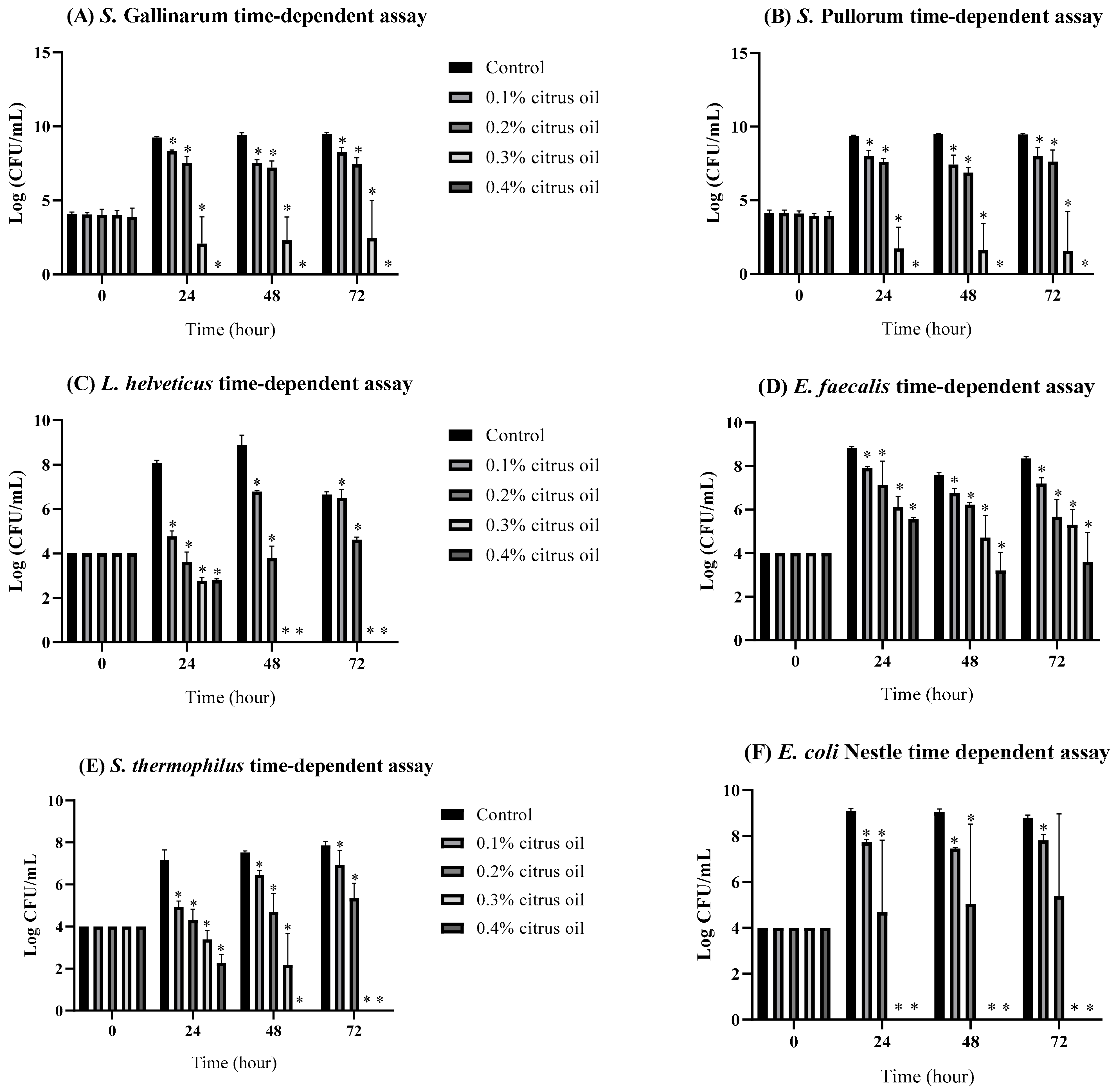
Figure 1. Growth performance of (A) S. Gallinarum, (B) S. Pullorum, (C) L. helveticus, (D) E. faecalis, (E) S. thermophilus, (F) E. coli Nissle in broth supplemented with 0.1%, 0.2%, 0.3%, or 0.4% CO. Bars marked with asterisks (*) indicate significant differences from the control (p < 0.05).
Animals 14 00023 g001
Figure 2. Growth performance of (A) S. Gallinarum, (B) S. Pullorum, in wooden chip as poultry floor simulation supplemented with 0.4% CO. Bars containing asterisks (*) are significantly different from the control (p < 0.05).
Animals 14 00023 g002
Figure 3. Gene expression of (A) S. Gallinarum, (B) S. Pullorum virulence genes after 24 h of treatment with either a control of LB broth supplemented with molecular water, or with 0.1%, 0.2%, 0.3% CO in LB broth. Asterisks (*) denote statistically significant differences from baseline.
Animals 14 00023 g003
Figure 4. Gene expression of (A) S. Gallinarum, (B) S. Pullorum virulence genes after 24 h in wooden chip with either a control of water or treatment with 0.4% CO added. Asterisks (*) denote statistically significant differences from baseline.
Animals 14 00023 g004
Table 1. Primers used in this study of virulence genes.
PrimerPrimer Sequences (5′-3′)Product Sizes (bp)References
spiA-FCCAGGGGTCGTTAGTGTATTGCGTGAGATG550[33,34]
spiA-RCGCGTAACAAAGAACCCGTAGTGATGGATT
invA-FCTGGCGGTGGGTTTTGTTGTCTTCTCTATT1070[33,34]
invA-RAGTTTCTCCCCCTCTTCATGCGTTACC
spaN-FAAAAGCCGTGGAATCCGTTAGTGAAGT504[33,34]
spaN-RCAGCGCTGGGGATTACCGTTTTG
sitC-FCAGTATATGCTCAACGCGATGTGGGTCTCC768[33,34]
sitC-RCGGGGCGAAAATAAAGGCTGTGATGAAC
sifA-FTTTGCCGAACGCGCCCCCACACG449[33,34]
sifA-RGTTGCCTTTTCTTGCGCTTTCCACCCATCT
sipB-FGGACGCCGCCCGGGAAAAACTCTC875[33,34]
sipB-RACACTCCCGTCGCCGCCTTCACAA
Table 2. MIC and MBC of CO against S. Gallinarum and S. Pullorum.
IsolatesMICMBC
S. Gallinarum (CAT 375)0.2%0.2%
S. Pullorum (ATCC 13036)0.4%0.4%
S. Gallinarum (isolate from organic retail chicken sample)0.4%0.4%
S. Pullorum (isolate from farm sample)0.4%0.4%
Disclaimer/Publisher’s Note: The statements, opinions and data contained in all publications are solely those of the individual author(s) and contributor(s) and not of MDPI and/or the editor(s). MDPI and/or the editor(s) disclaim responsibility for any injury to people or property resulting from any ideas, methods, instructions or products referred to in the content.