Article

Composition of the Midgut Microbiota Structure of Haemaphysalis longicornis Tick Parasitizing Tiger and Deer

1
Research Center for Parasites & Vectors, College of Veterinary Medicine, Hunan Agricultural University, Changsha 410128, China
2
Changsha Ecological Zoo, Changsha 410128, China
*
Authors to whom correspondence should be addressed.
Animals 2024, 14(11), 1557; https://doi.org/10.3390/ani14111557
Submission received: 11 April 2024 / Revised: 21 May 2024 / Accepted: 22 May 2024 / Published: 24 May 2024
(This article belongs to the Section Animal Genetics and Genomics)

Abstract

:

Simple Summary

Haemaphysalis longicornis is a predominant tick species in East Asia and infests humans, cattle and wildlife. It is also a common vector for pathogen transmission. The microbial community in tick midguts not only affects the transmission of tick-borne pathogens but also indirectly affects the development, metabolism, reproduction, and other physiology of ticks. There have been rare reports on the effects of different types of hosts on the microbial composition of tick midguts. Here, we conducted a midgut microbiome of H. longicornis collected from tiger and deer by high-throughput sequencing and showed that the relative abundance of each bacterium varied greatly at different classification levels.

Abstract

Haemaphysalis longicornis is a common tick species that carries several pathogens. There are few reports on the influence of different hosts on the structure of midgut microflora in H. longicornis. In this study, midgut contents of fully engorged female H. longicornis were collected from the surface of tiger (Panthera tigris) and deer (Dama dama). The bacterial genomic DNA of each sample was extracted, and the V3–V4 regions of the bacterial 16S rRNA were sequenced using the Illumina NovaSeq sequencing. The diversity of the bacterial community of the fully engorged female H. longicornis on the surface of tiger was higher than that of deer. In total, 8 phyla and 73 genera of bacteria annotations were detected in the two groups. At the phylum level, the bacterial phyla common to the two groups were Proteobacteria, Firmicutes, and Actinobacteriota. At the genus level, there were 20 common bacterial genera, among which the relative abundances of Coxiella, Morganella, Diplorickettsia, and Acinetobacter were high. The Morganella species was further identified to be Morganella morganii. The alpha diversity index indicated that the bacterial diversity of the tiger group was higher than that of the deer group. Bacteroidota, Patescibacteria, Desulfobacterota, Verrucomicrobiota, and Cyanobacteria were solely detected in the tiger group. A total of 52 bacterial genera were unique in the tiger group, while one bacterial genus was unique in the deer group. This study indicates that there are differences in the structure of the gut bacteria of the same tick species among different hosts. Further culture-based methods are needed to provide a more comprehensive understanding of the tick microbiota parasitizing different hosts.

Graphical Abstract

1. Introduction

The Haemaphysalis longicornis tick belongs to the genus Haemaphysalis of the ixodes family. H. longicornis is distributed in the United States, Korea, Japan, Russia, New Zealand, Australia, and other countries [1,2]. It is also a common tick species in northeastern and central China [2]. H. longicornis has a wide range of hosts; these hosts mainly are deer, cattle, sheep, brown rabbits, and dogs [3]. The direct harms of these ticks to hosts include paralysis, anemia, emaciation, and decreased fur quality [4]. More importantly, they act as vectors for several pathogens that can be transmitted to humans and animals during the blood-sucking process [5]. In addition, some pathogens, including tick-borne encephalitis virus (TBEV) [6], severe fever with thrombocytopenia syndrome virus (SFTSV) [7], Anaplasma [8], Rickettsia, Borrelia burgdorferi, and other pathogens [9], have been detected in H. longicornis. Thus, pathogen species and their abundance with in ticks need a concerned investigation for ensuring the public health.
As the largest organ in ticks, the midgut is the key interface for pathogen survival, colonization, and infection [10,11]. Simultaneously, symbiotic bacteria in ticks are active in midgut tissues and can provide nutrients for the growth and development of ticks [12]. The pathogens, symbiotic bacteria, and others together constitute the midgut microbiota of ticks. The diversity of the tick midgut microbiome may be affected by the geographical environment, host, temperature, developmental stage, sex, and other factors [13]. The structure of H. longicornis microflora has been reported in previous studies. Proteobacteria was identified to be the dominant phylum among H. longicornis [14]. Coxiella is the most abundant bacterial genus, and its relative abundance gradually increases with the blood-sucking process of H. longicornis at different developmental stages [14]. These results provide a reliable basis for the recognition of midgut pathogens and symbiotic species of H. longicornis. The microbiome from H. longicornis ticks is affected by different habitats, hosts, sampling sites, and other factors [15]. However, the influence of different hosts on the midgut microflora structure of H. longicornis has not studied extensively.
In this study, H. longicornis ticks from different hosts were collected, and their midgut microflora structure was analyzed using the Illumina NovaSeq sequencing platform. The resulting double-terminal reads were spliced and filtered to obtain amplicon sequence variants (ASVs), followed by species annotation, abundance, and alpha diversity analyses to reveal the midgut microbiome of H. longicornis parasitizing tiger and deer. Therefore, the purpose of this study was to investigate whether there were differences in the structure of the gut bacteria of the same tick species among different hosts.

2. Materials and Methods

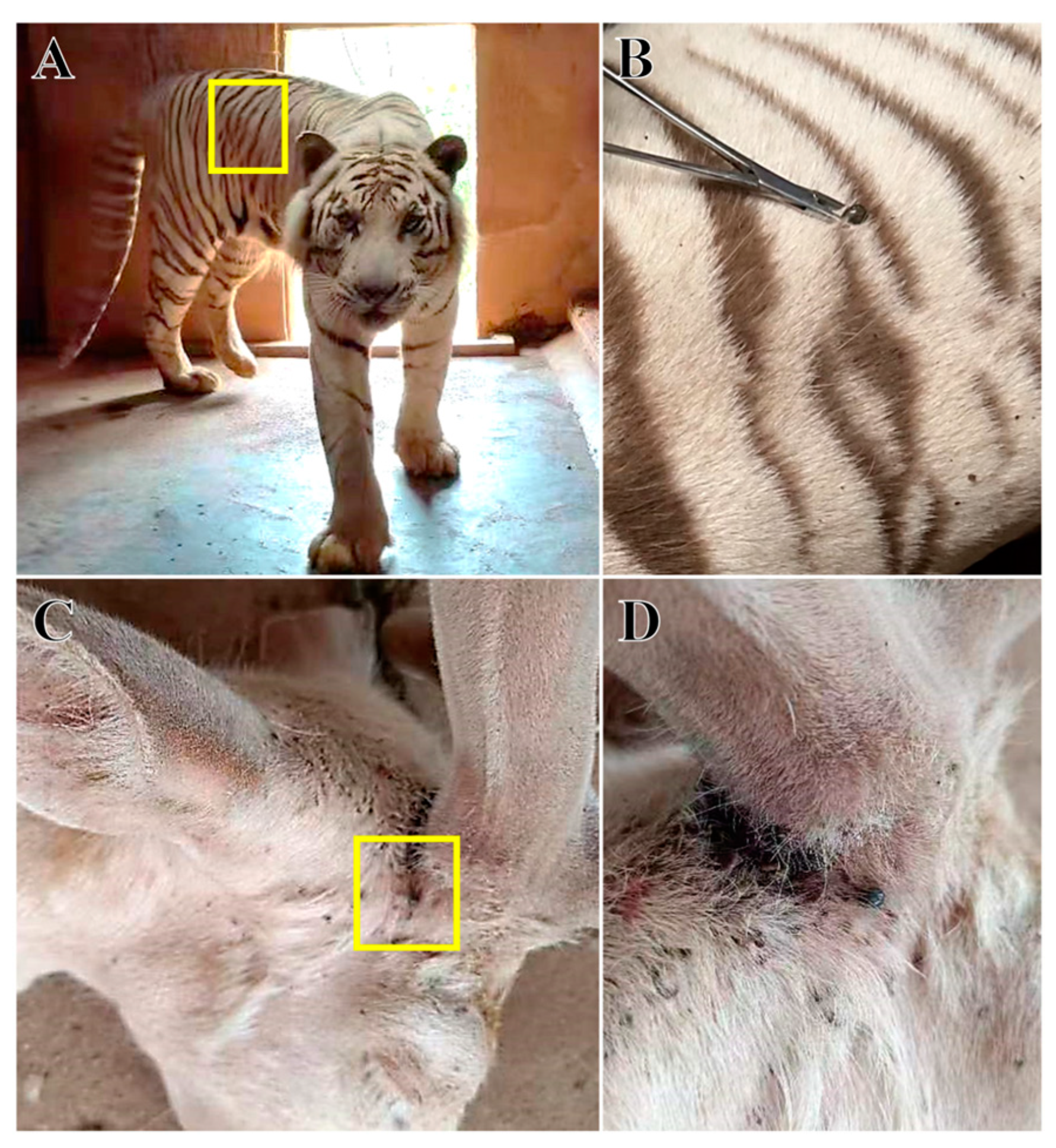
2.1. Collection and Identification of Tick Samples

Fifty female ticks with different satiated blood statuses were collected from the surface of tiger (Figure 1A,B) and deer (Figure 1C,D) at Changsha Ecological Zoo (28°1′ N, 113°0′ E), Hunan Province, China. The sample size referred to a recent similar study [16]. The tigers and deer lived in a stable environment within the same zoo, but two types of animals were housed in a relatively distant area (>0.5 km). Tigers were free-ranging in an outdoor enclosure with trees and shrubs as the main vegetation. In contrast, deer were kept in a smaller enclosed area with a cement-hardened ground and a few grasses. The zookeepers reported the tick infection during the weekly routine inspection. All tick samples were collected from naturally infected hosts at the same time.
A total of 5 mL of host blood was collected using disposable intravenous needles by practicing veterinarians. The blood sample of tiger was collected from the caudal vein, and the blood sample of deer was collected from the jugular vein. Tick samples and host blood samples were immediately sent to the Research Center for Parasites & Vectors, Hunan Agricultural University for further analysis.

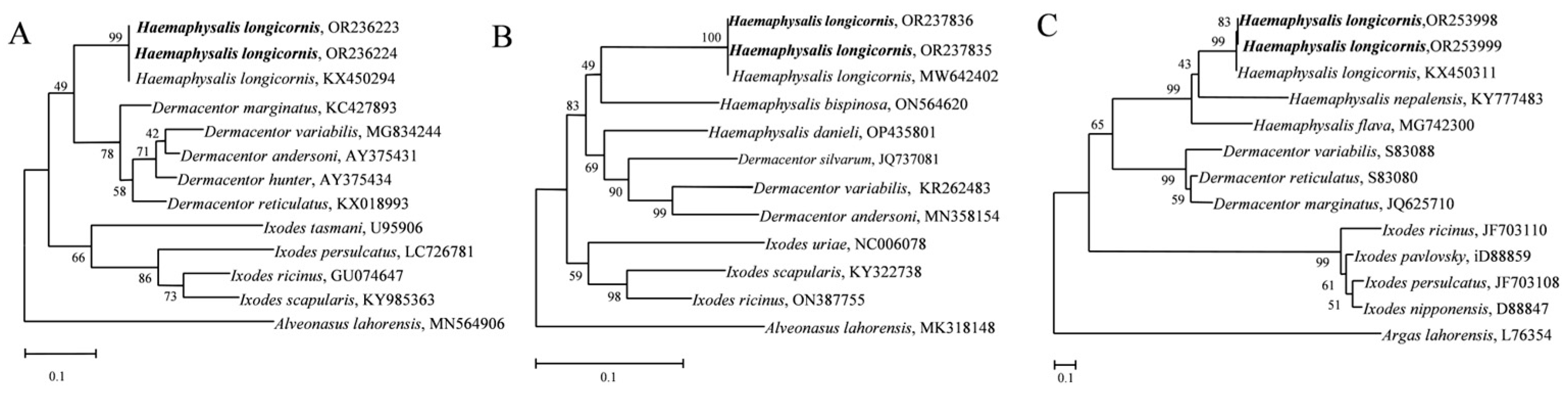
2.2. Identification of Tick Species

The ticks were observed under a stereomicroscope (Nikon, Tokyo, Japan). According to the morphological description by Teng and Jiang (1991), tick species collected in this study were preliminarily identified to be H. longicornis [17].
According to previous reports [18,19], amplification primers for the tick 16S rDNA and COX1 genes and ITS sequences were designed and synthesized by Sangon Biotech (Shanghai, China). The sequences of primers used are listed in Table 1. Tick DNA was extracted using a genomic DNA extraction kit (TianGen Biotech, Beijing, China). The PCR amplification system was as follows: DNA template1 μL, upper and downstream primers (10 mM) 1 μL, 2 × Easy TaqPCR Super Mix (Accurate Biotech, Changsha, China) 25 μL, and ddH2O 22 μL. The amplification conditions for the COX1 gene were as follows: initial denaturation at 95 °C for 5 min, followed by 40 cycles of 95 °C for 30 s, 55 °C for 60 s, 72 °C for 60 s, and a final elongation at 72 °C for 5 min. The amplification conditions for the ITS sequences were as follows: initial denaturation at 95 °C for 5 min, followed by 35 cycles of 95 °C for 30 s, 54 °C for 90 s, and 72 °C for 60 s, and a final elongation at 72 °C for 5 min. The amplification conditions for the 16S rDNA gene were as follows: initial denaturation at 95 °C for 5 min, followed by 35 cycles at 95 °C for 30 s, 56 °C for 30 s, and 72 °C for 60 s, and a final elongation at 72 °C for 5 min. The amplification products (4 μL) were subjected to electrophoresis on a 15 g/L agarose gel. The target bands were cut, and the PCR products were recovered using a DNA rapid recovery and purification kit (TianGen Biotech). The recovered products were connected to the pEASY-T1 cloning vector (TransGen Biotech, Beijing, China), and positive clones were sequenced by Sangon Biotech (Shanghai, China). The sequences obtained were subjected to multiple sequence analysis to identify tick species. MEGA7 software (v7.0.26) was used to construct a phylogenetic tree of each gene via the neighbor-joining (NJ) and maximum-likelihood (ML) methods.

2.3. DNA Extraction of the Midgut Microbiota and Blood Extraction

Five fully engorged ticks (0.280 ± 0.005 g) were used for each group. The ticks were wiped and disinfected with 75% alcohol and washed with sterile water three times to remove the impurities on body surface. The last wash water was used as the negative control. The marginal groove of the ticks was cut off, and the dorsum of the ticks was removed to avoid the outflow of midgut content. Then, the ticks and their organs were suspended in saline solution and exposed under a stereomicroscope. The other organs were separated from the midguts by a needle. The midgut tissues and contents were transferred to sterilized centrifuge tubes containing 1 mL of 3.8% sodium citrate sterile saline solution. The supernatant was centrifuged at 8609× g for 1 min. The supernatant was discarded, and the deposits were retained. The microbial genomic DNA of the deposits and the host whole blood samples were extracted using a Genomic DNA Extraction Kit (TianGen Biotech).

2.4. 16S rRNA Gene Amplification and High-Throughput Sequencing

The V3-V4 regions of bacterial 16S rRNA were amplified using primers 341F (5′-CCT AYG GGR BGC ASC AG-3′) and 806R (5′-GGA CTA CNN GGG TAT CTA AT-3′) with different barcodes. The bacterial genomic DNA of the two groups, the host blood genomic DNA, and the last wash water were used as templates for PCR amplification. The PCR amplification system was as follows (50 μL in total): DNA template 1 μL, Phusion® High-Fidelity PCR Master Mix (New England Biolabs, Ipswich, MA, USA) 25 μL, upper and downstream primers (10 mM) 2 μL, and ddH2O 20 μL. The amplification conditions were as follows: 98 °C for 1 min; 98 °C for 10 s, 50 °C for 30 s, and 72 °C for 30 s for a total of 30 cycles; and 72 °C for 5 min. The PCR products were detected by 2% agarose gel electrophoresis. In parallel with each amplification, a negative control (distilled water) was included. Electrophoresis of the host blood genomic DNA and the wash water revealed negative results. The results of the bacterial genomic DNA of the two host groups were positive. Because the host blood genomic DNA groups and wash water groups were unavailable for library construction, they were excluded from further study. Then, 16S rRNA gene amplicons from the two groups were recovered using a rapid DNA fragment purification and recovery kit (Qiagen, Düsseldorf, Germany). The NEBNext® Ultra™ II DNA Library Prep Kit (New England Biolabs) was used for library construction. Each amplicon was amplified three times. After the libraries were quantified by a Qubit@ 2.0 fluorometer (Thermo Scientific, Waltham, MA, USA) and quantitative real-time polymerase chain reaction, sequencing was performed using the Illumina NovaSeq platform (Thermo Fisher, Waltham, MA, USA).

2.5. Data Analysis

2.5.1. Sequencing Data Processing

After the barcode and primer sequences were removed, reads were spliced using FLASH software (version 1.2.11, http://ccb.jhu.edu/software/FLASH/, accessed on 1 August 2022) to obtain the raw tags. Then, fastp software (https://github.com/OpenGene/fastp, accessed on 1 August 2022) was used to control the raw tags, and high-quality clean tags were obtained. Finally, Vsearch software (https://github.com/torognes/vsearch, accessed on 1 August 2022) was used to compare with the database to detect and remove chimeras to obtain the final data, that is, the effective tags.

2.5.2. Amplicon Sequence Variant Noise Reduction and Species Annotation

For the effective tags, the DADA2 [20] module in QIIME2 software (https://qiime2.org/, accessed on 1 August 2022) was used to filter out sequences with an abundance of less than 5 to obtain the final Amplicon sequence variants ASVs corresponding to OTUs (which represent sequences) and the feature list. Then, the classify-sklearn [21,22] module in QIIME2 software was used to identify the ASVs [23]. The Silva138.1 database was used to annotate species information for each of the amplicon sequence variants (ASVs) [24]. The community composition of the two groups was determined at different levels of classification.

2.5.3. Alpha Diversity Analysis

QIIME2 software was used to calculate the observed ASVs, and Shannon, Simpson, Chao1, and Good’s coverage indices were employed to analyze the intergroup differences in alpha diversity. The rarefaction curves were displayed using Image GP (Version 1.0) software following these indices.

2.5.4. Beta Diversity Analysis

Based on phylogenetic group ASVs, the unweighted unifrac distance, Jaccard distance and Bray Curtis distance were calculated to measure the coefficient of difference between two groups through QIME2 platform.

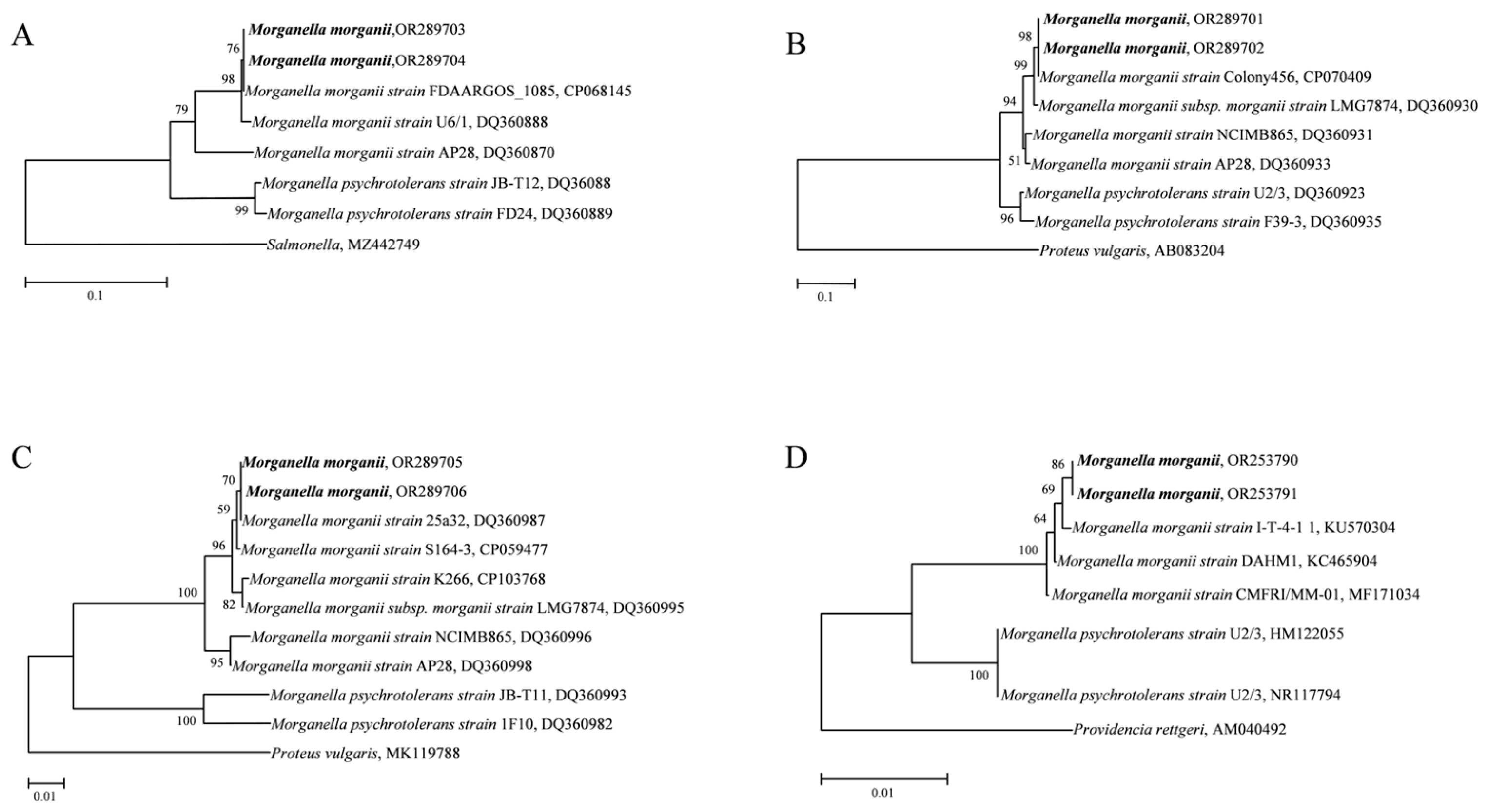
2.5.5. Identification of Morganella in Ticks

According to a previous study [25], the amplification primers for the 16S rDNA, dnaN, tuf, and hdc genes of Morganella were designed and synthesized by Sangon Biotech (Shanghai, China). The sequences of primers used are listed in Table 1. The 16S rDNA, dnaN, tuf, and hdc genes were amplified using PCR reaction as follows: total DNA template of tick midgut bacteria 1 μL, primers for the upper and downstream regions 1 μL (10 mM), 2 × EasyTaq PCR Super Mix (Accurate Biotech) 25 μL, ddH2O 22 μL. In parallel, a negative control (distilled water as a template) was included.
The amplification conditions for the 16S rDNA gene were as follows: initial denaturation at 95 °C for 30 s, followed by 34 cycles of 95 °C for 20 s, 51 °C for 20 s, and 72 °C for 60 s, and a final elongation at 72 °C for 5 min. The amplification conditions for the dnaN gene were as follows: initial denaturation at 95 °C for 30 s, followed by 34 cycles of 95 °C for 20 s, 51 °C for 20 s, and 72 °C for 60 s, and a final elongation at 72 °C for 5 min. The amplification conditions for the tuf gene were as follows: initial denaturation at 95 °C for 30 s, followed by 34 cycles of 95 °C for 20 s, 52 °C for 20 s, 72 °C for 60 s, and a final elongation at 72 °C for 5 min. The amplification conditions for the hdc gene were as follows: initial denaturation at 95 °C for 30 s, followed by 34 cycles of 95 °C for 20 s, 55 °C for 20 s, and 72 °C for 60 s, and a final elongation at 72 °C for 5 min. Amplified products of 5 μL were obtained and subjected to electrophoresis on a 15 g/L agarose gel. The target bands were cut, and the PCR products were recovered using a DNA rapid recovery and purification kit. The recovered products were connected to the pEASY-T1 cloning vector (TransGen Biotech, Beijing, China), and the positive clones were sequenced by Sangon Biotech. The sequences obtained were compared and analyzed with existing sequences in GenBank. Based on the homology of each gene sequences, MEGA7 software (v7.0.26) was used to construct a phylogenetic tree of each gene via the NJ and ML methods.

3. Results

3.1. Identification of Ticks

The ticks in the tiger and deer groups were morphologically identified as H. longicornis. Further molecular identification of the two groups were performed using 16S rDNA and COX1 genes and ITS sequences, in which the 16S rDNA gene was successfully amplified at 347 bp (tiger) and 372 bp (deer). COX1 was successfully amplified at 777 bp (tiger) and 775 bp (deer), and the ITS sequences were also successfully amplified at 1773 bp (tiger) and 1802 bp (deer) (Figure S1). The obtained gene sequences were uploaded to the NCBI database with the following accession numbers for 16S rDNA: OR236224 and OR236223; COX1: OR237835 and OR237836; and ITS: OR253998 and OR253999. Phylogenetic trees were constructed using the above gene sequences. All phylogenetic trees had a well aggregation, thus supporting the identification of ticks to be H. longicornis in this study (Figure 2). Morphological and molecular biology analyses showed that the ticks on the surface of tigers (Figure 1A,B) and deer (Figure 1C,D) in the Changsha Ecological Zoo in Hunan Province were the H. longicornis ticks.

3.2. Data Processing and ASVs Statistical Results

In the original sequence obtained by sequencing, the two groups generated 147,802 raw reads on the Illumina NovaSeq sequencing platform. After removing the barcode and primer sequences, 46,597 and 71,331 effective sequences were obtained from tiger and deer groups for next analyses, respectively. After noise reduction, 138 ASVs were obtained for the tiger group and 28 ASVs were obtained for the deer group, among which 22 ASVs were shared by the two groups, and six ASVs were unique to the deer group.

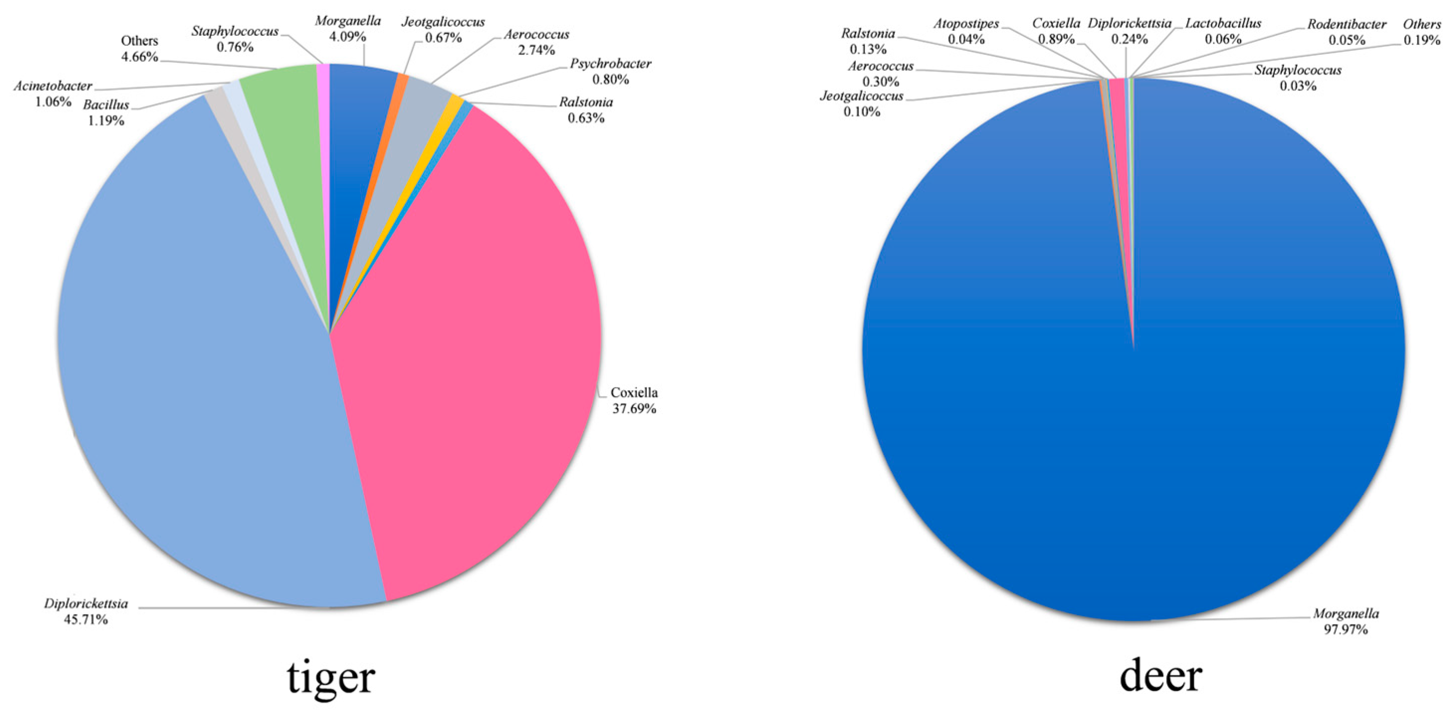
3.3. Analysis of Microbial Compositions

The alpha diversity indices of the tiger and deer groups were analyzed (Table 2). The results show that the Goods coverage of tiger and deer were 1.000, indicating that the sequencing depth in two groups were sufficient, and the data volume could be used for the corresponding analysis. The Chao1, Shannon, and Simpson indices of the tiger group were higher than those of the deer group, indicating that there were a large number of low-abundance species in the midgut community of H. longicornis parasitizing tigers. The rarefaction curve (Figure 3A) showed that the bacterial diversity of tiger group was higher than that of deer group. Unweighted unifrac distance (Figure 3B), Jaccard distance (Figure 3C), and Bray curtis distance (Figure 3D) showed that there were significant differences in the bacterial diversity between the tiger and deer group.

3.4. Difference Analysis of Midgut Microbiota Structure

After species annotation of ASVs, eight phyla were found in two groups and the relative abundance of each phylum is shown in Figure 4. There were three common phyla in the two groups, among which Proteobacteria had the highest relative abundance, accounting for 99.4% (deer) and 90.9% (tiger) of the total phyla. In addition, Firmicutes and Actinobacteriota were also commensal phyla taxa in two groups. In addition to common bacteria, Bacteroidota, Patescibacteria, Desulfobacterota, Verrucomicrobiota, and Cyanobacteria were unique bacteria in the tiger group.
A total of 73 bacterial genera were detected in H. longicornis from both groups listed in Table S1. The distribution characteristics of the top 10 common bacterial genera with relative abundances in the samples of the two groups are shown in Figure 5, and the relative abundances of other common bacterial genera are shown in Table S1. Diplorickettsia, Coxiella, Morganella, Aerococcus, Bacillus, and Acinetobacter were the high-abundance bacterial genera (above 1%) in the tiger group; Morganella, Coxiella, Aerococcus, Diplorickettsia, Ralstonia, and Jeotgalicoccus were the high-abundance ones in the deer group; Diplorickettsia, Coxiella, Morganella, Aerococcus were high-abundance ones in both groups. Diplorickettsia was the highest in the tiger group, accounting for 45.7%, while Morganella was the highest in the deer group, accounting for 97.9%. A total of 52 bacterial genera were unique in the tiger group, and one bacterial genus was unique in the deer group.

3.5. Identification of Morganella

The 16S rDNA, dnaN, tuf, and hdc genes were used to identify the Morganella specie in the two groups. Phylogenetic trees of the four genes showed that the amplified Morganella genes in both the deer and tiger groups polymerized with M. morganii to form a branch, and M. psychrotolerans formed a branch alone. All four phylogenetic trees were topologically stable. The high confidence indicates that the genus Morganella in the two groups was M. morganii (Figure 6). All sequences were uploaded to the GenBank database (16S rDNA: OR253790, OR253791; dnaN: OR289703, OR289704; tuf: OR289705, OR289706; hdc: OR289701, OR289702.)

4. Discussion

Ticks can acquire different bacteria from the host, reproductive process, and environment throughout their life cycle, forming a relatively stable microflora structure in the midgut [26]. The tick microbiome includes disease-causing symbiotes that can influence tick biology and vector capacity to regulate pathogen infection, reproduction, and spreading [27]. The diversity of a tick midgut microbial community is influenced by its host [13].
Therefore, we performed a high-throughput sequencing targeting distinction of the microbial community of H. longicornis, which were collected from tiger and deer. There were 8 and 73 bacterial taxa in the two groups at the phylum and genus levels, respectively. The bacterial taxa shared by the two groups were 3 and 20 at the phylum and genus levels, respectively. Besides the commensal taxa, 5 phyla and 52 genera were unique in the tiger group. The alpha diversity index indicates that midgut community diversity from the tiger group was higher than that from the deer group. The gut microbiome structure in the tiger and deer groups varied greatly in terms of the relative abundance of each bacterium at different taxonomic levels. The tiger group showed a large number of low-abundance species in the midgut community and higher bacterial diversity. This variation may be due to differences in the host genotype, health status, or host environment [15].
Ticks can carry obligate and facultative symbionts that are passed from the mother to the offspring [28]. Both types of symbionts are associated with the health of ticks, and the elimination of symbionts has adverse effects on laying, egg hatching, and survival of ticks [29]. In addition, these symbionts also affect the acquisition, colonization, and spread of tick-borne pathogens [30,31].
Coxiella was detected in the midgut of H. longicornis ticks. Currently, Coxiella burnetii and Coxiella endosymbionts have been found in ticks. C. burnetii is a lethal bacterium that causes Q fever and is considered a medically significant zoonotic pathogen worldwide [32]. This pathogen was first isolated from hard ticks Dermacentor andersoni and H. humerosa [33]. C. burnetii has been found in over 40 species of ticks, including Rhipicephalus spp. and Dermacentor spp., suggesting that these tick species are vectors for the transmission of Q fever [34]. Coxiella endosymbiont is common in tick species, including Amblyomma americanum [35], Ornithodoros rostratus [36], Rhipicephalus turanicus, R. sanguineus [37], Ornithodoros (Carios) capensis (Argasidae) [38] and H. longicornis [39]. Ticks can feed exclusively on vertebrate blood because of the ability of intracellular bacterial symbionts to synthesize B vitamins [40,41]. These nutritional endosymbionts are essential to the life cycle of ticks. In the case of the Coxiella-like endosymbiont of H. longicornis, metagenomics has shown that Coxiella-like endosymbionts can produce the three key B vitamins folic acid, biotin, and riboflavin required for tick well-being. Coxiella-like endosymbiont also plays an important role in determining the reproductive adaptability of ticks [42].
Acinetobacter is an aerobic saprophyte with an oxidation metabolism that is widely distributed in water, soil, and other external environments. Currently, 53 Acinetobacter species have been identified and classified, including opportunistic pathogens and potentially pathogenic strains [43]. Acinetobacter in Ixodes holocyclus, Rhipicephalus (Boophilus) decoloratus, Acinetobacter triguttatum, Aponomma fimbriatum, Bovicola spp. (ex-goat), and Bovicola spp. (ex-alpaca) have been extensively studied in blood-sucking arthropods [44]. In a study by Clayton et al. (2015), Acinetobacter was found to be one of the major symbionts in the midgut and salivary glands of Dermacentor andersoni. The amount of Acinetobacter in ticks may depend on the length of time that it was exposed to the surface of the host [45]. In this study, Acinetobacter was detected in both the deer and tiger groups, and its relative abundance in tiger group was higher than that in deer group. These results indicate that Acinetobacter can stably colonize the midgut of H. longicornis and that its abundance can change among different hosts.
Staphylococcus includes at least 66 effective species, including coagulase-negative Staphylococcus (CoNS), S. lentus, S. saprophyticus, S. aureus, S. epidermidis and others [46]. Huang et al. (2019) reported that Staphylococcus had a high abundance of 8.04–98.76% among ticks in the Yuyao area of China [47]. Staphylococcus is often found in A. americanum and R. microplus and may be derived from the skin and fur of the host [48,49]. It is common for ticks to carry Staphylococcus from those reports. Our results show that Staphylococcus was present in the tiger and deer groups, and relative abundance of Staphylococcus in the tiger samples was higher than that in the deer sample. S. lentus was the reported cause of endometrial infection in European hedgehogs, which are a common host of H. longicornis [50,51].
Morganella was the dominant genus in the deer group, and its relative abundance was the highest. Morganella is a gram-negative bacterium that is widely distributed in nature, and it colonizes mucous membranes of the gastrointestinal tract in humans and animals [52]. Morganella is currently are classified into M. morganii specie and M. psychrotolerans specie. M. morganii are classified into two subspecies, Morganella morganii and M. sibonii [53]. In this study, the relative abundance of Morganella in the deer group was higher than that in tigers, and the genus Morganella in both groups of samples was identified as M. morganii. M. morganii is considered to be an increasingly important pathogen due to its enhancing virulence and resistance to β-lactam antibiotics [54]. A fox had abundant ear discharge and secondary infections of M. morganii, which were suspected to be from ear mite parasites [55]. Klubal et al. (2016) detected Morganella in Ixodes ricinus, accounting for 3% [56]. These results indicate that M. morganii can colonize the midgut of H. longicornis.
In a similar study, R. microplus ticks were collected from cattle and goats in Jiangxi, Hunan, Guizhou and other provinces in China, and their intestinal microbes were analyzed [57]. Rickettsia peacockii and Coxiella spp. were the dominant strains of the R. microplus ticks from cattle, while Uncultured bacterium and Pseudomonas were the most abundant in all samples from goats [57]. Also, the bacterial diversity in the sample from Hunan was the highest among all tick samples, indicating that the diversity of tick intestinal microbes was affected by the types of tick hosts and geographical locations [57]. In addition, R. microplus was collected from two distinct ecological regions in Northern and Middle Magdalena, and the bacterial diversity of their salivary glands and intestines was studied by culture-dependent methods [58]. The results show that Proteobacteria were dominant in Northern Magdalena, while Firmicutes were the most abundant in Middle Magdalena, suggesting that the diversity of bacteria might be affected by geographical distribution [58].
The bacteria compositions of the H. longicornis parasitizing tiger and deer were shown to be different by high-throughput sequencing technology. However, this study did not take into account of the effects of host’s immune status, physiological habits, and the interactions between ticks and hosts on the structure of microbiomes. The host animals were found to be physiologically normal and had not been vaccinated against tick-borne diseases, but the other immune statuses of the hosts was unknown. Also, the present study only used a sequencing-based approach that may not capture the entire microbial diversity present in the tick sample. For instance, the fungal component of the tick microbiota was neglected; it plays an important role in tick ecology and may interact with bacteria to influence host–pathogen dynamics. Culture-based methods are further needed to provide a more comprehensive understanding of the tick microbiota.

5. Conclusions

The relative abundance of bacteria in the midgut microbiota structure of blood-saturated H. longicornis parasitized tiger and deer was significantly different at the phylum and genus levels.

Supplementary Materials

The following supporting information can be downloaded at: https://www.mdpi.com/article/10.3390/ani14111557/s1, Figure S1: Electrophoretogram of the COX1 and 16S rDNA genes and ITS sequences from fully engorged female Haemaphysalis longicornis parasitizing the surface of tiger and deer. M. DNA marker. 1, 2. Products of the COX1 gene from the female Haemaphysalis longicornis parasitizing the surface of tiger. 3, 4. Products of the COX1 gene from the female Haemaphysalis longicornis parasitizing the surface of deer. 5, 6. Products of the ITS sequences from the female Haemaphysalis longicornis parasitizing the surface of tiger. 7, 8. Products of the ITS sequences from female Haemaphysalis longicornis parasitizing the surface of deer. 9, 10. Products of the 16S rDNA gene from the female Haemaphysalis longicornis parasitizing the surface of tiger. 11, 12. Products of the 16S rDNA gene from the female Haemaphysalis longicornis parasitizing the surface of deer. N1, N2, N3. Negative control; Table S1: The relative abundances of 10 common bacterial genera and all unique bacterial genera in the two samples.

Author Contributions

Conceptualization, Z.-L.L., D.-Y.D. and L.L.; methodology, Z.-L.L. and T.-Y.C.; software, Z.-L.L.; validation, G.-H.L., L.L., D.-Y.D. and Z.-L.L.; formal analysis, Z.-L.L.; investigation, Q.-G.Q., G.-H.L. and D.-Y.D.; resources, Q.-G.Q. and T.-Y.C.; data curation, Z.-L.L.; writing—original draft preparation, Z.-L.L.; writing—review and editing, D.-Y.D. and L.L.; visualization, L.L.; supervision, L.L., G.-H.L. and D.-Y.D.; project administration, L.L.; funding acquisition, L.L. All authors have read and agreed to the published version of the manuscript.

Funding

This research was supported by grants from the National Natural Science Foundation of China (Beijing, China, No. 32273090), the Science Fund for Distinguished Young Scholars of Changsha City, China (Changsha, China, No. kq2206033), the Science and Technology Innovation Program of Hunan Province (Changsha, China, No. 2022RC1161), and the National Natural Science Foundation of China (Beijing, China, No. 31902294).

Institutional Review Board Statement

This experiment conforms to the biomedical Research Ethics Committee of Hunan Agricultural University (No. 2024 (29)).

Informed Consent Statement

Written and informed consent has been obtained from the owner of the animals involved in this study.

Data Availability Statement

The raw tags were deposited in the Sequence Read Archive (SRA) of the NCBI under BioProject accession number PRJNA1010648. The individual run files received accession numbers SRR25820263 and SRR25820262.

Conflicts of Interest

The authors declare that the research was conducted in the absence of any commercial or financial relationships that could be construed as a potential conflict of interest.

References

  1. Pritt, B.S. Haemaphysalis longicornis is in the United States and biting humans: Where do we go from here? Clin. Infect. Dis. 2020, 70, 317–318. [Google Scholar] [CrossRef] [PubMed]
  2. Rainey, T.; Occi, J.L.; Robbins, R.G.; Egizi, A. Discovery of Haemaphysalis longicornis (Ixodida: Ixodidae) parasitizing a sheep in New Jersey, United States. J. Med. Entomol. 2018, 55, 757–759. [Google Scholar] [CrossRef] [PubMed]
  3. Heath, A. Biology, ecology and distribution of the tick, Haemaphysalis longicornis Neumann (Acari: Ixodidae) in New Zealand. N. Z. Vet. J. 2016, 64, 10–20. [Google Scholar] [CrossRef] [PubMed]
  4. Haji, I.; Simuunza, M.; Jiang, N.; Chen, Q. Tick populations and molecular detection of selected tick-borne pathogens in questing ticks from northern and central Tanzania. Exp. Appl. Acarol. 2023, 90, 389–407. [Google Scholar] [CrossRef] [PubMed]
  5. Ferrell, A.M.; Brinkerhoff, R.J.; Bernal, J.; Bermúdez, S.E. Ticks and tick-borne pathogens of dogs along an elevational and land-use gradient in Chiriquí province, Panamá. Exp. Appl. Acarol. 2017, 71, 371–385. [Google Scholar] [CrossRef] [PubMed]
  6. Talactac, M.R.; Yoshii, K.; Hernandez, E.P.; Kusakisako, K.; Galay, R.L.; Fujisaki, K.; Mochizuki, M.; Tanaka, T. Synchronous Langat virus infection of Haemaphysalis longicornis using anal pore microinjection. Viruses 2017, 9, 189. [Google Scholar] [CrossRef]
  7. Zhuang, L.; Sun, Y.; Cui, X.; Tang, F.; Hu, J.; Wang, L.; Cui, N.; Yang, Z.; Huang, D.; Zhang, X.; et al. Transmission of severe fever with thrombocytopenia syndrome virus by Haemaphysalis longicornis ticks. China Emerg. Infect. Dis. 2018, 24, 868–871. (In Chinese) [Google Scholar] [CrossRef]
  8. Qin, X.; Han, F.; Luo, L.; Zhao, F.; Han, H.; Zhang, Z.; Liu, J.; Xue, Z.; Liu, M.; Ma, D.; et al. Anaplasma species detected in Haemaphysalis longicornis tick from China. Ticks Tick Borne Dis. 2018, 9, 840–843. [Google Scholar] [CrossRef]
  9. Stanley, H.M.; Ford, S.L.; Snellgrove, A.N.; Hartzer, K.; Smith, E.B.; Krapiunaya, I.; Levin, M.L. The ability of the invasive Asian longhorned tick Haemaphysalis longicornis (Acari: Ixodidae) to acquire and transmit Rickettsia rickettsii (Rickettsiales: Rickettsiaceae), the agent of Rocky Mountain spotted fever, under laboratory conditions. J. Med. Entomol. 2020, 57, 1635–1639. [Google Scholar] [CrossRef]
  10. Cirimotich, C.M.; Ramirez, J.L.; Dimopoulos, G. Native microbiota shape insect vector competence for human pathogens. Cell Host Microbe 2011, 10, 307–310. [Google Scholar] [CrossRef]
  11. Weiss, B.; Aksoy, S. Microbiome influences on insect host vector competence. Trends Parasitol. 2011, 27, 514–522. [Google Scholar] [CrossRef] [PubMed]
  12. Zhu, Z.Q.; Chen, J.W.; Xia, M.Y. Growth kinetics of systemic Lyme disease spirochetes in blood-feeding Ixodes ricinus females. Chin. J. Zoonoses 1998, 14, 16–20. (In Chinese) [Google Scholar]
  13. Dillon, R.J.; Dillon, V.M. The gut bacteria of insects: Nonpathogenic interactions. Annu. Rev. Entomol. 2004, 49, 71–92. [Google Scholar] [CrossRef] [PubMed]
  14. Zhang, R.; Huang, Z.; Yu, G.; Zhang, Z. Characterization of the bacterial community in Haemaphysalis longicornis (Acari: Ixodidae) throughout developmental stages. Exp. Appl. Acarol. 2019, 77, 173–186. [Google Scholar]
  15. Li, S.; Zhang, X.; Zhou, X.; Chen, K.; Masoudi, A.; Liu, J.; Zhang, Y. Bacterial microbiota analysis demonstrates that ticks can acquire bacteria from habitat and host blood meal. Exp. Appl. Acarol. 2022, 87, 81–95. [Google Scholar] [CrossRef] [PubMed]
  16. Qiu, H.; Lv, Q.; Chang, Q.; Ju, H.; Wu, T.; Liu, S.; Li, X.; Yan, Y.; Gao, J.; Wang, C. Microbiota Community Structure and Interaction Networks within Dermacentor silvarum, Ixodes persulcatus, and Haemaphysalis concinna. Animals 2022, 12, 3237. [Google Scholar] [CrossRef] [PubMed]
  17. Teng, G.F.; Jiang, Z.J. Acari: Ixodidae. Economicinsect Fauna of China; Science Press: Beijing, China, 1991. (In Chinese) [Google Scholar]
  18. DUAN, D.; LIU, G.; CHENG, T. Morphological and molecular markers characteristics of Argas persicus. Chin. Vet. Sci. 2019, 49, 990–997. (In Chinese) [Google Scholar]
  19. Zhou, H.; Cheng, T.; Duan, D. Comparison of morphological structure and molecular markers between Dermacentor nuttal and Dermacentor silvarum. Chin. Vet. Sci. 2019, 49, 317–323. (In Chinese) [Google Scholar]
  20. Callahan, B.J.; McMurdie, P.J.; Rosen, M.J.; Han, A.W.; Johnson, A.J.; Holmes, S.P. DADA2: High-resolution sample inference from illumina amplicon data. Nat. Methods 2016, 13, 581–583. [Google Scholar] [CrossRef]
  21. Bokulich, N.A.; Kaehler, B.D.; Rideout, J.R.; Dillon, M.; Bolyen, E.; Knight, R.; Huttley, G.A.; Gregory-Caporaso, J. Optimizing taxonomic classification of marker-gene amplicon sequences with QIIME2’s q2-feature-classifier plugin. Microbiome 2018, 6, 90. [Google Scholar] [CrossRef]
  22. Bolyen, E.; Rideout, J.R.; Dillon, M.R. Reproducible, interactive, scalable, and extensible microbiome data science using QIIME 2. Nat. Biotechnol. 2019, 37, 852–857. [Google Scholar] [CrossRef] [PubMed]
  23. Callahan, B.J.; Wong, J.; Heiner, C.; Oh, S.; Theriot, C.M.; Gulati, A.S.; McGill, S.K.; Dougherty, M.K. High-throughput amplicon sequencing of the full-length 16S rRNA gene with single-nucleotide resolution. Nucleic Acids Res. 2019, 47, e103. [Google Scholar] [CrossRef] [PubMed]
  24. Quast, C.; Pruesse, E.; Yilmaz, P.; Gerken, J.; Schweer, T.; Yarza, P.; Peplies, J.; Glöckner, F.O. The SILVA ribosomal RNA gene database project: Improved data processing and web-based tools. Nucleic Acids Res. 2013, 41, D590–D596. [Google Scholar] [CrossRef] [PubMed]
  25. Emborg, J.; Dalgaard, P.; Ahrens, P. Morganella psychrotolerans sp. nov., a histamine-producing bacterium isolated from various seafoods. Int. J. Syst. Evol. Microbiol. 2006, 56, 2473–2479. [Google Scholar] [CrossRef]
  26. Guizzo, M.G.; Dolezelikova, K.; Neupane, S.; Frantova, H.; Hrbatova, A.; Pafco, B.; Fiorotti, J.; Kopacek, P.; Zurek, L. Characterization and manipulation of the bacterial community in the midgut of Ixodes ricinus. Parasites Vectors 2022, 15, 248. [Google Scholar] [CrossRef] [PubMed]
  27. Bonnet, S.I.; Binetruy, F.; Hernández-Jarguín, A.M.; Duron, O. The tick microbiome: Why non-pathogenic microorganisms matter in tick biology and pathogen transmission. Front. Cell. Infect. Microbiol. 2017, 7, 236. [Google Scholar] [CrossRef] [PubMed]
  28. Ohtaka, C.; Ishikawa, H. Effects of heat treatment on the symbiotic system of an aphid mycetocyte. Symbiosis 1991, 11, 19–30. [Google Scholar]
  29. Smith, T.A.; Driscoll, T.; Gillespie, J.J.; Raghavan, R. A Coxiella-like endosymbiont is a potential vitamin source for the Lone Star tick. Genome Biol. Evol. 2015, 7, 831–838. [Google Scholar] [CrossRef] [PubMed]
  30. Greay, T.L.; Evasco, K.L.; Evans, M.L.; Oskam, C.L.; Magni, P.A.; Ryan, U.M.; Irwin, P.J. Illuminating the bacterial microbiome of Australian ticks with 16S and Rickettsia-specific next-generation sequencing. Curr. Res. Parasitol. Vector Borne Dis. 2021, 1, 100037. [Google Scholar] [CrossRef]
  31. Grostieta, E.; Zazueta-Islas, H.M.; Cruz-Valdez, T.; Ballados-González, G.G.; Álvarez-Castillo, L.; García-Esparza, S.M.; Cruz-Romero, A.; Romero-Salas, D.; Aguilar-Domínguez, M.; Becker, I.; et al. Molecular detection of Coxiella-like endosymbionts and absence of Coxiella burnetii in Amblyomma mixtum from Veracruz, Mexico. Exp. Appl. Acarol. 2022, 88, 113–125. [Google Scholar] [CrossRef]
  32. Abdel-Moein, K.A.; Hamza, D.A. The burden of Coxiella burnetii among aborted dairy animals in Egypt and its public health implications. Acta Trop. 2017, 166, 92–95. [Google Scholar] [CrossRef] [PubMed]
  33. Smith, D.J.W.; Derrick, E.H. Studies in the epidemiology of Q fever: 1. The isolation of six strains of Rickettsia burneti from the tick Haemaphysalis humerosa. Aust. J. Exp. Biol. Med. Sci. 1940, 18, 1–8. [Google Scholar] [CrossRef]
  34. Borawski, K.; Dunaj, J.; Czupryna, P.; Pancewicz, S.; Świerzbińska, R.; Żebrowska, A.; Moniuszko-Malinowska, A. Assess-ment of Coxiella burnetii presence after tick bite in north-eastern Poland. Infection 2020, 48, 85–90. [Google Scholar] [CrossRef] [PubMed]
  35. Klyachko, O.; Stein, B.D.; Grindle, N.; Clay, K.; Fuqua, C. Localization and visualization of a Coxiella-type symbiont within the lone star tick, Amblyomma americanum. Appl. Environ. Microbiol. 2007, 73, 6584–6594. [Google Scholar] [CrossRef] [PubMed]
  36. Almeida, A.P.; Marcili, A.; Leite, R.C.; Nieri-Bastos, F.A.; Domingues, L.N.; Martins, J.R.; Labruna, M.B. Coxiella symbiont in the tick Ornithodoros rostratus (Acari: Argasidae). Ticks Tick Borne Dis. 2012, 3, 203–206. [Google Scholar] [CrossRef] [PubMed]
  37. Lalzar, I.; Friedmann, Y.; Gottlieb, Y. Tissue tropism and vertical transmission of Coxiella in Rhipicephalus sanguineus and Rhipicephalus turanicus ticks. Environ. Microbiol. 2014, 16, 3657–3668. [Google Scholar] [CrossRef]
  38. Duron, O.; Jourdain, E.; McCoy, K.D. Diversity and global distribution of the Coxiella intracellular bacterium in seabird ticks. Ticks Tick Borne Dis. 2014, 5, 557–563. [Google Scholar] [CrossRef] [PubMed]
  39. Zhang, X.; Li, S.; Chen, K.; Yang, C.; Zhou, X.; Liu, J.; Zhang, Y. Growth dynamics and tissue localization of a Coxiella-like endosymbiont in the tick Hemaphysalis longicornis. Ticks Tick Borne Dis. 2022, 13, 102005. [Google Scholar] [CrossRef] [PubMed]
  40. Guizzo, M.G.; Parizi, L.F.; Nunes, R.D.; Schama, R.; Albano, R.M.; Tirloni, L.; Oldiges, D.P.; Vieira, R.P.; Oliveira, W.H.C.; Leite, M.S.; et al. A Coxiella mutualist symbiont is essential to the development of Rhipicephalus microplus. Sci. Rep. 2017, 14, 17554. [Google Scholar] [CrossRef]
  41. Duron, O.; Gottlieb, Y. Convergence of Nutritional Symbioses in Obligate Blood Feeders. Trends Parasitol. 2020, 36, 816–825. [Google Scholar] [CrossRef]
  42. Buysse, M.; Duron, O. Evidence that microbes identified as tick-borne pathogens are nutritional endosymbionts. Cell 2021, 29, 2259–2260. [Google Scholar] [CrossRef] [PubMed]
  43. Zeng, X. Study on Molecular Biological Strain Identification of Acinetobacter and Its Clinical Application. Master’s Thesis, University of South China, Hengyang, China, 2018. (In Chinese). [Google Scholar]
  44. Murrell, A.; Dobson, S.J.; Yang, X.; Lacey, E.; Barker, S.C. A survey of bacterial diversity in ticks, lice and fleas from Australia. Parasitol. Res. 2003, 89, 326–334. [Google Scholar] [CrossRef] [PubMed]
  45. Clayton, K.A.; Gall, C.A.; Mason, K.L.; Scoles, G.A.; Brayton, K.A. The characterization and manipulation of the bacterial microbiome of the Rocky Mountain wood tick, Dermacentor andersoni. Parasites Vectors 2015, 8, 632. [Google Scholar] [CrossRef] [PubMed]
  46. Piette, A.; Verschraegen, G. Role of coagulase-negative Staphylococci in human disease. Vet. Microbiol. 2009, 134, 45–54. [Google Scholar] [CrossRef] [PubMed]
  47. Huang, S.; Zhang, Y.; Luo, X. Analysis of bacterial diversity of ticks in Yuyao City, Zhejiang Province based on high-throughput sequencing technology. Dis. Surveill. 2019, 35, 642–645. (In Chinese) [Google Scholar]
  48. Budachetri, K.; Williams, J.; Mukherjee, N.; Sellers, M.; Moore, F.; Karim, S. The microbiome of neotropical ticks parasitizing on passerine migratory birds. Ticks Tick Borne Dis. 2017, 8, 170–173. [Google Scholar] [CrossRef] [PubMed]
  49. Menchaca, A.C.; Visi, D.K.; Strey, O.F.; Teel, P.D.; Kalinowski, K.; Allen, M.S.; Williamson, P.C. Preliminary assessment of microbiome changes following blood-feeding and survivorship in the Amblyomma americanum nymph-to-adult transition using semiconductor Sequencing. PLoS ONE 2013, 8, e67129. [Google Scholar] [CrossRef] [PubMed]
  50. Qi, Y.; Ai, L.; Zhu, C.; Lu, Y.; Lv, R.; Mao, Y.; Lu, N.; Tan, W. Co-existence of Multiple Anaplasma Species and Variants in Ticks Feeding on Hedgehogs or Cattle Poses Potential Threats of Anaplasmosis to Humans and Livestock in Eastern China. Front. Microbiol. 2022, 13, 913650. [Google Scholar] [CrossRef] [PubMed]
  51. Garcês, A.; Poeta, P.; Soeiro, V.; Lóio, S.; Cardoso-Gomes, A.; Torres, C.; Pires, I. Pyometra Caused by Staphylococcus lentus in a Wild European Hedgehog (Erinaceus europaeus). J. Wildl. Dis. 2019, 55, 724–727. [Google Scholar] [CrossRef]
  52. Guerra, M.; Marado, D. Fulminans Purpura due to Morganella morganii. Eur. J. Case Rep. Intern. Med. 2022, 9, 003670. [Google Scholar] [CrossRef]
  53. Podeur, G.; Dalgaard, P.; Leroi, F.; Prévost, H.; Emborg, J.; Martinussen, J.; Hansen, L.H.; Pilet, M.F. Development of a real-time PCR method coupled with a selective pre-enrichment step for quantification of Morganella morganii and Morganella psychrotolerans in fish products. Int. J. Food Microbiol. 2015, 203, 55–62. [Google Scholar] [CrossRef] [PubMed]
  54. O’Hara, C.M.; Brenner, F.W.; Miller, J.M. Classification, identification, and clinical significance of Proteus, Providencia, and Morganella. Clin. Microbiol. Rev. 2000, 13, 534–546. [Google Scholar] [CrossRef] [PubMed]
  55. Briceño, C.; González-Acuña, D.; Jiménez, J.E.; Bornscheuer, M.L.; Funk, S.M.; Knapp, L.A. Ear Mites, Otodectes Cynotis, on Wild Foxes (Pseudalopex spp.) in Chile. J. Wildl. Dis. 2020, 56, 105–112. [Google Scholar] [CrossRef] [PubMed]
  56. Klubal, R.; Kopecky, J.; Nesvorna, M.; Sparagano, O.A.; Thomayerova, J.; Hubert, J. Prevalence of pathogenic bacteria in Ixodes ricinus ticks in Central Bohemia. Exp. Appl. Acarol. 2016, 68, 127–137. [Google Scholar] [CrossRef] [PubMed]
  57. Xu, X.L.; Cheng, T.Y.; Yang, H.; Yan, F. Identification of intestinal bacterial flora in Rhipicephalus microplus ticks by conventional methods and PCR-DGGE analysis. Exp. Appl. Acarol. 2015, 66, 257–268. [Google Scholar] [CrossRef]
  58. Segura, J.A.; Isaza, J.P.; Botero, L.E.; Alzate, J.F.; Gutiérrez, L.A. Assessment of bacterial diversity of Rhipicephalus microplus ticks from two livestock agroecosystems in Antioquia, Colombia. PLoS ONE 2020, 15, e0234005. [Google Scholar] [CrossRef]
Figure 1. Sample collection. (A), The tiger host; (B), further details showing ticks on the surface of tiger; (C), the deer host; (D) further details showing ticks on the surface of deer.
Animals 14 01557 g001
Figure 2. Phylogenetic tree based on 16S rDNA, COX1 and ITS sequences. (A), 16S rDNA; (B), COX1; (C), ITS. Phylogenetic trees were constructed based on neighbor-joining (NJ; 1000 bootstrap replicates) and maximum-likelihood (ML; 1000 bootstrap replicates) analyses using MEGA7. The scale bar represents the inferred substitutions per nucleotide site. The relative support for clades in the tree was produced from the NJ and ML analyses.
Animals 14 01557 g002
Figure 3. Rarefaction curve, unweighted unifrac distance, Jaccard distance, Bray Curtis distance of midgut samples from fully engorged female adult H. longicornis parasitizing tiger and deer. (A) Rarefaction curves; (BD) unweighted unifrac distance (dissimilarity value = 0.82), Jaccard distance (dissimilarity value = 0.84), Bray Curtis distance (dissimilarity value = 0.94).
Animals 14 01557 g003
Figure 4. Relative abundances of bacteria at the phylum level. ‘Others’ indicates the sum of the relative abundances of the phyla except for the top 8 ones.
Animals 14 01557 g004
Figure 5. Relative abundances of bacteria at the genus level (top 10). ‘Others’ indicates the sum of the relative abundances of the genera except for the top 10 ones.
Animals 14 01557 g005
Figure 6. Phylogenetic trees based on dnaN, hdc, tuf and 16S rDNA sequences of Morganella. (A). dnaN. (B). hdc. (C). tuf. (D). 16S rDNA. The phylogenetic trees were constructed on the basis of neighbor-joining (NJ; 1000 bootstrap replicates) and maximum-likelihood (ML; 1000 bootstrap replicates) analyses using MEGA7. The scale bar represents the inferred substitutions per nucleotide site. The relative support for clades in the tree was produced from the NJ and ML analyses.
Animals 14 01557 g006
Table 1. PCR primer sequences for identification of ticks and Morganella.
Primer NamesPrimer Sequences (5′→3′)
16S rDNA-forward 15′-CTGCTCAATGATTTTTTAAATTGCTGTGG-3′
16S rDNA-reverse 15′-CCGGTCTGAACTCAGATCAAGT-3′
COX1-forward5′-GGAACAATATATTTAATTTTTGG-3′
COX1-reverse5′ATCTATCCCTACTGTAAATATATG-3′
ITS-forward5′-CGAGACTTGGTGTGAATTGCA-3′
ITS-reverse5′-TCCCATACACCACATTTCCCG-3′
16S rDNA-forward 25′-AGAGTTTGATCCTGGCTCAG-3′
16S rDNA-reverse 25′-GGTTACCTTGTTACGACTT-3′
dnaN-foward5′-ATGAAATTTACCGTTGAACGTGA-3′
dnaN-reverse5′-CCATCCACCAGCTTCGAGGT-3′
tuf-foward5′-TTGTTGCTGCAACTGATGG-3′
tuf-reverse5′-CACCTTCATCTTTGCTCAG-3′
hdc-foward5′-TCHATYARYAACTGYGGTGACTGGRG-3′
hdc-reverse5′-CCCACAKCATBARWGGDGTRTGRC-3′
1 Primer sequences were used to identify ticks. 2 Primer sequences were used to identify Morganella.
Table 2. Indices of microbial abundance and diversity in two groups.
GroupChao1Good’s CoverageShannonSimpson
Deer host31.0001.0001.5260.573
Tiger host138.0001.0002.3190.647
Disclaimer/Publisher’s Note: The statements, opinions and data contained in all publications are solely those of the individual author(s) and contributor(s) and not of MDPI and/or the editor(s). MDPI and/or the editor(s) disclaim responsibility for any injury to people or property resulting from any ideas, methods, instructions or products referred to in the content.

