Article

Significant Differences in Intestinal Bacterial Communities of Sympatric Bean Goose, Hooded Crane, and Domestic Goose

1
School of Resources and Environmental Engineering, Anhui University, Hefei 230601, China
2
Anhui Shengjin Lake Wetland Ecology National Long-Term Scientific Research Base, Chizhou 247230, China
3
International Collaborative Research Center for Huangshan Biodiversity and Tibetan Macaque Behavioral Ecology, Hefei 230601, China
*
Authors to whom correspondence should be addressed.
Animals 2024, 14(11), 1688; https://doi.org/10.3390/ani14111688
Submission received: 7 May 2024 / Revised: 25 May 2024 / Accepted: 1 June 2024 / Published: 5 June 2024
(This article belongs to the Section Poultry)

Abstract

:

Simple Summary

The gut microbiota plays important roles for maintaining the health of the host. In this study, the results revealed significant differences in the gut bacterial communities among bean geese, hooded cranes, and domestic geese. In comparison to domestic geese, the gut bacterial community of bean geese and hooded cranes had a greater capacity for energy metabolism, suggesting that wild birds may rely more on their gut microbiota to survive in cold conditions. Furthermore, pathogens were discovered to overlap among the three hosts, reminding us to monitor the potential for pathogen transmission between poultry and wild birds. Hooded cranes showed the highest diversity and relative abundance of pathogens compared to the other two species. Due to its vulnerable status, more focus should be paid to the protection of the hooded crane species. These findings could help us gain a deeper understanding of the structure of gut bacterial and pathogenic communities in poultry and wild birds.

Abstract

The host’s physiological well-being is intricately associated with the gut microbiota. However, previous studies regarding the intestinal microbiota have focused on domesticated or captive birds. This study used high-throughput sequencing technology to identify the gut bacterial communities of sympatric bean geese, hooded cranes, and domestic geese. The results indicated that the gut bacterial diversity in domestic geese and hooded cranes showed considerably higher diversity than bean geese. The gut bacterial community compositions varied significantly among the three hosts (p < 0.05). Compared to the hooded crane, the bean goose and domestic goose were more similar in their genotype and evolutionary history, with less difference in the bacterial community composition and assembly processes between the two species. Thus, the results might support the crucial role of host genotypes on their gut microbiota. The gut bacteria of wild hooded cranes and bean geese had a greater capacity for energy metabolism compared to domestic geese, suggesting that wild birds may rely more on their gut microbiota to survive in cold conditions. Moreover, the intestines of the three hosts were identified as harboring potential pathogens. The relative abundance of pathogens was higher in the hooded crane compared to the other two species. The hooded crane gut bacterial community assemblage revealed the least deterministic process with the lowest filtering/selection on the gut microbiota, which might have been a reason for the highest number of pathogens result. Compared to the hooded crane, the sympatric bean goose showed the least diversity and relative abundance of pathogens. The intestinal bacterial co-occurrence network showed the highest stability in the bean goose, potentially enhancing host resistance to adverse environments and reducing the susceptibility to pathogen invasion. In this study, the pathogens were also discovered to overlap among the three hosts, reminding us to monitor the potential for pathogen transmission between poultry and wild birds. Overall, the current findings have the potential to enhance the understanding of gut bacterial and pathogenic community structures in poultry and wild birds.

1. Introduction

The gut microbiota plays a crucial role in maintaining the well-being of various animal species. In addition to aiding in digestion [1], regulating metabolism, and bolstering immunity [2], microbial communities may also protect against pathogen invasion [3]. Changes in the gut microbiota increase the host’s susceptibility to diseases [4]. Furthermore, multiple factors have the potential to modify the structure and function of the gut microbiome, such as dietary choices [5], environmental conditions [6], gender differences [7], and fluctuations in the seasons [8]. Previous studies have claimed that the host genotype has a great impact on the composition of the gut microbiota [9].
In contrast to other vertebrates, the gut microbiota of migratory birds is markedly different due to their distinctive physiological characteristics [10]. The homeostasis of the intestinal microbiota plays a vital role in the growth and well-being of avian species [11]. It is essential for proper nutrient absorption [12], maintaining a healthy immune system [13], and maintaining different physiological functions [14]. The intestinal microbiota comprises crucial symbionts that profoundly influence the host’s life [15], while the host is also an essential factor influencing the intestinal microbiota [16]. However, migratory birds may be susceptible to disease. Wild birds inhabit various habitats during long-distance migration, which raises the potential for pathogen invasion. With overlapping niches, migratory birds are capable of spreading their intestinal pathogens to other sympatric host species [17,18]. The transmission of these pathogens to poultry, humans, and other wild birds can occur as a result of bird migration [19,20]. Therefore, a better understanding of the potential pathogens present in the intestinal tracts of migratory birds may contribute to the elucidation of cross-species pathogen transmission.
The hooded crane, scientifically known as Grus monacha, is a large waterfowl species that undertakes long-distance migrations. It is classified as vulnerable on the International Union for the Conservation of Nature (IUCN) red list of threatened species [21]. Another large wintering migratory wild bird is the bean goose (Anser fabalis). The bean goose is one of the most abundant wintering birds in the Yangtze River floodplain. The wintering period of these two migratory birds is from October to April in the Yangtze River floodplain. Anthropic activities trigger the rapid degradation of lake wetlands, leading to a significant reduction in food availability. Shengjin Lake, an internationally important wetland, is a river-connected shallow lake in the middle of the Yangtze River floodplain. During winter, the Shengjin Lake provides suitable foraging habitats for wild migratory birds, making it a significant wetland [22]. More than 12,000 bean geese and ≥350 hooded cranes perish annually in Shengjin Lake [23,24]. Wild birds typically assemble on farmland to rest and forage during winter [25]. However, there is a substantial population of poultry in farmland. The potential for the increased cross-transmission of pathogens exists due to the shared foraging niches of the bean goose, hooded crane, and domestic goose (Anser anser). The current study aimed to elucidate the composition of the gut bacterial communities and identify possible pathogens in bean geese, hooded cranes, and domestic geese during the wintering period in Shengjin Lake.
In this study, we used high-throughput sequencing technology to investigate the gut bacterial communities and infer the potential pathogens of sympatric bean geese, hooded cranes, and domestic geese at Shengjin Lake. We propose the following hypotheses: (a) the differences in the gut bacterial communities could be shaped by multiple factors, such as host genotype, diet, and living environment; (b) wild birds (i.e., bean geese and hooded cranes) might carry more diverse potential pathogens due to their harsh living environment relative to domestic geese.

2. Materials and Methods

2.1. Ethics Statement

Non-invasive fecal sample collection did not involve the hunting of experimental animals. Permission for this study was obtained from the Committee on Laboratory Animal Ethics and Regulation of Anhui University (IACUC-2024-043).

2.2. Sample Collection

In this study, the samples were collected via a non-invasive sampling method (fecal sampling techniques). Bean geese, hooded cranes, and domestic geese often forage together in rice paddies [23,25]. The diets of the three hosts are shown in Table S1 [26,27,28,29,30,31]. At the same rice paddies, fecal samples were collected from bean geese and hooded cranes. The area was inhabited by approximately 100 hooded cranes, 400 bean geese, and 60 domestic geese. Fecal samples were collected from bean geese, hooded cranes, and domestic geese on the 15th of November 2018. First, a telescope was used to find areas where bean geese and hooded cranes mixed flocks for foraging. The fresh fecal samples were collected immediately after the completion of the birds’ foraging activity. The fecal samples of domestic geese were then obtained in the yard of a farmer. To reduce the risk of contamination, the outermost layer of each sample was removed. Each fresh fecal sample was collected separately in a 15 mL centrifuge tube (sterilized), with ≥2 g per sample. As a precaution against sample contamination, polyethylene gloves were discarded and replaced after collecting the fecal samples. Moreover, to avoid redundancy, the samples were obtained at 10 m intervals. The feces were temporarily stored in an insulated container with ice packs after collection. They were returned to the laboratory within 12 h and maintained at −20 °C.

2.3. Bird Species Determination

The Total DNA was extracted from the fecal samples using the Qiagen DNA Stool Mini Kit (Hilden, Germany). The bird species were identified via the sequencing of the mitochondrial COI gene [32,33]. This section’s specific details are included in the Supplementary Materials. After species determination, a total of 60 samples were used in this study, with 20 samples per species (i.e., bean geese, hooded cranes, and domestic geese).

2.4. PCR and Amplicon Library Preparation

The bacterial 16S rRNA gene fragment of the V4-V5 hypervariable regions was amplified before sequencing using the primer sets F515/R907 [34,35]. The Supplementary Materials comprise the details of this section.

2.5. Bioinformatics

The raw bacterial data were examined via qiime2-2021.2 software [36]. Low-quality sequences were removed using the “deblur” algorithm [37]. The remaining high-quality sequences were grouped into amplicon sequence variations (ASVs) with 100% similarity. The evaluation of the chimeras was performed using the “vsearch” technique [38]. The annotation process was performed via the “classify-sklearn” algorithm using the Silva database v. 132. The subsets of 15,000 sequences per sample were randomly selected for further analyses.

2.6. Statistical Analysis

The normal distribution was examined using Shapiro–Wilk’s test for the gut bacterial data (i.e., the bacterial diversity and relative abundance; Table S2). The non-normal distribution data were transformed using a square root transformation to achieve a normal distribution (Table S2). A one-way analysis of variance (ANOVA) was performed to examine the differences in the gut bacterial data. The three hosts were comparatively analyzed using linear discriminant analysis effect size (LEfSe) and the non-parametric Kruskal–Wallis rank-sum test (p ≤ 0.05; LDA threshold ≥ 2) to detect the biomarkers in the gut bacterial taxa [39]. The community composition was analyzed using non-metric multidimensional scaling (NMDS) and a similarity analysis (ANOSIM) (permutations = 999) in R-Studio (V.4.2.1) using a vegan package (V.2.6-4). The identification of the unique and common gut bacterial ASVs among the three hosts was carried out using a Venn diagram (https://bioinfogp.cnb.csic.es/tools/venny/index.html, accessed on 24 May 2024). The labdsv package (V.2.1-0) in R-Studio (V.4.2.1) was used to examine the indicator species of the gut bacteria in each host. The beta nearest taxon index (betaNTI) was calculated using the picante package and Phylocom 4.2 to analyze the gut bacterial community assembly of the three hosts [40]. A PICRUSt analysis was used to predict the gut bacterial function [41,42]. A co-occurrence network analysis was conducted using R-Studio (V.4.2.1) and Gephi v.0.9.2 software [43]. The potential pathogens were identified via the functional annotation of prokaryotic taxa (FAPROTAX) database [44] and searched as keywords in the Web of Science to identify their hosts and symptoms [45,46,47,48,49,50,51,52,53,54,55,56,57,58,59,60,61,62,63,64]. The information of pathogens was shown in Table S3.

3. Results

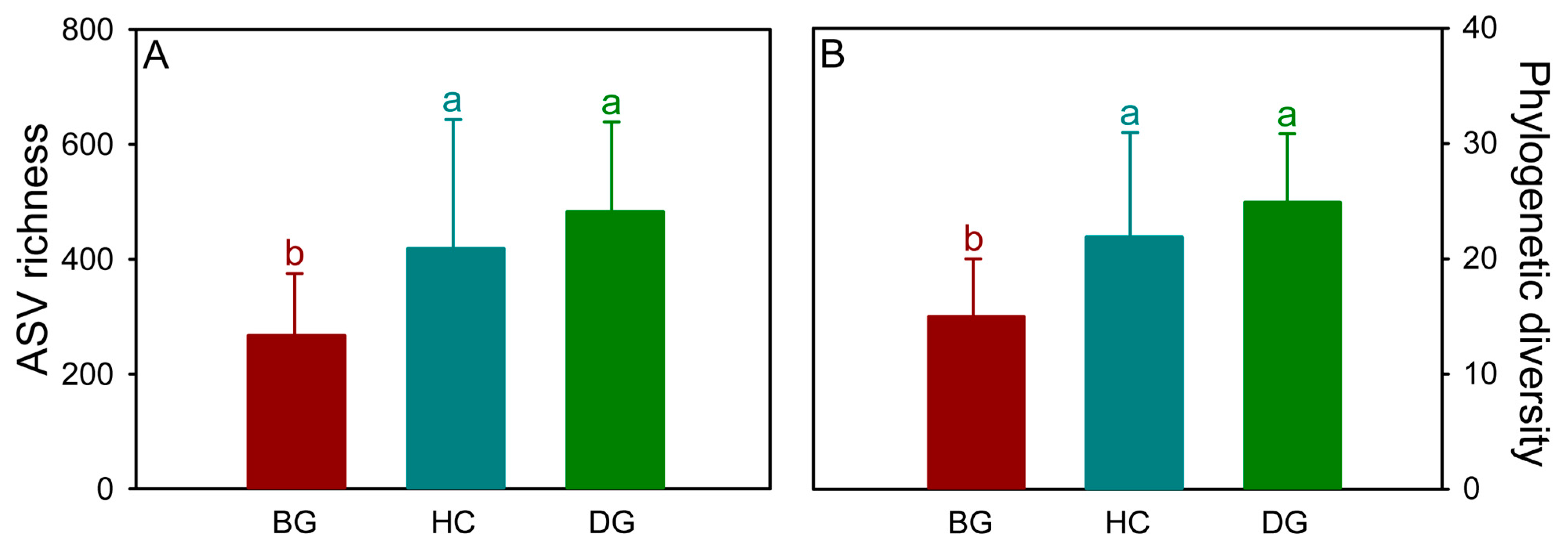
3.1. Bacterial Diversity

In total, 1,628,677 bacterial sequences passed the quality filter, and 2,466 bacterial ASVs were discovered in all samples. The number of sequences per sample ranged from 15,790 to 50,660, while the number of ASVs per sample ranged from 65 to 714. Among the three species, a total of 545 ASVs, representing 23.7% of the total, were shared in their guts (Figure S1). The number of distinct bacterial ASVs was 155 (6.7%) for the bean goose, 359 (15.6%) for the hooded crane, and 487 (21.2%) for the domestic goose. The phylogenetic diversity and ASV richness were used to evaluate the bacterial alpha diversity. The diversity of the hooded crane and domestic goose was significantly higher than that of the bean goose (Figure 1).

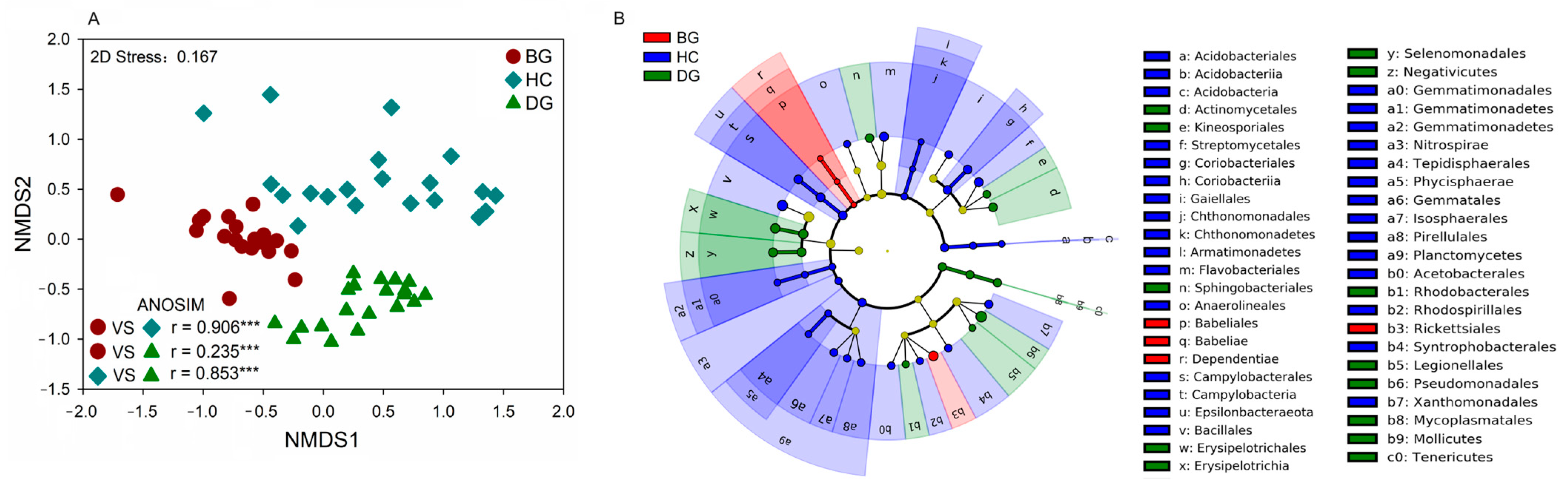
3.2. Bacterial Community Composition

The dominant gut bacterial phyla were Firmicutes (49.6%), Proteobacteria (18.8%), Actinobacteria (6.00%), and Bacteroidetes (1.5%). The dominant gut bacterial genera were Lactobacillus (32.5%), Pseudomonas (6.60%), Enterococcus (4.26%), and Bacillus (1.49%). The hooded crane showed a greater relative abundance of Actinobacteria and Bacillus. In comparison to the other two species, the relative abundance of Proteobacteria and Pseudomonas was greater in the domestic goose (Figures S2 and S3). The domestic goose had a substantially lower intestinal Firmicutes/Bacteroidetes ratio (F/B) than the bean goose and hooded crane (Figure S4).
The community compositions of the intestinal bacteria varied considerably among the three species (Figure 2A). The LEfSe showed that the bacteria from one phylum (Dependentiae), one class (Babeliae), and two orders (Babeliales and Rhckettsiales) were enriched in the gut of the bean goose. The bacteria from six phyla (Nitrospirae, Planctomycetes, Armatimonadetes, etc.), six classes (Coriobacteriia, Chthonomonadetes, Campylobacteria, etc.), and eight orders (Coriobacteriales, Campylobacterales, Bacillales, etc.) were enriched in the gut of the hooded crane. One phylum (Tenericutes), three classes (Mollicutes, Negativicutes, and Erysipelotrichales), and nine orders (Actinomycetales, Kineosporiales, and Tenericutes) were enriched in the domestic goose (Figure 2B and Figure S5).
Bacterial species related to species specificity were identified via an indicator analysis. There were five (e.g., Streptococcus suis, Kaistia geumhonensis, Agaricicola taiwanensis, etc.), ten (e.g., Paenibacillus xylanilyticus, Campylobacter canadensis, Allorhizobium vitis, etc.) and seventeen (e.g., Lactobacillus aviarius, Pseudomonas lundensis, Clostridium akagii, etc.) indicator bacterial species in the guts of the bean goose, hooded crane, and domestic goose, respectively (Table 1).

3.3. Gut Bacterial PICRUSt Analysis

Based on the KEGG database, gut bacteria have various important roles, such as energy metabolism, cofactor and vitamin metabolism, amino acid metabolism, transcription, and cell motility. Based on the PICRUSt analysis, it was found that the bean goose and hooded crane displayed higher capacities for energy metabolism and the metabolism of cofactors and vitamins compared to the domestic goose (Figure 3). The domestic geese showed improved amino acid metabolism and transcription capacity in comparison to the bean goose and hooded crane. The bean geese had a higher potential for energy metabolism than the hooded crane. The hooded crane showed greater cell motility and amino acid metabolism capacity compared to the bean goose (Figure 3).

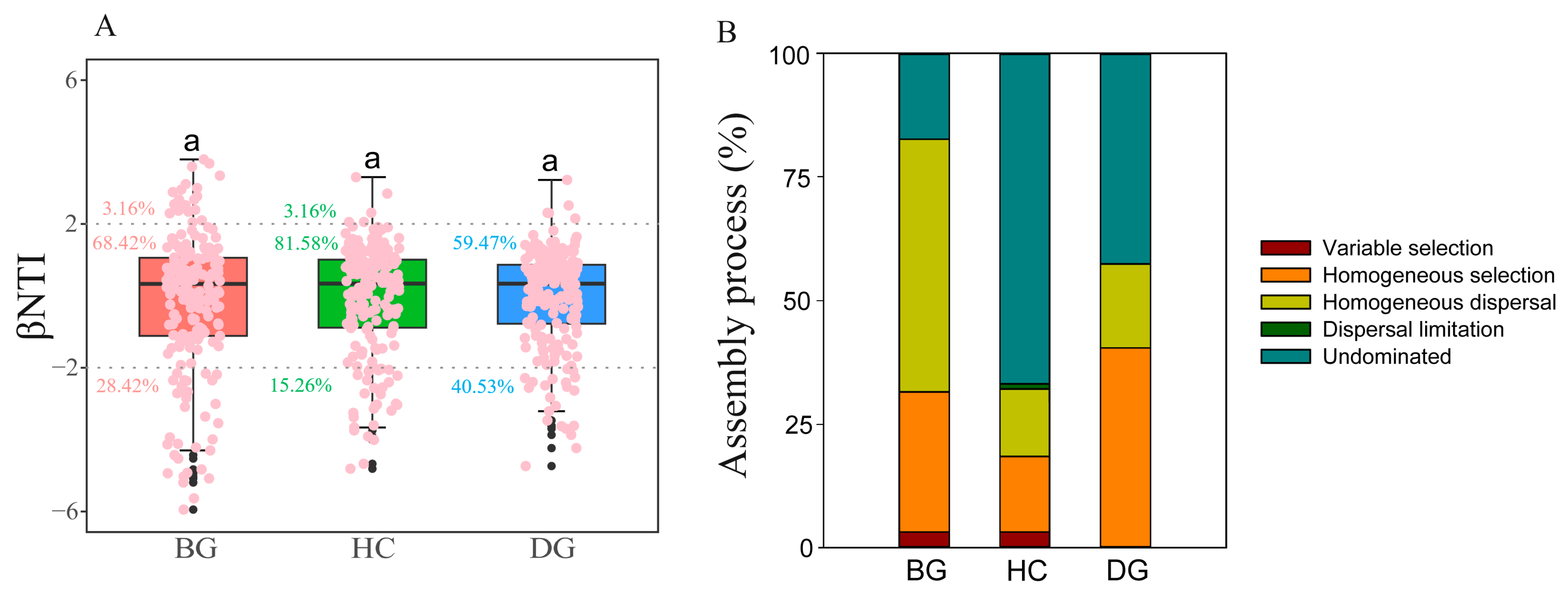
3.4. Gut Bacterial Community Assembly

The values of the betaNTI (beta nearest taxon index) within the range from −2 to 2 suggest a stochastic process, while values ≥ 2 or ≤−2 indicate deterministic processes. In the guts of bean geese, hooded cranes, and domestic geese, most of the betaNTI scores for the bacterial community assembly dropped within the range from −2 to 2. This indicates that stochastic assembly played a significant role in shaping the dynamics of the gut bacterial communities in these three species (Figure 4A). The assembly of the gut bacterial community in hooded cranes was associated with the highest stochastic assembly (81.58%) and the least deterministic process (18.42%). In the gut of the bean goose, homogeneous dispersal comprised the prevailing stochastic assembly process at 51.05%. Conversely, in the intestines of hooded cranes (66.84%) and domestic geese (42.63%), the undominated stochastic assembly process was predominant (Figure 4B).

3.5. Gut Bacterial Network Analysis

The interrelationships between the bacterial taxa in the guts of the bean goose, hooded crane, and domestic goose were investigated via a co-occurrence network analysis. Most of the interactions within the intestinal network were linked to Proteobacteria (Figure S6). In contrast to the domestic goose and hooded crane, the bean goose demonstrated the least average path length and modularity while also showing the maximum clustering coefficient and average degree (Table S4). In comparison to the domestic goose and hooded crane, the bean goose showed the highest level of network stability as assessed using natural connectivity (Figure S7).

3.6. Gut Potential Pathogens

A total of sixteen potentially pathogenic ASVs were identified, of which 6.3% (one ASV) was shared by three species, 12.5% (two ASVs) by bean geese and domestic geese, 12.5% (two ASVs) by hooded cranes and domestic geese, and 6.3% (one ASV) by bean geese and hooded cranes (Figure 5A). The distinct pathogenic ASVs identified in the guts of the bean goose, hooded crane, and domestic goose were one, six, and three, respectively (Figure 5A). The gut pathogenic community composition of the hooded crane exhibited a significant variation compared to the other species (Figure 5B). It appeared that the gut of the hooded crane had a higher diversity and relative abundance of potential pathogens compared to the other two species (Figure 5C,D).

4. Discussion

The focus on avian gut microbiota has been growing, emphasizing the important role of the host species in the formation of the gut microbiota [65,66]. Previous studies have shown that host species with close phylogenetic relationships are responsible for providing the same ecological niche for microbial communities [67,68]. Species with similar genotypes tend to have similar gut microbiota [69]. The bean geese and domestic geese are classified under the same genus, Anser, in the coevolutionary branch of the avian phylogenetic tree [70], with less difference in the bacterial community composition and assembly processes between the two species (r = 0.235) relative to the hooded crane (r = 0.906 for the bean goose and hooded crane; r = 0.853 for the domestic goose and hooded crane) (Figure 2A). Thus, the results might support the crucial role of the host phylogenetic relationship (i.e., the genotype) on their gut microbiota [71]. However, different living environments may lead to significant differences in the gut microbiota of closely related species [68]. The living environment of domestic geese is different from that of bean geese, triggering differences in their gut bacterial community composition. In addition, the gut microbiota could be regulated by the host’s diet [72]. The three hosts have different diets during the wintering period (Table S1), which might be another important reason for the differences in the gut bacterial communities among the three species (Figure 1, Figure 2A and Figure 4).
Consistent with the findings from studies on other avian species, such as Emberiza spodocephala [73], Falco tinnuculus [74], and Larus relictus [75], the avian gut microbiota was predominately composed of Firmicutes and Proteobacteria (Figure S2). In contrast to the hooded crane and bean goose, the domestic goose showed a significantly lower ratio of Firmicutes to Bacteroidetes (Figure S4). A larger ratio of Firmicutes to Bacteroidetes contributes to more effective calorie absorption and provides protection against intestinal infections for hosts [76]. The domestic goose displayed abundant Lactobacillus aviarius and Pseudomonas lundensis (Table 1). Previous studies have shown that these two specific species can inhibit the growth of pathogens in the host [77,78,79,80]. These results suggest that wild birds and domestic poultry might adopt different strategies to prevent pathogen invasion. The present study employed the PICRUSt analysis to predict the functions of the gut bacteria. The results indicated that wild birds have great potential for energy metabolism (Figure 3), which was consistent with the previous findings demonstrating a higher ratio of Firmicutes to Bacteroidetes and efficient calorie absorption in wild birds (Figure S4). Wild birds residing in natural habitats throughout the winter season experience harsh cold conditions, necessitating strong energy metabolism. The living conditions of domestic geese are superior to those of wild birds. Thus, wild birds may rely more extensively on their gut flora to enhance energy metabolism.
Moreover, 10 potential pathogen ASVs that can infect humans and/or animals were identified in three distinct hosts (Table S3). The gut pathogenic community composition showed little difference between the bean goose and domestic goose (Figure 5B). When sampling, mixed flocks with very similar niches were usually found between the two species, resulting in pathogenic transmission and similar pathogenic community composition. The intestinal pathogenic composition of the hooded crane was significantly distinct from that of the other two species (Figure 5B). The hooded crane also showed great diversity and relative abundance of gut animal pathogens (Figure 5C,D). The assembly of the gut bacterial community in hooded cranes was associated with the least deterministic process (Figure 4), leading to a reduced filtration effect on their gut microbiota and resulting in higher pathogens [66]. The hooded crane is classified as an endangered species. Therefore, it is crucial to prioritize the protection of this vulnerable species.
However, the relative abundance and diversity of pathogens were least prevalent in the bean goose compared to the other species. Bean geese appeared healthier than hooded cranes despite inhabiting a comparable niche in a harsh environment during the winter conditions. The population size was found to be more pronounced for the bean goose (≥12,000) compared to the hooded crane (≤350), indicating that bean geese are more suitable for surviving in wintering conditions compared to hooded cranes. Further analyses demonstrated the highest stability along with the greatest complexity in the gut bacterial co-occurrence network of bean geese (Figure S7). A network that has greater stability could potentially contribute to enhanced resilience against unfavorable conditions and facilitate multifunctionality [81]. This, in turn, may assist the bean goose in reducing the risk of pathogen invasion.
During winter, both wild migrating birds and domestic poultry use the same ecological niche for foraging in fields. This study also revealed the presence of a specific proportion of total pathogen ASVs shared in wild birds and poultry (Figure 5A). Previous studies have demonstrated the transmission of diseases between hosts through niche overlaps or close contact [82]. Thus, there might be cross-transmission of pathogens among the bean goose, hooded crane, and domestic goose. Most of the pathogens identified in the guts of the three species have the potential to induce diseases in humans (Table S2). Thus, the possibility of foraging ecological niche overlap may contribute to the transmission of diseases from avian species to poultry, which results in human transmission [18,31]. It is crucial to closely monitor the pathways of pathogenic transmission between wild birds and poultry to accurately assess the potential for disease outbreaks in future studies.

5. Conclusions

In conclusion, the results revealed significant differences in the bacterial communities within the guts of the bean goose, hooded crane, and domestic goose. In comparison to domestic geese, wild birds may rely more heavily on their gut microbiota to enhance energy metabolism and adapt to harsh winter conditions. Furthermore, pathogens were detected in the guts of the three hosts, with the hooded crane having the highest diversity and relative abundance of pathogens. Due to its vulnerable status, more focus should be paid to the protection of the hooded crane species. The bean goose displayed less variation and relative prevalence of diseases, indicating that bean geese possess a robust capacity to adapt to their surroundings. These results indicate that it is crucial to prioritize further studies into the transmission of gut pathogens between wild birds and poultry. However, there were certain limitations in this study. Firstly, only three species were included in this study. Secondly, we did not measure the detailed information on the niche overlap degree between different species. These limitations should be clarified in future studies.

Supplementary Materials

The following supporting information can be downloaded at: https://www.mdpi.com/article/10.3390/ani14111688/s1, Table S1: The different diets of three hosts, Table S2: The distribution of data was analyzed by Shapiro-Wilk’s test. Table S3: The potential pathogens were detected in guts of bean goose, hooded crane and domestic goose, Table S4: Co-occurrence network topological features, Figure S1: The Venn diagram showing the unique and shared gut bacterial ASVs in guts of bean goose, hooded crane and domestic goose, Figure S2: Relative abundance of the dominant bacterial phyla, Figure S3: Relative abundance of the dominant bacterial genus, Figure S4: The ratio of relative abundance of Firmicutes and Bacteroidetes, Figure S5: Identified phylotype biomarkers ranked by effect size with the alpha value was <0.05 in different hosts. The phylotype biomarkers were identified as significantly abundant when samples in bean goose, hooded crane and domestic goose were compared, Figure S6: The co-occurrence network structure of gut bacterial community for bean goose, hooded crane and domestic goose. Figure S7: The stability of co-occurrence network. References [20,26,27,28,29,30,31,32,33,34,35,45,46,47,48,49,50,51,52,53,54,55,56,57,58,59,60,61,62,63,64] are cited in the Supplementary Materials.

Author Contributions

Conceptualization, X.X., Z.C. and J.Y.; data curation, J.Y., D.Y., Z.X. and Y.W.; formal analysis, X.X., J.Y. and D.Y.; funding acquisition, X.X.; investigation, Z.C.; methodology, X.X. and Z.C.; writing—original draft, J.Y.; writing—review and editing, X.X. and J.Y. All authors have read and agreed to the published version of the manuscript.

Funding

This research was funded by the “National Natural Science Foundation of China” (31801989) and the Outstanding Youth Research Project of Anhui Province for Xingjia Xiang (2022AH030015).

Institutional Review Board Statement

The animal study was reviewed and approved by the Committee on Laboratory Animal Ethics and Regulation of Anhui University (IACUC-2024-043).

Informed Consent Statement

Not applicable.

Data Availability Statement

The raw data were submitted to the Sequence Read Archive (SRA) of NCBI under the accession number SAMN38055501-SAMN38055502-SAMN38055503.

Acknowledgments

We thank Rong Fu from Anhui University for their assistance in sample collection.

Conflicts of Interest

The authors declare they have no conflicts of interest.

References

  1. Gao, P.; Liu, Y.; Le, B.; Qin, B.; Liu, M.; Zhao, Y.; Duan, Z. A comparison of dynamic distributions of intestinal microbiota between Large White and Chinese Shanxi Black pigs. Arch. Microbiol. 2019, 201, 357–367. [Google Scholar] [CrossRef] [PubMed]
  2. Choi, H.; Rao, M.C.; Chang, E.B. Gut microbiota as a transducer of dietary cues to regulate host circadian rhythms and metabolism. Nat. Rev. Gastroenterol. Hepatol. 2021, 18, 679–689. [Google Scholar] [CrossRef] [PubMed]
  3. Vogt, S.L.; Finlay, B.B. Gut microbiota-mediated protection against diarrheal infections. J. Travel Med. 2017, 24, S39–S43. [Google Scholar] [CrossRef] [PubMed]
  4. Lewis, W.B.; Moore, F.R.; Wang, S. Characterization of the gut microbiota of migratory passerines during stopover along the northern coast of the Gulf of Mexico. J. Avian Biol. 2016, 47, 659–668. [Google Scholar] [CrossRef]
  5. Zhao, M.; Cao, L.; Klaassen, M.; Zhang, Y.; Fox, A.D. Avoiding competition? Site use, diet and foraging behaviours in two similarly sized geese wintering in China. Ardea 2015, 103, 27–38. [Google Scholar] [CrossRef]
  6. Chang, C.W.; Huang, B.H.; Lin, S.M.; Huang, C.L.; Liao, P.C. Changes of diet and dominant intestinal microbes in farmland frogs. BMC Microbiol. 2016, 16, 33. [Google Scholar] [CrossRef] [PubMed]
  7. Yoon, K.; Kim, N. Roles of sex hormones and gender in the gut microbiota. J. Neurogastroenterol. Motil 2021, 27, 314. [Google Scholar] [CrossRef] [PubMed]
  8. Zhang, F.; Xiang, X.; Dong, Y.; Yan, S.; Song, Y.; Zhou, L. Significant differences in the gut bacterial communities of Hooded Crane (Grus monacha) in different seasons at a stopover site on the flyway. Animals 2020, 10, 701. [Google Scholar] [CrossRef] [PubMed]
  9. Spor, A.; Koren, O.; Ley, R. Unravelling the effects of the environment and host genotype on the gut microbiome. Nat. Rev. Microbiol. 2011, 9, 279–290. [Google Scholar] [CrossRef]
  10. Song, S.J.; Sanders, J.G.; Delsuc, F.; Metcalf, J.; Amato, K.; Taylor, M.W.; Knight, R. Comparative analyses of vertebrate gut microbiomes reveal convergence between birds and bats. MBio 2020, 11, 10–1128. [Google Scholar] [CrossRef]
  11. Shang, Y.; Kumar, S.; Oakley, B.; Kim, W.K. Chicken gut microbiota: Importance and detection technology. Front. Vet. Sci. 2018, 5, 254. [Google Scholar] [CrossRef] [PubMed]
  12. Diaz Carrasco, J.M.; Casanova, N.A.; Fernández Miyakawa, M.E. Microbiota, gut health and chicken productivity: What is the connection? Microorganisms 2019, 7, 374. [Google Scholar] [CrossRef]
  13. Wilkinson, N.; Hughes, R.J.; Bajagai, Y.S.; Aspden, W.J.; Van, T.T.H.; Moore, R.J.; Stanley, D. Reduced environmental bacterial load during early development and gut colonisation has detrimental health consequences in Japanese quail. Heliyon 2020, 6, e03213. [Google Scholar] [CrossRef] [PubMed]
  14. Grond, K.; Sandercock, B.K.; Jumpponen, A.; Zeglin, L.H. The avian gut microbiota: Community, physiology and function in wild birds. J. Avian Biol. 2018, 49, e01788. [Google Scholar] [CrossRef]
  15. Feng, Y.; Liu, D.; Liu, Y.; Yang, X.; Zhang, M.; Wei, F.; Guo, Y. Host-genotype-dependent cecal microbes are linked to breast muscle metabolites in Chinese chickens. Iscience 2022, 25, 104469. [Google Scholar] [CrossRef] [PubMed]
  16. Benskin, C.M.H.; Wilson, K.; Jones, K.; Hartley, I.R. Bacterial pathogens in wild birds: A review of the frequency and effects of infection. Biol. Rev. 2009, 84, 349–373. [Google Scholar] [CrossRef] [PubMed]
  17. Mahtab, N.; Wu, Y.; Yin, J.; Lu, J.; Zhou, L.; Xiang, X. Comparison of the gut fungal communities among Hooded crane (Grus monacha), Greater white-fronted goose (Anser albifrons), and Bean goose (Anser fabalis) at Shengjin Lake, China. Glob. Ecol. Conserv. 2024, 49, e02767. [Google Scholar] [CrossRef]
  18. Wu, Y.; Fan, X.; Yu, J.; Liu, T.; Cui, R.; Xiang, X. Characteristics of cross transmission of gut fungal pathogens between wintering hooded cranes and sympatric domestic geese. Avian Res. 2023, 14, 100142. [Google Scholar] [CrossRef]
  19. Sun, W.; Zhao, M.; Yu, Z.; Li, Y.; Zhang, X.; Feng, N.; Gao, Y. Cross-species infection potential of avian influenza H13 viruses isolated from wild aquatic birds to poultry and mammals. Emerg. Microbes Infect. 2023, 12, e2184177. [Google Scholar] [CrossRef] [PubMed]
  20. Zheng, M.; Zhou, L.; Zhao, N.; Xu, W. Effects of variation in food resources on foraging habitat use by wintering Hooded Cranes (Grus monacha). Avian Res. 2015, 6, 11. [Google Scholar] [CrossRef]
  21. Cheng, L.; Xue, B.; Yao, S.; Liu, J. Response of Cladocera fauna to environmental change based on sediments from Shengjin Lake, a Yangtze River-connected lake in China. Quat. Int. 2020, 536, 52–59. [Google Scholar] [CrossRef]
  22. Zhang, N.; Zhou, L.; Yang, Z.; Gu, J. Effects of food changes on intestinal bacterial diversity of wintering Hooded Cranes (Grus monacha). Animals 2021, 11, 433. [Google Scholar] [CrossRef] [PubMed]
  23. Yang, Z.; Zhou, L. Is intestinal bacterial diversity enhanced by trans-species spread in the mixed-species flock of hooded crane (Grus monacha) and bean goose (Anser fabalis) wintering in the lower and middle Yangtze River floodplain? Animals 2021, 11, 233. [Google Scholar] [CrossRef] [PubMed]
  24. Mi, C.; Møller, A.P.; Guo, Y. Annual spatio-temporal migration patterns of Hooded Cranes wintering in Izumi based on satellite tracking and their implications for conservation. Avian Res. 2018, 9, 23. [Google Scholar] [CrossRef]
  25. Fu, R.; Xiang, X.; Dong, Y.; Cheng, L.; Zhou, L. Comparing the intestinal bacterial communies of sympatric wintering Hooded Crane (Grus monacha) and Domestic Goose (Anser anser domesticus). Avian Res. 2020, 11, 13. [Google Scholar] [CrossRef]
  26. Zhao, K.; Zhou, D.; Ge, M.; Zhang, Y.; Li, W.; Han, Y.; Shi, S. Intestinal Microbiota of Anser fabalis Wintering in Two Lakes in the Middle and Lower Yangtze River Floodplain. Animals 2023, 13, 707. [Google Scholar] [CrossRef] [PubMed]
  27. Zhao, M.; Cao, L.; Fox, A.D. Distribution and diet of wintering Tundra Bean Geese Anser fabalis serrirostris at Shengjin Lake. Yangtze River floodplain, China. Wildfowl 2010, 60, 52–63. [Google Scholar]
  28. Cao, L.; Barter, M.; Zhao, M.; Meng, H.; Zhang, Y. A systematic scheme for monitoring waterbird populations at Shengjin Lake, China: Methodology and preliminary results. Avian Res. 2011, 2, 1–17. [Google Scholar] [CrossRef]
  29. Zhang, P.; Zou, Y.; Xie, Y.; Zhang, S.; Chen, X.; Li, F. Hydrology-driven responses of herbivorous geese in relation to changes in food quantity and quality. Ecol. Evol. 2020, 10, 5281–5292. [Google Scholar] [CrossRef] [PubMed]
  30. Cui, Y.; Tang, Y.; Yang, S.; Wu, W.; Feng, X.; Ma, Q.; Ma, Z. Changes in wintering Hooded Cranes and their habitats at Chongming Dongtan over the past 20 years. Avian Res. 2023, 14, 100083. [Google Scholar] [CrossRef]
  31. Xiang, X.; Jin, L.; Yang, Z.; Zhang, N.; Zhang, F. Dramatic shifts in intestinal fungal community between wintering Hooded Crane and Domestic Goose. Avian Res. 2021, 12, 1–6. [Google Scholar] [CrossRef]
  32. Hebert, P.D.N.; Stoeckle, M.Y.; Zemlak, T.S.; Francis, C.M. Identification of birds through DNA barcodes. PLoS Biol. 2004, 2, e312. [Google Scholar] [CrossRef] [PubMed]
  33. Xiang, X.; Zhang, F.; Fu, R.; Yan, S.; Zhou, L. Significant differences in bacterial and potentially pathogenic communities between sympatric hooded crane and greater white-fronted goose. Front. Microbiol. 2019, 10, 163. [Google Scholar] [CrossRef] [PubMed]
  34. Biddle, J.F.; Fitz-Gibbon, S.; Schuster, S.C.; Brenchley, J.E.; House, C.H. Metagenomic signatures of the Peru Margin subseafloor biosphere show a genetically distinct environment. Proc. Natl. Acad. Sci. USA 2008, 105, 10583–10588. [Google Scholar] [CrossRef] [PubMed]
  35. Salter, S.J.; Cox, M.J.; Turek, E.M.; Calus, S.T.; Cookson, W.O.; Moffatt, M.F.; Walker, A.W. Reagent and laboratory contamination can critically impact sequence-based microbiome analyses. BMC Biol. 2014, 12, 87. [Google Scholar] [CrossRef] [PubMed]
  36. Bolyen, E.; Rideout, J.R.; Dillon, M.R.; Bokulich, N.A.; Abnet, C.C.; Al-Ghalith, G.A. Reproducible, interactive, scalable and extensible microbiome data science using QIIME 2. Nat. Biotechnol. 2019, 37, 852–857. [Google Scholar] [CrossRef] [PubMed]
  37. Amir, A.; McDonald, D.; Navas-Molina, J.A.; Kopylova, E.; Morton, J.T.; Zech Xu, Z.; Knight, R. Deblur rapidly resolves single-nucleotide community sequence patterns. MSystems 2017, 2, 10–1128. [Google Scholar] [CrossRef]
  38. Rognes, T.; Flouri, T.; Nichols, B.; Quince, C.; Mahé, F. VSEARCH: A versatile open source tool for metagenomics. PeerJ 2016, 4, e2584. [Google Scholar] [CrossRef] [PubMed]
  39. Segata, N.; Izard, J.; Waldron, L.; Gevers, D.; Miropolsky, L.; Garrett, W.S.; Huttenhower, C. Metagenomic biomarker discovery and explanation. Genome Biol. 2011, 12, R60. [Google Scholar] [CrossRef] [PubMed]
  40. Webb, C.O. Exploring the phylogenetic structure of ecological communities: An example for rain forest trees. Am. Nat. 2000, 156, 145–155. [Google Scholar] [CrossRef]
  41. Langille, M.G.I.; Zaneveld, J.; Caporaso, J.G.; McDonald, D.; Knights, D.; Reyes, J.A. Predictive functional profiling of microbial communities using 16S rRNA marker gene sequences. Nat. Biotechnol. 2013, 31, 814–821. [Google Scholar] [CrossRef] [PubMed]
  42. Kanehisa, M.; Goto, S.; Sato, Y.; Kawashima, M.; Furumichi, M.; Tanabe, M. Data, information, knowledge and principle: Back to metabolism in KEGG. Nucleic Acids Res. 2014, 42, D199–D205. [Google Scholar] [CrossRef] [PubMed]
  43. Duan, Y.; Awasthi, M.K.; Yang, J.; Tian, Y.; Li, H.; Cao, S.; Ravindran, B. Bacterial community dynamics and co-occurrence network patterns during different stages of biochar-driven composting. Bioresour. Technol. 2023, 384, 129358. [Google Scholar] [CrossRef] [PubMed]
  44. Liang, S.; Deng, J.; Jiang, Y.; Wu, S.; Zhou, Y.; Zhu, W. Functional distribution of bacterial community under different land use patterns based on FaProTax function prediction. Pol. J. Environ. Stud. 2020, 29, 2. [Google Scholar] [CrossRef] [PubMed]
  45. Boamah, H.; Puranam, P.; Sandre, R.M. Disseminated Nocardia farcinica in an immunocompetent patient. IDCases 2016, 6, 9–12. [Google Scholar] [CrossRef] [PubMed]
  46. Wallace, R.J.; Tsukamura, M.; Brown, B.A.; Brown, J.; Steingrube, V.A.; Zhang, Y.S.; Nash, D.R. Cefotaxime-resistant Nocardia asteroides strains are isolates of the controversial species Nocardia farcinica. J. Clin. Microbiol. 1990, 28, 2726–2732. [Google Scholar] [CrossRef] [PubMed]
  47. Zhu, W.; Shu, Z.; Song, H. A rare case of a hard-to-heal ulcer caused by pulmonary Nocardia infection. J. Wound Care 2024, 33, 197–200. [Google Scholar] [CrossRef] [PubMed]
  48. Faccin, M.; Wiener, D.J.; Rech, R.R.; Santoro, D.; Rodrigues Hoffmann, A. Common superficial and deep cutaneous bacterial infections in domestic animals: A review. Vet. Pathol. 2023, 60, 796–811. [Google Scholar] [CrossRef] [PubMed]
  49. Hughes, J.M.; Wilson, M.E.; Wertheim, H.F.L.; Nghia, H.D.T.; Taylor, W.; Schultsz, C. Streptococcus suis: An emerging human pathogen. Clin. Infect. Dis. 2009, 48, 617–625. [Google Scholar] [CrossRef]
  50. Tian, Z.; Lu, S.; Jin, D.; Yang, J.; Pu, J.; Lai, X.H.; Xu, J. Roseomonas wenyumeiae sp. nov., isolated from faeces of Tibetan antelopes (Pantholops hodgsonii) on the Qinghai–Tibet Plateau. Int. J. Syst. Evol. Microbiol. 2019, 69, 2979–2986. [Google Scholar] [CrossRef]
  51. Romano-Bertrand, S.; Bourdier, A.; Aujoulat, F.; Michon, A.L.; Masnou, A.; Parer, S.; Jumas-Bilak, E. Skin microbiota is the main reservoir of Roseomonas mucosa, an emerging opportunistic pathogen so far assumed to be environmental. Clin. Microbiol. Infect. 2016, 22, 737.e1–737.e7. [Google Scholar] [CrossRef] [PubMed]
  52. Marinella, M.A. Cellulitis and sepsis due to Sphingobacterium. JAMA 2002, 288, 1985. [Google Scholar] [CrossRef] [PubMed]
  53. Waldenström, J.; On, S.L.; Ottvall, R.; Hasselquist, D.; Harrington, C.S.; Olsen, B. Avian reservoirs and zoonotic potential of the emerging human pathogen Helicobacter canadensis. Appl. Environ. Microb. 2003, 69, 7523–7526. [Google Scholar] [CrossRef] [PubMed]
  54. Robino, P.; Tomassone, L.; Tramuta, C.; Rodo, M.; Giammarino, M.; Vaschetti, G.; Nebbia, P. Prevalence of Campylobacter jejuni, Campylobacter coli and enteric Helicobacter in domestic and free living birds in North-Western Italy. Schweiz. Arch. Tierheilkd. 2010, 152, 425. [Google Scholar] [CrossRef] [PubMed]
  55. Acheson, D.; Allos, B.M. Campylobacter jejuni infections: Update on emerging issues and trends. Clin. Infect. Dis. 2001, 32, 1201–1206. [Google Scholar] [CrossRef] [PubMed]
  56. LiPuma, J.J. Update on the Burkholderia cepacia complex. Curr. Opin. Pulm. Med. 2005, 11, 528–533. [Google Scholar] [CrossRef] [PubMed]
  57. Wilson, J.W. Nocardiosis: Updates and clinical overview. Mayo Clin. Proc. 2012, 87, 403–407. [Google Scholar] [CrossRef] [PubMed]
  58. Barash, J.R.; Arnon, S.S. A novel strain of Clostridium botulinum that produces type B and type H botulinum toxins. J. Infect. Dis. 2014, 209, 183–191. [Google Scholar] [CrossRef] [PubMed]
  59. Krüger, M.; Shehata, A.A.; Grosse-Herrenthey, A. Relationship between gastrointestinal dysbiosis and Clostridium botulinum in dairy cows. Anaerobe 2014, 27, 100–105. [Google Scholar] [CrossRef] [PubMed]
  60. Koshy, J.; Aronson, J.F.; Vishwanath, B.; Williams-Bouyer, N. Anaerobiospirillum succiniciproducens sepsis in an autopsy patient: A troublesome diagnostic workup. IDCases 2014, 1, 50–52. [Google Scholar] [CrossRef]
  61. Youssef, G.V.; Kar, P.; Mitra, S. Anaerobiospirillum succiniciproducens prosthetic joint infection. Anaerobe 2022, 79, 102689. [Google Scholar] [CrossRef] [PubMed]
  62. Schaumburg, F.; Dieckmann, R.; Schmidt-Bräkling, T.; Becker, K.; Idelevich, E.A. First description of an Anaerobiospirillum succiniciproducens prosthetic joint infection. New Microbes New Infect. 2017, 18, 1–2. [Google Scholar] [CrossRef] [PubMed]
  63. Rood, J.I. Virulence genes of Clostridium perfringens. Annu. Rev. Microbiol. 1998, 52, 333–360. [Google Scholar] [CrossRef] [PubMed]
  64. Immerseel, F.V.; Buck, J.D.; Pasmans, F.; Huyghebaert, G.; Haesebrouck, F.; Ducatelle, R. Clostridium perfringens in poultry: An emerging threat for animal and public health. Avian Pathol. 2004, 33, 537–549. [Google Scholar] [CrossRef] [PubMed]
  65. Yang, J.; Yu, Z.; Wang, B.; Ndayisenga, F. Gut region induces gastrointestinal microbiota community shift in Ujimqin sheep (Ovis aries): From a multi-domain perspective. Environ. Microbiol. 2021, 23, 7603–7616. [Google Scholar] [CrossRef] [PubMed]
  66. Wu, Y.; Li, Z.; Zhao, J.; Chen, Z.; Xiang, X. Significant differences in intestinal fungal community of hooded cranes along the wintering periods. Front. Microbiol. 2022, 13, 991998. [Google Scholar] [CrossRef] [PubMed]
  67. Ravigné, V.; Becker, N.; Massol, F.; Guichoux, E.; Boury, C.; Mahé, F.; Facon, B. Fruit fly phylogeny imprints bacterial gut microbiota. Evol. Appl. 2022, 15, 1621–1638. [Google Scholar] [CrossRef]
  68. Schmidt, E.; Mykytczuk, N.; Schulte-Hostedde, A.I. Effects of the captive and wild environment on diversity of the gut microbiome of deer mice (Peromyscus maniculatus). ISME J. 2019, 13, 1293–1305. [Google Scholar] [CrossRef]
  69. Kim, P.S.; Shin, N.R.; Lee, J.B.; Kim, M.S.; Whon, T.W.; Hyun, D.W.; Bae, J.W. Host habitat is the major determinant of the gut microbiome of fish. Microbiome 2021, 9, 166. [Google Scholar] [CrossRef]
  70. Honka, J.; Baini, S.; Searle, J.B.; Kvist, L.; Aspi, J. Genetic assessment reveals inbreeding, possible hybridization, and low levels of genetic structure in a declining goose population. Ecol. Evol. 2022, 12, e8547. [Google Scholar] [CrossRef]
  71. Youngblut, N.D.; Reischer, G.H.; Walters, W.; Schuster, N.; Walzer, C.; Stalder, G.; Farnleitner, A.H. Host diet and evolutionary history explain different aspects of gut microbiome diversity among vertebrate clades. Nat. Commun. 2019, 10, 2200. [Google Scholar] [CrossRef] [PubMed]
  72. Münger, E.; Montiel-Castro, A.J.; Langhans, W.; Pacheco-López, G. Reciprocal interactions between gut microbiota and host social behavior. Front. Integr. Neurosci. 2018, 12, 21. [Google Scholar] [CrossRef] [PubMed]
  73. Yan, R.; Lu, M.; Zhang, L.; Yao, J.; Li, S.; Jiang, Y. Effect of sex on the gut microbiota characteristics of passerine migratory birds. Front. Microbiol. 2022, 13, 917373. [Google Scholar] [CrossRef] [PubMed]
  74. Zhou, L.; Huo, X.; Liu, B.; Wu, H.; Feng, J. Comparative analysis of the gut microbial communities of the Eurasian kestrel (Falco tinnunculus) at different developmental stages. Front. Microbiol. 2020, 11, 592539. [Google Scholar] [CrossRef] [PubMed]
  75. Liu, L.; Du, C.; Liu, Y.; Gao, L. Comparative Analysis of the Fecal Microbiota of Relict Gull (Larus relictus) in Mu Us Desert (Hao Tongcha Nur) and Bojiang Haizi in Inner Mongolia, China. Front. Vet. Sci. 2022, 9, 860540. [Google Scholar] [CrossRef] [PubMed]
  76. Li, C.; Liu, Y.; Gong, M.; Zheng, C.; Zhang, C.; Li, H.; Liu, G. Diet-induced microbiome shifts of sympatric overwintering birds. Appl. Microbiol. Biotechnol. 2021, 105, 5993–6005. [Google Scholar] [CrossRef]
  77. He, S.; Zhang, Z.; Sun, H.; Zhu, Y.; Cao, X.; Ye, Y.; Cao, Y. Potential effects of rapeseed peptide Maillard reaction products on aging-related disorder attenuation and gut microbiota modulation in d-galactose induced aging mice. Food Funct. 2019, 10, 4291–4303. [Google Scholar] [CrossRef]
  78. MahmoudAl-Atiyat, R. Dynamic of Bacterial Diversity in Ileum Digesta Under Water Supplements of Antibiotics and Probiotics. Pak. J. Biol. Sci. 2017, 20, 372–381. [Google Scholar] [CrossRef]
  79. Liu, L.; Zhou, Z.; Hong, Y.; Jiang, K.; Yu, L.; Xie, X.; Li, J. Transplantion of predominant Lactobacilli from native hens to commercial hens could indirectly regulate their ISC activity by improving intestinal microbiota. Microb. Biotechnol. 2022, 15, 1235–1252. [Google Scholar] [CrossRef]
  80. Baráti-Deák, B.; Belák, Á.; Mohácsi-Farkas, C. Characterisation of Pseudomonas lundensis CP-P-5 as a potential antagonist of foodborne pathogenic bacteria. Acta Aliment. 2021, 50, 219–227. [Google Scholar] [CrossRef]
  81. Deng, Y.; Zhang, P.; Qin, Y.; Tu, Q.; Yang, Y.; He, Z.; Zhou, J. Network succession reveals the importance of competition in response to emulsified vegetable oil amendment for uranium bioremediation. Environ. Microbiol. 2016, 18, 205–218. [Google Scholar] [CrossRef] [PubMed]
  82. Xiang, X.; Wu, Y.; Zhang, F.; Kuang, Y.; Li, C.; Sun, R.; Hui, C. Evidence for cross transmission of pathogens between wild hooded cranes and domestic geese. J. Avian Biol. 2023, 2023, e03083. [Google Scholar] [CrossRef]
Figure 1. Intestinal bacterial ASV (amplicon sequence variant) richness (A) and phylogenetic diversity (B) in the three hosts. The letters represent significant differences from the one-way ANOVA (analysis of variance) test (p < 0.05; n = 20 for the BG, HC, and DG, respectively). BG: bean goose; HC: hooded crane; DG: domestic goose; ASV: amplicon sequence variation; ANOVA: analysis of variance.
Animals 14 01688 g001
Figure 2. The intestinal bacterial community composition among three hosts (A). The LEfSe (linear discriminant analysis effect size) analysis showed intestinal bacterial biomarkers with an effect size > 2, and the alpha value was <0.05 associated with each species (B). (***: p < 0.001; n = 20 for the BG, HC, and DG, respectively); ANOSIM: analysis of similarity; BG: bean goose; HC: hooded crane; DG: domestic goose.
Animals 14 01688 g002
Figure 3. The difference in gut bacterial potential functions predicted using a PICRUSt (phylogenetic investigation of communities by reconstruction of unobserved states) analysis among three different hosts (p < 0.05; n = 20 for the BG, HC, and DG, respectively). BG: bean goose; HC: hooded crane; DG: domestic goose.
Animals 14 01688 g003
Figure 4. The bacterial community assembly processes in the guts of three species. The values of the weighted beta nearest taxon index (betaNTI) for bacterial communities (A). The percentage of deterministic processes (i.e., homogeneous and variable selection) and stochastic processes (i.e., dispersal limitation, homogenizing dispersal, and undominated) in the guts of three species (B). The letters represent significant differences from the one-way ANOVA (analysis of variance) test (p > 0.05; n = 20 for the BG, HC, and DG, respectively).
Animals 14 01688 g004
Figure 5. Venn diagram showing the unique and shared animal pathogen ASVs (amplicon sequence variants) in the guts of three hosts (A); pathogenic community composition among three species (B); animal pathogen diversity (i.e., pathogenic ASVs) (C); relative abundance of animal pathogens (D). The letters represent significant differences from the one-way ANOVA (p < 0.05; n = 20 for the BG, HC, and DG, respectively); ***: p < 0.001; ns: no significant difference. BG: bean goose; HC: hooded crane; DG: domestic goose; ASV: amplicon sequence variation.
Animals 14 01688 g005
Table 1. An indicator analysis was conducted to show the indicator species with relative abundances > 0.01% of each host (n = 20 for the BG, HC, and DG, respectively). BG: bean goose; HC: hooded crane; DG: domestic goose.
Indicator ValuepTaxonomyRelative Abundance (%)
BG0.386 0.022s_Streptococcus suis0.260
0.469 0.001s_Kaistia geumhonensis0.112
0.549 0.001s_Agaricicola taiwanensis0.068
0.599 0.001s_Methylobacterium hispanicum0.058
0.250 0.008s_Clostridium colinum0.010
HC0.905 0.001s_Paenibacillus xylanilyticus1.806
0.608 0.001s_Campylobacter canadensis0.896
0.638 0.001s_Allorhizobium vitis0.231
0.483 0.007s_Novosphingobium barchaimii0.086
0.404 0.024s_Xanthomonas sacchari0.082
0.426 0.031s_Clostridium gasigenes0.061
0.317 0.005s_Stenotrophomonas rhizophila0.044
0.300 0.005s_Anaerobiospirillum succiniciproducens0.038
0.401 0.001s_Mesorhizobium ciceri0.031
0.300 0.003s_Campylobacter jejuni0.012
DG0.539 0.003s_Lactobacillus aviarius78.87
0.732 0.001s_Pseudomonas lundensis11.46
0.597 0.001s_Clostridium akagii0.923
0.919 0.001s_Clostridium vincentii0.561
0.832 0.001s_Sanguibacter inulinus0.523
0.282 0.044s_Sphingobacterium faecium0.315
0.299 0.006s_Chryseobacterium balustinum0.312
0.365 0.004s_Pseudomonas graminis0.161
0.250 0.007s_Lactobacillus siliginis0.092
0.200 0.030s_Clostridium acidisoli0.056
0.230 0.025s_Sphingomonas faeni0.043
0.423 0.008s_Rhodococcus hoagii0.038
0.400 0.002s_Weissella soli0.030
0.237 0.047s_Pseudokineococcus lusitanus0.015
0.299 0.004s_Clostridium bovipellis0.011
0.350 0.001s_Massiliomicrobiota timonensis0.011
0.225 0.030s_Paenibacillus turicensis0.010
Disclaimer/Publisher’s Note: The statements, opinions and data contained in all publications are solely those of the individual author(s) and contributor(s) and not of MDPI and/or the editor(s). MDPI and/or the editor(s) disclaim responsibility for any injury to people or property resulting from any ideas, methods, instructions or products referred to in the content.

