Article

Effects of Low-Salinity Stress on Histology and Metabolomics in the Intestine of Fenneropenaeus chinensis

1
State Key Laboratory of Mariculture Biobreeding and Sustainable Goods, Yellow Sea Fisheries Research Institute, Chinese Academy of Fishery Sciences, Qingdao 266071, China
2
Jiangsu Key Laboratory of Marine Biotechnology, Jiangsu Ocean University, Lianyungang 222005, China
3
Laboratory for Marine Fisheries Science and Food Production Processes, Qingdao Marine Science and Technology Center, Qingdao 266237, China
4
College of Fisheries and Life Science, Dalian Ocean University, Dalian 116023, China
*
Authors to whom correspondence should be addressed.
Animals 2024, 14(13), 1880; https://doi.org/10.3390/ani14131880
Submission received: 15 May 2024 / Revised: 11 June 2024 / Accepted: 14 June 2024 / Published: 26 June 2024

Abstract

:

Simple Summary

Acute low-salinity stress can affect intestinal microbiota composition and differentially expressed genes in Fenneropenaeus chinensis. However, the effects of acute low-salinity stress on the metabolic profile of F. chinensis remain unclear. In this study, intestinal histological examination and untargeted metabonomic analysis of F. chinensis were conducted after exposure to low-salinity stress at different time points. Our results demonstrated that the intestinal morphological structure of F. chinensis and many differential intestinal metabolites were present in a low-salinity environment. Fourteen key metabolites were screened to predict their metabolism under stressful conditions. Furthermore, some related pathways, such as phenylalanine, tyrosine, and tryptophan biosynthesis; fatty acid and retinol metabolism; and ABC transporters, were significantly enriched, which played an important role in the low-salinity environment. This study contributes to our understanding of the molecular response mechanisms to low-salinity stress in shrimp.

Abstract

Metabolomics has been used extensively to identify crucial molecules and biochemical effects induced by environmental factors. To understand the effects of acute low-salinity stress on Fenneropenaeus chinensis, intestinal histological examination and untargeted metabonomic analysis of F. chinensis were performed after exposure to a salinity of 15 ppt for 3, 7, and 14 d. The histological examination revealed that acute stress resulted in most epithelial cells rupturing, leading to the dispersion of nuclei in the intestinal lumen after 14 days. Metabolomics analysis identified numerous differentially expressed metabolites (DEMs) at different time points after exposure to low-salinity stress, in which some DEMs were steadily downregulated at the early stage of stress and then gradually upregulated. We further screened 14 overlapping DEMs, in which other DEMs decreased significantly during low-salinity stress, apart from L-palmitoylcarnitine and vitamin A, with enrichments in phenylalanine, tyrosine and tryptophan biosynthesis, fatty acid and retinol metabolism, and ABC transporters. ABC transporters exhibit significant abnormalities and play a vital role in low-salinity stress. This study provides valuable insights into the molecular mechanisms underlying the responses of F. chinensis to acute salinity stress.

1. Introduction

The Chinese shrimp (Fenneropenaeus chinensis) is a well-established and migratory species in aquaculture in China, primarily distributed in the coastal regions of the Yellow and Bohai Seas in northern China [1,2,3]. This species is highly valued for its nutritional composition and ability to thrive in artificial culture environments [2]. However, in the process of pond culture, continuous heavy rainfall also causes rapid changes in the salinity of pond water, which disrupts the osmotic balance in crustaceans and causes a series of physiological adaptive responses, resulting in a stress response that, if prolonged in a state of stress, leads to a decline in the immunity of the organism and an increase in the incidence of disease [4,5]. Therefore, knowing how F. chinensis adapts to salinity fluctuations at different time points is vital to research.
Stress plays a pivotal role in modulating the immune response of shrimp, resulting in overall immunosuppressive effects and potentially impacting metabolic activities in some cases [6,7], particularly under salinity stress. However, the mechanisms of osmoregulation and molecular response to salinity stress in F. chinensis are unknown. Salinity stress enhances cells’ reactive oxygen species (ROS) content and creates oxidative stress [6], ultimately affecting crustaceans. Shrimps have developed a complex antioxidant defense system to mitigate ROS-mediated oxidative damage that includes low-molecular-mass antioxidants [6]. This system is an integrated intracellular protective network that utilizes specific enzymes to catalyze the breakdown or conversion of ROS and non-enzymatic antioxidants to directly scavenge free radicals, which work together to maintain the stability of the intracellular environment and the healthy state of the cell [8]. Enzymes involved in the antioxidant defense system are glutathione-S-transferase (GST), catalase (CAT), glutathione peroxidase (GPx), superoxide dismutase (SOD), and others. Vitamin C, vitamin A, and reduced glutathione (GSH), are non-enzymatic defense systems [9]. Additional energy may help to maintain osmolarity in cells for homeostasis under stressful conditions.
Untargeted metabolomics aims to provide a holistic understanding of metabolic networks and fluctuation in metabolite levels by simultaneously tracking changes in all small-molecule metabolites [10]. Metabolomic profiling reveals the fundamental mechanisms between the host and metabolites using high-throughput analytical methods, which helps determine how metabolism is affected by different disease states linked to dysbiosis [11,12,13]. It is hoped that metabolomics can be used to identify key metabolic features by regulatory molecules related to metabolic pathways further to analyze the interaction mechanisms between hosts and metabolites.
Previous studies have demonstrated remarkable gene expression effects in F. chinensis in response to low-salinity stress [14]. However, it has been observed that different omics approaches provide different results, and relying only on a single omics dataset may not fully reflect the changes in various biomolecules during stress response [15,16]. In this study, liquid chromatography–mass spectrometry (LC-MS) technology was used to comprehensively investigate the changes in molecular metabolites in the intestine of F. chinensis, which is of great theoretical and practical significance for the development of new species for antiretroviral aquaculture, adjustment of aquaculture structure in watersheds, and development of sustainable fisheries.

2. Materials and Methods

2.1. Animals

A total of 180 healthy and standard individuals (weight 9.53 ± 1.55 g) were obtained from the Haifeng Fishery Technology Company (Changyi, China). They included randomly selected male and female shrimps above four months of age placed in a tank for seven days (salinity at 30 ppt). For other details regarding the animals, please refer to [14].

2.2. Low Salinity Stress Experiment and Sampling

The methods used for the low-salinity stress experiment and sampling were the same as those described in a previous study [14]. According to the results of another study [17], the mortality rate of F. chinensis was 100% after 72 h at a salinity of 10 ppt; whereas, the average survival rate of the shrimp at a salinity of 15 ppt was 65%. Therefore, in brief, after 7 days of acclimation, the shrimp were placed in a 71 × 50 × 43 cm (150 L) tank, then in a low-salinity environment (15 ppt) for 0 days (C0), 3 days (S3), 7 days (S7), and 14 days (S14), to represent different experimental time points. There were 9 shrimps per parallel experiment and 27 shrimps per stress time point. Intestinal samples were collected separately at different stress time points and then placed in liquid nitrogen for further metabolomic analysis; the shrimps were not fasted before sampling.

2.3. Histological Analyses of Intestine

In addition, 36 intestinal fragments (approximately 0.5 cm) from the shrimp midsection at different stress time points were collected and immediately fixed in 4% paraformaldehyde fixative for 24 h, and then stored in 70% ethanol for intestinal tissue section analysis. After rinsing in water, the tissues were dehydrated in a series of ethanol (70%, 80%, 90% and 100%), cleared with xylene, embedded in paraffin and cut in a microtome (Leica, RM2016, Wetzlar, Germany) at a thickness of 6 μm. After hematoxylin–eosin (HE) staining, the slides were observed and photographed using an optical microscope (Pannoramic MIDI II, Budapest, Hungary).

2.4. Untargeted Metabonomic Analysis

For sample preparation, 100 mg of intestinal tissue from each repeat with 27 shrimps was individually ground with liquid nitrogen, and the homogenate was resuspended in pre-chilled 80% methanol by vortexing. The samples were incubated on ice for 5 min and then were centrifuged at 15,000× g, 4 °C for 20 min. Some of the supernatant was diluted to a final concentration of 53% in methanol by LC-MS-grade water. Subsequently, the supernatant was clarified by centrifugation at 15,000× g, 4 °C for 15 min (D3024R, Scilogex, USA), which was then transformed into the LC-MS system analysis [18]. Quality control (QC) samples were prepared by mixing aliquots of all samples into one pooled sample.
Mass spectrometry conditions: The scan range was range from 100 to 1500 m/z; QExactiveTM HF-X mass spectrometer (Thermo Fisher, Dreieich, Germany) was operated in positive/negative polarity mode with SprayVoltage of 3.5 kV, Sheath gas flow rate of 35 psi, AuxGas flow rate of 10 L/min, Capillary Temp of 320 °C, S-lens RF level of 60, Aux gas heater temp of 350 °C.
The CompoundDiscoverer 3.1 (CD3.1, Thermo Fisher Scientific) was used for peak identification, data correction, and metabolite identification. The main parameters were set as follows: retention time, 0.2 min; actual mass, 5 ppm; signal intensity, 30%; signal/noise ratio, 3; and minimum intensity. The peaks were then matched using the mzCloud (https://www.mzcloud.org/ accessed on 23 May 2023), mzVault, and MassList databases to obtain accurate qualitative and quantitative results. Pearson correlation coefficients between QC samples were calculated based on the relative quantitative values of the metabolites [19].

2.5. Identification and Multivariate Statistical Analysis of Metabolites

Metabolites were annotated using the Kyoto Encyclopedia of Genes and Genomes (KEGG) (https://www.genome.jp/kegg/pathway.html accessed on 10 February 2018), Human Metabolome Database (HMDB) (https://hmdb.ca/metabolites accessed on 23 February 2022) and LIPIDMaps (http://www.lipidmaps.org/ accessed on 5 November 2021) databases. Metabolites with Variable Importance in Projection (VIP) [20] >1 from Partial Least Squares Discriminant Analysis (PLS-DA) [21,22] and p value < 0.05 (Student’s t-test) were considered significant metabolic perturbations between the two groups. To compare differences between groups, a discriminant analysis method using PLS-DA was used together with a permutation test to confirm that supervised learning methods to obtain classifications were not fortuitous. Clustering heat maps were plotted using the R package heatmap, and metabolite data were normalized using the z-score. Correlation analysis (Spearman’s correlation coefficient) between differentially expressed metabolites (DEMs) was performed using the Corrplot package in R. Bubble. We investigated metabolites’ function and metabolic pathways using the metaX [23], which were considered significantly enriched when p-values were <0.05.

3. Results

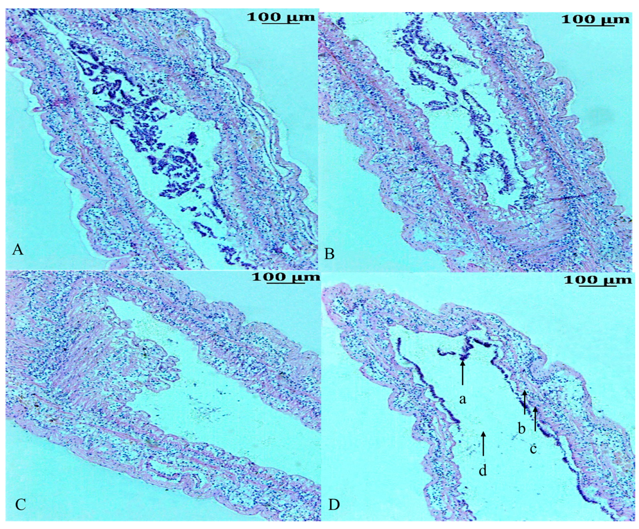
3.1. Histological Analysis of the Intestine

The morphological changes in the intestinal tissue of F. chinensis after low-salinity stress are shown in Figure 1. Histological observation showed that the intestinal tissue in shrimp in the control group (C0, Figure 1A) had a normal morphology, a well-developed network of villi almost filling the entire intestinal lumen, and epithelial cells with uniformly distributed columnar and cup-shaped nuclei. In S3 (Figure 1B), the changes in tissue morphology were not obvious. In S7 (Figure 1C), the epithelial cells were deformed, and vacuoles appeared. In contrast, under prolonged low-salinity stress, the S14 group (Figure 1D) displayed many intestinal alterations. For example, most epithelial cells were necrotic, and even ruptured; epithelial cells were detached from the basement membrane, and deformed, and loosely attached; nuclei were dispersed in the intestinal lumen; intestinal villi were gradually atrophied, and their height showed a tendency to decrease.

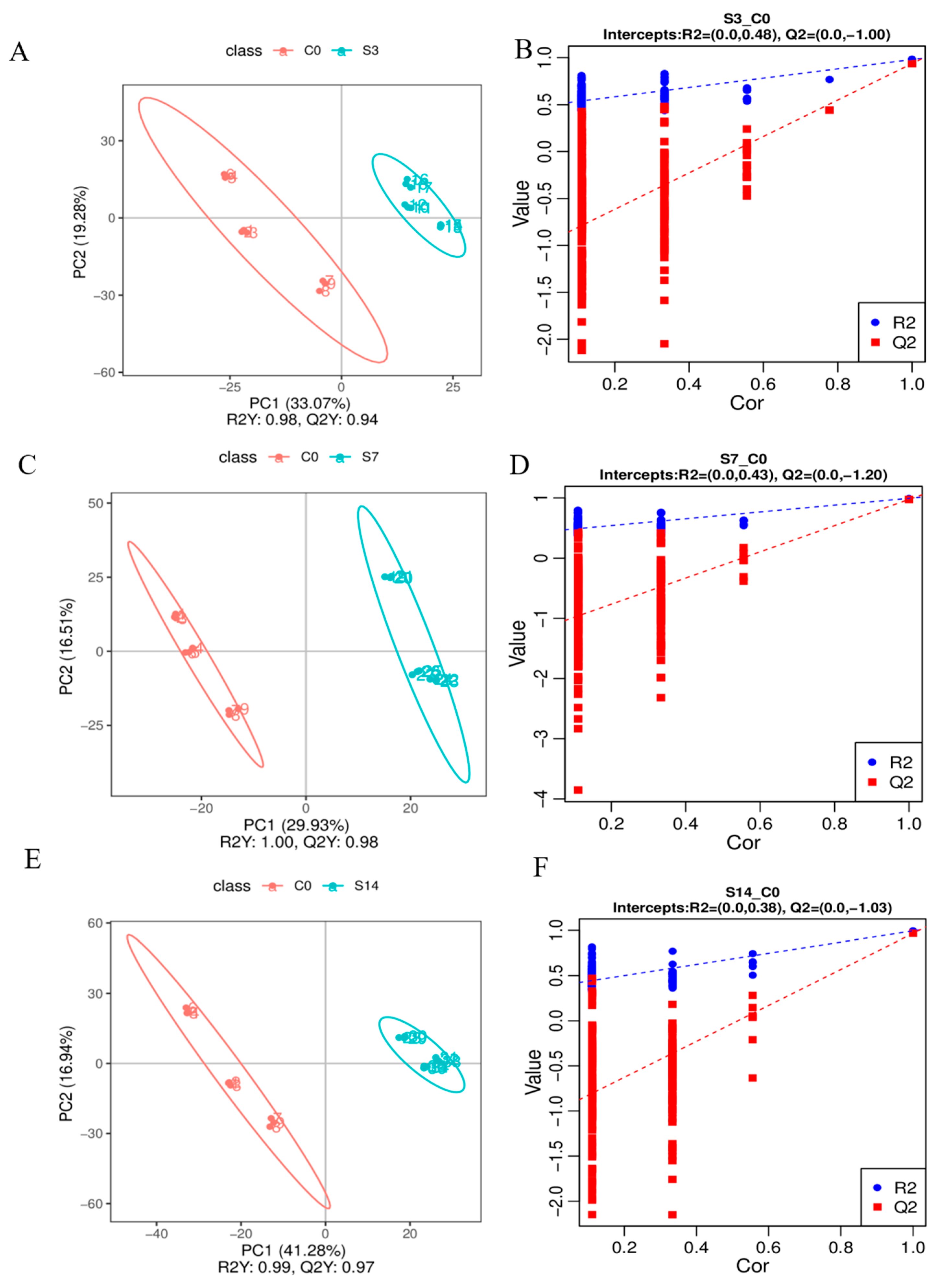
3.2. Identification of Metabolic Changes

A total of 1459 metabolic signals from 36 samples were detected, including 1050 and 409 metabolites in the positive and negative ion modes, respectively (Table S1), indicating their robustness and reliability. Typical total ion chromatograms of intestinal samples were obtained from F. chinensis in the positive mode (Figure S1). Pearson’s correlation showed that all the QC samples were of good quality (≥0.988 (Figure S2A). Principal Component Analysis (PCA) revealed some differences after low-salinity stress (Figure S2B). The PLS-DA model demonstrated a significant metabolic difference between the control and experimental groups, with Q2 values of 0.94, 0.98, and 0.97 in the S3, S7, and S14 groups, respectively, and R2 values of 0.98, 1.00, and 0.99 in the S3, S7, and S14 groups, respectively (Figure 2). Taken together, these results are consistent and can be further analyzed using metabolomics. Metabolites were divided into nine main categories: lipids and lipid-like molecules (50.90%) (Figure S2C). Lipid maps (Figure S3A) and KEGG pathway annotations (Figure S3B) were used to understand the functional properties and classifications of the identified metabolites.

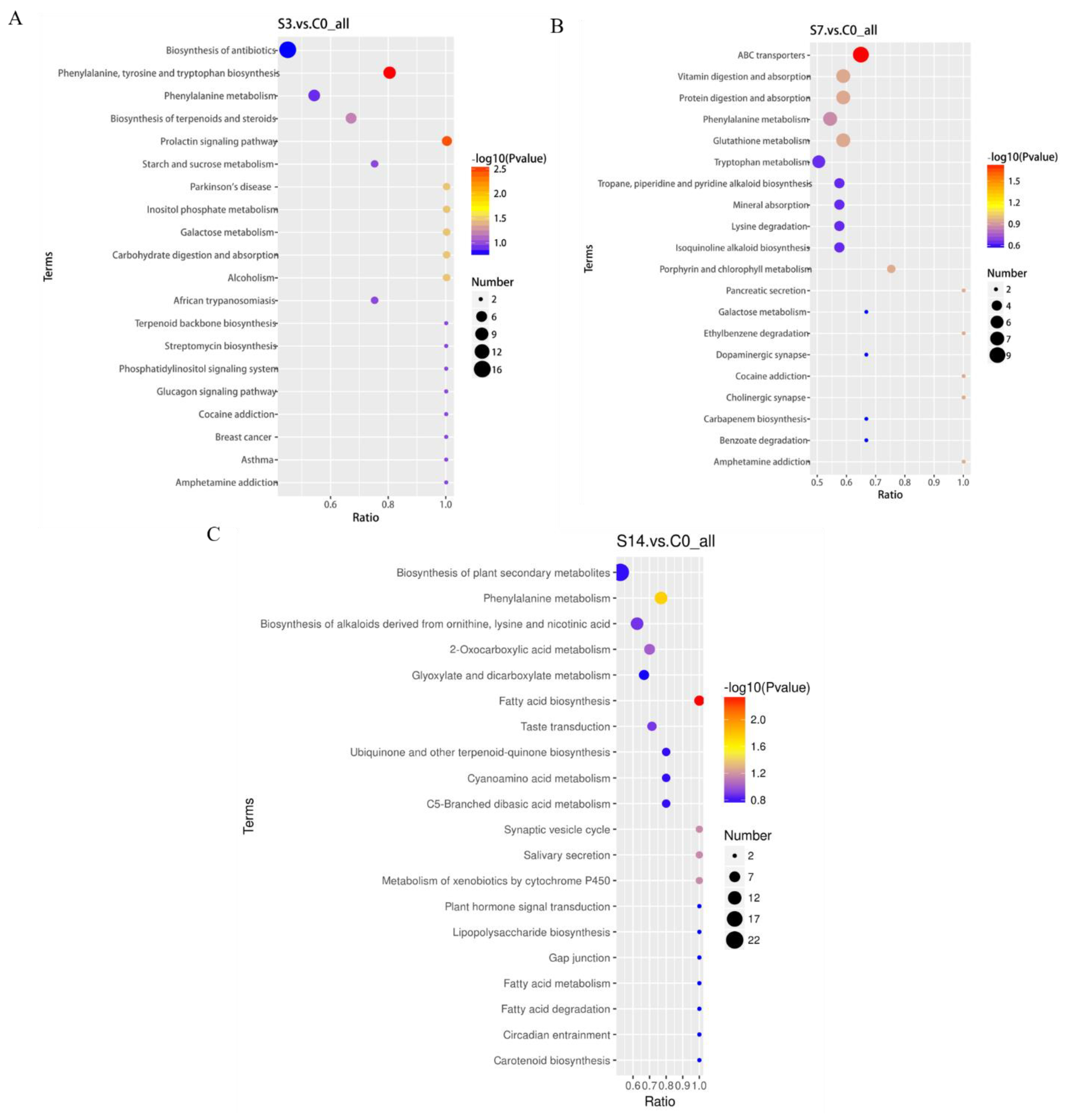
3.3. Identification and Enrichment Analysis of DEMs

In total, 382 (183 up-regulated and 199 down-regulated), 490 (213 up-regulated and 277 down-regulated), and 546 (274 up-regulated and 272 down-regulated) DEMs were identified in S3, S7, and S14, respectively (Figure 3A). Cluster analyses of DEMs portrayed different expression profiles among the three treatment groups (Figure 3B–D). Using a Venn diagram, a total of 134 DEMs from the two ion modes were simultaneously regulated by low-salinity stress (Figure 3E,F). To further analyze the biological pathways associated with metabolic changes in F. chinensis under low-salinity stress, all identified DEMs were mapped to the KEGG pathway database, and the top 20 enriched pathways were subsequently analyzed (Figure 4). Significant differences in the functional and metabolic pathways were observed at different time points. For example, as also shown in Table S2, intriguingly, in S3, we observed that acute salinity stress induced significant enrichment of 28 DEMs in seven pathways in comparison with C0, including phenylalanine, tyrosine, and tryptophan biosynthesis; galactose metabolism; inositol phosphate metabolism; carbohydrate digestion and absorption. In S7, some pathways, including ABC transporters, were enriched with metabolites. In S14, fatty acid biosynthesis and phenylalanine metabolism were significantly enriched.

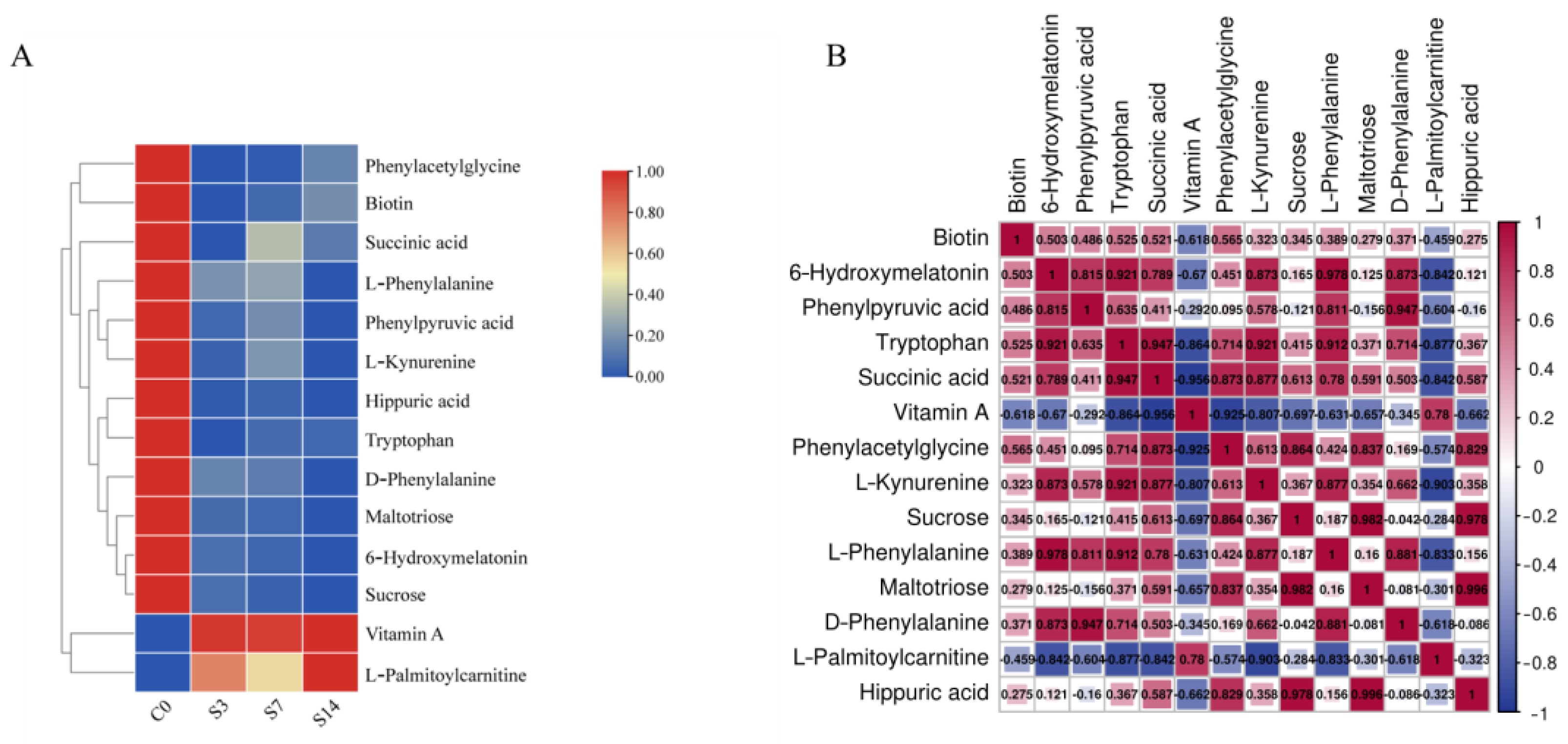
3.4. Identification and Analysis of 14 Key DEMs

To investigate the effects of low-salinity stress on F. chinensis, we performed an untargeted metabolomic analysis of the shrimp intestine using an LC-MS platform. Overlapping DEMs were enriched in phenylalanine metabolism; phenylalanine, tyrosine, and tryptophan biosynthesis; ABC transporters; retinol metabolism; and fatty acid degradation (Table S3). Among these pathways, 14 metabolites were selected as key DEMs and their information is shown in Table 1. Vitamin A and L-palmitoylcarnitine (L-Pcar) were up-regulated, whereas other DEMs were down-regulated.
Subsequently, we examined the relative changes (Figure 5A) and correlations (Figure 5B) among the 14 DEMs to further understand the metabolite changes in response to low salinity stress. The content of all these DEMs consistently decreased during low-salinity stress, apart from vitamin A and L-Pcar. Overall, the 14 DEMs were closely related, which is significant for explaining the pathogenesis of stress. For instance, Tryptophan (Trp) was more positively closely related to 6-Hydroxymelatonin (6-OHM), Succinic acid (SA), L-Kynurenine (L-Kyn), and L-Phenylalanine (L-Phe), in which L-Phe was more positively closely related to 6-OHM. Notably, Sucrose (SUC) was more positively closely related to Maltotriose (Mal) and Hippuric acid (HA). In contrast, L-Pcar and vitamin A were negatively related with the other metabolites.

4. Discussions

4.1. Low-Salinity Stress Induced Micromorphic and Metabolic Changes in Intestine

As a result of the decrease in arable land resources in developing countries, it is important to evaluate the salinity tolerance of shrimp species and their ability to grow in salinized environments [24]. As a key metabolic organ, the intestine governs energy metabolism such as glucose, lipid, and amino acid metabolism [25]. Consistent with the results of [26], compared to the control group (pH 8.3) under acute stress conditions, both low and high pH stresses disrupted the intestinal morphological structures. Previously, it was demonstrated that stress leads to intestinal structural damage, including goblet cells, and affects intestinal villus and wall integrity [27], suggesting that autoimmunity might be disrupted by stress in shrimp. In our study, intestinal epithelial cells were stripped from the basement membrane under low-salinity stress for 14 days, which is consistent with the result of Wang [28] and Duan et al. [29] under environmental stress. Consequently, episodes of tissue injury and incomplete healing of the intestinal epithelium are prerequisites for immune reactivation [30].
Although previous studies have examined the adaptive transcriptomic response of F. chinensis to low salinity [3,14,31,32], we used LC-MS-based metabolomics to determine its rapid adaptive and acute stimulatory response to low-salinity stress. Our study indicated that some DEMs were steadily down-regulated at the early stage of stress in S3 and S7, and these DEMs were more than the up-regulated metabolites during salinity stress, suggesting that acute low salinity exposure induced a drastic change in the metabolite content of the intestine. In a previous study, it led to the general depression of a large number of metabolites under stress exposure [33,34]. With the extension of stress time, F. chinensis gradually adapted to the saline environment through its regulation, and the number of up-regulated DEMs was approximately equal to the number of down-regulated metabolites in S14. These results align with our findings from transcriptome analyses of low-salinity stress [14]. Additionally, we screened 14 overlapping DEMs that regulated some KEGG pathways. Metabolic abnormalities in these pathways are the main causes of acute low-salinity stress, the effects of which on F. chinensis in low-salinity environments are elucidated below.

4.2. Effects of Low-Salinity Stress on Fatty Acid and Retinol Metabolism

Among the major nutritional components, lipids represent a complex group of biomolecules for the storage of energy and signal transduction, which maintain homeostasis in the presence of environmental stresses, and many fatty acids from lipid metabolism are essential for responding to ambient salinity change in animals [35,36,37]. A study demonstrated that the fatty acids metabolism plays a vital role in osmoregulation under long-term low-salinity stress [38]. Based on the results of this study, we conclude that fatty acids metabolism could be activated under acute low-salinity stress for 14 days.
Our study indicates that the role of L-Pcar is to regulate fatty acid degradation and metabolic pathways and increases content levels of this metabolite under salinity stress. The expression of up-regulated L-Pcar can increase the levels of palmitate and triacylglycerides and decrease the levels of free cholesterol, which can modulate signal transduction pathways [39]. This suggests that shrimp require extra energy from fatty acids to adapt and survive under low-salinity stress [32,39].
Enrichment analysis showed that retinol metabolism is also involved in low-salinity stress. As an essential nutrient, animals’ initial vitamin A status at the time of infection may be an important factor in their ability to respond to intestinal infection [40]. In the present study, F. chinensis enhanced innate and adaptive immunity by regulating retinol metabolism in the intestine and increasing the levels of vitamin A induced salinity stress to adjust to the environment. Hence, the upregulation of these DEMs in this study indicated their importance in the response of F. chinensis to acute low-salinity stress.

4.3. Effects of Low-Salinity Stress on Phenylalanine, Tyrosine, and Tryptophan Biosynthesis

Amino acid metabolism, one of the most enriched metabolic pathways, regulates essential cellular functions, such as complementary glucose and fatty acid-driven bioenergetic pathways, and can be metabolized to energy substances or used as signaling molecules to regulate cellular homeostasis [41,42]. Some amino acids that change in concentration during salinity acclimation play important roles in cell volume regulation [43]. These represented down-regulated metabolites of HA, Phenylpyruvic acid (PPA), Phenylacetylglycine (PAG), L/D-Phe, Trp, 6-OHM, and L-KYN. Again, these metabolites suggest that acute salinity induces disturbances in osmolality and energy metabolism compared to the metabolic changes induced by reduced salinity [44]. Previously, the concentrations of amino acids were reduced to balance the osmolarity under reduced seawater salinity [44]. While phenylalanine is an essential amino acid (or it cannot be synthesized by humans), Trp is considered a conditionally indispensable amino acid because it can be synthesized by the hydroxylation of phenylalanine by the enzyme phenylalanine hydroxylase [45]. It easily induces the biosynthesis pathways (phenylalanine, tyrosine, and tryptophan biosynthesis) in adverse conditions, which involves the regulation of energy metabolism [45,46,47]. Similar results were also observed in the present study, where the expression of eight metabolites, including phenylalanine and Trp significantly decreased the regulation of amino acid metabolism under low-salinity conditions. Trp was consistently suppressed by exposure to salinity stress, possibly reducing inflammatory factors and disease and enhancing immune function. In addition, PPA, PAG, SA, and HA play a vital role in adapting to ambient stress [48,49,50]. Kynurenine and 6-OHM is also an intimate relationship between tryptophan metabolism disorder and diseases [51,52,53,54,55]. SA is an endogenous isomer of methylmalonic acid that is widely distributed in human serum and urine [56]. SA plays a vital role in various metabolic pathways, including phenylalanine, tyrosine, and tryptophan metabolism and biosynthesis. It can prompt an immune response, resist ammonia stress, and suppress pathogenic infection in shrimp [50]; however, down regulation of SA resulted in low-salinity-stress-induced impairment of the immune system of F. chinensis. 6-OHM is an enzymatic metabolite and degradant of melatonin and is thought to be a direct free radical scavenger that provides tissue protection [55]. The use of 6-OHM enhances antioxidant and anti-inflammatory activity, which is the body’s defense mechanism against Alzheimer’s disease [57]. These results strongly suggested that DEMs related to amino acid metabolism are actively involved in energy generation processes for salinity acclimation in the intestine of F. chinensis at low salinity concentrations.

4.4. Effects of Low-Salinity Stress on ABC Transporters

Salinity stress also leads to osmotic imbalance, oxidative stress, and ionic disturbance, and even an increase in the amount and serious imbalance of ROS within animal cells [58]. Acute salinity induces disturbances in energy metabolism compared to the metabolic changes induced by reduced salinity and may interfere with normal energy intake and expenditure [44]. During this process, aquatic animals are forced to alter the expression of various enzymes and transporters to generate metabolic activities [59,60]. For instance, ABC transporters, a 48-member superfamily of membrane proteins, actively transport various biological substrates across lipid membranes (including exogenous substances) and are involved in the absorption of small molecules [61].
Previous studies have shown that ABC transporters play key roles in the physiology and development of plants’ responses to abiotic stresses [62,63,64], but few studies are concentrated on crustaceans. In the present study, some ion transporter metabolites were inhibited after F. chinensis was subjected to low-salinity stress, which reduced the intestinal absorption of ions, the ion loss, and ATP consumption during stress. DEMs, including Mal, SUC, and Biotin, are involved in inducing ABC transporters under low-salinity stress. Mal is largely resistant to the action of the body but is readily hydrolyzed to glucose by homogenates of the small intestinal mucosa [65]. As a metabolite, SUC plays a crucial role in development, stress response, and yield by promoting the growth and synthesis of essential compounds [66]. Biotin (Vitamin B7), a water-soluble B-vitamin, is an essential micronutrient for cellular functions, and its receptors are overexpressed in certain cancers [67]. In conclusion, ABC transporters play a key role in F. chinensis in response to low-salinity stress.

5. Conclusions

This is the first study to demonstrate that low-salinity stress causes apparent histological changes in the intestine of F. chinensis after 14 days of exposure. Further, untargeted metabolic profiling indicated that there were 14 overlapping DEMs enriched in phenylalanine, tyrosine, and tryptophan biosynthesis, ABC transporters, fatty acid, and retinol metabolism, in which the content of some DEMs was decreased, while the corresponding metabolites also increased to underlie the rapid adaptive and acute stimulatory responses to low-salinity stress. Key metabolic pathways are involved in the regulation of adverse effects caused by low-salinity stress in the intestine of F. chinensis. These findings provide new insights into the regulatory mechanisms of the intestine in F. chinensis and essential information for the breeding F. chinensis tolerant to low-salinity stress.

Supplementary Materials

The following supporting information can be downloaded at: https://www.mdpi.com/article/10.3390/ani14131880/s1, Figure S1: Representative liquid chromatography–mass spectrometry (LC–MS) total ion chromatograms (TICs) of quality control (QC) in F. chinensis; Figure S2: Information of between all QC sample and initial data analysis; Figure S3: Annotation of intestine metabolites in F. chinensis under low-salinity stress; Table S1: Metabolite differential screening results; Table S2: Significant differential metabolic pathways in the intestine of F. chinensis in response to low-salinity stress; Table S3: Results of KEGG significant enrichment analysis of 14 overlapping DEMs.

Author Contributions

Methodology, C.T. and H.S.; validation, C.T., T.G. and Y.H.; data curation, Q.W. and Y.H.; visualization, C.T.; formal analysis, C.T.; writing—original draft, C.T.; writing—review and editing, Q.W., J.L. and Y.H.; supervision, Y.H.; funding acquisition, Y.H.; roject administration, J.L. and Y.H. All authors have read and agreed to the published version of the manuscript.

Funding

This work was supported by National Key R & D Program of China (No. 2022YFD2400104-03), the earmarked fund for CARS-48, National Natural Science Foundation of China (No. 42376111), and Funding for Basic Research Operations of Chinese Academy of Fisheries Sciences (No. 2023TD50).

Institutional Review Board Statement

All animal treatment and test methods described in the present study were approved by the Institutional Animal Care and Use Committee (IACUC) of the Yellow Sea Fisheries Research Institute (CAFS) (Qingdao, China) (Approval No.: YSFRI-2022022).

Informed Consent Statement

Not applicable.

Data Availability Statement

Data will be provided on request.

Conflicts of Interest

The authors declare no conflicts of interest.

References

  1. Li, Z.; Li, J.; Wang, Q.; He, Y.; Liu, P. Aflp-Based Genetic Linkage Map of Marine Shrimp Penaeus (Fenneropenaeus chinensis). Aquaculture 2006, 261, 463–472. [Google Scholar] [CrossRef]
  2. Li, X.; Meng, X.; Luan, S.; Luo, K.; Cao, B.; Chen, B.; Kong, J. Isolation and Characterization of a Raf Gene from Chinese Shrimp Fenneropenaeus chinensis in Response to White Spot Syndrome Virus Infection. Fish Shellfish. Immunol. 2018, 83, 341–347. [Google Scholar] [CrossRef]
  3. Liu, J.; Zhang, D.; Zhang, L.; Wang, Z.; Shen, J. New Insight on Vitality Differences for the Penaeid Shrimp, Fenneropenaeus chinensis, in Low Salinity Environment Through Transcriptomics. Front. Ecol. Evol. 2022, 10, 716018. [Google Scholar] [CrossRef]
  4. Gao, W.; Tian, L.; Huang, T.; Yao, M.; Hu, W.; Xu, Q. Effect of Salinity on the Growth Performance, Osmolarity and Metabolism-Related Gene Expression in White Shrimp Litopenaeus vannamei. Aquac. Rep. 2016, 4, 125–129. [Google Scholar] [CrossRef]
  5. Pan, L.Q.; Luan, Z.H.; Jin, C.X. Effects of Na+/K+ and Mg2+/Ca2+ Ratios in Saline Groundwaters On Na+–K+-Atpase Activity, Survival and Growth of Marsupenaeus Japonicus Postlarvae. Aquaculture 2006, 261, 1396–1402. [Google Scholar] [CrossRef]
  6. Wang, Y.; Li, H.; Wei, J.; Hong, K.; Zhou, Q.; Liu, X.; Hong, X.; Li, W.; Liu, C.; Zhu, X.; et al. Multi-Effects of Acute Salinity Stress on Osmoregulation, Physiological Metabolism, Antioxidant Capacity, Immunity, and Apoptosis in Macrobrachium rosenbergii. Antioxidants 2023, 12, 1836. [Google Scholar] [CrossRef]
  7. Yu, Y.-Y.; Ding, L.-G.; Huang, Z.-Y.; Xu, H.-Y.; Xu, Z. Commensal Bacteria-Immunity Crosstalk Shapes Mucosal Homeostasis in Teleost Fish. Rev. Aquac. 2021, 13, 2322–2343. [Google Scholar] [CrossRef]
  8. Li, Y.; Wei, L.; Cao, J.; Qiu, L.; Jiang, X.; Li, P.; Song, Q.; Zhou, H.; Han, Q.; Diao, X. Oxidative Stress, Dna Damage and Antioxidant Enzyme Activities in the Pacific White Shrimp (Litopenaeus vannamei) When Exposed to Hypoxia and Reoxygenation. Chemosphere 2016, 144, 234–240. [Google Scholar] [CrossRef] [PubMed]
  9. Ragunathan, M. Vicissitudes of Oxidative Stress Biomarkers in the Estuarine Crab Scylla Serrata with Reference to Dry and Wet Weather Conditions in Ennore Estuary, Tamil Nadu, India. Mar. Pollut. Bull. 2017, 116, 113–120. [Google Scholar] [CrossRef]
  10. Shi, H.; Jiang, C.; Ye, T.; Tan, D.-X.; Reiter, R.J.; Zhang, H.; Liu, R.; Chan, Z. Comparative Physiological, Metabolomic, and Transcriptomic Analyses Reveal Mechanisms of Improved Abiotic Stress resistance in Bermudagrass [Cynodon dactylon (L). Pers.] by Exogenous Melatonin. J. Exp. Bot. 2014, 66, 681–694. [Google Scholar] [CrossRef]
  11. Fernández-García, J.; Altea-Manzano, P.; Pranzini, E.; Fendt, S.-M. Stable Isotopes for Tracing Mammalian-Cell Metabolism In Vivo. Trends Biochem. Sci. 2020, 45, 185–201. [Google Scholar] [CrossRef] [PubMed]
  12. Hosseinkhani, F.; Heinken, A.; Thiele, I.; Lindenburg, P.W.; Harms, A.C.; Hankemeier, T. The Contribution of Gut Bacterial Metabolites in the Human Immune Signaling Pathway of Non-Communicable Diseases. Gut Microbes 2021, 13, 1882927. [Google Scholar] [CrossRef] [PubMed]
  13. Ursell, L.K.; Haiser, H.J.; Van Treuren, W.; Garg, N.; Reddivari, L.; Vanamala, J.; Dorrestein, P.C.; Turnbaugh, P.J.; Knight, R. The Intestinal Metabolome: An Intersection Between Microbiota and Host. Gastroenterology 2014, 146, 1470–1476. [Google Scholar] [CrossRef]
  14. Tian, C.; Wang, Q.; Wang, J.; Li, J.; Guan, C.; He, Y.; Gao, H. Integrated Analysis of the Intestinal Microbiota and Transcriptome of Fenneropenaeus chinensis Response to Low-Salinity Stress. Biology 2023, 12, 1502. [Google Scholar] [CrossRef] [PubMed]
  15. Huang, L.-B.; Peng, L.-N.; Yan, X.-H. Multi-Omics Responses of Red Algae Pyropia haitanensis to Intertidal Desiccation during Low Tides. Algal Res. 2021, 58, 102376. [Google Scholar] [CrossRef]
  16. Wang, T.; Long, X.; Chen, X.; Liu, Y.; Liu, Z.; Han, S.; Yan, S. Integrated Transcriptome, Proteome and Physiology Analysis of Epinephelus coioides After Exposure to Copper Nanoparticles Or Copper Sulfate. Nanotoxicology 2017, 11, 236–246. [Google Scholar] [CrossRef] [PubMed]
  17. Zhou, Y.J. Effects of Saline and Alkaline Stress on Growth, Reproduction and Immune Function of Fenneropenaeus chinensis. Master’s Thesis, Dalian Ocean University, Dalian, China. 2023.
  18. Want, E.J.; Masson, P.; Michopoulos, F.; Wilson, I.D.; Theodoridis, G.; Plumb, R.S.; Shockcor, J.; Loftus, N.; Holmes, E.; Nicholson, J.K. Global Metabolic Profiling of Animal and Human Tissues via UPLC-MS. Nat. Protoc. 2013, 8, 17–32. [Google Scholar] [CrossRef]
  19. Rao, G.; Sui, J.; Zhang, J. Metabolomics Reveals Significant Variations in Metabolites and Correlations Regarding the Maturation of Walnuts (Juglans regia L.). Biol. Open 2016, 5, 829–836. [Google Scholar] [CrossRef] [PubMed]
  20. Heischmann, S.; Quinn, K.; Cruickshank-Quinn, C.; Liang, L.-P.; Reisdorph, R.; Reisdorph, N.; Patel, M. Exploratory Metabolomics Profiling in the Kainic Acid Rat Model Reveals Depletion of 25-Hydroxyvitamin D3 during Epileptogenesis. Sci. Rep. 2016, 6, 31424. [Google Scholar] [CrossRef]
  21. Boulesteix, A.-L.; Strimmer, K. Partial Least Squares: A Versatile Tool for the Analysis of High-Dimensional Genomic Data. Brief. Bioinform. 2006, 8, 32–44. [Google Scholar] [CrossRef]
  22. Wang, J.-B.; Pu, S.-B.; Sun, Y.; Li, Z.-F.; Niu, M.; Yan, X.-Z.; Zhao, Y.-L.; Wang, L.-F.; Qin, X.-M.; Ma, Z.-J.; et al. Metabolomic Profiling of Autoimmune Hepatitis: The Diagnostic Utility of Nuclear Magnetic Resonance Spectroscopy. J. Proteome Res. 2014, 13, 3792–3801. [Google Scholar] [CrossRef] [PubMed]
  23. Wen, B.; Mei, Z.; Zeng, C.; Liu, S. Metax: A Flexible and Comprehensive Software for Processing Metabolomics Data. BMC Bioinform. 2017, 18, 183. [Google Scholar] [CrossRef] [PubMed]
  24. Roy, L.A.; Davis, D.A.; Saoud, I.P.; Boyd, C.A.; Pine, H.J.; Boyd, C.E. Shrimp Culture in Inland Low Salinity Waters. Rev. Aquac. 2010, 2, 191–208. [Google Scholar] [CrossRef]
  25. Andrews, N. Intestinal Iron Absorption: Current Concepts Circa 2000. Dig. Liver Dis. 2000, 32, 56–61. [Google Scholar] [CrossRef] [PubMed]
  26. Duan, Y.; Wang, Y.; Liu, Q.; Zhang, J.; Xiong, D. Changes in the Intestine Barrier Function of Litopenaeus vannamei in Response to pH Stress. Fish Shellfish. Immunol. 2019, 88, 142–149. [Google Scholar] [CrossRef] [PubMed]
  27. Chen, J.; Xu, P.; Wen, H.; Xue, M.; Wang, Q.; He, J.; He, C.; Su, S.; Li, J.; Yu, F.; et al. Hypothermia-Mediated Oxidative Stress Induces Immunosuppression, Morphological Impairment and Cell Fate Disorder in the Intestine of Freshwater Drum, Aplodinotus grunniens. Aquaculture 2023, 575, 739805. [Google Scholar] [CrossRef]
  28. Wang, Z.; Zhou, J.; Li, J.; Lv, W.; Zou, J.; Fan, L. A New Insight into the Intestine of Pacific White Shrimp: Regulation of Intestinal Homeostasis and Regeneration in Litopenaeus vannamei during Temperature Fluctuation. Comp. Biochem. Physiol. Part D Genom. Proteom. 2020, 35, 100687. [Google Scholar] [CrossRef] [PubMed]
  29. Duan, Y.; Dong, H.; Wang, Y.; Li, H.; Liu, Q.; Zhang, Y.; Zhang, J. Intestine oxidative stress and immune response to sulfide stress in Pacific white shrimp Litopenaeus vannamei. Fish Shellfish. Immunol. 2017, 63, 201–207. [Google Scholar] [CrossRef] [PubMed]
  30. Rath, E.; Haller, D. Intestinal Epithelial Cell Metabolism at the Interface of Microbial Dysbiosis and Tissue Injury. Mucosal Immunol. 2022, 15, 595–604. [Google Scholar] [CrossRef]
  31. Tang, D.; Wu, Y.; Huang, S.; Wu, L.; Luo, Y.; Wang, Z. Transcriptome Reveals the Mechanism of Immunity in the Low Salinity Stress of the Chinese Shrimp (Fenneropenaeus chinensis). Thalass. Int. J. Mar. Sci. 2022, 38, 977–987. [Google Scholar] [CrossRef]
  32. Lu, H.; Chen, W.; Peng, K.; Huang, M.; Zhao, J.; Chen, X.; Sun, Y.; Ruan, Z.; Li, C.; Liu, D.; et al. Rapid Adaptive and Acute Stimulatory Responses to Low Salinity Stress in Pacific White Shrimp (Litopenaeus vannamei): Insights from Integrative Transcriptomic and Proteomic Analysis. Comp. Biochem. Physiol. Part D Genom. Proteom. 2023, 48, 101149. [Google Scholar] [CrossRef]
  33. Liu, C.; Li, C.; Bing, H.; Li, L.; Sun, P.; Li, T.; Du, D.; Zhao, J.; Wang, X.; Xiang, W. Integrated Physiological, Transcriptomic, and Metabolomic Analysis Reveals the Mechanism of Guvermectin Promoting Seed Germination in Direct-Seeded Rice under Chilling Stress. J. Agric. Food Chem. 2023, 71, 7348–7358. [Google Scholar] [CrossRef]
  34. Meng, X.; Jayasundara, N.; Zhang, J.; Ren, X.; Gao, B.; Li, J.; Liu, P. Integrated physiological, transcriptome and metabolome analyses of the hepatopancreas of the female swimming crab Portunus trituberculatus under ammonia exposure. Ecotoxicol. Environ. Saf. 2021, 228, 113026. [Google Scholar] [CrossRef]
  35. Li, E.; Wang, X.; Chen, K.; Xu, C.; Qin, J.G.; Chen, L. Physiological Change and Nutritional Requirement of Pacific White Shrimp Litopenaeus vannamei at Low Salinity. Rev. Aquac. 2017, 9, 57–75. [Google Scholar] [CrossRef]
  36. Sato, T.; Yamamoto, H.; Sawada, N.; Nashiki, K.; Tsuji, M.; Muto, K.; Kume, H.; Sasaki, H.; Arai, H.; Nikawa, T.; et al. Restraint Stress Alters the Duodenal Expression of Genes Important for Lipid Metabolism in Rat. Toxicology 2006, 227, 248–261. [Google Scholar] [CrossRef]
  37. Snaebjornsson, M.T.; Janaki-Raman, S.; Schulze, A. Greasing the Wheels of the Cancer Machine: The Role of Lipid Metabolism in Cancer. Cell Metab. 2020, 31, 62–76. [Google Scholar] [CrossRef]
  38. Xu, C.; Li, E.; Liu, Y.; Wang, X.; Qin, J.G.; Chen, L. Comparative Proteome Analysis of the Hepatopancreas from the Pacific White Shrimp Litopenaeus vannamei under Long-Term Low Salinity Stress. J. Proteom. 2017, 162, 1–10. [Google Scholar] [CrossRef]
  39. Nałęcz, K.A.; Szczepankowska, D.; Czeredys, M.; Kulikova, N.; Grześkiewicz, S. Palmitoylcarnitine Regulates Estrification of Lipids and Promotes Palmitoylation of Gap-43. FEBS Lett. 2007, 581, 3950–3954. [Google Scholar] [CrossRef]
  40. Chai, Z.; Lyu, Y.; Chen, Q.; Wei, C.-H.; Snyder, L.M.; Weaver, V.; Sebastian, A.; Albert, I.; Li, Q.; Cantorna, M.T.; et al. RNAseq Studies Reveal Distinct Transcriptional Response to Vitamin A Deficiency in Small Intestine Versus Colon, Uncovering Novel Vitamin A-Regulated Genes. J. Nutr. Biochem. 2021, 98, 108814. [Google Scholar] [CrossRef] [PubMed]
  41. Suzuki, R.; Sato, Y.; Obeng, K.A.; Suzuki, D.; Komiya, Y.; Adachi, S.-I.; Yoshizawa, F.; Sato, Y. Energy Metabolism Profile of the Effects of Amino Acid Treatment On Skeletal Muscle Cells: Leucine Inhibits Glycolysis of Myotubes. Nutrition 2020, 77, 110794. [Google Scholar] [CrossRef]
  42. Suzuki, A.; Iwata, J. Amino Acid Metabolism and Autophagy in Skeletal Development and Homeostasis. Bone 2021, 146, 115881. [Google Scholar] [CrossRef]
  43. Jarvis, P.L.; Ballantyne, J.S. Metabolic Responses to Salinity Acclimation in Juvenile Shortnose Sturgeon Acipenser Brevirostrum. Aquaculture 2003, 219, 891–909. [Google Scholar] [CrossRef]
  44. Wu, H.; Zhang, X.; Wang, Q.; Li, L.; Ji, C.; Liu, X.; Zhao, J.; Yin, X. A Metabolomic Investigation On Arsenic-Induced Toxicological Effects in the Clam Ruditapes philippinarum under Different Salinities. Ecotoxicol. Environ. Saf. 2013, 90, 1–6. [Google Scholar] [CrossRef]
  45. Dai, X.; Liu, M.; Xu, S.; Zhao, H.; Li, X.; Bai, Y.; Zou, Y.; An, Y.; Fan, F.; Zhang, J.; et al. Metabolomics Profile of Plasma in Acute Diquat-Poisoned Patients Using Gas Chromatography-Mass Spectrometry. Food Chem. Toxicol. 2023, 176, 113765. [Google Scholar] [CrossRef]
  46. Du, Y.; Zhao, J.; Teng, J.; Ren, J.; Shan, E.; Zhu, X.; Zhang, W.; Wang, L.; Hou, C.; Wang, Q. Combined Effects of Salinity and Polystyrene Microplastics Exposure On the Pacific Oysters Crassostrea gigas: Oxidative Stress and Energy Metabolism. Mar. Pollut. Bull. 2023, 193, 115153. [Google Scholar] [CrossRef]
  47. Mani, K.; Vitenberg, T.; Ben-Mordechai, L.; Schweitzer, R.; Opatovsky, I. Comparative Untargeted Metabolic Analysis of Natural- And Laboratory-Reared Larvae of Black Soldier Fly, Hermetia illucens (L.) (Diptera: Stratiomyidae). Comp. Biochem. Physiol. Part B Biochem. Mol. Biol. 2023, 266, 110851. [Google Scholar] [CrossRef]
  48. Rocchetti, G.; Bhumireddy, S.R.; Giuberti, G.; Mandal, R.; Lucini, L.; Wishart, D.S. Edible Nuts Deliver Polyphenols and their Transformation Products to the Large Intestine: An in Vitro Fermentation Model Combining Targeted/Untargeted Metabolomics. Food Res. Int. 2019, 116, 786–794. [Google Scholar] [CrossRef]
  49. Valerio, F.; Di Biase, M.; Lattanzio, V.M.; Lavermicocca, P. Improvement of the Antifungal Activity of Lactic Acid Bacteria by Addition to the Growth Medium of Phenylpyruvic Acid, a Precursor of Phenyllactic Acid. Int. J. Food Microbiol. 2016, 222, 1–7. [Google Scholar] [CrossRef]
  50. Duan, Y.F.; Wang, Y.; Zhang, J.S.; Sun, X.X.Y.; Wang, J. Dietary Effects of Succinic Acid On the Growth, Digestive Enzymes, Immune Response and Resistance to Ammonia Stress of Litopenaeus vannamei. Fish Shellfish Immunol. 2018, 78, 10–17. [Google Scholar] [CrossRef]
  51. Cheng, S.; Zhu, Z.; Li, H.; Wang, W.; Jiang, Z.; Pan, F.; Liu, D.; Ho, R.C.; Ho, C.S. Rifaximin Ameliorates Depression-Like Behaviour in Chronic Unpredictable Mild Stress Rats by Regulating Intestinal Microbiota and Hippocampal Tryptophan Metabolism. J. Affect. Disord. 2023, 329, 30–41. [Google Scholar] [CrossRef]
  52. Liu, Y.; Pei, Z.; Pan, T.; Wang, H.; Chen, W.; Lu, W. Indole Metabolites and Colorectal Cancer: Gut Microbial Tryptophan Metabolism, Host Gut Microbiome Biomarkers, and Potential Intervention Mechanisms. Microbiol. Res. 2023, 272, 127392. [Google Scholar] [CrossRef]
  53. Parolisi, S.; Montanari, C.; Borghi, E.; Cazzorla, C.; Zuvadelli, J.; Tosi, M.; Barone, R.; Bensi, G.; Bonfanti, C.; Vici, C.D.; et al. Possible Role of Tryptophan Metabolism Along the Microbiota-Gut-Brain Axis On Cognitive & Behavioral Aspects in Phenylketonuria. Pharmacol. Res. 2023, 197, 106952. [Google Scholar] [CrossRef]
  54. Xue, C.; Li, G.; Zheng, Q.; Gu, X.; Shi, Q.; Su, Y.; Chu, Q.; Yuan, X.; Bao, Z.; Lu, J.; et al. Tryptophan Metabolism in Health and Disease. Cell Metab. 2023, 35, 1304–1326. [Google Scholar] [CrossRef]
  55. Sakano, K.; Oikawa, S.; Hiraku, Y.; Kawanishi, S. Oxidative Dna Damage Induced by a Melatonin Metabolite, 6-Hydroxymelatonin, Via a Unique Non-O-Quinone Type of Redox Cycle. Biochem. Pharmacol. 2004, 68, 1869–1878. [Google Scholar] [CrossRef]
  56. Ma, X.; Zou, Y.; Tang, Y.; Wang, D.; Zhou, W.; Yu, S.; Qiu, L. High-throughput analysis of total homocysteine and methylmalonic acid with the efficiency to separate succinic acid in serum and urine via liquid chromatography tandem mass spectrometry. J. Chromatogr. B 2022, 1193, 123135. [Google Scholar] [CrossRef]
  57. Maharaj, D.S.; Walker, R.B.; Glass, B.D.; Daya, S. 6-Hydroxymelatonin protects against cyanide induced oxidative stress in rat brain homogenates. J. Chem. Neuroanat. 2003, 26, 103–107. [Google Scholar] [CrossRef]
  58. Singh, M.; Singh, V.P.; Prasad, S.M. Responses of Photosynthesis, Nitrogen and Proline Metabolism to Salinity Stress in Solanum lycopersicum under Different Levels of Nitrogen Supplementation. Plant Physiol. Biochem. 2016, 109, 72–83. [Google Scholar] [CrossRef]
  59. Tseng, Y.L.; Hwang, P. Some Insights Into Energy Metabolism for Osmoregulation in Fish. Comp. Biochem. Physiol. Part C Toxicol. Pharmacol. 2008, 148, 419–429. [Google Scholar] [CrossRef]
  60. Chen, K.; Li, E.; Li, T.; Xu, C.; Wang, X.; Lin, H.; Qin, J.G.; Chen, L. Transcriptome and Molecular Pathway Analysis of the Hepatopancreas in the Pacific White Shrimp Litopenaeus vannamei under Chronic Low-Salinity Stress. PLoS ONE 2015, 10, e0131503. [Google Scholar] [CrossRef]
  61. Moore, J.M.; Bell, E.L.; Hughes, R.O.; Garfield, A.S. ABC Transporters: Human Disease and Pharmacotherapeutic Potential. Trends Mol. Med. 2023, 29, 152–172. [Google Scholar] [CrossRef]
  62. Moons, A. Ospdr9, Which Encodes a Pdr-Type Abc Transporter, is Induced by Heavy Metals, Hypoxic Stress and Redox Perturbations in Rice Roots11the Nucleotide Sequence Reported in this Paper Has Been Submitted to the Embl, Genbank and Ddbj Nucleotide Sequence Databases with the Accession Number Ay271618. FEBS Lett. 2003, 553, 370–376. [Google Scholar] [PubMed]
  63. Nguyen, V.N.T.; Moon, S.; Jung, K.-H. Genome-Wide Expression Analysis of Rice Abc Transporter Family Across Spatio-Temporal Samples and in Response to Abiotic Stresses. J. Plant Physiol. 2014, 171, 1276–1288. [Google Scholar] [CrossRef]
  64. Yazaki, K. Abc Transporters Involved in the Transport of Plant Secondary Metabolites. FEBS Lett. 2006, 580, 1183–1191. [Google Scholar] [CrossRef]
  65. Messer, M.; Kerry, K.R. Intestinal Digestion of Maltotriose in Man. Biochim. Biophys. Acta Enzymol. 1967, 132, 432–443. [Google Scholar] [CrossRef]
  66. Rompicharla, S.V.K.; Kumari, P.; Bhatt, H.; Ghosh, B.; Biswas, S. Biotin Functionalized Pegylated Poly(Amidoamine) Dendrimer Conjugate for Active Targeting of Paclitaxel in Cancer. Int. J. Pharm. 2019, 557, 329–341. [Google Scholar] [CrossRef]
  67. Zhang, N.; Yang, Y.; Li, C.; Zhang, K.; Gao, X.; Shen, J.; Wang, Y.; Cheng, D.; Lv, J.; Sun, J. Based On 1H Nmr and Lc-Ms Metabolomics Reveals Biomarkers with Neuroprotective Effects in Multi-Parts Ginseng Powder. Arab. J. Chem. 2023, 16, 104840. [Google Scholar] [CrossRef]
Figure 1. Intestine histological morphology of F. chinensis under low-salinity stress. (A) Intestine structure of C0, Χ400; (B) intestine structure of S3, Χ400; (C) intestine structure of S7, Χ400; (D) intestine structure of S14, Χ400. (a) Nuclei, (b) epithelium, (c) brush border, (d) lumen, rule: 100 μm.
Animals 14 01880 g001
Figure 2. Multivariate modeling of LC-MS data in response to low-salinity stress in F. chinensis. (A) PCA score plot and (B) validation PLS-DA score plot in S3; (C) PCA score plot and (D) validation PLS-DA score plot in S7; (E) PCA score plot and (F) validation PLS-DA score plot in S14.
Animals 14 01880 g002
Figure 3. Information of intestine differentially expressed metabolites (DEMs) of F. chinensis under low-salinity stress. (A) numbers of DEMs in S3, S7, and S14 vs. C0. (B) DEMs are expressed by a heatmap in S3. (C) DEMs are expressed by a heatmap in S7. (D) DEMs are expressed by a heatmap in S14. DEMs indicated by a Venn diagram in the positive (E) and negative (F) iron mode.
Animals 14 01880 g003
Figure 4. The top 20 KEGG enrichment analysis of DEMs in response to low-salinity stress. (A) enrichment analysis of DEMs in S3 vs. C0. (B) enrichment analysis of DEMs in S7 vs. C0. (C) enrichment analysis of DEMs in S14 vs. C0.
Animals 14 01880 g004
Figure 5. Information of 14 key DEMs in the intestine of F. chinensis in response to low-salinity stress. (A) Heatmap and (B) Spearman’s correlation analysis of DEMs. Numbers represent the correlation coefficient values between different DEMs; red and blue indicate positive and negative correlation.
Animals 14 01880 g005
Table 1. Metabolite differential screening results.
ModeNameUp/DownFormulaRT [min]m/zp Value in S3, S7, S14
PosHippuric acid (HA)DownC9H9NO35.131180.06570.0087253180.0160197710.005866128
Phenylpyruvic acid (PPA)DownC9H8O35.035165.05472.66021 × 10−50.000598331.03938 × 10−5
L-Phenylalanine (L-Phe)DownC9H11NO25.012166.08650.0024370220.0052382818.70015 × 10−5
D-Phenylalanine (D-Phe)DownC9H11NO210.229166.08680.005345960.0037186510.000552025
6-Hydroxymelatonin (6-OHM)DownC13H16N2O35.442249.12451.4535 × 10−63.12169 × 10−71.54119 × 10−7
L-Kynurenine (L-Kyn)DownC10H12N2O35.272209.09250.0013328620.0104330430.0011305
Vitamin AUpC20H30O9.36287.23690.0007731570.0013748980.004364512
L-Palmitoylcarnitine (L-Pcar)UpC23H45NO410.234400.34290.0079615630.0108618240.001583441
NegSuccinic acid (SA)DownC4H6O42.469117.01820.0004170820.0239976580.002328271
Phenylacetylglycine (PAG)DownC10H11NO35.606192.0660.0009715550.00086220.005389868
Tryptophan (Trp)DownC11H12N2O25.219203.08230.0001429690.0008694030.000486337
Maltotriose (Mal)DownC18H32O161.385539.13810.0047086510.0070460870.001962056
Sucrose (SUC)DownC12H22O111.364341.11010.0023283820.0016751350.000357143
BiotinDownC10H16N2O3S5.394243.08091.13455 × 10−63.62306 × 10−55.47528 × 10−5
Disclaimer/Publisher’s Note: The statements, opinions and data contained in all publications are solely those of the individual author(s) and contributor(s) and not of MDPI and/or the editor(s). MDPI and/or the editor(s) disclaim responsibility for any injury to people or property resulting from any ideas, methods, instructions or products referred to in the content.

