Simple Summary
Ticks are ectoparasites with medical significance. They inhabit diverse environments and maintain close interactions with numerous vertebrate hosts. Ixodes ticks can transmit various pathogens to animals and humans. The aim here was to examine Ixodes ticks from Bosnia and Herzegovina to check for specific pathogens. This study found Rickettsia spp., Babesia spp., Anaplasma phagocytophilum, and Borrelia burgdorferi sensu lato in ticks from domestic animals. These findings highlight the need for the ongoing monitoring of ticks and tick-borne pathogens to protect animal and public health. Additionally, this study provides valuable insights into the occurrence and spread of these pathogens, emphasizing the importance of broader surveillance and control measures. Effective prevention, surveillance, and control of tick-borne diseases require urgent regional and international collaboration.
Abstract
Limited information is available regarding the presence of tick-borne pathogens and their distribution within Ixodes species in Bosnia and Herzegovina. This study aimed to identify Rickettsia spp., Babesia spp., Anaplasma phagocytophilum, and Borrelia burgdorferi sensu lato (s.l.) in Ixodes ticks collected from domestic and wild animals and vegetation in different regions across Bosnia and Herzegovina. A total of 7438 adult ticks, including 4526 Ixodes ricinus, Ixodes canisuga, and Ixodes hexagonus, were collected. Real-time PCR screening of 450 pooled I. ricinus samples revealed a 22.1% infection rate with at least one pathogen. Rickettsia spp. (6.3%) were found in ticks from dogs, cats, and goats, Babesia spp. (3.1%) in ticks from dogs and cattle, A. phagocytophilum (8.8%) in ticks from dogs, goats, and cattle, and B. burgdorferi s.l. (3.4%) in ticks from dogs and cats. Mixed infections with B. burgdorferi s.l. and A. phagocytophilum, as well as B. burgdorferi s.l. and Rickettsia spp., were found in two pools of I. ricinus from dogs and cats, respectively. Additionally, co-infection with Rickettsia spp. and A. phagocytophilum was confirmed in three tick pools from dogs and goats. Each tick from these pooled samples was individually retested to confirm the presence of pathogens. In the examined pooled samples of I. canisuga (1) and I. hexagonus (6), none of the tested pathogens were detected. Our findings represent the first detection of Rickettsia spp., Babesia spp., A. phagocytophilum, and B. burgdorferi s.l. in I. ricinus collected from domestic animals and vegetation in Bosnia and Herzegovina. Considering the established infection rates, the detection of tick-borne pathogens in adult ticks collected from domestic animals and vegetation enriches the current knowledge of the presence of tick-borne pathogens at the local, regional, national, and broader levels.
1. Introduction
Alongside mosquitoes, ticks are considered the primary vectors of the most important infectious diseases in humans and animals worldwide [1]. In recent years, the spread of tick-borne pathogens (TBPs) has increased, and the diseases they transmit are appearing in new regions or re-emerging within endemic areas, posing growing concerns for veterinary and public health, as well as biodiversity conservation [2]. Evidence indicates that zoonotic tick-borne diseases (TBDs) are expanding their geographical range, with infection rates posing a potential future public health crisis [3]. Given these factors, the study of TBPs and their occurrence in different tick species is crucial for understanding TBDs in humans, livestock, pets, and wildlife [3].
In Europe, Ixodes ricinus is the most widely distributed species, with its range extending across the continent and infesting numerous animal species and humans [4]. It is considered a serious health concern due to its extensive host population and ability to transmit various pathogens, including the tick-borne encephalitis virus (TBEV), Borrelia burgdorferi sensu lato (s.l.), the causative agent of Lyme borreliosis, and other pathogens such as Anaplasma phagocytophilum, as well as the pathogenic species of Rickettsia, Bartonella, Francisella, and Babesia [5,6,7,8]. In most European countries, Ixodes canisuga is commonly found infesting medium-sized Mustelidae and Canidae, such as badgers (Meles meles), jackals (Canis aureus), red foxes (Vulpes vulpes), and European hedgehogs (Erinaceus europaeus) [9,10]. Like I. canisuga, Ixodes hexagonus is also distributed throughout Europe and is associated with hedgehogs, Erinaceus europaeus, and Erinaceus roumanicus, as well as a wide range of medium-sized burrow-inhabiting mammals such as red foxes, badgers, European pine martens (Martes martes), stoats (Mustela erminea), European polecats (Mustela putorius), and occasionally otters (Lutra lutra) [9,11]. However, both species have been recorded biting humans and their companion animals; for example, I. canisuga is often found feeding on dogs, while I. hexagonus commonly feeds on pet cats and dogs and less frequently on humans [12,13,14]. Furthermore, pathogens such as Rickettsia, A. phagocytophilum, Babesia, and B. burgdorferi s.l. have been confirmed in both I. canisuga and I. hexagonus [15,16,17,18,19,20].
In previous studies conducted in Bosnia and Herzegovina, the presence of four tick species from the genus Ixodes has been recorded, namely I. ricinus, I. canisuga, I. hexagonus, and Ixodes vespertilionis [21,22,23,24,25]. Previous research has confirmed the dominant presence of I. ricinus [22,24,25] collected from domestic and wild animals, with a prevalence of 70.5% and 63.8% in 2008 and 2022, respectively. Ixodes canisuga and I. hexagonus were found infesting animals (primarily dogs and red foxes), with prevalence rates of 0.3% and 0.1%, respectively [24,25,26]. In Bosnia and Herzegovina, there are data on pathogens confirmed in I. ricinus in several studies, where the presence of B. burgdorferi s.l. complex has been confirmed in ticks collected from humans [27]. Additionally, in I. ricinus collected from animals, the presence of Babesia divergens, Rickettsia monacensis, Rickettsia Helvetica, and TBEV has been established [28,29,30]. Furthermore, no other studies have been encountered that reveal the presence of TBPs in other Ixodes species.
Data on TBPs in Bosnia and Herzegovina are limited. Therefore, this study aims to investigate the presence and distribution of Ixodes ticks and to identify pathogens including Rickettsia spp., Babesia spp., A. phagocytophilum, and B. burgdorferi s.l. in ticks collected from domestic and wild animals as well as from vegetation across diverse regions of Bosnia and Herzegovina.
2. Materials and Methods
2.1. Study Area and Tick Collection
The collection of ticks was conducted across the entire territory of Bosnia and Herzegovina from 2017 to 2023. The methods of tick collection and sampling procedures were carefully tailored to match the geographical and bioclimatic characteristics of the study area. Sampling was based on prior knowledge of tick distribution and abundance in Bosnia and Herzegovina, as depicted in Figure 1.
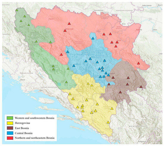
Figure 1.
Geographical overview of the regions and sampling sites of Bosnia and Herzegovina included in the study. The study map was developed using ArcGIS® Pro software (ESRI, Redlands, California, United States of America), version 3.2.2.
The sampling of ticks involved the direct collection of adult ticks from domestic and wild animals by veterinarians and animal owners in defined areas. The adult ticks were carefully isolated from the host body using tweezers to avoid any external damage to the specimens. Collection of adult ticks from vegetation, leaf litter, or other substrates was conducted once between May and August by dragging a white flannel blanket [31] over the leaf litter and low vegetation near farms and enclosures where the animals from which ticks were previously sampled resided. Ticks were then collected from the fabric using forceps or a tick removal tool. All tick species encountered, whether questing or feeding, were collected without any specific selection using the previously described sampling methods (n = 7438).
2.2. Morphological Identification of Ticks and Tick Pooling Procedure
After collection, the adult ticks were stored refrigerated at +4 °C and transported to the Laboratories of the University of Sarajevo-Veterinary faculty (accredited by BAS EN ISO/IEC 17025:2018 [32]). The ticks were counted, pooled, and identified based on morphological characteristics using a Zeiss Stemi 508 stereomicroscope (magnification 0.63–5.0×), following established identification keys [33,34].
Once the ticks were morphologically identified (7438), only Ixodes species (4526) were selected for this study. Subsequently, adult ticks were grouped and classified according to various characteristics such as engorgement status, sex, sampling location, vegetation, and host from which they were collected. Of the collected adult Ixodes species, 4490 were identified as I. ricinus, 32 as I. canisuga, and 4 as I. hexagonus. From the identified ticks, 450 pools were selected for molecular analysis out of a total of 2250 identified I. ricinus ticks (50.1%). Among these, 431 pools originated from animal hosts and 19 from vegetation. Additionally, analysis was conducted on 6 pools out of 32 collected I. canisuga ticks and 1 pool out of 4 identified I. hexagonus ticks.
Pooling criteria were based on geographical origin and representativeness, ensuring an adequate quantity of ticks from various regions of Bosnia and Herzegovina, particularly concerning I. ricinus (covering different species of wild and domestic animals, including livestock and pets). Similarly, due to the smaller number of collected I. canisuga and I. hexagonus, only one pool of I. hexagonus and 6 pools of I. canisuga were examined. Grouped samples contained up to 5 adult ticks, and sample selection was adjusted based on the number of ticks collected from the same animal or vegetation. In cases where ≥10 adult ticks were collected from a single host or herd, every other tick was selected for sampling. Likewise, if ≥20 ticks were collected under similar conditions, every fourth tick was chosen. Ticks were transferred from individual vials to sterile tubes using fine-tipped forceps, ensuring specimen integrity. The number of ticks per pool was recorded, and tubes were labeled for tracking and analysis. Prepared samples were then stored at −20 °C until further testing.
2.3. Molecular Identification of Pathogens
Nucleic acid extraction from samples utilized a QIAamp DNA Mini kit (Qiagen, Hilden, Germany), following the manufacturer’s instructions. Before DNA extraction, all ticks were carefully dissected and halved using sterile scissors, ensuring that sterile conditions were maintained throughout the process. The dissected halves of ticks were individually homogenized in lysis buffer using a TissueLyser II (Qiagen, Hilden, Germany) to effectively disrupt tick tissues and release DNA. Homogenization was conducted at high speed to ensure thorough tissue disruption. Following this, DNA extraction was performed from the homogenized samples using the standard protocol provided by the manufacturer. The remaining dissected tick halves were frozen at deep temperatures as a precaution for potential future studies. DNA amplification was conducted using the QuantiFast Pathogen PCR + IC Kit (Qiagen, Hilden, Germany) on the Mic qPCR Cycler platform (Bio Molecular Systems, Upper Coomera, Queensland, Australia). The specific primers, probes, and relevant information for the investigated pathogens are detailed in Table 1.

Table 1.
Primers and probes used for molecular detection of pathogens (Real-Time PCR).
A multiplex PCR approach was employed for selected pathogens, involving two duplex PCRs for Rickettsia spp. and Babesia spp., as well as A. phagocytophilum and B. burgdorferi s.l. Identification of Rickettsia spp., based on the amplification of a segment of the gltA gene, and Babesia spp., based on the amplification of a segment of the 18S rRNA gene, followed established protocols from previous studies [35,36]. Protocols for amplifying the ospA gene segment of B. burgdorferi s.l. and Msp2 genes of A. phagocytophilum were also based on previously published studies [37,38]. Cycling conditions for both duplex PCRs were identical, involving initial denaturation at 95 °C for 5 min, followed by 40 cycles of denaturation at 95 °C for 15 s, and annealing and elongation at 60 °C for 30 s. Fluorescence reading and graphical plotting were automatically conducted at 60 °C at the conclusion of each extension/elongation phase using micPCR software version 2.8.13 (Bio Molecular Systems, Upper Coomera, Queensland, Australia).
2.4. Data Analysis
The prevalence of Rickettsia spp., Babesia spp., A. phagocytophilum, and B. burgdorferi s.l. infection in positive adult ticks collected at a certain region was estimated using the Minimum Infection Rate (MIR), i.e., the minimum infected proportion expressed as a percentage: MIR = (p/N) × 100%, where p = the number of positive pools and N = the total number of ticks tested. This method assumes that only one infected tick is present in each positive pool [39,40].
3. Results
3.1. Tick Species Distribution
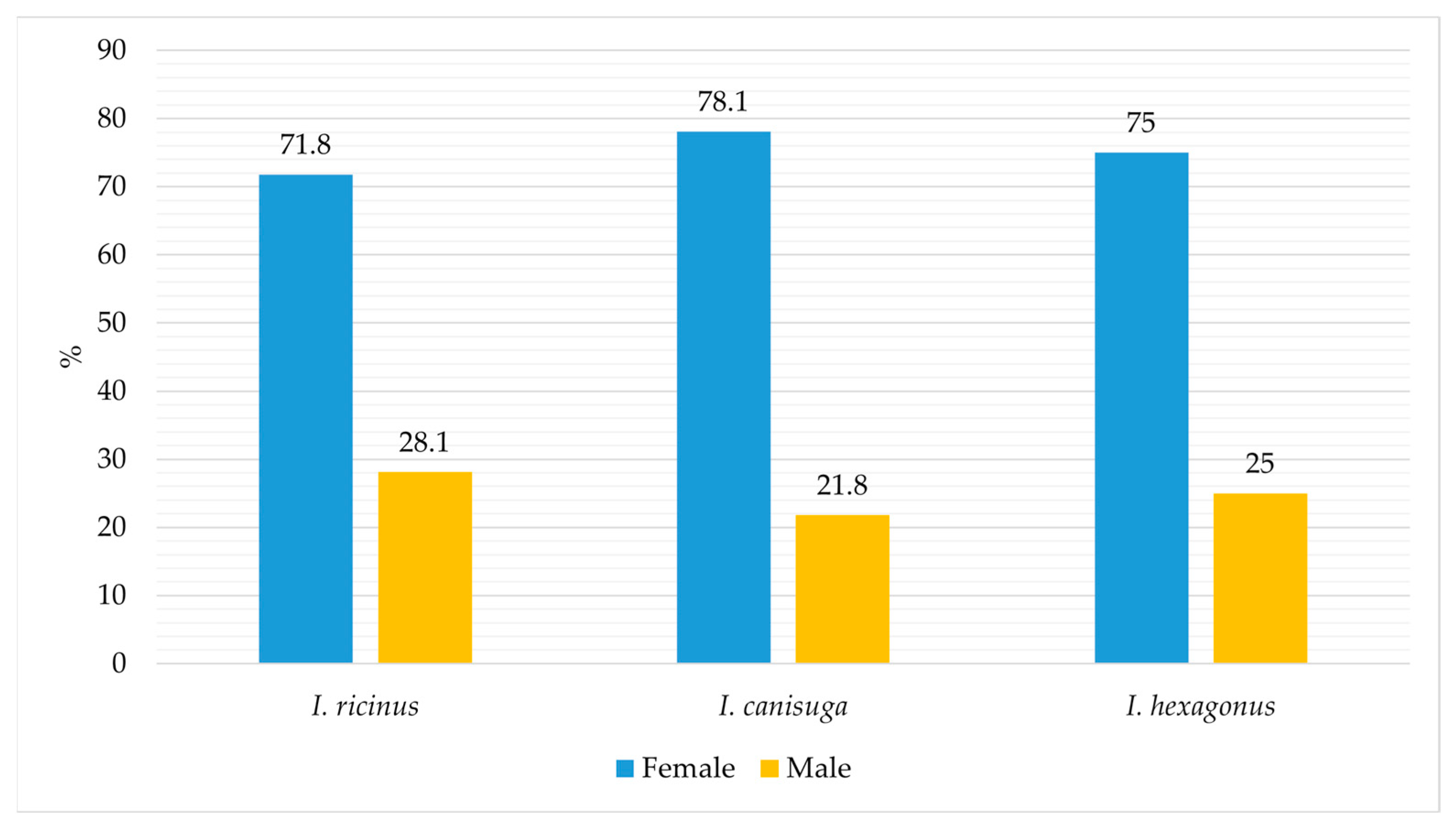
The proportion of I. ricinus out of the total collected adult ticks from 2017 to 2023 was 60.3% (4490/7438). It was 0.4% (32/7438) and 0.05% (4/7438) for I. canisuga and I. hexagonus, respectively. The percentage of collected females and males of I. ricinus, I. canisuga, and I. hexagonus, relative to the total number of each species collected from all regions, animal species, and vegetation, is shown in Figure 2.

Figure 2.
Collected females and males of I. ricinus, I. canisuga, and I. hexagonus ticks.
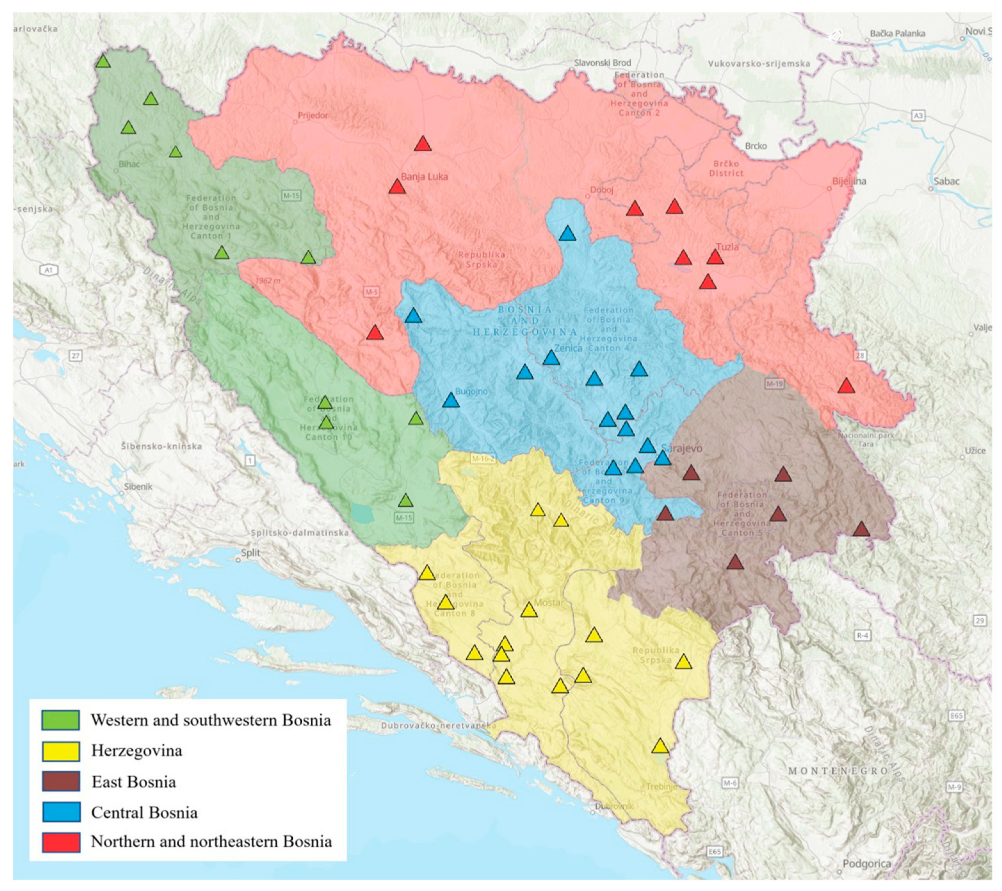
Figure 3 illustrates the distribution of I. ricinus, I. canisuga, and I. hexagonus according to the hosts and vegetation from which they were collected. Ixodes ricinus was predominantly collected from dogs, goats, and cattle, while to a slightly lesser extent from cats, sheep, foxes, roe deer, and vegetation, and from all defined sites in Bosnia and Herzegovina (Figure 3). Similarly, I. hexagonus was only collected from dogs, while I. canisuga was collected from foxes and dogs, specifically from the areas of Western and Southwestern Bosnia (Figure 4). Most of the collected Ixodes ticks were from the Central Bosnia and Herzegovina region.
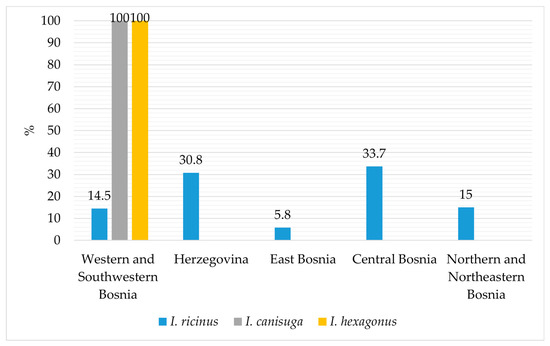
Figure 3.
Distribution of collected I. ricinus, I. canisuga, and I. hexagonus according to the hosts and vegetation.

Figure 4.
Spatial distribution of I. ricinus, I. canisuga, and I. hexagonus across different regions of Bosnia and Herzegovina.
3.2. Pathogen Detection and Identification of Tick Pools
The molecular analysis of 450 I. ricinus tick pools revealed an MIR of 22.1%. None of the examined I. canisuga and I. hexagonus pools tested positive for any of the pathogens analyzed. Detailed information regarding the MIRs (minimum infection rates) of pathogens in I. ricinus ticks according to locality and animal host can be found in Table 2. Detailed information regarding the MIRs of pathogens in I. ricinus ticks from vegetation according to locality can be found in Table 3.
Table 2.
The total number of I. ricinus pools tested and MIRs of pathogens detected in tick pools according to the locality and animal host.
Table 3.
The total number of I. ricinus pools tested and MIRs of pathogens detected in tick pools from vegetation according to the locality.
Rickettsia spp. was detected in I. ricinus ticks collected from dogs, cats, sheep, and goats from all sites except Eastern Bosnia (Table 2). The overall MIR was 6.3% in pools of all ticks collected from animals across all locations. Specifically, the MIRs for Western and Southwestern Bosnia, Herzegovina, Central Bosnia, Northern and Northeastern Bosnia were 10.4%, 10.6%, 3.6%, and 6.6%, respectively.
Babesia spp. was detected in I. ricinus ticks from dogs and cattle in Northern and Northeastern Bosnia with an MIR of 3.1% and in ticks collected from vegetation in Western and Southwestern Bosnia with an MIR of 3.6% (Table 3). Anaplasma phagocytophilum was confirmed in I. ricinus ticks from all locations and hosts, including dogs, goats, and cattle (Table 2). The overall infection rate for A. phagocytophilum was 8.8%, the highest among the four pathogens tested. The individual MIRs by locality were 13% in Western and Southwestern Bosnia, 8.2% in Herzegovina, 35.2% in Central Bosnia, 54.4% in Eastern Bosnia, and 7% in Northern and Northeastern Bosnia.
Borrelia burgdorferi s.l. was detected in Central Bosnia, Eastern Bosnia, and Northern and Northeastern Bosnia in I. ricinus from dogs and cats, with an overall MIR of 3.4% (Table 2). The individual infection rates by locality were 22.5% in Central Bosnia, 24.4% in Eastern Bosnia, and 13.1% in Northern and Northeastern Bosnia.
A total of 1.1% (5/450) of pooled samples tested positive for at least two investigated pathogens. Co-infection of B. burgdorferi s.l. and A. phagocytophilum was detected in one pooled sample originating from dogs in the Eastern Bosnia region, while in two pooled samples from dogs in the Herzegovina and Western and Southwestern Bosnia regions, the presence of two pathogens, Rickettsia spp. and A. phagocytophilum, was confirmed (Table 4). Each tick from these pooled samples was individually retested to confirm the presence of pathogens, revealing three ticks originating from three pooled dog samples as carriers of both pathogens.
Table 4.
Co-infection of pooled I. ricinus ticks.
Co-infection of B. burgdorferi s.l. and Rickettsia spp. was detected in one pooled sample originating from cats in Central Bosnia. The ticks from the positive cat pool were individually tested, and within the pool, the presence of both pathogens was confirmed in two ticks. Additionally, in one pooled sample from goats in the Northern and Northeastern Bosnia region, the presence of Rickettsia spp. and A. phagocytophilum was confirmed (Table 4). Similarly, ticks from the positive pool were individually tested, revealing co-infection in one tick.
4. Discussion
Bosnia and Herzegovina exhibit high climatic and habitat heterogeneity conducive to ticks and the spread of TBDs, which is increasingly becoming a concern for veterinary and public health. The results of this study highlight the frequent occurrence of TBPs in I. ricinus collected from various domestic animal species and vegetation; given that it is the most prevalent tick species in Bosnia and Herzegovina [22,24], this research holds significant importance. Considering that I. hexagonus and I. canisuga rarely infest humans and that this study also determined a low frequency of these ticks in both domestic and wild animals, their significance in the context of veterinary and public health is relatively minor. However, they can certainly play an important role as maintenance vectors in silent enzootic transmission cycles.
This study represents the first comprehensive examination of a larger number of I. ricinus ticks collected from domestic and wild animals and vegetation for the pathogens under investigation. Moreover, to the best of the authors’ knowledge, this study represents the first investigation into the presence of pathogens in I. canisuga and I. hexagonus ticks in Bosnia and Herzegovina.
Given that the majority of ticks were collected from animals (97.4%) rather than vegetation (2.6%), it is noteworthy that the proportion of females compared to males is higher in all three Ixodes species, with 71.8%, 78.1%, and 75% for I. ricinus, I. canisuga, and I. hexagonus, respectively (Figure 2). The overall proportion of I. ricinus among all collected tick species was 60.3%, which represents a slightly lower frequency compared to the results of previous studies conducted in Bosnia and Herzegovina, where the overall frequency was reported as 63.8% [22] and 70.5% [24]. Our findings indicate that I. ricinus ticks were predominantly identified on dogs, consistent with previous research in Bosnia and Herzegovina [22,24]. However, for the first time, this species was identified on deer and red foxes (Figure 3). Ixodes ricinus was most frequently encountered in the regions of Central Bosnia (33.8%) and Herzegovina (31%), followed by the regions of Northern and Northeastern Bosnia (15%) and Western and Southwestern Bosnia (14.5%). In this study, I. ricinus was the least prevalent in the Eastern Bosnia region (5.8%) (Figure 4). This distribution pattern is consistent with the results of the earlier investigation conducted in Bosnia and Herzegovina [22,24].
Furthermore, this study confirmed the presence of I. canisuga and I. hexagonus on animals at frequencies of 0.4% and 0.05%, respectively. Ixodes canisuga sampled from red foxes and dogs had a frequency of 0.4%, while I. hexagonus was found in 0.05% of samples collected from four dogs. Their low frequencies are consistent with previous research on ticks in Bosnia and Herzegovina, where 0.09% of I. hexagonus was sampled from dogs, while I. canisuga was sampled from red foxes at a frequency of 0.3% [24,25,26]. Both species were sampled in locations within the Western and Southwestern Bosnia region (Figure 4), and their presence was also confirmed in previous studies conducted in the Southwestern Bosnia region [22,24].
DNA from Rickettsia spp. was detected in 6.3% (95% CI 5.0–7.5) of I. ricinus pools collected from animals. The highest rate of infection was confirmed in the Herzegovina region (10.6%), while no presence of Rickettsia spp. was confirmed in ticks from the Eastern Bosnia region. The majority of positive ticks originated from dogs with an MIR ranging from 2.4% in Northern and Northeastern Bosnia to 14.2% in Herzegovina (Table 2). Additionally, the presence of Rickettsia spp. was confirmed for the first time in ticks from goats (7.14%) in Northern and Northeastern Bosnia, cats (4.7%) in Central Bosnia, and sheep (9.5%) in Herzegovina (Table 2). In this study, ticks collected from vegetation were not positive for the presence of Rickettsia spp. Data about the occurrence of Rickettsia in ticks are limited in Bosnia and Herzegovina. However, there are reports of finding Rickettsia spp. in ticks collected from vegetation in Bosnia and Herzegovina. Hodžić et al. [28] molecularly identified R. monacensis (1.1%) and R. helvetica (5.7%) in 21.8% of I. ricinus ticks in Bosnia and Herzegovina. Additionally, in studies conducted on people in Bosnia and Herzegovina, antibodies against Rickettsia were detected in blood samples from individuals from the northwestern part of Bosnia and Herzegovina. Of the 231 sera tested using the complement fixation test (CFT), 61.5% were positive for Rickettsia typhi, 4.3% for Rickettsia prowazekii, and 1.7% for Rickettsia conorii. Meanwhile, 183 serum samples tested using IFAT showed that 37.7% were positive for R. typhi and 1.6% for R. conorii [41]. The detection of Rickettsia at the recorded infection rates in tick pools (MIRs of 2.4–14.3%), particularly in ticks from dogs, underscores the need for ongoing surveillance and research to monitor and reduce the risks of TBDs in Bosnia and Herzegovina.
In the present study, the MIR for Babesia spp. was 3.1% (95% CI 2.2–3.9) in female I. ricinus pools. Babesia spp. was identified in eight pools (100%; eight/eight) originating from vegetation in Western and Southwestern Bosnia, but it was not detected in tick pools from animals from this region (Table 3). However, the small sample size of ticks from vegetation in this study makes it difficult to draw general conclusions, highlighting the need for a larger investigation. Of the examined ticks from animals in Northern and Northeastern Bosnia, Babesia spp. was detected in six I. ricinus pools sampled from cattle (17.1%) and forty pools from dogs (16%) (Table 2). Studies on babesiosis in Bosnia and Herzegovina in sheep and cattle were conducted in previous research by examining blood and ticks collected from animals. Babesia divergens was confirmed in the blood of clinically ill cattle in central Bosnia using blood smears and molecular detection methods [30,38]. Additionally, in the same study, B. divergens was confirmed in two female I. ricinus ticks (25%; two/eight) collected from animals [30]. In a study on Babesia ovis in sheep blood from 2022 in the area of Eastern Bosnia and Herzegovina, out of 192 tested sheep, B. ovis was confirmed in 70 (36.4%). The lack of reports on babesiosis in ruminants in the area of Bosnia and Herzegovina is likely due to the fact that mild and subclinical cases often go undiagnosed [30,38,42]. Given that bovine babesiosis has been present in cattle grazing on pastures where I. ricinus is reported as the dominant tick species, it is likely that Babesia is maintained in the environment with endemic stability; although, Babesia spp. were not detected in ticks originating from wild animals in our study. Previous research confirmed Babesia vulpes and Babesia canis in spleen samples of red foxes with frequencies of 31.9% and 0.8%, respectively [43]. Babesia spp. “Badger type A” was identified in a European wildcat (Felis silvestris) in Bosnia and Herzegovina [44].
Additionally, as this is the first examination of the presence of Babesia spp. from ticks on dogs, it is noteworthy that Babesia spp. was not confirmed in I. ricinus collected from dogs in Bosnia and Herzegovina. However, in previous research conducted in Bosnia and Herzegovina, Babesia spp. has been confirmed in a larger number of studies in blood. Peripheral blood samples taken from one hundred and thirty-four dogs in the northeastern part of Bosnia confirmed the presence of B. canis in seven dogs (5.2%) [45]. Furthermore, a study on blood smears from the peripheral blood of 183 dogs in Northern Bosnia confirmed a slightly higher prevalence of Babesia spp. infection (30.6%) [46]. A lower prevalence of infection was reported by Hrvat [47], where, in the northeastern part of Bosnia, Babesia spp. was identified in 11.8% of 432 dogs. Babesia canis was confirmed by molecular detection methods in dog blood in the study by Ćoralić et al. [48], with a frequency of 85% out of 80 dogs with symptoms of babesiosis. Furthermore, Babesia vogeli was identified in two dogs in Central Bosnia and one dog in the Western and Southwestern Bosnia region (0.7%) [49].
Anaplasma phagocytophilum is recognized as an emerging TBP that is important for animals and humans [50]. This study provides the first confirmation of A. phagocytophilum in I. ricinus in Bosnia and Herzegovina, with an MIR of 8.8% (95% CI 7.4–10.4). It was detected in all regions of Bosnia and Herzegovina, at various locations and in different animal species. The A. phagocytophilum-positive male and female pools were detected in I. ricinus sampled from dogs in all regions, except Northern and Northeastern Bosnia, with an infection rate ranging from 0.9% to 23.8% (Table 2). In this study, ticks collected from vegetation were not positive for the presence of A. phagocytophilum. In previous studies conducted in Bosnia and Herzegovina, Anaplasma was mainly confirmed in the blood of infected animals. Colella et al. [49] collected blood samples from 408 dogs and tested them using a microfluidic RT-PCR test for 43 different pathogens. The study revealed the presence of single and mixed infections. Furthermore, Anaplasma platys was confirmed in one dog in the Herzegovina region (0.2%) [49]. In this study, A. phagocytophilum was also detected in I. ricinus species collected from ruminants for the first time in I. ricinus from cattle in Northeastern Bosnia (20%) and goats in Eastern Bosnia (20.4%). Additionally, mixed infection with Rickettsia spp. and A. phagocytophilum was confirmed in three pools of I. ricinus collected from dogs and goats.
DNA from B. burgdorferi s.l. was detected in I. ricinus pools collected from dogs and cats in Central Bosnia, Eastern Bosnia, and Northern and Northeastern Bosnia, with an infection rate of 3.4% (95% CI 2.5–4.3). In this study, ticks collected from vegetation were not positive for the presence of B. burgdorferi s.l. Additionally, this study confirmed a mixed infection in one tick pool with B. burgdorferi s.l. and A. phagocytophilum originating from dogs and B. burgdorferi s.l. and Rickettsia spp. in cats. These findings are in agreement with other studies that confirmed the presence of these pathogens as single and/or coinfection in ticks, dogs, and cats [51]. In previous studies conducted in Bosnia and Herzegovina, Lasić et al. [27] found that out of 48 I. ricinus collected from human patients in the Central Bosnia region, 32 (66.7%) tested positive for the B. burgdorferi s.l. complex. Over the past 10 years, a slight increase in reported cases of Lyme borreliosis in humans has been observed in Bosnia and Herzegovina. According to statistical data from the Institute of Public Health in Bosnia and Herzegovina, the total number of reported cases of Lyme borreliosis in humans from 2002 to 2018 was 1081 [52,53]. Furthermore, another study on B. burgdorferi s.l. and other pathogens in questing ticks was conducted by Hodžić et al. in 2015 [44]. However, none of the 30 examined I. ricinus ticks tested positive for B. burgdorferi s.l. [44].
Ticks may also carry multiple pathogens simultaneously, increasing the likelihood of co-transmission to their hosts. Given the observed co-infections in individual ticks in our study, our findings emphasize the need for vigilance regarding potential public health or veterinary issues in Bosnia and Herzegovina. Our study involved the individual testing of ticks in cases where co-infections or mutual infections were suspected based on initial sample screening results; however, this approach may have limitations. The selective nature of individual testing could have led to underestimation or missed detection of certain pathogens, particularly those with low prevalence or those not identified during initial sample screening. Therefore, future research should consider the feasibility of comprehensive individual testing of ticks to improve the accuracy and reliability of pathogen detection.
5. Conclusions
This is the first comprehensive study of a larger number of I. ricinus ticks for various pathogens in the area of Bosnia and Herzegovina, as well as the first investigation of I. canisuga and I. hexagonus. The results of this study provide valuable insights into the dynamics of TBPs. These findings underscore the importance of broader surveillance and control measures for the spread of Ixodes species, both in Bosnia and Herzegovina and in areas with similar ecological conditions. Moreover, the discovery of pathogens in I. ricinus raises concerns about their potential transmission to humans as well as the threat to financial losses in livestock production. Given the interconnectedness of ecosystems and the mobility of hosts and vectors, urgent collaboration among researchers, public health authorities, and policymakers is needed to develop and implement integrated strategies for the prevention, surveillance, and control of these diseases.
Author Contributions
Conceptualization, J.O., T.G. and N.K.; methodology, J.O., N.K., Š.G., A.S., I.T., E.Š., V.Š., D.K.S. and T.G.; software, N.K.; validation, J.O., T.G. and N.K.; formal analysis, J.O., N.K., Š.G., A.S., I.T., E.Š., V.Š., D.K.S. and T.G.; resources, J.O. and T.G.; writing—original draft preparation, N.K. and J.O.; writing—review and editing, Š.G., A.S., I.T., E.Š., V.Š., D.K.S. and T.G.; visualization, N.K.; supervision, J.O. and T.G.; project administration, J.O.; funding acquisition, J.O. and T.G. All authors have read and agreed to the published version of the manuscript.
Funding
This research was funded by the Ministry of Science, Higher Education, and Youth of Canton Sarajevo. Scientific project: Investigation of the causative agents of infectious and parasitic diseases in ticks (Grant no. 11-05-14-26437-1/18).
Institutional Review Board Statement
The authors declare that no experimentation on animals has been conducted to obtain the data presented in this paper. The study was performed in accordance with the Veterinary Law of Bosnia and Herzegovina (“OJ BiH” no: 34/02), the Game Law of Bosnia and Herzegovina (“OJ BiH”, no. 4/06), the Animal Protection Law in Bosnia and Herzegovina (“OJ BiH”, no. 9/18) and the Law on Scientific Research Activity (“OJ BiH”, no. 26/16). Additionally, we affirm that all sample collection procedures adhered to institutional, national, and international guidelines. Field studies were conducted in compliance with local legislation, and it is noted that no additional approvals were required for field activities or specimen collection. Informed consent was obtained from the owners prior to specimen collection.
Informed Consent Statement
Written informed consent was obtained from the owners of the animals involved in this study.
Data Availability Statement
The data presented in this study are available on request from the corresponding author.
Acknowledgments
The authors express their gratitude to the staff of the Laboratory for Molecular Genetic and Forensic Investigations, as well as the staff of the Laboratory for Parasitology, Veterinary Institute of the University of Sarajevo—Veterinary faculty.
Conflicts of Interest
The authors declare that they have no known competing financial interests or personal relationships that could have appeared to influence the work reported in this paper.
References
- Parola, P.; Raoult, D. Ticks and Tickborne Bacterial Diseases in Humans: An Emerging Infectious Threat. Clin. Infect. Dis. 2001, 32, 897–928. [Google Scholar] [CrossRef] [PubMed]
- Ogden, N.H.; Mechai, S.; Margos, G. Changing Geographic Ranges of Ticks and Tick-Borne Pathogens: Drivers, Mechanisms and Consequences for Pathogen Diversity. Front. Cell. Infect. Microbiol. 2013, 3, 46. [Google Scholar] [CrossRef] [PubMed]
- Estrada-Pena, A.; de la Fuente, J. The ecology of ticks and epidemiology of tick-borne viral diseases. Antivir. Res. 2014, 108, 104–128. [Google Scholar] [CrossRef] [PubMed]
- Estrada-Peña, A.; Farkas, R.; Jaenson, T.G.T.; Koenen, F.; Madder, M.; Pascucci, I.; Salman, M.; Tarrés-Call, J.; Jongejan, F. Association of Environmental Traits with the Geographic Ranges of Ticks (Acari: Ixodidae) of Medical and Veterinary Importance in the Western Palearctic. A Digital Data Set. Exp. Appl. Acarol. 2012, 59, 351–366. [Google Scholar] [CrossRef] [PubMed]
- Zubriková, D.; Wittmann, M.; Hönig, V.; Švec, P.; Víchová, B.; Essbauer, S.; Dobler, G.; Grubhoffer, L.; Pfister, K. Prevalence of Tick-Borne Encephalitis Virus and Borrelia burgdorferi sensu lato in Ixodes ricinus Ticks in Lower Bavaria and Upper Palatinate, Germany. Ticks Tick Borne Dis. 2020, 11, 101375. [Google Scholar] [CrossRef] [PubMed]
- Smith, F.D.; Wall, L.E.R. Prevalence of Babesia and Anaplasma in Ticks Infesting Dogs in Great Britain. Vet. Parasitol. 2013, 198, 18–23. [Google Scholar] [CrossRef] [PubMed]
- Rizzoli, A.; Silaghi, C.; Obiegala, A.; Rudolf, I.; Hubálek, Z.; Földvári, G.; Plantard, O.; Vayssier-Taussat, M.; Bonnet, S.; Špitalská, E.; et al. Ixodes ricinus and Its Transmitted Pathogens in Urban and Peri-Urban Areas in Europe: New Hazards and Relevance for Public Health. Front. Public Health 2014, 2, 251. [Google Scholar] [CrossRef] [PubMed]
- Petersen, J.M.; Mead, P.S.; Schriefer, M.E. Francisella tularensis: An Arthropod-Borne Pathogen. Vet. Res. 2008, 40, 07. [Google Scholar] [CrossRef]
- Krumpálová, Z.; Mangová, B.; Purgatová, S.; Didyk, Y.M.; Kazimírová, M. Molecular Characterisation of Three Ixodes (Pholeoixodes) Species (Ixodida, Ixodidae) and the First Record of Ixodes (Pholeoixodes) kaiseri from Slovakia. ZooKeys 2023, 1158, 147–162. [Google Scholar] [CrossRef]
- Santos-Silva, M.M.; Beati, L.; Santos, A.S.; De Sousa, R.; Núncio, M.S.; Melo, P.; Santos-Reis, M.; Fonseca, C.; Formosinho, P.; Vilela, C.; et al. The Hard-Tick Fauna of Mainland Portugal (Acari: Ixodidae): An Update on Geographical Distribution and Known Associations with Hosts and Pathogens. Exp. Appl. Acarol. 2011, 55, 85–121. [Google Scholar] [CrossRef]
- Karbowiak, G.; Stanko, M.; Miterpaková, M.; Hurníková, Z.; Víchová, B. Ticks (Acari: Ixodidae) Parasitizing Red Foxes (Vulpes vulpes) in Slovakia and New Data About Subgenus Pholeoixodes Occurrence. Acta Parasitol. 2020, 65, 636–643. [Google Scholar] [CrossRef] [PubMed]
- Abdullah, S.; Helps, C.; Tasker, S.; Newbury, H.; Wall, R. Ticks Infesting Domestic Dogs in the UK: A Large-Scale Surveillance Programme. Parasites Vectors 2016, 9, 391. [Google Scholar] [CrossRef] [PubMed]
- Jameson, L.J.; Medlock, J.M. Tick Surveillance in Great Britain. Vector Borne Zoonotic Dis. 2011, 11, 403–412. [Google Scholar] [CrossRef] [PubMed]
- Ogden, N.H.; Cripps, P.; Davison, C.C.; Owen, G.; Parry, J.M.; Timms, B.J.; Forbes, A.B. The Ixodid Tick Species Attaching to Domestic Dogs and Cats in Great Britain and Ireland. Med. Vet. Entomol. 2000, 14, 332–338. [Google Scholar] [CrossRef] [PubMed]
- Gern, L.; Rouvinez, E.; Toutoungi, L.N.; Godfroid, E. Transmission Cycles of Borrelia burgdorferi sensu lato Involving Ixodes ricinus and/or I. hexagonus Ticks and the European Hedgehog, Erinaceus Europaeus, in Suburban and Urban Areas in Switzerland. Folia Parasitol. 1997, 44, 309–314. [Google Scholar]
- Estrada-Peña, A.; Oteo, J.A.; Estrada-Peña, R.; Gortázar, C.; Osácar, J.J.; Moreno, J.A.; Castell, J. Borrelia burgdorferi sensu lato in Ticks (Acari: Ixodidae) from Two Different Foci in Spain. Exp. Appl. Acarol. 1995, 19, 173–180. [Google Scholar] [CrossRef] [PubMed]
- Boldis, V.; Kocianová, E.; Strus, J.; Tusek-Znidaric, M.; Sparagano, O.A.; Stefanidesová, K.; Spitalska, E. Rickettsial agents in Slovakian ticks (Acarina, Ixodidae) and their ability to grow in Vero and L929 cell lines. Ann. N. Y. Acad. Sci. 2008, 1149, 281–285. [Google Scholar] [CrossRef] [PubMed]
- Barradas, P.F.; Mesquita, J.R.; Mateus, T.L.; Ferreira, P.; Amorim, I.; Gärtner, F.; de Sousa, R. Molecular detection of Rickettsia spp. in ticks and fleas collected from rescued hedgehogs (Erinaceus europaeus) in Portugal. Exp. Appl. 2021, 83, 449–460. [Google Scholar] [CrossRef] [PubMed]
- Najm, N.A.; Meyer-Kayser, E.; Hoffmann, L.; Herb, I.; Fensterer, V.; Pfister, K.; Silaghi, C. A molecular survey of Babesia spp. and Theileria spp. in red foxes (Vulpes vulpes) and their ticks from Thuringia, Germany. Ticks Tick Borne Dis. 2014, 5, 386–391. [Google Scholar] [CrossRef]
- Keyte, S.; Abdullah, S.; James, K.; Newbury, H.; Helps, C.; Tasker, S.; Wall, R. Prevalence and distribution of Anaplasma phagocytophilum in ticks collected from dogs in the United Kingdom. Vet Rec. 2021, 188, 1–7. [Google Scholar] [CrossRef]
- Kapo, N.; Bogdanović, I.Z.; Gagović, E.; Žekić, M.; Veinović, G.; Sukara, R.; Mihaljica, D.; Adžić, B.; Kadriaj, P.; Cvetkovikj, A.; et al. Ixodid Ticks and Zoonotic Tick-Borne Pathogens of the Western Balkans. Parasites Vectors 2024, 17, 45. [Google Scholar] [CrossRef] [PubMed]
- Omeragić, J.; Haračić, S.Š.; Soldo, D.K.; Kapo, N.; Fejzić, N.; Škapur, V.; Medlock, J. Distribution of Ticks in Bosnia and Herzegovina. Ticks Tick-Borne Dis. 2022, 13, 101870. [Google Scholar] [CrossRef] [PubMed]
- Krčmar, S. Diversity, Ecology, and Seasonality of Hard Ticks (Acari: Ixodidae) in Eastern Croatia. J. Vector Ecol. 2019, 44, 18–29. [Google Scholar] [CrossRef]
- Omeragić, J. Investigation of Ticks of the Ixodidae Family in Bosnia and Herzegovina. Ph.D. Thesis, University of Sarajevo—Veterinary Faculty, Sarajevo, Bosnia and Herzegovina, 2008. [Google Scholar]
- Omeragić, J. Ixodid ticks in Bosnia and Herzegovina. Exp. Appl. Acarol. 2011, 53, 301–309. [Google Scholar] [CrossRef] [PubMed]
- Omeragić, J.; Zuko, A.; Jazić, A. The effect of various factors for the development of ticks and tick-borne diseases in Bosnia and Herzegovina. In Proceedings of the 2nd Conference on Neglected Vectors and Vector-Borne Diseases (EURNEGVEC) with Management Committee and Working Group Meetings of the Cost Action TD1303, Izmir, Turkey, 31 March–2 April 2015. [Google Scholar]
- Lasić, L.; Ušanović, L.; Ćakić, S.; Hanjalić, J.; Kalamujić Stroil, B. First molecular detection of Borrelia burgdorferi sensu lato in Ixodes ricinus ticks collected from humans in the Sarajevo Canton (Bosnia and Herzegovina). Syst. Appl. Acarol. 2020, 25, 169172. [Google Scholar] [CrossRef]
- Hodžić, A.; Fuehrer, H.P.; Duscher, G.G. First Molecular Evidence of Zoonotic Bacteria in Ticks in Bosnia and Herzegovina. Transbound. Emerg. Dis. 2016, 64, 1313–1316. [Google Scholar] [CrossRef] [PubMed]
- Zlobin, V.I.; Adeljšin, R.A.; Golubović, S.; Tešanović, M.; Verhozina, M.M.; Kozlova, I.V.; Belikov, S.I. Molecular epidemiology of tick-borne encephalitis virus. In Proceedings of the 3rd Congress of Infectious Disease Specialists in Bosnia and Herzegovina with International Participation, Banja Luka, Bosnia and Herzegovina, 2–5 November 2006; Naučne Medicinske Komunikacije: Banja Luka, Bosnia and Herzegovina, 2006; pp. 11–14. [Google Scholar]
- Stevanović, O.; Jurković, D.; Polkinghorne, A.; Ćeleš, A.; Ilić, T.; Dimitrijević, S.; Nedić, D.; Beck, R. Molecular Detection of Babesia divergens and Mycoplasma wenyonii Infection in Cattle from Bosnia and Herzegovina. Parasitol. Res. 2020, 119, 1423–1427. [Google Scholar] [CrossRef] [PubMed]
- Salomon, J.; Hamer, S.A.; Swei, A. A Beginner’s Guide to Collecting Questing Hard Ticks (Acari: Ixodidae): A Standardized Tick Dragging Protocol. J. Insect Sci. 2020, 20, 11. [Google Scholar] [CrossRef] [PubMed]
- General Requirements for the Competence of Testing and Calibration Laboratories. Available online: https://www.isbih.gov.ba/en/standard/305987 (accessed on 15 February 2024).
- Estrada-Pena, A.; Bouattour, A.; Camicas, J.L.; Walker, A.R. Ticks of Domestic Animals in the Mediterranean Region: A Guide to Identification of Species; University of Zaragoza: Zaragoza, Spain, 2004; p. 131. ISBN 8-4962141-84. [Google Scholar]
- Estrada-Peña, A.; Mihalca, A.D.; Petney, T.N. Ticks of Europe and North Africa: A Guide to Species Identification; Springer International Publishing: Berlin/Heidelberg, Germany, 2018. [Google Scholar] [CrossRef]
- Labruna, M.B.; Whitworth, T.; Horta, M.C.; Bouyer, D.H.; McBride, J.W.; Pinter, A.; Popov, V.; Gennari, S.M.; Walker, D.H. Rickettsia species infecting Amblyomma cooperi ticks from an area in the state of São Paulo, Brazil, where Brazilian spotted fever is endemic. J. Clin. Microbiol. 2004, 42, 90–98. [Google Scholar] [CrossRef]
- Øines, Ø.; Radzijevskaja, J.; Paulauskas, A.; Rosef, O. Prevalence and Diversity of Babesia spp. in Questing Ixodes ricinus Ticks from Norway. Parasites Vectors 2012, 5, 1–8. [Google Scholar] [CrossRef]
- Ivacic, L.; Reed, K.D.; Mitchell, P.D.; Ghebranious, N. A LightCycler TaqMan Assay for Detection of Borrelia burgdorferi sensu lato in Clinical Samples. Diagn. Microbiol. Infect. Dis. 2007, 57, 137–143. [Google Scholar] [CrossRef] [PubMed]
- Courtney, J.W.; Kostelnik, L.M.; Zeidner, N.S.; Massung, R.F. Multiplex Real-Time PCR for Detection of Anaplasma phagocytophilum and Borrelia burgdorferi. J. Clin. Microbiol. 2004, 42, 3164–3168. [Google Scholar] [CrossRef] [PubMed]
- Fracasso, G.; Grillini, M.; Grassi, L.; Gradoni, F.; Rold, G.D.; Bertola, M. Effective Methods of Estimation of Pathogen Prevalence in Pooled Ticks. Pathogens 2023, 12, 557. [Google Scholar] [CrossRef] [PubMed]
- Estrada-Peña, A.; Cevidanes, A.; Sprong, H.; Millán, J. Pitfalls in Tick and Tick-Borne Pathogens Research, Some Recommendations and a Call for Data Sharing. Pathogens 2021, 10, 712. [Google Scholar] [CrossRef] [PubMed]
- Punda-Polić, V.; Leko-Grbić, J.; Radulović, S. Prevalence of Antibodies to Rickettsiae in the North-Western Part of Bosnia and Herzegovina. Eur. J. Epidemiol. 1995, 11, 697–699. [Google Scholar] [CrossRef]
- Papić, L. Contribution to the Knowledge of Cattle Piroplasmosis and Its Vectors in the Bugojno Region. Master Thesis, University of Sarajevo—Veterinary Faculty, Sarajevo, Bosnia and Herzegovina, 1976. [Google Scholar]
- Hodžić, A.; Alić, A.; Fuehrer, H.P.; Harl, J.; Wille-Piazzai, W.; Duscher, G. A Molecular Survey of Vector-Borne Pathogens in Red Foxes (Vulpes vulpes) from Bosnia and Herzegovina. Parasites Vectors 2015, 8, 88. [Google Scholar] [CrossRef]
- Hodžić, A.; Alić, A.; Duscher, G.G. High Diversity of Blood-Associated Parasites and Bacteria in European Wild Cats in Bosnia and Herzegovina: A Molecular Study. Ticks Tick-Borne Dis. 2018, 9, 589–593. [Google Scholar] [CrossRef]
- Omeragić, J.; Hrvat, H.; Crnkić, Ć. Occurrence of protozoa in dogs in the area of Tuzla. In International Congress on One World-One Health-One Vision; VETistanbul Group: Sarajevo, Bosnia and Herzegovina, 2015; pp. 14–15. [Google Scholar]
- Majkić, M.; Kovačević, V.; Kovčević, D. The prevalence of canine babesiosis in the territory of the municipality of Teslić. Vet. J. Rebublic Srp. 2015, 2, 191–299. [Google Scholar]
- Hrvat, H. Study of Protozoa from the Class Sporozoea and Zoomastigophorea in Dogs in the Northeastern Bosnia Area. Master Thesis, University of Sarajevo, Sarajevo, Bosnia and Herzegovina, 2015. [Google Scholar]
- Ćoralić, A.; Gabrielli, S.; Zahirović, A.; Stojanović, N.M.; Milardi, G.L.; Jažić, A.; Zuko, A.; Čamo, D.; Otašević, S. First Molecular Detection of Babesia canis in Dogs from Bosnia and Herzegovina. Ticks Tick-Borne Dis. 2018, 9, 363–368. [Google Scholar] [CrossRef]
- Colella, V.; Huggins, L.; Hodžić, A.; Galon, C.; Traub, R.; Alić, A.; Iatta, R.; Halos, L.; Otranto, D.; Vayssier-Taussat, M.; et al. High-throughput Microfluidic Real-time PCR for the Simultaneous Detection of Selected Vector-borne Pathogens in Dogs in Bosnia and Herzegovina. Transbound. Emerg. Dis. 2022, 69, e2943–e2951. [Google Scholar] [CrossRef]
- Atif, F.A. Alpha proteobacteria of genus Anaplasma (Rickettsiales: Anaplasmataceae): Epidemiology and characteristics of Anaplasma species related to veterinary and public health importance. Parasitology 2016, 143, 659–685. [Google Scholar] [CrossRef] [PubMed]
- Probst, J.; Springer, A.; Fingerle, V.; Strube, C. Frequency of Anaplasma phagocytophilum, Borrelia spp., and coinfections in Ixodes ricinus ticks collected from dogs and cats in Germany. Parasites Vectors 2024, 17, 87. [Google Scholar] [CrossRef] [PubMed]
- The Institute of Public Health of the Federation of Bosnia and Herzegovina. Available online: https://www.zzjzfbih.ba/ (accessed on 15 February 2024).
- The Institute of Public Health of the Republic of Srpska. Available online: https://www.phi.rs.ba/ (accessed on 15 February 2024).


Disclaimer/Publisher’s Note: The statements, opinions and data contained in all publications are solely those of the individual author(s) and contributor(s) and not of MDPI and/or the editor(s). MDPI and/or the editor(s) disclaim responsibility for any injury to people or property resulting from any ideas, methods, instructions or products referred to in the content. |
© 2024 by the authors. Licensee MDPI, Basel, Switzerland. This article is an open access article distributed under the terms and conditions of the Creative Commons Attribution (CC BY) license (https://creativecommons.org/licenses/by/4.0/).