Article

Characterization of Bovine Papillomavirus Types Detected in Cattle Rumen Tissues from Amazon Region, Brazil

by
Paulo Henrique Gilio Gasparotto
1,*,
Igor Ribeiro dos Santos
2,
Jerônimo Viera Dantas Filho
3,4,
Mariana Soares da Silva
5,
Fernanda dos Anjos Souza
1,
Jennefer Caroline de Macedo Sousa
1,
David Driemeier
2,
Cláudio Wageck Canal
6,
Flavio Roberto Chaves da Silva
1 and
Cíntia Daudt
1
1
Laboratório de Virologia e Parasitologia, Universidade Federal do Acre (UFAC), Rio Branco 69920-900, Brazil
2
Setor de Patologia Veterinária, Universidade Federal do Rio Grande do Sul (UFRGS), Porto Alegre 91540-000, Brazil
3
Programa de Pós-Graduação em Ciências Ambientais, Universidade Federal de Rondônia (UNIR), Rolim de Moura 76801-058, Brazil
4
Department de Medicina Veterinária, Centro Universitário São Lucas Ji-Paraná (UniSL Afya), Ji-Paraná 76907-524, Brazil
5
Laboratório de Microbiologia Molecular, Universidade Feevale, Novo Hamburgo 93525-075, Brazil
6
Laboratório de Virologia Veterinária, Faculdade de Veterinária, Universidade Federal do Rio Grande do Sul (UFRGS), Porto Alegre 91540-000, Brazil
*
Author to whom correspondence should be addressed.
Animals 2024, 14(15), 2262; https://doi.org/10.3390/ani14152262
Submission received: 24 April 2024 / Revised: 23 May 2024 / Accepted: 24 May 2024 / Published: 4 August 2024
(This article belongs to the Section Animal Welfare)

Abstract

:

Simple Summary

Bovine papillomavirus (BPV) infection of the gastrointestinal tract (GIT) can induce the development of masses with high growth, resulting in breathing and eating obstructions leading to animal suffering and death. Beyond this, BPV is related to economic losses worldwide by depressing meat and milk production as well as cattle by-products. Using PCR followed by Sanger sequencing, we were able to identify the high-risk Delta BPVs and the BPV44 on rumen cattle samples collected in slaughterhouses. These results can contribute to future epidemiological studies and vaccine studies regarding BPV infections.

Abstract

The Bos Taurus Papillomavirus, commonly known as bovine papillomavirus (BPV), can cause lesions in the mucosa of the gastrointestinal tract (GIT) in cattle and induce the formation of papillomas in organs such as the pharynx, esophagus, rumen and reticulum. GIT papillomas can lead to feeding and breathing distress. Moreover, the sample collection is challenging, which reduces the BPV diagnosis in these organs. BPV can cause exophytic nodular, cauliflower-like, flat, filiform or atypical-shape papillomas at the epidermis. Histologically, the papillomas demonstrate orthokeratotic/parakeratotic hyperkeratosis and koilocytosis and, currently, BPV comprises 45 described types. The aim of this study was to carry out the genetic characterization of BPV present in rumen neoplastic lesions of cattle raised extensively in the Western Amazon region, Brazil. A total of 100 papillomatous ruminal samples were collected from animals slaughtered in Ji-Paraná and Urupá municipalities from the Rondônia state, Brazil. The samples were submitted to PCR using the primer pair FAP59/FAP64 and sequenced by the Sanger method. Histopathological analysis was performed on 24 samples, which had enough material for this purpose. As a result, samples were histologically classified as fibropapilloma and squamous papilloma. Among the samples analyzed, it was possible to identify the BPVs 2, 13 (Delta PVs) and 44, with one sample classified as a putative new subtype of BPV44. The present study could identify BPV13 and 44 types in cattle rumen tissues from the Brazilian Amazon region for the first time.

1. Introduction

Papillomaviruses (PVs) are a large and diverse group of double-stranded circular DNA viruses about 55-60 nm in diameter, lacking lipoprotein envelope and infecting a wide variety of animal species from fish to humans [1,2,3]. Bovine papillomavirus (BPV) induces papillomas and fibropapillomas in both mucosal and cutaneous epithelium, causing vulvovaginal, penile and ocular lesions as well as udder and teats papillomas, urinary bladder and gastrointestinal tract (GIT) cancers [4,5,6,7,8]. Currently, BPVs comprise 44 types classified into five genera (Delta, Xi, Epsilon, Dyokappa and Dyoxipapillomavirus). In addition, the BPV types 19, 21, 27 and tick-associated BPV are still not attributed to a genus [5,9,10,11]. PVs are classified based on the nucleotide sequence of the L1 gene, which differs between each PV genotype [9].
BPV1 and 2 are the most described BPV types in cattle worldwide but are commonly found infecting equids, giraffes, deer and other animal species [12]. In cattle, BPV1 and 2 have been described in different clinical specimens [13,14,15], being mainly associated with skin lesions [12,16,17] and urinary bladder cancer [7,8,12,18,19]. Infection of the gastrointestinal tract (GIT) of cattle by BPV induces the formation of lesions that affect organs such as the pharynx, esophagus, rumen and reticulum [20]. Small, nodular or thin papillomas associated with BPV1, 2 and 5 have already been described in the rumen mucosa [21]. Additionally, GIT papillomas have been primarily associated with BPV4, which can progress to carcinomas [22]. These lesions can lead to clinical signs such as rumen intermittent swelling, wheezing and drooling [22], resulting in feeding and breath difficulties [23]. In cattle and buffaloes, papillomas have been described in the rumen mucosa, ranging from high growth to small nodular, spherical and pedunculated lesions [21,23].
Bovine GIT papillomas are predominantly accidental findings at necropsy or in slaughterhouses. These lesions can progress to squamous cell carcinoma (SCC) frequently associated to the chronic consumption of Pteridium spp. (Dennstaedtiaceae) [8,24]. Benign tumors usually show spontaneous regression; however, they can remain and, in the presence of environmental and/or genetic cofactors, evolve into a malignant lesion [8,24,25]. Despite being a relevant etiological agent in cattle farming, the detection and characterization studies of PV in animals are still deficient, especially regarding cattle rumen lesions [26,27]. BPV infection of the GIT can extend from the mouth, tongue, rumen and reticulum and can present high mass growth, which can result in breathing and eating obstructions and animal suffering and death [21]. Beyond animal suffering, these viruses cause worldwide economic losses by depressing meat and milk production as well as cattle by-products [10,11].
Recently, molecular biology techniques have allowed for the identification and characterization of several new and putative new types (PNTs) of BPV in bovine skin papilloma lesions in the Western Amazon region and in other regions of Brazil [5,6,12]. Herein, we aimed to genetically and histologically characterize papillomatous lesions from the upper gastrointestinal tract of beef cattle raised extensively in the Western Amazon region, Brazil, providing data for future epidemiological studies of cattle GIT lesion etiologies.

2. Material and Methods

2.1. Research Ethics

The approval by the Ethics Committee in the Use of Animals is not required once the specimens were collected from cattle slaughtered in slaughterhouses registered by the Brazilian sanitary inspection (Brazilian Law No 11794/2008). All rumen samples were collected by a veterinarian.

2.2. Sample Collection

All gross neoplastic lesions were collected from 100 animals, totaling 100 samples of papillomatous lesions from the bovine rumen (one sample per animal) (Figure 1) in slaughterhouses in the central region of Rondônia state, Northern Brazil. The samples came from two slaughterhouses in Ji-Paraná and Urupá municipalities, which receive cattle from Ji-Paraná, Alvorada do Oeste, Urupá, Teixeirópolis and Mirante da Serra municipalities, Brazil. The sampling was performed using a sterile scalpel and tweezers for each lesion. Subsequently, half of each sample was conditioned in 10% formalin (according to tissue availability) and the other half was refrigerated and stored at −20 °C.

2.3. PCR, Sequencing and Sequence Analysis

DNA extraction was performed using the commercial Purelink® Genomic DNA Mini kit (Invitrogen, Carlsbad, CA, USA), according to the manufacturer’s instructions. FAP59/FAP64 primer pairs were used for partial amplification of the L1 gene [28]. The PCR reactions were performed with 2 µL of the extracted DNA, 0.2 μM of each primer, 1 unit of Taq DNA polymerase (Invitrogen, Carlsbad, CA, USA), 2.5 μL of 10× PCR buffer, 0.38 mM MgCl2, 0.05 mM of each dNTP and sterile ultrapure water, to the final volume of 25 µL. Amplifications were performed in a thermocycler under the following time and temperature conditions: 5 min at 95 °C, followed by 40 cycles of 1 min at 94 °C, 1 min at 50 °C, 1 min at 72 °C and a final extension of 7 min at 72 °C. Afterwards, 5 µL aliquots of the amplification reactions were subjected to electrophoresis in a 2% agarose gel, using Gel Red (Quatro G Biotechnology, Porto Alegre, Brazil), and visualized in a transilluminator UV LTB HE (Loccus, Cotia, Brazil).
The PCR positive samples were purified using the commercial kit NucleoSpin Extract II (Macherey—Nagel, Duëren, Germany) and subsequently sequenced using the automatic sequencer ABI-PRISM 3100 Genetic Analyzer armed with 50 cm capillaries and POP6 polymer (Applied Biosystems, Waltham, MA, USA), with forward and reverse primers. The sequences were edited using Geneious Prime software (version 2023.1.2). BLASTn tool (http://www.ncbi.nlm.nih.gov/BLAST, accessed on 5 April 2024) was used to compare the identity of the sequences obtained in this study with the sequences deposited in public databases (GenBank).

2.4. Phylogenetic Analysis

All BPVs reference genomes, as well as the sequences most similar to those obtained in this study, were retrieved from the NCBI (https://www.ncbi.nlm.nih.gov) for phylogenetic analysis. The alignment was performed with Clustal W [6,28,29] and the phylogenetic tree was built with the maximum likelihood method (ML) with the most suitable nucleotide substitution model, according to the “Find Best DNA/Protein Model” tool available in MEGA X (version 10.2.6) [30,31,32]. The reliability of the tree was tested with 1000 nonparametric bootstrap analyses.

2.5. Histopathological Analysis

Samples of papillomatous lesions from 100 animals were fixed in 10% buffered formalin and routinely processed for histology. For this, the samples were trimmed, dehydrated in increasing concentrations of alcohol, clarified in xylene solution and embedded in paraffin wax. Tissue sections (3–5 μm) were stained with hematoxylin and eosin (HE) and evaluated under light microscopy. Based on histological findings, the tumors were classified as fibropapilloma or squamous papilloma [33].

3. Results

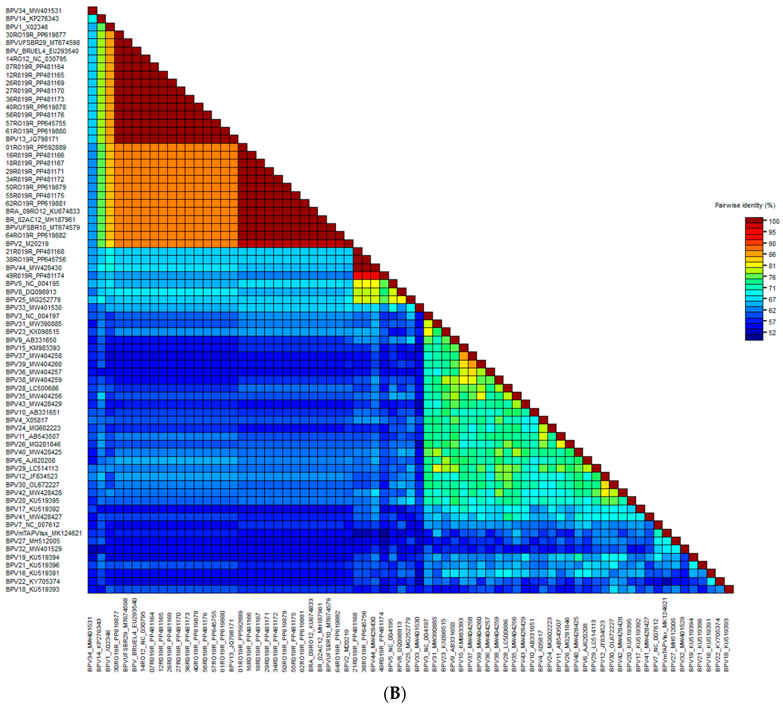
3.1. PCR, Genetic Sequencing and Phylogenetic Analysis

For the 100 rumen samples subjected to PCR with the FAP 59/64 primer pair, papillomavirus (PV) DNA was amplified in 41 samples and not amplified in 59 samples using the well-established PCR with FAP59/FAP64 primer pair (Table 1 and Table 2). Phylogenetic analysis was performed for 22 samples that presented high-quality sanger sequencing (electropherogram showing a single peak to a single nucleotide). In the generated phylogenetic tree, the vast majority of sequences clustered in the genus Deltapapillomavirus (19 sequences) and three sequences grouped in an unclassified genus (Figure 2A,B), in the BPV44 cluster.
The samples belonging to genus Delta PV were classified as BPV2 (40.91%; 9/22) and BPV13 (45.45%; 10/22). Samples classified as BPV2 showed a high degree of identity with each other (100%) and with the reference BPV2 (98.02%). The sequences that clustered with BPV13 (100% identity) also showed 100% identity with each other.
The 21RO19R and 38RO19R sequences (100% similarity between them) have a common ancestor, namely BPV44. Greater phylogenetic distance was observed between the study sequence 49RO19R and BPV44 (only 94.42% of similarity), which can be classified as a possible new viral subtype. Results are shown in Figure 2 and Table 1, Table 2 and Table 3. The average age and weight of the slaughtered animals sampled were 33 months-old and 480 kg, respectively.

3.2. Histopathological Analysis

Histopathological analysis was performed on 22 samples, which had enough material for this purpose. The results show that nineteen (79.10%) can be histologically classified as fibropapilloma and two as squamous papilloma. Additionally, two samples (8.4%) did not show any histopathological alteration and one sample (4.10%) was inconclusive (Table 3). Fibropapillomas were characterized by mild mucosal hyperplasia, which had a wavy surface and formed long epithelial pins (rete pegs) toward the proliferated submucosa (Figure 3A).
The cell proliferation in submucosa was fusiform and formed bundles arranged in random flows, supported by a mild-to-moderate myxoid or collagenous stroma (Figure 3B). There was moderate anisocytosis and anisokaryosis and no mitotic activity. Squamous papilloma consisted of marked mucosal hyperplasia and ortho or parakeratotic hyperkeratosis, with a finger-like or slightly flat surface (Figure 3C). Keratinocytes often had eccentric pyknotic nuclei and a perinuclear halo (consistent with koilocytes). Enlarged keratohyaline granules and intranuclear amphophilic inclusions (Figure 3D) were observed in only two cases.

4. Discussion

Herein, it was possible to amplify PV sequences in 41/100 rumen papillomatous lesions using primer pair FAP59/64. Moreover, we report the occurrence of BPV44 in cattle rumen, which was first identified in bovine teat papilloma lesions using rolling circle amplification followed by high-throughput sequencing [6].
From the 59 negative papillomatous samples, 22 were histologically diagnosed as squamous papilloma or fibropapillomas. This fact may be due to the fact that the FAP primer pair is not able to amplify some types of BPV that were already detected in this region due to the lower affinity for the primers [34,35].
The primer pair FAP59/64 allowed for the amplification of BPV mostly from the Deltapapillomavirus genus (BPV2 and 13). The FAP primer pair has been widely and satisfactorily used in previous BPV screening studies [17,32]. It is important to point that these primers were originally designed to amplify human PVs and have been widely used to amplify PV from a vast range of species worldwide, which made it possible to describe several new PV types from distinct animal species [1,5,12,17,28,29,30,31,32,33,34,35].
Furthermore, two samples were classified as BPV44 and one as a possible new subtype of this recently described viral type. BPV44 was previously isolated in cattle teat lesions in southern Brazil [6], but we describe here the first detection of this viral type in ruminal lesions.
The histological findings of most samples in the current study are consistent with those typically seen in bovine cutaneous papilloma lesions [17,27,36,37]. BPVs of the Deltapapillomavirus genus (BPV1, 2, 13 and 14) usually induce the formation of fibropapilloma, as they infect the epidermis and dermis [5]. In this study, BPV2 and 13 were detected in papillomatous lesions mostly classified as fibropapilloma.
Delta PV have already been detected in papillomatous skin lesions of cattle in Rondônia state [12,17,34], in Southern Brazil [5,7,34,35,36,37,38] and in southeast Brazil [38,39]. Additionally, BPVs 1, 2 and 5 were detected in the mouth, esophagus, rumen and reticulum fibropapillomas of cattle and buffaloes in India, Japan and Brazil [21,39,40], corroborating to the findings of this study. Delta PVs are considered high-risk since they are frequently detected in neoplasms of the upper gastrointestinal tract and urinary bladder, especially when they are associated with the consumption of Pteridium species [7,8,12,41]. In the present study, it was not possible to obtain data on the farms. However, the region presents data on the incidence of the Pteridium species and can be a damaging factor; in addition, the biodiversity of other toxic plants may be related to the incidence [41].
Other studies detected BPV4 (Xi-PV) in GIT papillomatous lesions of cattle in the UK [22] and Italy [42,43]. On the other hand, members of the Xipapillomavirus genus, such as BPV12, were described in a cattle tongue in Japan [44], although these viral types were not detected in this study. BPV4, which has already been found in the GIT, is probably not a predominant viral type in northern Brazil, as it was not detected in any previous study carried out on cattle in the Amazon region, not even using high-throughput sequencing [17,34]. Other studies of GIT papillomas in bovines have already been associated with BPV1, 2, 4 and 5 [21,22,42]. However, regression may not occur in chronically immunodepressed animals and some types of PV have been linked to malignancy, especially in synergism with some chemical or environmental carcinogens [21]. These infections can extend from the mouth and tongue to the esophagus, rumen and reticulum and present low to high growth, with a nodular, spherical and pedunculated shape, which can result in difficulty in feeding and breathing, obstructive bloat and cause the death of the animal [21,22,23,44,45,46,47].
BPV1 DNA is commonly found in skin warts, bovine peripheral blood, placenta, amniotic fluid and bovine colostrum [12,14,19,39,48] and it has been suggested that the bloodstream contributes to viral dissemination [39,49,50,51,52]. Similarly, the identification of BPV2 and BPV13 in the present study could also be explained by bloodstream dissemination. Although the pathogenesis of the PVs involves epitheliotropism, its genetic material can be found in several different tissues in the same animal and also in the peripheral blood of their offspring [46,52].

5. Conclusions

Here, we were able to identify Delta papillomaviruses and the recently identified BPV44 infecting the rumen of cattle from the western Amazon, Brazil. This study shows that ruminal papillomatosis is a post-mortem finding, emphasizing the importance of this type of study for a better understanding of the pathologies caused by BPV.

Author Contributions

The authors thank the participants who gave their time to the trial. C.D. and F.R.C.d.S. conceived the research. P.H.G.G., F.d.A.S., J.C.d.M.S. and I.R.d.S. performed fieldwork and data collection. M.S.d.S., C.D., C.W.C. and D.D. analyzed data. P.H.G.G. and J.V.D.F. prepared the manuscript. All authors contributed to subsequent drafts. All authors have read and agreed to the published version of the manuscript.

Funding

The Acre State Research Support Foundation (FAPAC) provided partial financial support, Notice 003 of 2019.

Institutional Review Board Statement

Ethical review and approval were waived for this study due to as they are animals slaughtered in slaughterhouses following legislation Brazilian Law No 11794/2008.

Informed Consent Statement

Not applicable.

Data Availability Statement

Data are contained within the article and genetic sequences can be accessed by the GenBank accession numbers provided.

Conflicts of Interest

The authors declare no conflicts of interest.

References

  1. Antonsson, A.; Hansson, B.G. Healthy Skin of Many Animal Species Harbors Papillomaviruses Which Are Closely Related to Their Human Counterparts. J. Virol. 2002, 76, 12537–12542. [Google Scholar] [CrossRef]
  2. de Villiers, E.-M.; Fauquet, C.; Broker, T.R.; Bernard, H.-U.; zur Hausen, H. Classification of papillomaviruses. Virology 2004, 324, 17–27. [Google Scholar] [CrossRef] [PubMed]
  3. López-Bueno, A.; Mavian, C.; Labella, A.M.; Castro, D.; Borrego, J.J.; Alcami, A.; Alejo, A. Concurrence of Iridovirus, Polyomavirus, and a Unique Member of a New Group of Fish Papillomaviruses in Lymphocystis Disease-Affected Gilthead Sea Bream. J. Virol. 2016, 90, 8768–8779. [Google Scholar] [CrossRef]
  4. Borzacchiello, G.; Roperto, F. Bovine papillomaviruses, papillomas and cancer in cattle. Vet. Res. 2008, 39, 45. [Google Scholar] [CrossRef]
  5. Bianchi, R.M.; Alves, C.D.B.T.; Schwertz, C.I.; Panziera, W.; De Lorenzo, C.; da Silva, F.S.; de Cecco, B.S.; Daudt, C.; Chaves, F.R.; Canal, C.W.; et al. Molecular and pathological characterization of teat papillomatosis in dairy cows in southern Brazil. Braz. J. Microbiol. 2019, 51, 369–375. [Google Scholar] [CrossRef]
  6. Sauthier, J.T.; Daudt, C.; da Silva, F.R.C.; Alves, C.D.B.T.; Mayer, F.Q.; Bianchi, R.M.; Driemeier, D.; Streit, R.S.A.; Staats, C.C.; Canal, C.W.; et al. The genetic diversity of “papillomavirome” in bovine teat papilloma lesions. Anim. Microbiome 2021, 3, 51. [Google Scholar] [CrossRef]
  7. de Alcântara, B.K.; Lunardi, M.; Agnol, A.M.D.; Alfieri, A.F.; Alfieri, A.A. Detection and Quantification of the E6 Oncogene in Bovine Papillomavirus Types 2 and 13 From Urinary Bladder Lesions of Cattle. Front. Vet. Sci. 2021, 8, 673189. [Google Scholar] [CrossRef]
  8. Medeiros-Fonseca, B.; Abreu-Silva, A.L.; Medeiros, R.; Oliveira, P.A.; Gil da Costa, R.M. Pteridium spp. and Bovine Papillomavirus: Partners in Cancer. Front. Vet. Sci. 2021, 8, 758720. [Google Scholar] [CrossRef]
  9. Van Doorslaer, K.; Li, Z.; Xirasagar, S.; Maes, P.; Kaminsky, D.; Liou, D.; Sun, Q.; Kaur, R.; Huyen, Y.; McBride, A.A. Papilomavírus Episteme: Uma grande atualização para o banco de dados de sequência de papilomavírus. Res. Nucleic Acids 2016, 45, D499–D506. [Google Scholar] [CrossRef]
  10. Yamashita-Kawanishi, N.; Tsuzuki, M.; Kasuya, F.; Chang, H.-W.; Haga, T. Genomic characterization of a novel bovine papillomavirus type 28. Virus Genes 2020, 56, 594–599. [Google Scholar] [CrossRef]
  11. Yamashita-Kawanishi, N.; Ito, S.; Ishiyama, D.; Chambers, J.K.; Uchida, K.; Kasuya, F.; Haga, T. Characterization of Bovine papillomavirus 28 (BPV28) and a novel genotype BPV29 associated with vulval papillomas in cattle. Vet. Microbiol. 2020, 250, 108879. [Google Scholar] [CrossRef]
  12. Daudt, C.; Da Silva, F.R.C.; Lunardi, M.; Alves, C.B.D.T.; Weber, M.N.; Cibulski, S.P.; Alfieri, A.A.; Canal, C.W. Papillomaviruses in ruminants: An update. Transbound. Emerg. Dis. 2018, 65, 1381–1395. [Google Scholar] [CrossRef]
  13. Stocco-Dos-Santos, R.C.; Lindsey, C.J.; Ferraz, O.P.; Pinto, J.R.; Mirandola, R.S.; Benes, F.J.; Birgel, E.H.; Bragança, C.A.P.; Beçak, W. Bovine papillomavirus transmission and chromosomal aberrations: An experimental model. J. Gen. Virol. 1998, 79, 2127–2135. [Google Scholar] [CrossRef] [PubMed]
  14. de Freitas, A.C.; de Carvalho, C.; Brunner, O.; Birgel-Junior, E.H.; Dellalibera, A.M.M.P.; Benesi, F.J.; Gregory, L.; Beçak, W.; Santos, R.d.C.S.d. Viral DNA sequences in peripheral blood and vertical transmission of the virus: A discussion about BPV-1. Braz. J. Microbiol. 2003, 34, 76–78. [Google Scholar] [CrossRef]
  15. Wosiacki, S.R.; Claus, M.P.; Alfieri, A.F.; Alfieri, A.A. Bovine papillomavirus type 2 in the urinary bladder of cattle with chronic enzootic haematuria. Memórias Do Inst. Oswaldo Cruz-Fiocruz 2006, 101, 635–638. [Google Scholar] [CrossRef]
  16. Araldi, R.; Carvalho, R.; Melo, T.; Diniz, N.; Sant’ana, T.; Mazzuchelli-De-Souza, J.; Spadacci-Morena, D.; Beçak, W.; Stocco, R. Bovine papillomavirus in beef cattle: First description of BPV-12 and putative type BAPV8 in Brazil. Genet. Mol. Res. 2014, 13, 5644–5653. [Google Scholar] [CrossRef]
  17. da Silva, F.R.C.; Daudt, C.; Streck, A.F.; Weber, M.N.; Filho, R.V.L.; Driemeier, D.; Canal, C.W. Genetic characterization of Amazonian bovine papillomavirus reveals the existence of four new putative types. Virus Genes 2015, 51, 77–84. [Google Scholar] [CrossRef] [PubMed]
  18. Tessele, B.; Barros, C.S.L. Tumors in cattle found in slaughterhouses. Braz. Vet. Res. 2016, 36, 145–160. [Google Scholar] [CrossRef]
  19. Russo, V.; Roperto, F.; De Biase, D.; Cerino, P.; Urraro, C.; Munday, J.S.; Roperto, S. Bovine Papillomavirus Type 2 Infection Associated with Papillomatosis of the Amniotic Membrane in Water Buffaloes (Bubalus bubalis). Pathogens 2020, 9, 262. [Google Scholar] [CrossRef]
  20. Campo, M. Animal models of papillomavirus pathogenesis. Virus Res. 2002, 89, 249–261. [Google Scholar] [CrossRef]
  21. Kumar, P.; Nagarajan, N.; Saikumar, G.; Arya, R.S.; Somvanshi, R. Detection of bovine papilloma viruses in wart-like lesions of upper gastrointestinal tract of cattle and buffaloes. Transbound. Emerg. Dis. 2015, 62, 264–271. [Google Scholar] [CrossRef] [PubMed]
  22. Tsirimonaki, E.; O’Neil, B.; Williams, R.; Campo, M. Extensive papillomatosis of the bovine upper gastrointestinal tract. J. Comp. Pathol. 2003, 129, 93–99. [Google Scholar] [CrossRef] [PubMed]
  23. Haga, T.; Dong, J.; Zhu, W.; Burk, R.D. The many unknown aspects of bovine papillomavirus diversity, infection and pathogenesis. Vet. J. 2013, 197, 122–123. [Google Scholar] [CrossRef] [PubMed]
  24. Reis, M.O.; Slaviero, M.; Lorenzet, M.P.; Cruz, R.A.S.; Guimarães, L.L.B.; Pavarini, S.P.; Driemeier, D.; Sonne, L. Bovine neoplasms diagnosed at the Veterinary Pathology Sector of UFRGS, Porto Alegre (2005–2014). Braz. Vet. Res. 2017, 37, 105–109. [Google Scholar] [CrossRef]
  25. Martano, M.; Roperto, F.; Stocco, R.d.C.; Russo, V.; Borzacchiello, G.; Paciello, O.; Iovane, V.; Leonardi, L.; Maiolino, P.; Restucci, B.; et al. Bovine Papillomavirus Type 2 Infection and a Series of Mesenchymal Tumors of the Urinary Bladder in Cattle. BioMed Res. Int. 2013, 2013, 1–9. [Google Scholar] [CrossRef] [PubMed]
  26. Rector, A.; Van Ranst, M. Animal papillomaviruses. Virology 2013, 445, 213–223. [Google Scholar] [CrossRef] [PubMed]
  27. Munday, J.S. Bovine and Human Papillomaviruses: A Comparative Review. Vet. Pathol. 2014, 51, 1063–1075. [Google Scholar] [CrossRef] [PubMed]
  28. Forslund, O.; Antonsson, A.; Nordin, P.; Stenquist, B.; Hansson, B.G. A broad range of human papillomavirus types detected with a general PCR method suitable for analysis of cutaneous tumours and normal skin. J. Gen. Virol. 1999, 80, 2437–2443. [Google Scholar] [CrossRef]
  29. Larkin, M.A.; Blackshields, G.; Brown, N.P.; Chenna, R.; McGettigan, P.A.; McWilliam, H.; Valentin, F.; Wallace, I.M.; Wilm, A.; Lopez, R.; et al. Clustal W and Clustal X version 2.0. Bioinformatics 2007, 23, 2947–2948. [Google Scholar] [CrossRef]
  30. Kumar, S.; Stecher, G.; Li, M.; Knyaz, C.; Tamura, K. MEGA X: Molecular evolutionary genetics analysis across computing platforms. Mol. Biol. Evol. 2018, 35, 1547–1549. [Google Scholar] [CrossRef]
  31. Bernard, H.U.; Burk, R.D.; Chen, Z.; van Doorslaer, K.; Hausen, H.; zur de Villiers, E.M. Classification of papillomaviruses (PVs) based on 189 PV types and proposal of taxonomic amendments. Virology 2022, 401, 70–79. Available online: https://pubmed.ncbi.nlm.nih.gov/20206957/ (accessed on 15 February 2020). [CrossRef] [PubMed]
  32. Edgar, R.C. MUSCLE: Multiple sequence alignment with high accuracy and high throughput. Nucleic Acids Res. 2004, 32, 1792–1797. [Google Scholar] [CrossRef] [PubMed]
  33. Munday, J.S.; Löhr, C.V.; Kiupel, M. Tumors of the digestary tract. In Tumors in Domestic Animals, 5th ed.; Meuten, D.J., Ed.; Wiley Blackwell: Ames, United Arab Emirates, 2017; pp. 499–601. [Google Scholar]
  34. Daudt, C.; da Silva, F.R.C.; Streck, A.F.; Weber, M.N.; Mayer, F.Q.; Cibulski, S.P.; Canal, C.W. How many papillomavirus species can go undetected in papilloma lesions? Sci. Rep. 2016, 6, 36480. [Google Scholar] [CrossRef] [PubMed]
  35. Ogawa, T.; Tomita, Y.; Okada, M.; Shinozaki, K.; Kubonoya, H.; Kaiho, I.; Shirasawa, H. Broad-spectrum detection of papillomaviruses in bovine teat papillomas and healthy teat skin. J. Gen. Virol. 2004, 85, 2191–2197. [Google Scholar] [CrossRef] [PubMed]
  36. Araldi, R.P.; Giovanni, D.N.S.; Melo, T.C.; Diniz, J.N.; Maz-Zuchelli-De-Souza, T.A.; Sant’Ana, R.F.; Carvalho, W.; Beçak, R.C.; Stocco, C. A Novel Simple and Versatile Method for the Papillomavirus Isolation through Ultracentrifugation. J. Virol. Methods 2014, 208, 119–124. [Google Scholar] [CrossRef] [PubMed]
  37. Claus, M.P.; Lunardi, M.; Alfieri, F.A.; Ferracin, M.L.; Fungaro, P.H.M.; Alfieri, A.A. Identification of unreported putative new bovine papillomavirus types in Brazilian cattle herds. Vet. Microbiol. 2008, 132, 396–401. [Google Scholar] [CrossRef] [PubMed]
  38. Daudt, C.; da Silva, F.R.; Cibulski, S.P.; Weber, M.N.; Mayer, F.Q.; Varela, A.P.M.; Roehe, P.M.; Canal, C.W. Complete genome sequence of Deltapapillomavirus 4 (bovine papillomavirus 2) from a bovine papillomavirus lesion in Amazon Region, Brazil. Memórias Do Inst. Oswaldo Cruz–FioCruz 2016, 111, 277–279. [Google Scholar] [CrossRef] [PubMed]
  39. Tozato, C.C.; Lunardi, M.; Alfieri, A.F.; Otonel, R.A.; Di Santis, G.W.; de Alcântara, B.K.; Headley, S.A.; Alfieri, A.A. Teat papillomatosis associated with bovine papillomavirus types 6, 7, 9, and 10 in dairy cattle from Brazil. Braz. J. Microbiol. 2013, 44, 905–909. [Google Scholar] [CrossRef]
  40. Lunardi, M.; Alfieri, A.A.; Otonel, R.A.A.; de Alcântara, B.K.; Rodrigues, W.B.; de Miranda, A.B.; Alfieri, A.F. Genetic characterization of a novel bovine papillomavirus member of the Deltapapillomavirus genus. Vet. Microbiol. 2013, 162, 207–213. [Google Scholar] [CrossRef]
  41. Crespo, S.E.I.; Lunardi, M.; Otonel, R.A.A.; Headley, S.A.; Alfieri, A.F.; Alfieri, A.A. Genetic characterization of a putative new type of bovine papillomavirus in the Xipapillomavirus 1 species in a Brazilian dairy herd. Virus Genes 2019, 55, 682–687. [Google Scholar] [CrossRef]
  42. Borzacchiello, G.; Ambrosio, V.; Roperto, S.; Poggiali, F.; Tsirimonakis, E.; Venuti, A.; Campo, M.; Roperto, F. Bovine papillomavirus type 4 in oesophageal papillomas of cattle from the south of Italy. J. Comp. Pathol. 2003, 128, 203–206. [Google Scholar] [CrossRef] [PubMed]
  43. Savini, F.; Gallina, L.; Alberti, A.; Müller, M.; Scagliarini, A. Bovine papillomavirus type 7 in Italy: Complete genomes and sequence variants. Virus Genes 2016, 52, 253–260. [Google Scholar] [CrossRef] [PubMed]
  44. Shimakura, H.; Dong, J.; Zhu, W.; Chambers, J.K.; Uchida, K.; Kiriki, K.; Uematsu, M.; Goto, Y.; Yasuda, M.; Yamashita-Kawanishi, N.; et al. Full genome analysis of bovine papillomavirus type 1 derived from a calf with severe cutaneous multiple papillomatosis. J. Vet. Med Sci. 2018, 80, 1691–1695. [Google Scholar] [CrossRef] [PubMed]
  45. Leroy, L.; Barbosa, J.A.R.G.; de Prat-Gay, G.; Polikarpov, I.; Pinheiro, C.B. The structure of the extended E2 DNA-binding domain of the bovine papillomavirus-1. Proteins Struct. Funct. Bioinform. 2020, 88, 106–112. [Google Scholar] [CrossRef]
  46. Freitas, A.C.; Silva, M.A.R.; Carvalho, C.C.R.; Birgel, E.H.; Santos, J.F.; Beçak, W.; Stocco, R.C. Papillomavírus DNA detection in non-epithelial tissues: A discussion about Bovine Papillomavírus. In Communicating Current Research and Educational Topics and Trends in Applied Microbiology; Mendez-Villas, A., Ed.; Formatex: Helsinki, Finland, 2007; pp. 697–704. [Google Scholar]
  47. Thomson, J.A. Morphological and genomicodiversity in the genus Pteridium (Dennstaedtiaceae). Ann. Bot. 2000, 85 (Suppl. S2), 77–99. [Google Scholar] [CrossRef]
  48. Dong, J.; Zhu, W.; Haga, T. Papillomavirus in yaks: The isolates of bovine papillomavirus type 1 have a high possibility of belonging to a novel type. J. Vet. Med Sci. 2016, 78, 1059–1061. [Google Scholar] [CrossRef]
  49. Faccin, T.C.; Cargnelutti, J.F.; Rodrigues, F.d.S.; de Menezes, F.R.; Piazer, J.V.M.; de Melo, S.M.P.; Lautert, B.F.; Flores, E.F.; Kommers, G.D. Bovine upper alimentary squamous cell carcinoma associated with bracken fern poisoning: Clinical-pathological aspects and etiopathogenesis of 100 cases. PLoS ONE 2018, 13, e0204656. [Google Scholar] [CrossRef]
  50. Bocaneti, F.; Altamura, G.; Corteggio, A.; Velescu, E.; Roperto, F.; Borzacchiello, G. Bovine Papillomavirus: New Insights into an Old Disease. Transbound. Emerg. Dis. 2014, 63, 14–23. [Google Scholar] [CrossRef] [PubMed]
  51. Campo, M. Bovine papillomavirus and cancer. Vet. J. 1997, 154, 175–188. [Google Scholar] [CrossRef]
  52. Hamad, M.A.; Al-Shammari, A.M.; Odisho, S.M.; Yaseen, N.Y. Molecular Epidemiology of Bovine Papillomatosis and Identification of Three Genotypes in Central Iraq. Intervirology 2018, 60, 156–164. [Google Scholar] [CrossRef]
Figure 1. Papillomatous lesions found at bovine rumen. Exophytic lesions with filiform (A) and nodular (B) shape.
Animals 14 02262 g001
Figure 2. (A) Phylogenetic tree of Bos taurus Papillomavirus (BPV) sequences detected in the gastrointestinal tract (GIT). Dataset based on partial L1 sequences. The study samples and the most similar sequences, as well as the reference sequences of each type of BPV, were included in the analysis, totaling 73 sequences. Evolutionary analysis inferred by the maximum likelihood method, 3-parameter Tamura model, discrete Gamma distribution and bootstrap of 1000 replicates. The probability that taxa cluster together is shown next to the branches. Bootstraps <50% have been suppressed. Highlighted study sequences. (B) Nucleotide identity matrix between sequences generated by the alignment of partial L1 fragments of the BPVs of the study. The figure was generated in SDT software (version 1.2).
Animals 14 02262 g002aAnimals 14 02262 g002b
Figure 3. Papillomatous lesions from bovine rumen. (A) Fibropapilloma. Submucosal proliferation (asterisk) closely associated with mildly hyperplastic mucosa, which exhibits evident epidermal rete pegs (arrowheads). The mucosa is covered by orthokeratotic hyperkeratosis. Bar, 200 µm. (B) The proliferated cells of the submucosa show moderate pleomorphism and are supported by collagenous stroma (asterisk). Bar, 100 µm. (C) Squamous papilloma. Squamous mucosal epithelial proliferation with multiple exophytic projections and orthokeratotic hyperkeratosis (arrowhead). Bar, 200 µm. (D) Keratinocytes showing an increase in keratohyaline granules (arrowheads) or intranuclear amphophilic inclusion (arrow). Bar, 100 µm.
Animals 14 02262 g003
Table 1. Negative samples in PCR and positive in histopathological diagnosis.
Municipalities *SexSampleHistopathological Diagnosis
Alvorada do Oeste02RO19RFibropapilloma
03RO19RFibropapilloma
04RO19RFibropapilloma
05RO19RFibropapilloma
06RO19RFibropapilloma
08RO19RFibropapilloma
10RO19RSquamous papilloma
11RO19RSquamous papilloma
14RO19RFibropapilloma
15RO19RFibropapilloma
17RO19RFibropapilloma
19RO19RFibropapilloma
20RO19RSquamous papilloma
Ji-Paraná22RO19RSquamous papilloma
28RO19RFibropapilloma
31RO19RSquamous papilloma
35RO19RSquamous papilloma
37RO19RFibropapilloma
Mirante da Serra39RO19RFibropapilloma
49RO19RSquamous papilloma
51RO19RFibropapilloma
52RO19RFibropapilloma
53RO19RFibropapilloma
63RO19RSquamous papilloma
65RO19RSquamous papilloma
70RO19RSquamous papilloma
Urupá71RO19RFibropapilloma
72RO19RFibropapilloma
73RO19RSquamous papilloma and Fibropapilloma
74RO19RFibropapilloma
84RO19RFibropapilloma
85RO19RSquamous papilloma
95RO19RFibropapilloma
98RO19RSquamous papilloma
Subtitle: * Municipalities from Rondônia state, Brazil.
Table 2. Negative samples in PCR and negative/inconclusive in the histopathological diagnosis.
Municipalities *SexSampleHistopathological Diagnosis
Alvorada do Oeste09RO19RNo Change
13RO19RNo Change
25RO19RInconclusive **
Ji-Paraná36RO19RNo Change
42RO19RNo Change
43RO19RNo Change
Mirante da Serra46RO19RNo Change
47RO19RNo Change
58RO19RNon-diagnostic sample
66RO19RNo Change
67RO19RNo Change
68RO19RNo Change
69RO19RNon-diagnostic sample
71RO19RInconclusive **
72RO19RInconclusive **
Urupá76RO19RInconclusive **
77RO19RNo Change
78RO19RInconclusive **
79RO19RInconclusive **
81RO19RInconclusive **
82RO19RInconclusive **
83RO19RNo Change
86RO19RInconclusive **
87RO19RInconclusive **
88RO19RInconclusive **
89RO19RNo Change
90RO19RInconclusive **
92RO19RNon-diagnostic sample
94RO19RNo Change
99RO19RInconclusive **
100RO19RInconclusive **
Subtitle: * Municipalities from Rondônia state, Brazil; ** Inconclusive: Samples with histological lesions of mild acanthosis and hyperkeratosis.
Table 3. Data on the sampled cattle, types of BPV and new putative types of BPV found in this study.
BPV TypeGenusMunicipalities *SexIdentity **SampleHistopathological DiagnosisGenBank Access No.
BPV2
M20219
DeltaAlvorada do Oeste98.02%01RO19RFibropapillomaPP592889
98.02%16RO19RFibropapillomaPP481166
98.02%18RO19RSquamous papillomaPP481167
Ji-Paraná98.02%29RO19RInconclusivePP481171
98.02%34RO19RFibropapillomaPP481172
98.02%50RO19RFibropapillomaPP481168
98.02%55RO19RFibropapillomaPP619879
98.02%62RO19RFibropapillomaPP619881
98.02%64RO19RFibropapillomaPP619882
BPV 13
JQ798171
DeltaAlvorada do Oeste100%07RO19RFibropapillomaPP481164
100%12RO19RFibropapillomaPP481165
100%26RO19RFibropapillomaPP481169
Ji-Paraná100%27RO19RFibropapillomaPP481170
100%30RO19RFibropapillomaPP619877
100%36RO19RNo ChangePP481173
100%40RO19RFibropapillomaPP619878
Mirante da Serra100%56RO19RFibropapillomaPP481176
100%57RO19RFibropapillomaPP645755
100%61RO19RFibropapillomaPP619880
BPV44 MW543422UnclassifiedJi-Paraná100%38RO19RSquamous papillomaPP645756
Alvorada do Oeste100%21RO19RNo ChangePP481168
94.42%49RO19RSquamous papillomaPP481174
Subtitle: * Municipalities from Rondônia state, Brazil; Sex: ♂ Male and ♀ Female. ** Based on reference BPV genome.
Disclaimer/Publisher’s Note: The statements, opinions and data contained in all publications are solely those of the individual author(s) and contributor(s) and not of MDPI and/or the editor(s). MDPI and/or the editor(s) disclaim responsibility for any injury to people or property resulting from any ideas, methods, instructions or products referred to in the content.

