Article

Dynamics of Gill Responses to a Natural Infection with Neoparamoeba perurans in Farmed Tasmanian Atlantic Salmon

by
Max Charles Vallarino
1,
Sarah L. Dagen
1,
Eoin Costelloe
1,
Shalom Inioluwa Oyenekan
1,
John Tinsley
2,
Victoria Valdenegro
3,
Elżbieta Król
1,
Patricia Noguera
1,4,* and
Samuel A. M. Martin
1,*
1
Scottish Fish Immunology Research Centre, School of Biological Sciences, University of Aberdeen, Aberdeen AB24 2TZ, Scotland, UK
2
BioMar Ltd, Grangemouth FK3 8UL, Scotland, UK
3
BioMar Australia, Wesley Vale, TAS 7307, Australia
4
Aquaculture and Marine Environment, Marine Scotland Science, Aberdeen AB11 9DB, Scotland, UK
*
Authors to whom correspondence should be addressed.
Animals 2024, 14(16), 2356; https://doi.org/10.3390/ani14162356
Submission received: 17 June 2024 / Revised: 7 August 2024 / Accepted: 12 August 2024 / Published: 15 August 2024
(This article belongs to the Section Aquatic Animals)

Abstract

:

Simple Summary

This study focuses on how Tasmanian sea-farmed Atlantic salmon respond to a natural infection with a parasite that causes amoebic gill disease (AGD), a major health issue for salmon globally. The research aimed to understand the progression of the disease by examining the salmon’s gills at different stages of the outbreak and evaluating their response to freshwater treatment. The study employed both macroscopic and microscopic examinations of gills, as well as molecular tools to measure the abundance of amoebae and the expression of specific genes that indicate inflammation. Key findings showed a strong correlation between different measures of gill health and significant differences between distinct stages of AGD. After treatment, a varied response among the fish was observed, indicating that management strategies need to account for individual genetic, environmental, and health factors. The expression patterns of some inflammation-related genes highlight their potential as biomarkers for early detection of gill damage in salmon aquaculture worldwide. The results of this research are important as they can help develop better ways to detect and manage this disease early, potentially reducing losses in salmon farming and ensuring the sustainability of this vital food resource.

Abstract

Gill health has become a significant global challenge for Atlantic salmon (Salmo salar) aquaculture, particularly during the marine phase of farming. The increasing prevalence of gill pathologies has been linked to rising seawater temperatures, underscoring the need to evaluate existing tools for monitoring gill health and to develop novel approaches for early detection. In this study, we investigated the gill responses of commercially farmed Atlantic salmon to natural infection with Neoparamoeba perurans during an outbreak of amoebic gill disease (AGD) in Tasmania. Our focus spanned the low AGD prevalence, high AGD prevalence, and post-freshwater treatment stages of the outbreak. Evaluations of gill tissue included assessments of the gross AGD score, histopathological score, abundance of N. perurans (measured by 18S rRNA gene expression), and expression levels of inflammation-related transcripts. We demonstrated a strong correlation between different measures of AGD-related gill pathology and significant differences between distinct stages of the N. perurans outbreak. Post-treatment, fish exhibited considerable variability in their responses to the freshwater bath, highlighting the necessity for personalized management strategies that consider genetic, environmental, and health status factors. The expression patterns of angiogenin-1 (ANG1) and complement C1q tumour necrosis factor-related protein 3-like (C1QTNF3) emphasize their potential as biomarkers for early detection of gill damage in salmon aquaculture worldwide.

1. Introduction

Atlantic salmon (Salmo salar), extensively farmed in temperate regions worldwide, faces cultivation limits due to extreme summer and winter temperatures. The industry’s growth is further constrained by infectious and non-infectious diseases, with gill health being a significant concern. This is primarily due to infectious agents such as Neoparamoeba perurans, which causes amoebic gill disease (AGD) [1,2], and various agents contributing to complex gill disease (CGD) [3,4]. The increasing prevalence of gill pathologies has been linked to rising seawater temperatures associated with climate change [5,6,7].
The gill of teleost fish, including Atlantic salmon, is vital for osmoregulation, gas exchange, excretion of nitrogenous waste, and immune function [8,9]. Its extensive surface area maintains constant contact with the aquatic environment. Each gill arch consists of filaments branching into highly vascularized lamellae [10]. Gill-associated lymphoid tissues (GIALT) at the filament bases play crucial immune roles [11,12,13], including pathogen and parasite prevention [14]. When compromised, the gill undergoes non-specific pathologies such as inflammation, lamellar fusion, and epithelial hyperplasia, leading to dysfunction and reduced performance [4,15,16,17].
AGD, first identified in the 1980s [1], is a major threat to the global salmon industry [18,19]. Initially reported in Tasmania [1] and the USA [20], AGD has spread to major salmon-producing regions worldwide, including Chile, Ireland, Scotland, and Norway [21,22,23,24]. Caused by the marine amoeba N. perurans [2], the disease triggers gill inflammation, manifested by epithelial hyperplasia, hypertrophy, lamellar fusion, and increased mucus production [24,25,26]. Clinically, AGD presents as white mucoid lesions on the gills, with affected fish exhibiting respiratory distress, cardiac dysfunction, anorexia, lethargy, and surface congregation [20,27,28]. The disease spreads rapidly in warmer months, significantly reducing salmon’s ability to withstand environmental stressors [21,29,30]. Without timely treatment, AGD can cause up to 50% mortality [1], highlighting the need for vigilant monitoring for N. perurans infection.
Monitoring gill health for AGD involves non-destructive, gross morphological examination of gill arches using an AGD scoring system to evaluate white mucoid lesions [31]. Some fish also undergo histopathological examination with a scoring system for gill histopathology in sea-farmed Atlantic salmon [16,32,33]. N. perurans presence is confirmed microscopically or via PCR assays targeting the 18S ribosomal RNA gene [34,35,36,37]. Recent gill transcriptomics studies have identified potential biomarkers for gill inflammation, useful for monitoring gill health [9]. Each technique has limitations: gross morphological scoring can be inconsistent, especially early in the disease [9]; histopathological examination is labour-intensive and time-consuming [33]; and detecting N. perurans does not reveal overall gill health. The biomarker panel, developed for Scottish Atlantic salmon, may not be applicable elsewhere [9]. More research is needed to compare monitoring tools for AGD and gill health, which are crucial for early detection, treatment strategies, and treatment evaluation.
Commercial treatments for AGD include freshwater or hydrogen peroxide baths, lasting 15 min to 4 h depending on the type [24,38,39], generally administered when the average gross AGD gill score exceeds 2 [31]. Freshwater baths, used in Tasmania [38,40], parts of Ireland [36], and Norway [23], involve immersing fish in freshwater for 2 to 4 h, significantly reducing amoebae and excess mucus [38,41], though limited by freshwater availability. Hydrogen peroxide baths, effective in cooler regions like Scotland, Ireland, and Norway, use 1000–1400 mg/L for 18–22 min but are unsafe above 13.5 °C or when AGD scores reach 3 [39,42]. Rising global water temperatures make freshwater bathing the predominant AGD treatment [43,44], necessitating further research.
In this study, we investigated the gill responses of commercially farmed Atlantic salmon during a natural infection with N. perurans amidst an AGD outbreak in Tasmania. Fish were monitored and sampled at three stages: low AGD prevalence, high AGD prevalence, and post-freshwater treatment. We analysed gill tissues for gross AGD scores, histopathology scores, N. perurans abundance (18S rRNA gene expression), and gene expression of inflammation-related transcripts, including angiogenin-1 (ANG1), ladderlectin (LL), interleukin 8 (IL8), glutathione peroxidase 6-like (GPX6), and complement C1q tumour necrosis factor-related protein 3-like (C1QTNF3). The purpose of this research was to enhance understanding of AGD progression and improve management strategies for gill health in aquaculture. Our findings showed significant correlations between AGD-related gill pathology measures and infection stages. Post-treatment variability highlighted the need for personalized management strategies. ANG1 and C1QTNF3 emerged as potential early biomarkers for gill damage in salmon aquaculture.

2. Materials and Methods

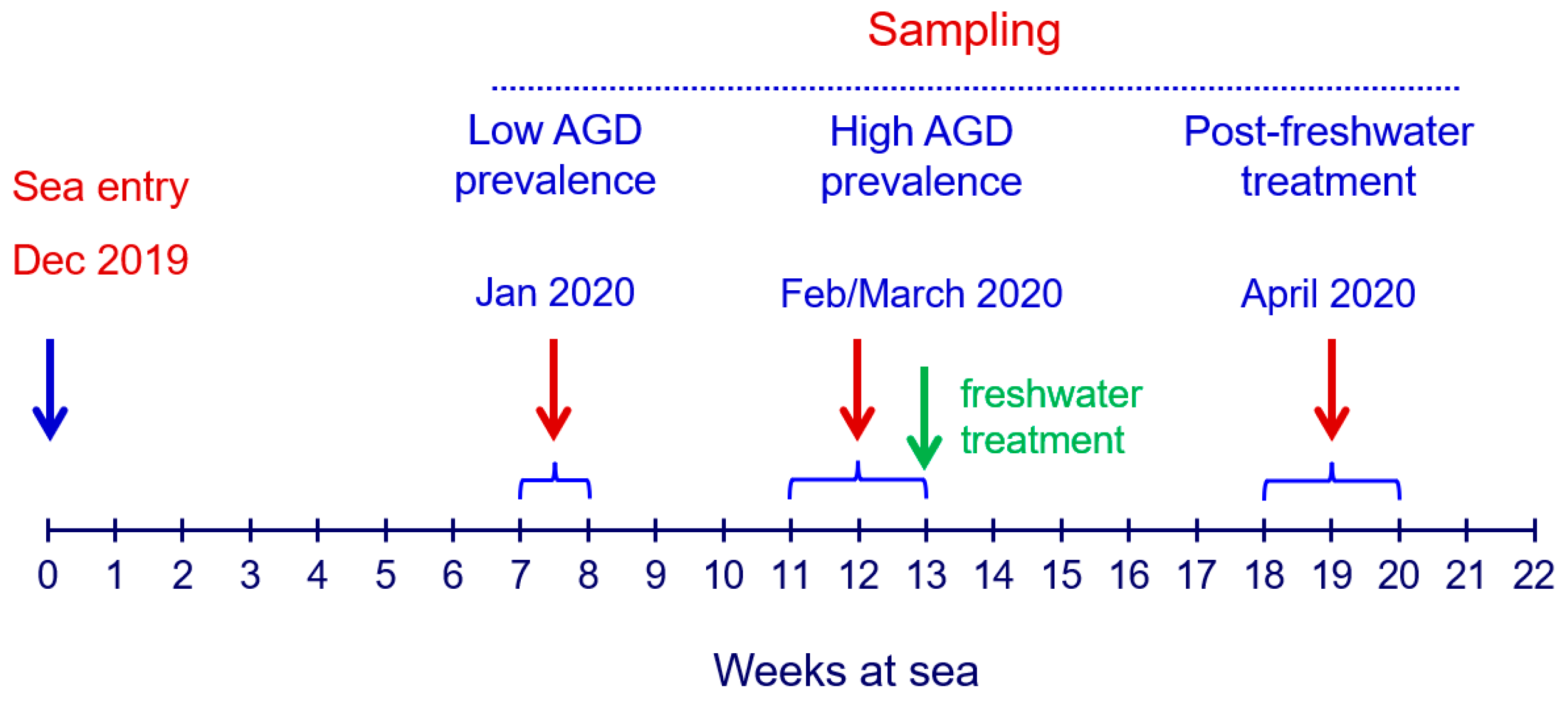
2.1. Fish Husbandry and Sampling Regime

The study focused on sea-farmed Atlantic salmon (Salmo salar) experiencing an outbreak of AGD in Tasmania. The fish were cultivated within a semi-commercial open sea-based system utilizing 18 net pens (5 m L × 5 m W × 5 m D), spanning from December 2019 to May 2020. During this period, the mean weekly water temperature, salinity, and dissolved oxygen concentration varied from 13.9 to 16.6 °C, 32.5 to 33.5 ppt, and 6.0 to 9.9 mg/L, respectively, with all parameters measured at a depth of 5 m. Pens were stocked with 270 fish at the start, each weighing 165–170 g, with a survival rate of over 86%. Weekly surveillance for AGD signs was conducted through visual inspection and non-destructive scoring of gill tissue for gross morphology abnormalities. This assessment, carried out by fish health professionals, was based primarily on the proportion of the gill surface occupied by mucus patches, hyperplasia, and other gross morphological changes indicative of AGD progression. The semi-quantitative 6-grade scoring system for AGD ranged from 0 (indicating no observable macroscopic pathology) to 5 (reflecting severe macroscopic pathology), as previously outlined [31]. Each of the eight gill arches (four on the left and four on the right side) underwent separate scoring, resulting in a total of eight AGD scores attributed to each fish. The AGD scores from the eight gill arches were then averaged to generate a single AGD score per fish.
The progression of AGD scores facilitated the identification of different stages within the outbreak, including periods of low and high AGD prevalence, and informed the optimal timing for AGD treatment. A commercial 3-h freshwater bath (salinity < 3 ppt) was administered in a small net pen enclosed with a tarpaulin when average AGD scores exceeded 2. All sampling procedures were conducted by personnel from BioMar Australia. Sampling occurred on three distinct occasions: during the low AGD prevalence stage (at week 7 post-seawater transfer), during the high AGD prevalence stage (weeks 11–13 post-seawater transfer, preceding freshwater treatment), and post-freshwater treatment at the conclusion of the production cycle (weeks 18–20 post-seawater transfer) (Figure 1).
At each sampling point, 4 out of the 18 net pens were selected, and 5 fish were sacrificed from each pen, resulting in 20 fish per sampling and 60 fish in total across the study. The selection of pens was random: 4 of the 18 pens for the first sampling, 4 of the remaining 14 pens for the second sampling, and 4 of the previously used 8 pens for the third sampling, including 2 from the first sampling and 2 from the second sampling (Table S1). The fish from each pen were sampled using a box net, which is the standard approach for subsampling from marine salmon cages. After being caught, the fish were placed in an anaesthetic bath (~20 g of MS-222/150 L) for 5–10 min, followed by macroscopic scoring of gill tissue for AGD manifestation (Table S1). Post-scoring, fish were promptly bled, and the gill arch exhibiting the highest AGD score was excised for subsequent gene expression profiling and histopathological examination, as previously described [9]. Specifically, for gene expression profiling, three transverse sections from the dorsal, medial, and ventral regions of the gill were submerged in RNAlater (Sigma-Aldrich, St. Louis, MO, USA), subjected to an overnight equilibration period at 4 °C, and subsequently stored at −80 °C until RNA extraction. The remaining tissue from the same gill arch was immersed in freshly prepared seawater Davidson’s fixative [45] for 24 h, followed by preservation in 10% neutral buffered formalin before undergoing tissue processing for histopathological examination.
Additionally, the body weights of sampled fish were recorded (Table S1), with averages of 1.1 ± 0.2 kg, 1.4 ± 0.3 kg, and 2.2 ± 0.4 kg observed for the low AGD prevalence, high AGD prevalence, and post-freshwater treatment stages of the outbreak, respectively (n = 20, mean ± standard deviation). Following the collection of all gill samples (n = 60), they were transferred to the University of Aberdeen (Scotland, UK) for subsequent processing.

2.2. Gill Histopathology

Gill tissue was subjected to standard histological procedures, including dehydration in ethanol, equilibration in xylene, and embedding in paraffin wax, following established protocols [46]. Sagittal sections (3 µm) of the gill arch were cut using a microtome and subsequently affixed onto microscope slides. These sections underwent haematoxylin and eosin (H&E) staining to facilitate detailed examination. Utilizing the Olympus dotSlide 2.1 Virtual Slide System (Olympus Corporation, Tokyo, Japan), all stained sections were digitized at a magnification of 40×. To ensure unbiased evaluation, the resultant images were randomized before being subjected to scoring. For this purpose, we employed a semi-quantitative scoring system specifically designed for assessing gill histopathology in sea-farmed Atlantic salmon [16], with minor adaptations from previous work [9].
The gill histopathology scoring system comprised four index criteria and twelve ancillary criteria. Each criterion was assigned a score ranging from 0 (indicating no pathological changes) to 4 (indicating severe changes affecting more than 50% of the gill tissue), with intermediate scores reflecting varying degrees of severity (Table S1). Of particular interest were the index criteria, which included lamellar hyperplasia (LH), lamellar fusion (LF), lamellar oedema (LO), and cellular anomalies (CA), as these features were closely associated with AGD-related lesions [25] (for representative images of gill histopathology see Figure S1). For each fish, the individual scores for LH, LF, LO, and CA were totalled to provide an overall representation of gill histopathology. This approach ensured a robust assessment of the gill tissue condition in relation to AGD. One of the 60 gill samples yielded no histological results due to suboptimal sectioning, which rendered the slide unreadable (Table S1).

2.3. RNA Extraction and cDNA Synthesis

Upon thawing, all gill samples (n = 60) were trimmed to remove arch tissue while retaining full-length filaments for subsequent processing. Total RNA extraction involved homogenizing ~100 mg of gill filaments (representing the dorsal, medial, and ventral regions of the gill) in TRI Reagent® (Sigma-Aldrich, London, UK), utilizing 3 mm tungsten carbide beads and the TissueLyser II Disruption System (Qiagen GmbH, Hilden, Germany). Following isolation, RNA quantification was performed using spectrophotometry (NanoDrop Technologies, Wilmington, DE, USA), with validation of integrity via electrophoresis (Agilent Technologies, Santa Clara, CA, USA). Subsequently, all RNA samples were subjected to reverse transcription using the QuantiTect RT Kit (Qiagen GmbH, Hilden, Germany) and following the manufacturer’s guidelines. The resulting cDNA samples were diluted to a working concentration of 500 ng/μL with molecular grade water and stored at −20 °C prior to gene expression analysis by qPCR.

2.4. Detection of N. perurans and Expression of Host Genes in Gill Tissue

We detected and quantified N. perurans in infected gill tissues using targeted amplification of the 18S ribosomal RNA (rRNA) gene, following previously described methods [35,37]. Additionally, we assessed the expression levels of seven salmon genes, including five associated with gill inflammation in Scottish farmed fish [9] and two commonly used as housekeeping genes (Table 1). The expression levels of N. perurans and salmon genes were analysed using qPCR.
Briefly, qPCR reactions were performed in 96-well plates, with each reaction containing 7.5 μL of 2× SYBR Green (Agilent Technologies, Cedar Creek, TX, USA), 1.5 μL of forward and reverse primer each (500 nM), 1 μL of molecular grade water, and 5 μL of cDNA (for primer sequence see Table 1). The qPCR was performed using an Mx3005P qPCR System (Agilent Technologies, Santa Clara, CA, USA) with the following cycling parameters: 95 °C for 3 min followed by 40 cycles of 95 °C for 20 s and 64 °C for 20 s (a two-step qPCR). A melting curve was added to the end of every run to confirm the presence of a single PCR product. Expression data were pre-processed and analysed using GenEx Pro Version 5.3.6 (MultiD Analyses AB, Goteborg, Sweden). The cycle threshold (Ct) values of target genes were normalised to the expression of housekeeping genes by ΔCt method. Specifically, the N. perurans 18S rRNA gene was normalised to EF1A, while the salmon genes associated with inflammation were normalised to EF1A and RPS13. Relative expression data were log-transformed for statistical analysis. Some qPCR reactions did not reach the quality threshold due to unsatisfactory melting curves and were removed from further analysis (Table S1).

2.5. Statistical Analysis

Gill scores are reported as median and interquartile range. Differences in gill scores between fish at the low AGD prevalence, high AGD prevalence, and post-freshwater treatment sampling points were assessed using the Kruskal-Wallis H test, followed by Dunn’s test for subsequent post hoc pairwise comparisons between the groups. The association between gross AGD gill scores and gill histopathology scores, as well as the association between gill scores and the abundance of N. perurans as determined by qPCR, was evaluated using Spearman’s rank correlation test.
Gene expression data were assessed for normality using the Shapiro-Wilk test. Due to the non-normal distribution of the data, differences in gene expression across groups were analysed using the Kruskal-Wallis H test. Where necessary, Dunn’s post hoc test was employed for pairwise comparisons. All statistical analyses were conducted using R version 4.2.2 [47]. Statistical significance was established at a threshold of p < 0.05.

3. Results

3.1. Gill Scores

The median gross AGD gill scores of fish sampled during the low AGD prevalence, high AGD prevalence, and post-treatment stages of the outbreak were 0.1 (0.0–0.3), 2.3 (2.0–2.4), and 1.9 (1.3–2.4), respectively (Figure 2A). Statistical analysis revealed significant differences in AGD scores between the fish groups (K-W chi-squared = 36.4, p < 0.001). Specifically, fish at both the high AGD prevalence and post-treatment sampling points had significantly higher AGD scores compared to fish at the low AGD prevalence sampling point (low vs. high, Z = −5.8, p < 0.001; low vs. post-treatment, −Z = 4.3, p < 0.001). Despite exposure to freshwater treatment (Figure 1), the AGD scores in the high AGD prevalence and post-treatment groups remained indistinguishable (Z = 1.5, p > 0.05), likely attributed to heterogeneity of fish sampled at the post-treatment time point. Notably, the post-treatment group comprised individuals with gills ranging from healthy (average AGD score of 0) to visibly compromised (average AGD score > 2.5) (Table S1).
The median gill histopathology scores (the sum of index criteria) for fish sampled during the low AGD prevalence, high AGD prevalence, and post-treatment time points were 3.0 ± (1.5–4.0), 6.5 (5.0–7.3), and 7.0 (4.0–9.0), respectively, with a sample size of 19–20 individuals per group (Figure 2B). Differences in gill histopathology scores between the groups were significant (K-W chi-squared = 25.9, p < 0.001). Similarly to the AGD scores, the histopathology scores for fish sampled during the high AGD prevalence and post-treatment time points were significantly higher than those in the low AGD prevalence group (low vs. high, Z = −4.5, p < 0.001; low vs. post-treatment, Z = −4.4, p < 0.001). Differences in gill histopathology scores between the high AGD prevalence and post-treatments groups were not significant (Z = −0.1, p > 0.05).
The association between gross AGD gill scores and gill histopathology scores was assessed by pooling data from all sampled fish across three time points (n = 59). Analysis revealed a strong and significant positive correlation between AGD gill scores and gill histopathology scores (Spearman’s p = 0.76, p < 0.001), indicating that fish with higher AGD gill scores tended to exhibit more severe gill histopathology (Figure 3).

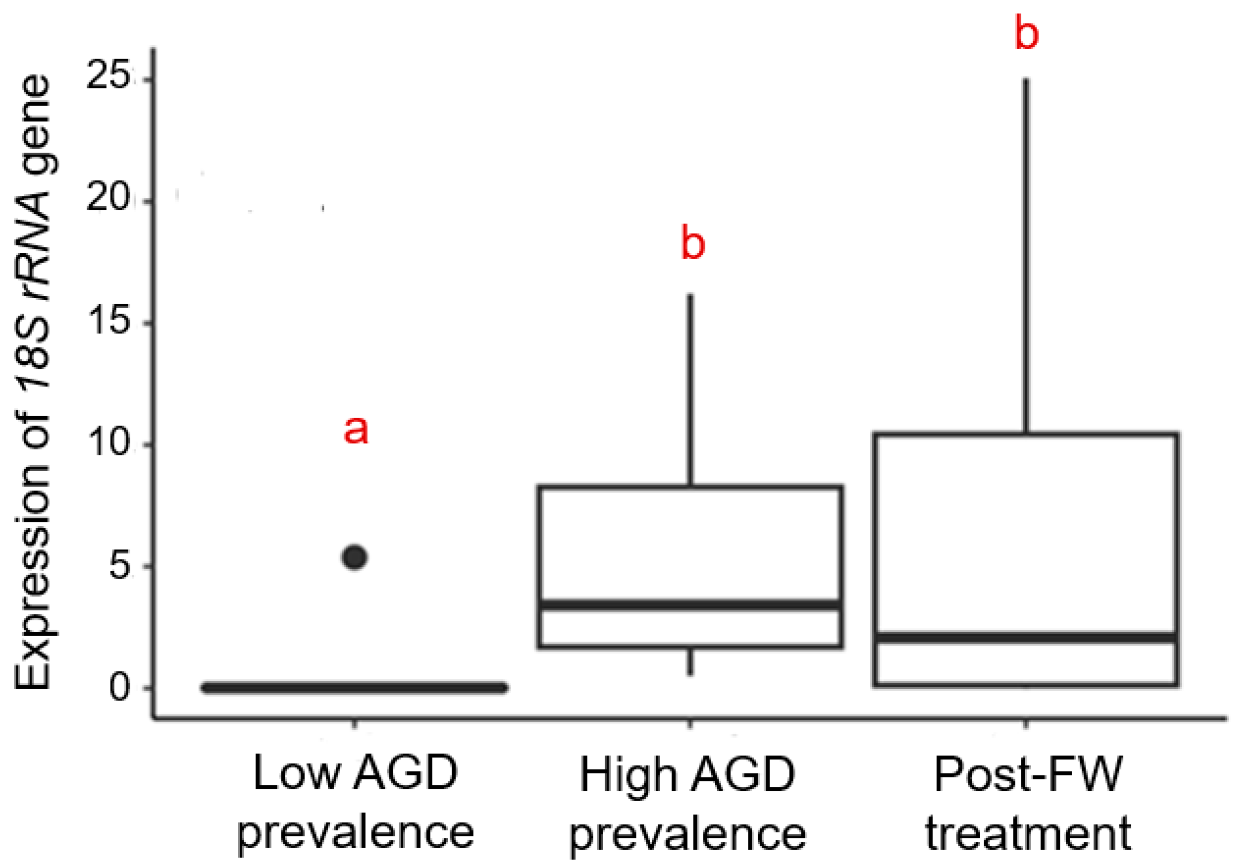
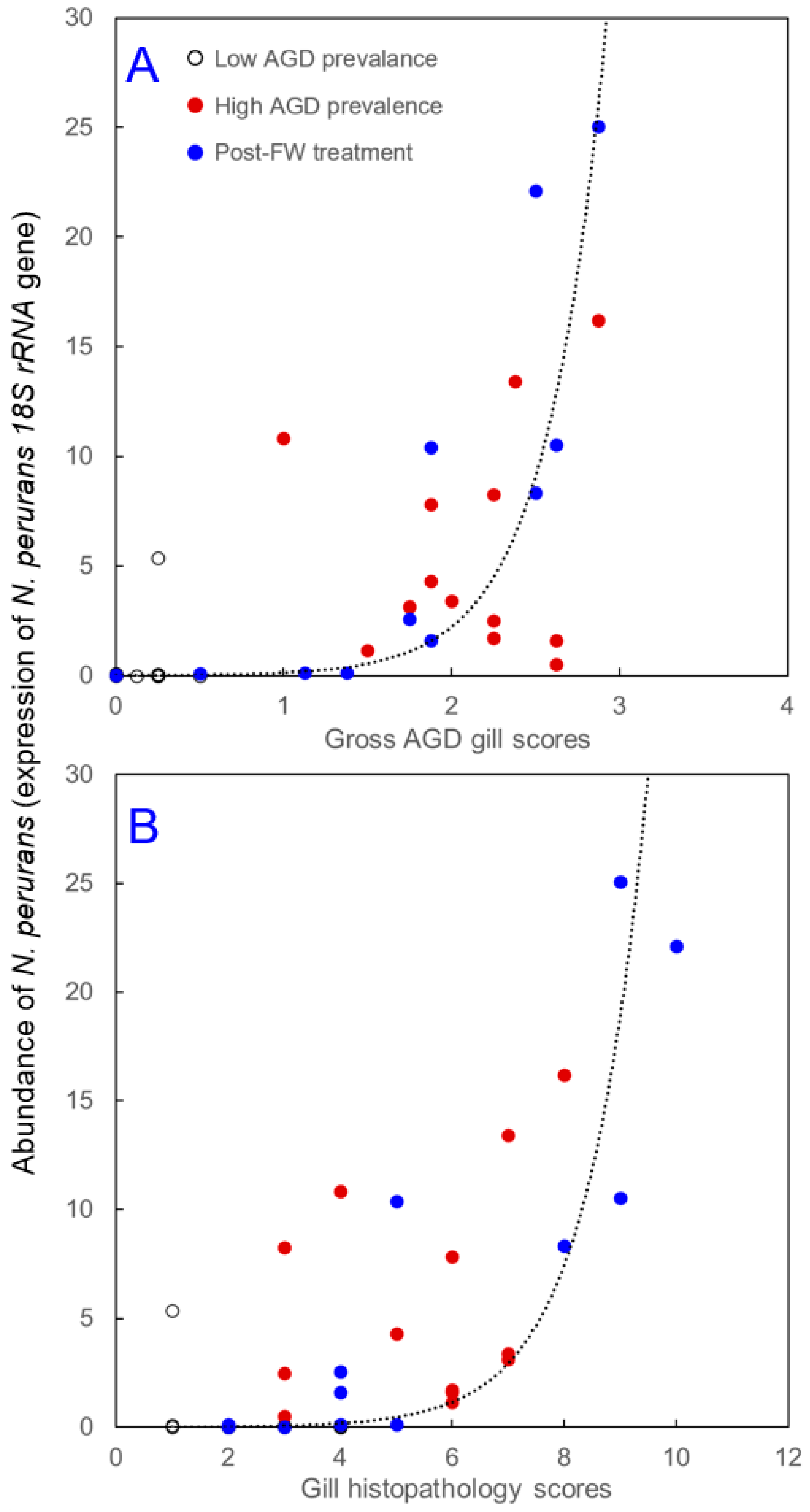
3.2. Abundance of N. perurans in Gill Tissue

The abundance of N. perurans in the gill tissue was quantified by measuring the expression levels of the N. perurans 18S rRNA gene, normalised to the expression of the housekeeping gene EF1A (Table 1). Fish sampled at three distinct time points exhibited significantly different levels of the 18S rRNA transcript (K-W chi-squared = 20.5, p < 0.001). Specifically, expression levels of the 18S rRNA transcript were nearly undetectable at the low AGD prevalence sampling point but increased significantly during the high AGD prevalence and post-treatment time points (low vs. high, Z = −4.2, p < 0.001; low vs. post-treatment, Z = −3.3, p = 0.001). However, the differences between the high AGD prevalence and post-treatment sampling points were not statistically significant (Z = 0.8, p > 0.05) (Figure 4).
Pooling data from all sampled fish across three time points revealed significant correlations between the abundance of N. perurans in gill tissue and gill scores. Fish with higher gross AGD gill scores exhibited a higher abundance of N. perurans (Spearman’s ρ = 0.80, p < 0.001, n = 41) (Figure 5A). A similar association was also found for gill histopathology scores (Spearman’s ρ = 0.66, p < 0.001, n = 40) (Figure 5B).

3.3. Expression of Salmon Genes

Three of the five inflammation-related genes showed significant differences in the expression levels between fish sampled at different stages of N. perurans infection. These genes included ANG1 (K-W chi-squared = 7.0, p = 0.031, n = 55), C1QTNF3 (K-W chi-squared = 12.0, p = 0.003, n = 51) and GPX6 (K-W chi-squared = 8.9, p = 0.011, n = 52) (Figure 6). In contrast, the two remaining genes (LL and IL8) showed no such differences (K-W chi-squared < 4.2, p > 0.05, n = 53–55).
The most prominent feature of the observed gene expression changes was an increase in gene expression from relatively lower levels during the low AGD prevalence stage to relatively higher levels during the high AGD prevalence stage of N. perurans outbreak. For genes ANG1 and C1QTNF3, the increase was significant (ANG1, low vs. high, Z = −2.6, p = 0.016; C1QTNF3, low vs. high, Z = −3.1, p = 0.003), while for GPX6, the increase did not reach significance (low vs. high, Z = −1.9, p > 0.05). Post-treatment, the transcript levels were either indistinguishable from the low and high AGD prevalence levels (ANG1; low vs. post-treatment, Z = −0.7, p > 0.05; high vs. post-treatment, Z = 1.8, p > 0.05), remained elevated (C1QTNF3; low vs. post-treatment, Z = −2.9, p = 0.005; high vs. post-treatment, Z = 0.2, p > 0.05), or dropped down to the low AGD prevalence levels (GPX6; low vs. post-treatment, Z = 1.2, p > 0.05; high vs. post-treatment, Z = 3.0, p = 0.005) (Figure 6).

4. Discussion

Atlantic salmon aquaculture faces increasing challenges from climatic shifts and disease outbreaks, particularly those affecting gill health [5,6,7,48]. As the industry intensifies, it is crucial to enhance measures for monitoring, controlling, and maintaining fish health. Diseases impacting the gills compromise physiological functions, leading to significant economic losses from poor feed conversion, reduced growth rates, treatment costs, and increased mortality [21,24]. Handling and crowding exacerbate gill pathologies, highlighting the need for regular, non-destructive monitoring of sea-farmed fish [49,50]. AGD has become a significant global concern, necessitating effective surveillance and response strategies amid ongoing environmental changes [18,19,51]. This study rigorously assesses gill tissue from sea-farmed Atlantic salmon during an AGD outbreak in Tasmania, addressing knowledge gaps and identifying gene expression patterns associated with AGD-related pathology.
In this study, fish were sampled at three stages of N. perurans outbreak: low AGD prevalence, high AGD prevalence, and post-freshwater treatment (Figure 1). Evaluations included gross AGD scores, histopathological scores, N. perurans abundance by qPCR, and inflammation-related gene expression. While some of these metrics are established for monitoring AGD [16,31,32,33,34,35,36,37], they are rarely applied concurrently. This study addresses this gap and also pioneers gene expression analysis to establish a robust panel of biomarkers for gill health, enhancing the understanding and management of gill pathologies in aquaculture [9].
When different measures of AGD-related gill pathologies were applied concurrently to fish experiencing an outbreak of AGD, they exhibited a remarkably similar pattern of temporal changes. Specifically, the gross AGD gill scores, gill histopathology scores, and the abundance of N. perurans were relatively low in fish sampled during the low AGD prevalence stage of the outbreak, then significantly increased during the high AGD prevalence stage (Figure 2 and Figure 4). Interestingly, while the freshwater treatment seemed to halt the further increase in these measured parameters, it did not return them to the low AGD prevalence levels. Instead, the freshwater bath led to increased variability in both gill scores and N. perurans abundance among individual fish around the group median, as evidenced by the expanded interquartile ranges and the broader spread of minimum and maximum values in the boxplots (Figure 2 and Figure 4). Across all three stages of N. perurans outbreak, the gross AGD gill scores and gill histopathology scores were highly and positively correlated with each other (Figure 3). This finding contrasts with our earlier results on the limited association between gross gill morphology and gill histopathology in Atlantic salmon farmed in Scotland [9]. The gills of Scottish-farmed salmon were macroscopically scored for multifactorial gill pathologies, whereas the fish in the current study were specifically scored for AGD, explaining the observed differences. Previously, gross gill scores have been challenging to interpret when non-AGD pathologies such as CGD are present [15,52]. Finally, our study demonstrated that both macroscopic and microscopic gill scores correlate well with the abundance of N. perurans (Figure 5), consistent with previous research [32,34].
Post-freshwater treatment, fish exhibited large variability in their responses to the freshwater bath, ranging from individuals with healthy gills to those with significantly compromised gills (Figure 2 and Figure 4). Specifically, the gross AGD gill scores, gill histopathology scores, and N. perurans abundance varied from 0 to 2.9, 2 to 10, and 0 to 25.1, respectively (Table S1). This variability can be attributed to several factors that not only influence the health outcomes of individual fish but also their propensity for reinfection following treatment [41]. Firstly, subtle genetic differences among the fish may lead to varied immune responses to N. perurans, affecting their susceptibility to AGD and recovery post-treatment [53]. Secondly, environmental conditions within the aquaculture system, such as water quality, temperature, and oxygen levels, can differ slightly from one area to another. These microenvironmental variations can significantly impact disease dynamics and the effectiveness of the freshwater treatment across different groups of fish [54]. Thirdly, previous health history may also play crucial roles. Fish that have encountered previous stressors or health challenges may have compromised gill structures, making them more susceptible to severe AGD manifestations and less responsive to treatment [55]. Moreover, the stage of infection at the time of treatment can greatly influence outcomes. Fish treated at an earlier stage of infection might display minimal pathological changes and recover more rapidly, whereas those treated at more advanced stages might not recover as effectively, leading to higher variability in gill scores and pathogen abundance [38]. Understanding this high degree of variability is crucial for optimizing treatment protocols and improving overall fish health management in aquaculture settings.
While scoring gill tissue for macroscopic and microscopic features serves as a reliable monitoring tool for AGD-related pathologies, such gill scores above 0 indicate tissue that is already visibly damaged [16,31,33]. Recognizing the limitations of these traditional methods, recent studies have demonstrated that changes in gene expression can precede visible tissue damage [9,56,57]. This suggests that monitoring gene expression in gill tissue could serve as an effective early warning system, potentially allowing for pre-emptive interventions. Our earlier work identified a panel of potential biomarkers of gill health developed for Scottish-farmed Atlantic salmon experiencing multifactorial gill pathologies [9]. In the current study, we evaluated five of these gill inflammation-related transcripts (Table 1). Three of them (ANG1, C1QTNF3 and GPX6) showed differences across various stages of N. perurans outbreak, but only two transcripts (ANG1 and C1QTNF3) were differentially expressed between the low and high AGD prevalence stages (Figure 6), which correspond to the most significant changes in gill pathology and pathogen abundance (Figure 2 and Figure 4). The higher gene expression of ANG1 during the high AGD prevalence stage (Figure 6) is consistent with higher protein levels of angigenin-1 in the gills of Atlantic salmon following four successive infections with N. perurans [58]. In fish, angiogenin-related proteins are involved in tissue regeneration and repair, immune responses, and possibly in the development of blood vessels [59]. Given its functions in other vertebrates, ANG1 might also contribute to physiological processes like wound healing and response to infection in fish [9,60]. Similarly to ANG1, the expression of C1QTNF3 increased from relatively low levels during the low AGD prevalence stage to significantly higher levels during the high AGD prevalence stage (Figure 6). The C1QTNF3 gene encodes a protein that is crucial to the complement system, a fundamental part of the innate immune response [61]. This protein primarily functions to inhibit the complement system, preventing its overactivation and thus reducing excessive inflammation and subsequent tissue damage. Notable changes in C1QTNF3 gene expression have been demonstrated in the gill epithelia of tilapia following salinity challenges [62], and in the fins of Atlantic salmon infested with sea lice [63]. The consistent upregulation of ANG1 and C1QTNF3 transcripts in the gill tissues of Scottish-farmed Atlantic salmon with multifactorial gill pathologies [9] and Tasmanian Atlantic salmon affected by AGD (this study) underscores their potential as biomarkers for early detection of gill damage in salmon aquaculture worldwide.

5. Study Limitations

One limitation of our study is the time gap between the freshwater treatment and the sampling, which was 5–7 weeks. This delay could have influenced the results, as the potential for reinfection during this period might have contributed to the variability in the gross AGD gill scores, gill histopathology scores, and the abundance of N. perurans. While we acknowledged the possibility of reinfection, the impact of this time gap on distinguishing between acute and chronic infections was not fully explored. Future studies should aim to sample more frequently post-treatment to better understand the immediate and long-term effects of freshwater treatments on AGD.
Another limitation is the absence of a control group that did not receive freshwater treatment. Without this control group, it is difficult to assert definitively that the observed stabilization in measured parameters was due to the treatment itself. The values might have stabilized even without the treatment. Including a control group in future studies would help in drawing more concrete conclusions about the efficacy of freshwater treatments.
Additionally, our study did not include initial control values before the onset of low AGD prevalence. This absence makes it challenging to compare the initial values with the low AGD values and to use these parameters as reliable indicators for early detection. Future studies should ensure the inclusion of baseline measurements before the onset of AGD to establish a clear reference point. This would enable a more accurate assessment of disease progression and the effectiveness of early detection of measures for AGD.

6. Conclusions

Our critical evaluation of AGD progression through a combined approach of gross morphological scoring, histopathological assessments, and molecular diagnostics, including qPCR and gene expression profiling, not only enhances our understanding of the disease dynamics but also facilitates the development of targeted intervention strategies. Specifically, the identification of potential biomarkers of gill health such as ANG1 and C1QTNF3 offers promising avenues for the early detection and management of gill pathologies, potentially enabling pre-emptive actions before the onset of critical disease stages.
Moreover, the variability in the responses of individual fish to freshwater treatment underscores the need for personalized management strategies that consider genetic, environmental, and health status factors. Integrating comprehensive monitoring systems alongside traditional and emerging treatment methodologies is imperative to improve resilience against AGD. Future research should focus on optimizing these strategies to mitigate the impact of AGD and ensure the sustainability of salmon farming under changing climatic conditions. The insights gained from this study contribute significantly to the body of knowledge required to safeguard the health of farmed Atlantic salmon and enhance the economic viability of the aquaculture industry.

Supplementary Materials

The following supporting information can be downloaded at: https://www.mdpi.com/article/10.3390/ani14162356/s1, Table S1: Sampling details, gill scores and gene expression data for sea-farmed Atlantic salmon (n = 60) experiencing an outbreak of amoebic gill disease (AGD) in Tasmania. Figure S1: Representative images of gill histopathology in farmed Tasmanian Atlantic salmon at the high AGD prevalence sampling point.

Author Contributions

Conceptualization, S.A.M.M., P.N., E.K., J.T. and V.V.; methodology, M.C.V. and E.C.; formal analysis, M.C.V. and S.L.D.; investigation, P.N., S.I.O. and M.C.V.; writing—original draft preparation, M.C.V. and S.L.D.; writing—review and editing, S.A.M.M., P.N. and E.K.; supervision, S.A.M.M. and P.N.; funding acquisition, S.A.M.M., P.N., E.K., J.T. and V.V. All authors have read and agreed to the published version of the manuscript.

Funding

This research was funded by the Scottish Aquaculture Innovation Centre (SAIC grant SL_2019_06, ‘Spatial and Temporal Drivers of Gill Pathology in Atlantic Salmon’).

Institutional Review Board Statement

Ethical review and approval for this study were not required as the sampling of fish adhered to established protocols designed for routine health assessments.

Informed Consent Statement

Not applicable.

Data Availability Statement

All data are presented in Supplementary Materials, Table S1.

Acknowledgments

We thank fish farm personnel in Tasmania for accommodating our research, performing gross morphology scoring and helping with sampling.

Conflicts of Interest

John Tinsley and Victoria Valdenegro are employed by BioMar. The remaining authors declare that the research was conducted in the absence of any commercial or financial relationships that could be construed as a potential conflict of interest.

References

  1. Munday, B.L. Diseases of salmonids. In Proceedings of the Workshop on Diseases of Australian Fish and Shellfish; Humphrey, J.D., Langdon, J.A., Eds.; Victorian Development of Agriculture and Rural Affairs: Benalla, Australia, 1985; pp. 127–141. [Google Scholar]
  2. Young, N.D.; Crosbie, P.B.B.; Adams, M.B.; Nowak, B.F.; Morrison, R.N. Neoparamoeba perurans n. sp., an agent of amoebic gill disease of Atlantic salmon (Salmo salar). Int. J. Parasitol. 2007, 37, 1469–1481. [Google Scholar] [CrossRef] [PubMed]
  3. Rodger, H.D. Gill disorders: An emerging problem for farmed Atlantic salmon (Salmo salar) in the marine environment? Fish Vet. J. 2007, 9, 38–48. [Google Scholar]
  4. Herrero, A.; Thompson, K.D.; Ashby, A.; Rodger, H.D.; Dagleish, M.P. Complex gill disease: An emerging syndrome in farmed Atlantic salmon (Salmo salar L.). J. Comp. Pathol. 2018, 163, 23–28. [Google Scholar] [CrossRef]
  5. Foyle, K.L.; Hess, S.; Powell, M.D.; Herbert, N.A. What is gill health and what is its role in marine finfish aquaculture in the face of a changing climate? Front. Mar. Sci. 2020, 7, 400. [Google Scholar] [CrossRef]
  6. Calado, R.; Mota, V.C.; Madeira, D.; Leal, M.C. Summer is coming! Tackling ocean warming in Atlantic salmon cage farming. Animals 2021, 11, 1800. [Google Scholar] [CrossRef]
  7. Sajid, Z.; Gamperl, A.K.; Parrish, C.C.; Colombo, S.M.; Santander, J.; Mather, C.; Neis, B.; Holmen, I.M.; Filgueira, R.; McKenzie, C.H.; et al. An aquaculture risk model to understand the causes and consequences of Atlantic salmon mass mortality events: A review. Rev. Aquac. 2024, 1–22. [Google Scholar] [CrossRef]
  8. Evans, D.H.; Piermarini, P.M.; Choe, K.P. The multifunctional fish gill: Dominant site of gas exchange, osmoregulation, acid-base regulation, and excretion of nitrogenous waste. Physiol. Rev. 2005, 85, 97–177. [Google Scholar] [CrossRef]
  9. Król, E.; Noguera, P.; Shaw, S.; Costelloe, E.; Gajardo, K.; Valdengro, V.; Bickerdinke, R.; Douglas, A.; Martin, S.A.M. Integration of transcriptome, gross morphology and histopathology in gill of sea farmed Atlantic salmon (Salmo salar): Lessons from multi-site sampling. Front. Genet. 2020, 11, 610. [Google Scholar] [CrossRef] [PubMed]
  10. Wells, P.R.; Pinder, A.W. The respiratory development of Atlantic salmon: I. Morphometry of gills, yolk sac and body surface. J. Exp. Biol. 1996, 199, 2725–2736. [Google Scholar] [CrossRef]
  11. Koppang, E.O.; Fischer, U.; Moore, L.; Tranulis, M.A.; Dijkstra, J.M.; Köllner, B.; Aune, L.; Jirillo, E.; Hordvik, I. Salmonid T cells assemble in the thymus, spleen and in novel interbranchial lymphoid tissue. J. Anat. 2010, 217, 728–739. [Google Scholar] [CrossRef]
  12. Salinas, I. The mucosal immune system of teleost fish. Biology 2015, 4, 525–539. [Google Scholar] [CrossRef] [PubMed]
  13. Rességuier, J.; Dalum, A.S.; Du Pasquier, L.; Zhang, Y.; Koppang, E.O.; Boudinot, P.; Wiegertjes, G.F. Lymphoid tissue in teleost gills: Variations on a theme. Biology 2020, 9, 127–214. [Google Scholar] [CrossRef] [PubMed]
  14. Xu, Z.; Takizawa, F.; Casadei, E.; Shibasaki, Y.; Ding, Y.; Sauters, T.J.C.; Yu, Y.; Salinas, I.; Sunyer, J.O. Specialization of mucosal immunoglobulins in pathogen control and microbiota homeostasis occurred early in vertebrate evolution. Sci. Immunol. 2020, 5, eaay3254. [Google Scholar] [CrossRef] [PubMed]
  15. Mitchell, S.O.; Rodger, H.D. A review of infectious gill disease in marine salmonid fish. J. Fish Dis. 2011, 34, 411–432. [Google Scholar] [CrossRef] [PubMed]
  16. Mitchell, S.O.; Rodger, H.D.; Holland, Á.C.; Baxter, E.J. Development of a novel histopathological gill scoring protocol for assessment of gill health during a longitudinal study in marine-farmed Atlantic salmon (Salmo salar). Aquat. Int. 2012, 20, 813–825. [Google Scholar] [CrossRef]
  17. Gjessing, M.C.; Steinum, T.; Olsen, A.B.; Lie, K.I.; Tavornpanich, S.; Colquhoun, S.; Gjevre, A. Histopathological investigation of complex gill disease in sea farmed Atlantic salmon. PLoS ONE 2019, 14, e0222926. [Google Scholar] [CrossRef] [PubMed]
  18. Nowak, B.F.; Archibald, B.M. Opportunistic but lethal: The mystery of Paramoebae. Trends Parasitol. 2018, 34, 404–419. [Google Scholar] [CrossRef]
  19. Ajasa, A.A.; Boison, S.A.; Gjøen, H.M.; Lillehammer, M. Genome-assisted prediction of amoebic gill disease resistance in different populations of Atlantic salmon during field outbreak. Aquaculture 2024, 578, 740078. [Google Scholar] [CrossRef]
  20. Kent, M.L.; Sawyer, T.; Hedrick, R. Paramoeba pemaquidensis (Sarcomastigophora: Paramoebidae) infestation of the gills of coho salmon Oncorhynchus kisutch reared in sea water. Dis. Aquat. Org. 1988, 5, 163–169. [Google Scholar] [CrossRef]
  21. Steinum, T.; Kvellestad, A.; Rønneberg, L.B.; Nilsen, H.; Asheim, A.; Fiell, K.; Nygård, S.M.R.; Olsen, A.B.; Dale, O.B. First cases of amoebic gill disease (AGD) in Norwegian seawater farmed Atlantic salmon, Salmo salar L., and phylogeny of the causative amoeba using 18S cDNA sequences. J. Fish Dis. 2008, 31, 205–214. [Google Scholar] [CrossRef]
  22. Bustos, P.A.; Young, N.D.; Rozas, M.A.; Bohle, H.M.; Ildefonso, R.S.; Morrison, R.N.; Nowak, B.F. Amoebic gill disease (AGD) in Atlantic salmon (Salmo salar) farmed in Chile. Aquaculture 2011, 310, 281–288. [Google Scholar] [CrossRef]
  23. Powell, M.D.; Reynolds, P.; Kristensen, T. Freshwater treatment of amoebic gill disease and sea-lice in seawater salmon production: Considerations of water chemistry and fish welfare in Norway. Aquaculture 2015, 448, 18–22. [Google Scholar] [CrossRef]
  24. Oldham, T.; Rodger, H.; Nowak, B.F. Incidence and distribution of amoebic gill disease (AGD)—An epidemiological review. Aquaculture 2016, 457, 35–42. [Google Scholar] [CrossRef]
  25. Adams, M.B.; Nowak, B.F. Distribution and structure of lesions in the gills of Atlantic salmon, Salmo salar L., affected with amoebic gill disease. J. Fish Dis. 2001, 24, 535–542. [Google Scholar] [CrossRef]
  26. Adams, M.B.; Nowak, B.F. Amoebic gill disease: Sequential pathology in cultured Atlantic salmon, Salmo salar L. J. Fish Dis. 2003, 26, 601–614. [Google Scholar] [CrossRef] [PubMed]
  27. Powell, M.D.; Nowak, B.F.; Adams, M.B. Cardiac morphology in relation to amoebic gill disease history in Atlantic salmon, Salmo salar L. J. Fish Dis. 2002, 25, 209–215. [Google Scholar] [CrossRef]
  28. Kube, P.D.; Taylor, R.S.; Elliott, N.G. Genetic variation in parasite resistance of Atlantic salmon to amoebic gill disease over multiple infections. Aquaculture 2012, 364, 165–172. [Google Scholar] [CrossRef]
  29. Benedicenti, O.; Pottinger, T.G.; Collins, C.; Secombes, C.J. Effects of temperature on amoebic gill disease development: Does it play a role? J. Fish Dis. 2019, 42, 1241–1258. [Google Scholar] [CrossRef]
  30. Bowden, A.J.; Adams, M.B.; Andrewartha, S.J.; Elliott, N.G.; Frappell, P.B.; Clark, T.D. Amoebic gill disease increases energy requirements and decreases hypoxia tolerance in Atlantic salmon (Salmo salar) smolts. Comp. Biochem. Physiol. Part A Mol. Integr. Physiol. 2022, 265, 111128. [Google Scholar] [CrossRef]
  31. Taylor, R.S.; Muller, W.J.; Cook, M.T.; Kube, P.D.; Elliott, N.G. Gill observations in Atlantic salmon (Salmo salar L.) during repeated amoebic gill disease (AGD) field exposure and survival challenge. Aquaculture 2009, 290, 1–8. [Google Scholar] [CrossRef]
  32. Downes, J.K.; Rigby, M.L.; Taylor, R.S.; Maynard, B.T.; MacCarthy, E.; O’Connor, I.; Marcos-Lopez, M.; Rodger, H.D.; Collins, E.; Ruane, N.M.; et al. Evaluation of non-destructive molecular diagnostics for the detection of Neoparamoeba perurans. Front. Mar. Sci. 2017, 4, 61. [Google Scholar] [CrossRef]
  33. Noguera, P.; Olsen, A.B.; Hoare, J.; Lie, K.I.; Marcos-López, M.; Poppe, T.T.; Rodger, H. Complex gill disorder (CGD): A histopathology workshop report. Bull. Eur. Ass. Fish Pathol. 2019, 39, 172–176. [Google Scholar]
  34. Bridle, A.R.; Crosbie, P.B.B.; Cadoret, K.; Nowak, B.F. Rapid detection and quantification of Neoparamoeba perurans in the marine environment. Aquaculture 2010, 309, 56–61. [Google Scholar] [CrossRef]
  35. Fringuelli, E.; Gordon, A.W.; Rodger, H.; Welsh, M.D.; Graham, D.A. Detection of Neoparamoeba perurans by duplex quantitative Taqman real-time PCR in formalin-fixed, paraffin-embedded Atlantic salmonid gill tissues. J. Fish Dis. 2012, 35, 711–724. [Google Scholar] [CrossRef] [PubMed]
  36. Downes, J.K.; Henshilwood, K.; Collins, E.M.; Ryan, A.; O’Connor, I.; Rodger, H.D.; MacCarthy, E.; Ruane, N.M. A longitudinal study of amoebic gill disease on a marine Atlantic salmon farm utilising a real-time PCR assay for the detection of Neoparamoeba perurans. Aquac. Environ. Interact. 2015, 7, 239–251. [Google Scholar] [CrossRef]
  37. Wynne, J.W.; Rigby, M.L.; Maynard, B.T.; Taylor, R.S. Improved environmental detection of Neoparamoeba perurans using sensitive RNA-based qPCR. J. Fish Dis. 2024, 47, e13879. [Google Scholar] [CrossRef]
  38. Parsons, H.; Nowak, B.; Fisk, D.; Powell, M. Effectiveness of commercial freshwater bathing as a treatment against amoebic gill disease in Atlantic salmon. Aquaculture 2001, 195, 205–210. [Google Scholar] [CrossRef]
  39. Rodger, H.D. Amoebic gill disease (AGD) in farmed salmon (Salmo salar) in Europe. Fish Vet. J. 2014, 14, 16–26. [Google Scholar]
  40. Munday, B.L.; Zilberg, D.; Findlay, V. Gill disease of marine fish caused by infection with Neoparamoeba pemaquidensis. J. Fish Dis. 2001, 24, 497–507. [Google Scholar] [CrossRef]
  41. Clark, G.; Powell, M.; Nowak, B. Effects of commercial fresh-water bathing on reinfection of Atlantic salmon, Salmo salar, with amoebic gill disease. Aquaculture 2003, 219, 135–142. [Google Scholar] [CrossRef]
  42. Adams, M.B.; Crosbie, P.B.B.; Nowak, B.F. Preliminary success using hydrogen peroxide to treat Atlantic salmon, Salmo salar L., affected with experimentally induced amoebic gill disease (AGD). J. Fish Dis. 2012, 35, 839–848. [Google Scholar] [CrossRef]
  43. Wynne, J.W.; Stratford, C.; Slinger, J.; Samsing, F.; Rigby, M.; McCulloch, R.; Quezada-Rodriguez, P.; Taylor, R.S. The interaction between temperature and dose on the efficacy and biochemical response of Atlantic salmon to hydrogen peroxide treatment for amoebic gill disease. J. Fish Dis. 2020, 43, 39–48. [Google Scholar] [CrossRef] [PubMed]
  44. Mahbub, P.; Yeager, T. Prospect of hydroxyl exposure during seawater bathing to treat amoebic gill disease in Atlantic salmon. Aquat. Res. 2023, 2023, 5074580. [Google Scholar] [CrossRef]
  45. Cadoret, K.; Bridle, A.R.; Leef, M.J.; Nowak, B.F. Evaluation of fixation methods for demonstration of Neoparamoeba perurans infection in Atlantic salmon, Salmo salar L., gills. J. Fish Dis. 2013, 36, 831–839. [Google Scholar] [CrossRef]
  46. Bancroft, J.D.; Gamble, M. Theory and Practice of Histological Techniques, 6th ed.; Churchill Livingstone Elsevier: London, UK, 2007; 744p. [Google Scholar]
  47. R Core Team, R. R: A Language and Environment for Statistical Computing; R Foundation for Statistical Computing: Vienna, Austria, 2018. [Google Scholar]
  48. Clinton, M.; Król, E.; Sepúlveda, D.; Andersen, N.R.; Brierley, A.S.; Ferrier, D.E.K.; Hansen, P.J.; Lorenzen, N.; Martin, S.A.M. Gill transcriptomic responses to toxin-producing alga Prymnesium parvum in rainbow trout. Front. Immunol. 2021, 12, 794593. [Google Scholar] [CrossRef]
  49. Ashley, P.J. Fish welfare: Current issues in aquaculture. Appl. Anim. Behav. Sci. 2007, 104, 199–235. [Google Scholar] [CrossRef]
  50. Hoem, K.S.; Tveten, A.K. Sea transfer and net pen cleaning induce changes in stress-related gene expression in commercial Atlantic salmon (Salmo salar) gill tissue. Aquacult. Int. 2023, 31, 2245–2262. [Google Scholar] [CrossRef]
  51. English, C.J.; Tyml, T.; Botwright, N.A.; Barnes, A.C.; Wynne, J.W.; Lima, P.C.; Cook, M.T. A diversity of amoebae colonise the gills of farmed Atlantic salmon (Salmo salar) with amoebic gill disease (AGD). Eur. J. Protistol. 2019, 67, 27–45. [Google Scholar] [CrossRef]
  52. Steinum, T.; Kvellestad, A.; Colquhoun, D.J.; Heum, M.; Mohammad, S.; Grøntvedt, R.N.; Falk, K. Microbial and pathological findings in farmed Atlantic salmon Salmo salar with proliferative gill inflammation. Dis. Aquat. Org. 2010, 91, 201–211. [Google Scholar] [CrossRef]
  53. Robledo, D.; Hamilton, A.; Gutiérrez, A.P.; Bron, J.E.; Houston, R.D. Characterising the mechanisms underlying genetic resistance to amoebic gill disease in Atlantic salmon using RNA sequencing. BMC Genom. 2020, 21, 271. [Google Scholar] [CrossRef]
  54. Oldham, T.; Dempster, T.; Crosbie, P.; Adams, M.; Nowak, B. Cyclic hypoxia exposure accelerates the progression of amoebic gill disease. Pathogens 2020, 9, 597. [Google Scholar] [CrossRef] [PubMed]
  55. Okon, E.M.; Okocha, R.C.; Taiwo, A.B.; Falana, B.M.; Adeniran, M.B. Dynamics of co-infection in fish: A review of pathogen-host interaction and clinical outcome. Fish Shellfish Immunol. Rep. 2023, 4, 100096. [Google Scholar] [CrossRef] [PubMed]
  56. Emam, M.; Caballero-Solares, A.; Xue, X.; Umasuthan, N.; Milligan, B.; Taylor, R.G.; Balder, R.; Rise, M.L. Gill and liver transcript expression changes associated with gill damage in Atlantic salmon (Salmo salar). Front. Immunol. 2022, 13, 806484. [Google Scholar] [CrossRef] [PubMed]
  57. Yang, S.; Li, D.; Feng, L.; Zhang, C.; Xi, D.; Liu, H.; Yan, C.; Xu, Z.; Zhang, Y.; Li, Y.; et al. Transcriptome analysis reveals the high temperature induced damage is a significant factor affecting the osmotic function of gill tissue in Siberian sturgeon (Acipenser baerii). BMC Genom. 2023, 24, 2. [Google Scholar] [CrossRef] [PubMed]
  58. Valdenegro-Vega, V.A.; Crosbie, P.; Bridle, A.; Leef, M.; Wilson, R.; Nowak, B.F. Differentially expressed proteins in gill and skin mucus of Atlantic salmon (Salmo salar) affected by amoebic gill disease. Fish Shellfish Immunol. 2014, 40, 69–77. [Google Scholar] [CrossRef]
  59. Pizzo, E.; Varcamonti, M.; Maro, A.D.; Zanfardino, A.; Giancola, C.; D’Alessio, G. Ribonucleases with angiogenic and bactericidal activities from the Atlantic salmon. FEBS J. 2008, 275, 1283–1295. [Google Scholar] [CrossRef]
  60. Shi, X.; Zhang, W.; Yin, L.; Chilian, W.M.; Krieger, J.; Zhang, P. Vascular precursor cells in tissue injury repair. Transl. Res. 2017, 184, 77–100. [Google Scholar] [CrossRef] [PubMed]
  61. Lv, C.; Zhang, Q.; Zhao, L.; Yang, J.; Zou, Z.; Zhao, Y.; Li, C.; Sun, X.; Lin, X.; Jin, M. African swine fever virus infection activates inflammatory responses through downregulation of the anti-inflammatory molecule C1QTNF3. Front. Immunol. 2022, 13, 1002616. [Google Scholar] [CrossRef]
  62. Campo, A.; Nitzan, T.; Slosman, T.; Doron-Faigenboim, A.; D’Cotta, H.; Baroiller, J.-F.; Cnaani, A. Different transcriptomic architecture of the gill epithelia in Nile and Mozambique tilapia after salinity challenge. Comp. Biochem. Physiol. Part D Genom. Proteom. 2022, 41, 100927. [Google Scholar] [CrossRef] [PubMed]
  63. Umasuthan, N.; Xue, X.; Caballero-Solares, A.; Kumar, S.; Westcott, J.D.; Chen, Z.; Fast, M.D.; Skugor, S.; Nowak, B.F.; Taylor, R.G.; et al. Transcriptomic profiling in fins of atlantic salmon parasitized with sea lice: Evidence for an early imbalance between chalimus-induced immunomodulation and the host’s defense response. Int. J. Mol. Sci. 2020, 21, 2417. [Google Scholar] [CrossRef]
Figure 1. Timeline of the study showing seawater entry, three sampling points and freshwater treatment in farmed Atlantic salmon experiencing an outbreak of AGD in Tasmania (for details see Table S1).
Animals 14 02356 g001
Figure 2. Gross AGD gill scores (A) and gill histopathology scores (B) in farmed Atlantic salmon at the low AGD prevalence, high AGD prevalence, and post-freshwater (FW) treatment sampling points. Data are presented as boxplots indicating the median, interquartile range (box), 1.5 times the interquartile range (whiskers), and outliers (closed circles). Different letters (a and b) indicate significant differences between the fish groups at p < 0.001.
Animals 14 02356 g002
Figure 3. Correlation between gross AGD gill scores and gill histopathology scores in farmed Atlantic salmon at the low AGD prevalence, high AGD prevalence and post-freshwater (FW) treatment sampling points. A linear trendline illustrates a positive association between the two types of gill scores.
Animals 14 02356 g003
Figure 4. Abundance of N. perurans in the gill tissue of Atlantic salmon at the low AGD prevalence, high AGD prevalence, and post-freshwater (FW) treatment sampling points, quantified as the expression levels of N. perurans 18S rRNA gene (relative units) by qPCR. Data are presented as boxplots indicating the median, interquartile range (box), 1.5 times the interquartile range (whiskers), and outliers (closed circles). Different letters (a and b) indicate significant differences between the fish groups at p ≤ 0.001.
Animals 14 02356 g004
Figure 5. Correlation between the abundance of N. perurans in gill tissue and gross AGD gill scores (A), and gill histopathology scores (B) in farmed Atlantic salmon at the low AGD prevalence, high AGD prevalence, and post-freshwater (FW) treatment sampling points. An exponential trendline illustrates a monotonic increase in N. perurans abundance corresponding to rising gill scores.
Animals 14 02356 g005
Figure 6. Gene expression levels of five inflammation-related genes (ANG1, C1QTNF3, GPX6, LL, IL8) in the gill tissue of farmed Atlantic salmon at the low AGD prevalence, high AGD prevalence, and post-freshwater (FW) treatment sampling points. Data are presented as boxplots indicating the median, interquartile range (box), 1.5 times the interquartile range (whiskers), and outliers (closed circles). The genes in the left panel (ANG1, C1QTNF3 and GPX6) showed different expression levels across the stages, with distinct letters (a and b) denoting statistically significant differences among the groups. The genes in the right panel (LL and IL8) showed no significant differences in expression between the groups.
Animals 14 02356 g006
Table 1. List of genes and primers used for qPCR in gill tissue of Atlantic salmon.
Gene Symbol 1Gene NameSpeciesNCBI Accession NumberEnsembl Accession NumberPrimers 5′-3′
(Forward and Reverse) 2
18S rRNA gene18S ribosomal RNA geneN. peruransEF216905n/aF:GTTCTTTCGGGAGCT
R:GAACTATCGCCGGCA
ANG1Angiogenin-1S. salarXM_014199172ENSSSAT00000023457F:ACTGTGGCAGATATTTGGGGAAGA
R:GTCACCCTGGACACCTGTGG
LLLadderlectinS. salarXM_014125761ENSSSAT00000132935F:GATCTACGTGCCGCAAAGGC
R:TTTGGTCCAACCTCCGGGAC
IL8Interleukin 8S. salarNM_001140710ENSSSAT00000013831F:GAATGTCAGCCAGCCTTGT
R:TCCAGACAAATCTCCTGACCG
GPX6Glutathione peroxidase 6-like S. salarXM_014155258ENSSSAT00000067910F:TAGCATGCAGGGTTACACAATGG
R:GAGCACCTTGCCCCTGTAGT
C1QTNF3Complement C1q tumour necrosis factor-related protein 3-likeS. salarXM_014134640ENSSSAT00000142506F:AGACGATGCTTCCTCTCCAGAT
R:ACACCCACAGAGTTGCGTGA
EF1AElongation factor 1-alpha S. salarXM_014177562ENSSSAT00000157959F:CAAGGATATCCGTCGTGGCA
R:ACAGCGAAACGACCAAGAGG
RPS13Ribosomal protein S13 S. salarBT059859ENSSSAT00000078374F:CCCTCTCAGATCGGTGTGATCC
R:TCCTGTCCTTTCTGTTCCTCTCC
1 ANG1, LL, IL8, GPX6 and C1QTNF3 were assayed due to their association with gill inflammation [9], EF1A and RPS13 served as housekeeping genes. 2 Primers for N. perurans 18S rRNA gene were previously published [35], primers for salmon genes were specifically designed for this study.
Disclaimer/Publisher’s Note: The statements, opinions and data contained in all publications are solely those of the individual author(s) and contributor(s) and not of MDPI and/or the editor(s). MDPI and/or the editor(s) disclaim responsibility for any injury to people or property resulting from any ideas, methods, instructions or products referred to in the content.

