Article

Identification of Aeromonas veronii as the Pathogen Associated with Massive Mortality in Bronze Gudgeon (Coreius heterodon)

1
Yangtze River Fisheries Research Institute, Chinese Academy of Fishery Sciences, Wuhan 430223, China
2
Graduate School of Chinese Academy of Agricultural Sciences, Beijing 100081, China
3
College of Fisheries and Life Sciences, Shanghai Ocean University, Shanghai 201306, China
4
Fisheries College, Hunan Agricultural University, Changsha 410128, China
*
Authors to whom correspondence should be addressed.
These authors contributed equally to this work.
Animals 2024, 14(16), 2440; https://doi.org/10.3390/ani14162440
Submission received: 1 July 2024 / Revised: 13 August 2024 / Accepted: 19 August 2024 / Published: 22 August 2024
(This article belongs to the Section Aquatic Animals)

Abstract

:

Simple Summary

An Aeromonas veronii strain, isolated from bronze gudgeon, was identified as a significant cause of morbidity and mortality. Through bacterial isolation, virulence gene analysis, and physiological characterization, we identified this bacterium as containing the act, alt, lip, LuxS, and ascV genes, which are distinct from those reported in infections of other aquatic animals. Designated as strain WH10, this is the first documented instance of Aeromonas veronii infecting bronze gudgeon. Our findings provide new insights into the mechanisms of bacterial transmission and underscore the necessity for further research into Aeromonas veronii pathogenesis.

Abstract

Aeromonas veronii, an opportunistic pathogen toward aquatic organisms, was identified as the causative pathogen (isolate WH10) in diseased bronze gudgeon via bacterial isolation, and morphological, physiological, biochemical, and molecular characterization. WH10 exerted its pathogenicity via five virulence genes, including those encoding cytotoxic enterotoxins (act and alt), lipase (lip), a quorum sensing-controlled virulence factor (LuxS), and a Type III secretion system inner membrane component (ascV). WH10 was shown to be sensitive to compound sulfamethoxazoles, cefothiophene, doxycycline, and sulfamethoxazole. Toward bronze gudgeon, WH10 had a median lethal dose (LD50) of 1.36 × 10⁶ colony forming units/mL. Analysis of blood parameters of diseased fish revealed significant increases in monocytes and neutrophils, but decreased numbers of lymphocytes. Serum aspartate aminotransferase activity and triglyceride concentration were significantly higher in diseased fish than in healthy fish. The reverse was noted for alkaline phosphatase, total protein, albumin, total cholesterol, and glucose. Thus, Aeromonas veronii is implicated as the causative agent of the mass mortality observed in bronze gudgeon, warranting further investigations into the diagnosis, epidemiology, prevention, and treatment of this infectious disease.

1. Introduction

The bronze gudgeon (Coreius heterodon Bleeker 1964) is an indigenous fish species in China, classified under the subfamily Gobioninae within the order Cypriniformes. It has important economic significance among freshwater fish and is the predominant species inhabiting the mid- and upstream regions of the Yangtze River [1,2]. Moreover, the bronze gudgeon is frequently utilized as an indicator species in ecological research to assess water temperature stress and investigate the impacts of reservoir and hydropower operations on fish spawning [3,4,5]. Previous studies on bronze gudgeon have primarily focused on aspects such as population dynamics [1], population genetics [6], the accumulation of metals [7], and body proximate compositions [8]; however, there is a general absence of documented instances of problematic diseases in this species.
However, in October 2023, an outbreak of a severe disease affecting bronze gudgeon occurred in Wuhan city, Hubei province. The diseased fish exhibited notable hemorrhage on their body surface and fins, together with ulcerated skin. The cumulative mortality rate was estimated to exceed 70%, representing the first reported occurrence of such an epidemic in bronze gudgeon. Subsequent investigation suggested that water disinfection or oral administration of antibacterial drugs could effectively control the disease, leading to the suspicion an etiological agent was a bacterium.
The present study aimed to comprehensively investigate an Aeromonas veronii [9] pathogen that was isolated from bronze gudgeon in China. We carried out bacterial isolation, bacterial identification, animal experiments, antibiotic susceptibility assays, differential leukocyte counts (DLC), biochemical analysis of serum samples, and screening of virulence-related genes. The isolated Aeromonas veronii strain, designated as WH10, was identified to be the bacterium that caused the epidemic affecting bronze gudgeon in Hubei province, China.

2. Materials and Methods

2.1. Experimental Bronze Gudgeon

Moribund bronze gudgeon displaying typical clinical signs, with a length ranging from 15 to 42 cm, were sampled from a bronze gudgeon farm in Wuhan city, Hubei province, in October 2023. The diseased bronze gudgeon were promptly preserved in oxygenated bags or on ice before transport to the laboratory for subsequent diagnostic assessment and pathogen identification.
Healthy bronze gudgeon measuring 15–20 cm in length were sourced from the breeding station of Institute of Hydroecology, MWR & CAS, which did not report any instances of the disease. Before experimental infection, the healthy control fish were allowed to acclimatize in recirculation rearing systems (2 m × 1.5 m × 1.5 m). The water in these systems was aerated and maintained at 25 °C. The fish received a regular commercial diet for 7 days to ensure proper acclimatization.

2.2. Histological Analysis

Tissue samples obtained from diseased and healthy bronze gudgeon were initially fixed using 4% paraformaldehyde for 24 h at 4 °C. Subsequently, the samples were washed using Dulbecco’s phosphate-buffered saline (DPBS, Sigma, St. Louis, MO, USA), followed by ethanol gradient (70%, 80%, 90%, 95%, and 100%) dehydration. After dehydration, the samples were subjected to optimum cutting temperature compound embedding. The embedded tissues were then cut into sections using a cryostat (CM1950, Leica, Wetzlar, Germany) at −20 °C. Following sectioning, the samples were stained using Hematoxylin–Eosin and examined under a light microscope equipped with a CCD imaging system (DM2500, Leica, Wetzlar, Germany) [10].

2.3. Bacterial Isolation

The diseased fish were sacrificed by employing 0.1% tricaine methane sulfonate (MS-222; Sigma, St. Louis, MO, USA) and sanitized using 75% ethanol before dissection. Tissue samples from the liver and kidney were then streaked separately on Brain Heart Infusion (BHI) agar plates (HopeBio, Qingdao, China) and incubated at 28 °C for 24 h. Following incubation, dominant and uniform bacterial colonies were selected and purified using BHI agar re-streaking. We isolated single colonies, which were preserved in glycerol at −80 °C for subsequent characterization.

2.4. Identification of the Bacteria

To characterize the bacterial isolates morphologically, pure colonies were suspended in DPBS, smeared onto glass slides, dried, and Gram stained using a kit (Jiangcheng, Nanjing, China) following the manufacturer’s instructions. An optical microscope (Olympus, Tokyo, Japan) was employed to observe the staining and morphological characteristics. To further analyze the bacteria morphologically, they were fixed using 2.5% glutaraldehyde. Subsequently, the bacteria were subjected to dehydration, drying, and examination under a scanning electron microscope (Hitachi, Tokyo, Japan) [11].
Biochemical tests were conducted utilizing a Biolog microbial identification system (Biolog, Hayward, CA, USA). Pure culture colonies were inoculated into IF-A inoculation solution using a sterile cotton swab. Subsequently, the inoculum of the pure clone was added to GEN III plates (Biolog) at 100 μL per well for analysis using the above-mentioned automatic microbial identification system. Analyses included dextrin, gentiobiose, sucrose, acidic PH PH6, α-D-Lactose, L-Glutamic Acid, Vancomycin, the Voges–Proskauer test (acetoin), raffinose, dulcitol, and fructose.
Identification of the WH10 strain was further conducted using 16S rRNA gene analysis. WH10 DNA was extracted using a Bacterial Genomic DNA Kit (Tiangen, Beijing, China) according to the supplier’s guidelines. The extracted DNA was employed as the template for PCR amplification. Primer pairs were designed as described previously [12] (Table 1). PCR amplification involved an initial denaturation at 95 °C for 5 min, followed by 35 cycles of denaturation for at 94 °C for 30 s, annealing at 55 °C for 45 s, and extension at 72 °C for 60 s. A final extension step was carried out at 72 °C for 7 min. The resulting amplicons were analyzed using 1.5% agarose gel electrophoresis stained with ethidium bromide for visualization. Following sequencing, the 16S rRNA sequences of the isolate clones were compared with sequences in the NCBI database using BLAST for sequence homology analysis. A phylogenetic tree was constructed using the Maximum-likelihood method in MEGA 7.0 (version 7.0, Mega Limited, Auckland, New Zealand).

2.5. WH10 Challenge Experiment

Healthy bronze gudgeon were divided randomly into six groups (n = 30 per group) and subjected to a pathogen challenge. The experimental groups comprised bronze gudgeon challenged via intraperitoneal (IP) injection with 0.3 mL of bacterial supernatant at titers of 2 × 104.0, 2 × 105.0, 2 × 106.0, 2 × 107.0, and 2 × 108.0 colony forming units (CFU)/mL, respectively. The control fish received an IP injection of 0.3 mL DPBS. All the fish were reared in tanks containing aerated water at 25 °C for the duration of the challenge assay. Every day, we monitored clinical signs and mortality, and randomly sampled three moribund or dead fish per group (including controls), which were processed for PCR analysis to determine whether the pathogen was present or not. The median lethal dose (LD50) of the WH10 strain for bronze gudgeon was determined utilizing the Reed Muench method [17]. Three replicate experiments were performed to ensure the robustness and reliability of the findings.

2.6. Susceptibility to Antibiotics Assay

The antibiotic susceptibility of WH10 was evaluated in triplicate using the standard disk diffusion method with compound sulfamethoxazoles, cefothiophene, doxycycline, sulfamethoxazole, gentamicin, amikacin, neomycin, florfenicol, enrofloxacin, and ciprofloxacin. Briefly, WH10 was cultured in BHI medium for 1 day. Thereafter, the WH10 solution was adjusted to 1 × 108.0 CFU/mL with sterile PBS and spread on BHI agar plates. Before the WH10 solution was completely absorbed, antibiotic-containing paper disks (Hangwei, Hangzhou, China) were placed on the agar surface. The plates were then placed in a thermostatic incubator (CIMO, Shanghai, China) for 24 h at 28 °C. The diameter of the inhibition zone around each antibiotic disk was then measured, which was used to determine the sensitivity of WH10 to each antibiotic following the supplier’s guidelines.

2.7. Screening of Virulence-Related Genes

Ten virulence genes were screened using conventional PCR with template DNA extracted from the WH10 strain. These genes included aha, exu, lip, ast, alt, act, LuxS, aerA, hlyA, and ascV. The primers utilized to amplify these genes are detailed in Table 1. Briefly, DNA extracted from WH10 (see Section 2.4) was subjected to PCR as follows: initial denaturation for 5 min at 95 °C; followed by 35 cycles of denaturation for 45 s at 94 °C, annealing for 60 s at 57 °C, extension for 90 s at 72 °C; and a last extension step for 7 min at 72 °C. After sequencing, the virulence-related gene sequences of isolate clones were searched in the NCBI database for sequence homology using BLAST.

2.8. Blood Parameters

Leukocytes were counted and classified employing the differential leukocyte counts (DLC) method. Diseased bronze gudgeon (n = 3) and healthy bronze gudgeon (n = 3) were anesthetized, and a sterile disposable syringe was used to collect blood samples at caudal vertebral vein of fish, which were added immediately to a slide to make a blood smear. From each dish, three blood smears were prepared, followed by Wright–Giemsa staining using the manufacturer’s recommendations (Baso, Zhuhai, China). On each blood smear, 100 white blood cells were classified and counted under the oil lens of a microscope. To quantify serum biochemical markers, 1.5 mL of fresh blood was collected from both diseased and healthy bronze gudgeon. Equal volumes of blood were aliquoted into separate tubes for subsequent analysis. This experiment was conducted in triplicate. The supernatant was obtained by centrifugation at 4000× g for 10 min after storage overnight at 4 °C, and then transferred to a new tube for further testing. The serum activity and content of glucose (GLU), total cholesterol (T-CHO), triglyceride (TG), alkaline phosphatase (ALP), alanine aminotransferase (ALT), aspartate aminotransferase (AST), albumin (ALB), and total protein (TP) were assessed using an automatic biochemical analyzer (Sysmex, Kobe, Japan).

2.9. Statistical Analysis

SPSS software was used to analyze the data (version 19.0, IBM Corp., Armonk, NY, USA) and statistical analysis was conducted using analysis of variance (ANOVA). Duncan’s multiple range test was employed to evaluate the significance of the differences between means. Differences were considered significant at p < 0.05. In this study, statistical analysis was conducted specifically on the bronze gudgeon blood sample data.

3. Results

3.1. Clinical Signs of the Disease

In October 2023, a disease outbreak occurred among cultured bronze gudgeon in Wuhan city, Hubei province, China. The disease was highly contagious and resulted in the mortality of bronze gudgeon of all sizes. Clinical manifestations included hemorrhages on the body surface (Figure 1A) and fins (Figure 1C), along with ulcerated skin (Figure 1B). Additionally, diseased fish revealed a dark and enlarged spleen, as well as a hemorrhagic liver (Figure 1A,D). Microscopic examination of the gills from diseased fish did not reveal the presence of any parasites.

3.2. Pathological Features

Histopathological analysis of the diseased fish revealed severe pathological changes in the liver, kidney, and spleen, including extensive necrosis, vacuolation, and focal hemorrhaging (Figure 2). The liver lesions were marked by hepatocyte necrosis and significant vacuolation (white arrow; Figure 2B). Some cell boundaries were not clear, and the membrane was disintegrated or even absent. Additionally, there was pronounced swelling and hemorrhaging in the liver blood sinuses (red arrow and blue triangular arrowheads; Figure 2B). The infected kidney tissues showed edema in the renal tubules (white arrow; Figure 2D) accompanied by lymphocyte infiltration in and around the affected areas (blue triangular arrowheads; Figure 2D). The spleen exhibited varying degrees of necrosis, vacuolation (white arrow; Figure 2F), and enlargement of melanomacrophage centers (blue triangular arrowheads; Figure 2F).

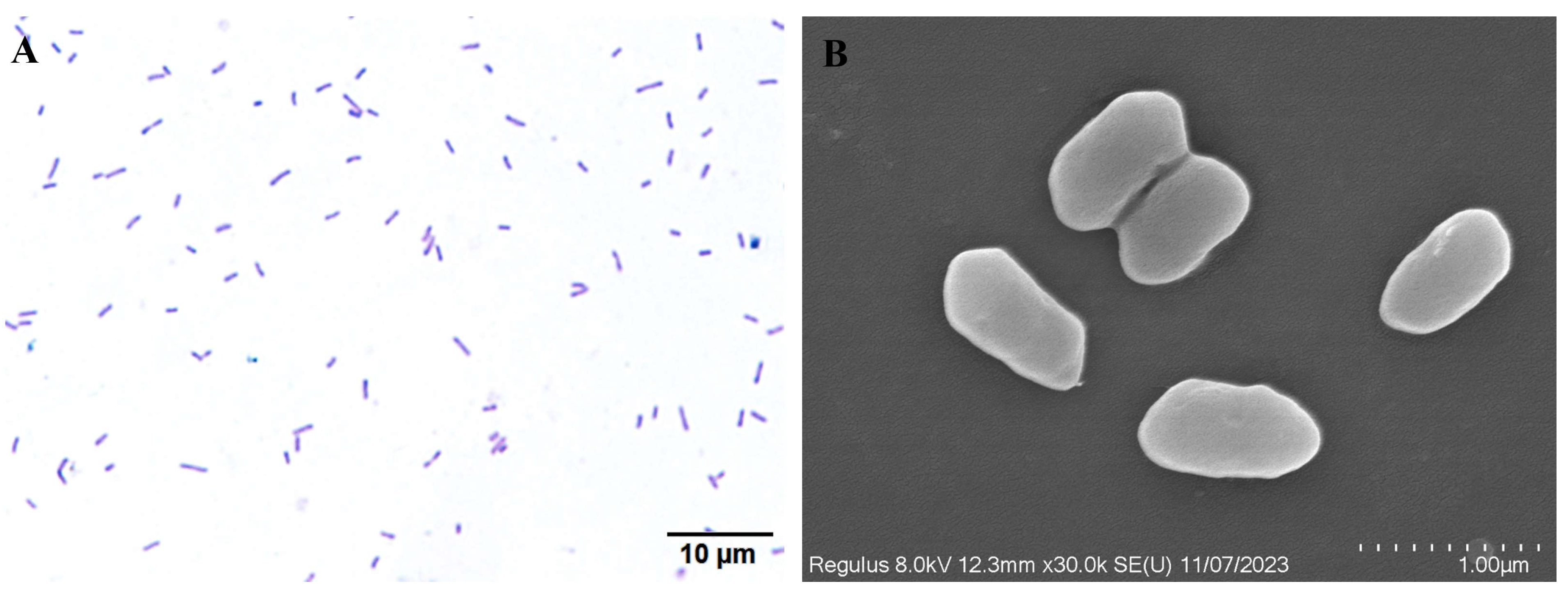
3.3. Morphological Characterization of WH10

The colonies of isolate WH10 were cultured on BHI agar, displaying a yellow hue. The surface of the colony appeared to be moist and glossy. The bacterium was Gram-negative, and was characterized by a rod-shaped structure, with no budding cells or pods (Figure 3A). Under scanning electron microscopy, the bacterium appeared arc-shaped and was approximately 1 µm in length (Figure 3B).

3.4. Sequence Analysis of the 16S rRNA Gene

BLAST searching in the NCBI GenBank database indicated that the identified sequences had significant similarity to Aeromonas veronii 16S rRNA gene sequences. The constructed phylogenetic tree showed that the WH10 isolate was closely related to Aeromonas veronii, thus confirming its taxonomic classification as Aeromonas veronii at the molecular level (Figure 4A).

3.5. Virulence Gene Detection

Herein, we screened the PCR profiles of ten virulence genes, which indicated the presence of the alt, act, lip, ascV, and LuxS genes in isolate WH10, while the remaining five genes were not detected (Figure 4B).

3.6. Bacterial Biochemical Identification

WH10 was subjected to biochemical tests using a biochemical analyzer (Table 2). WH10 was positive for N-Acetyl-D-Glucosamine, Dextrin, Sucrose, 1% NaCl, Acidic PH PH6, Niaproof 4, D-Galactonic Acid, D-Glucose-6-po4, Vancomycin, Nalidixic Acid, Rifamycin SV, Tween 40 reaction, Tetrazolium Blue, L-Malic Acid, and D-Fructose-6-po4, but was negative for Acidic PH PH5, D-Malic Acid, P-Hydroxy-Phenylacetic Acid, Tetrazolium Violet, N-Acetyl-D-Galactosamine, 4% NaCl, 8% NaCl, D-Ducrose, Inosine, Fusidic Acid, 3-Methyl Glucose, Lincomycin, D-Glucuronic Acid, Glucuronamide, D-Arabitol, D-Aspartic acid, Troleandomycin, Minocycline, Potassium Tellurite, Sodium Bromate, Sodium Butyrate, Formic Acid, Propionic Acid, and α-Keto-Butyric Acid (Table 2). According to the morphological and biochemical results, isolate WH10 was identified tentatively as Aeromonas veronii. Owing to the intricate diversity within Aeromonas veronii members and the restricted number of biochemical reactions assessed by the Biolog analysis system, the test successfully identified this isolate as the species Aeromonas veronii.

3.7. Antibiotic Susceptibility Analysis

In the antibiotic susceptibility analysis, WH10 showed varying susceptibilities to ten antibacterial drugs. Notably, it was sensitive to four antibacterial drugs: compound sulfamethoxazoles, cefothiophene, doxycycline, and sulfamethoxazole (Table 3).

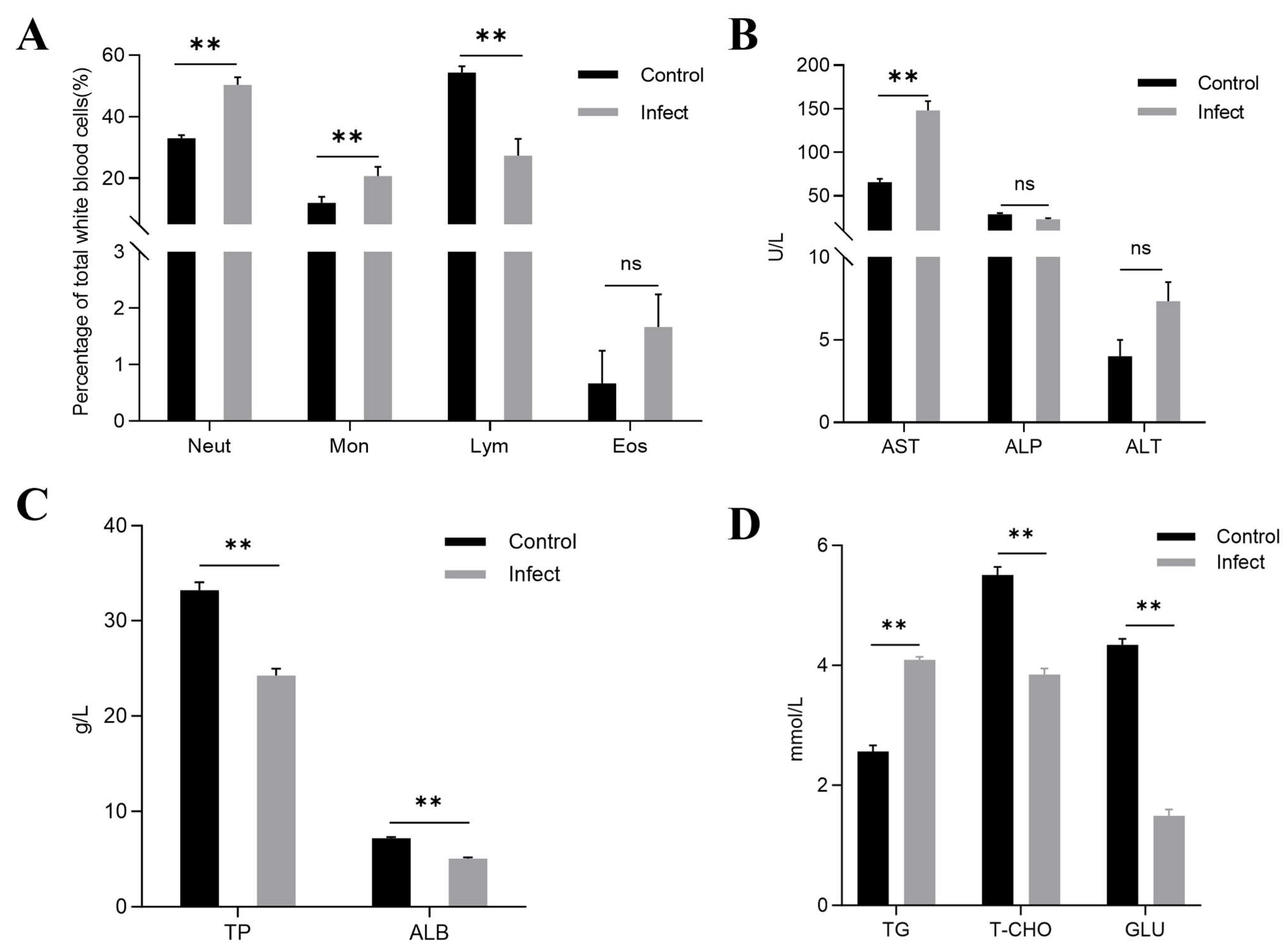
3.8. Differential Leukocyte Counts (DLC)

As depicted in Figure 5, infection with WH10 significantly impacted the blood parameters of bronze gudgeon. In the affected group, the percentage of neutrophils (50.3%) was significantly higher than that of the control group (p < 0.01). The percentage of monocytes also increased in infected group, reaching 20.7% (p < 0.01), in comparison with that of the control group. Conversely, the lymphocyte percentage in the infected group (27.3%) was markedly lower than that in the controls (p < 0.01). Furthermore, throughout the experimental process, the percentage of eosinophils in the infected group remained at basal levels compared with those in the control group (Figure 5A).

3.9. Serum Biochemical Analysis
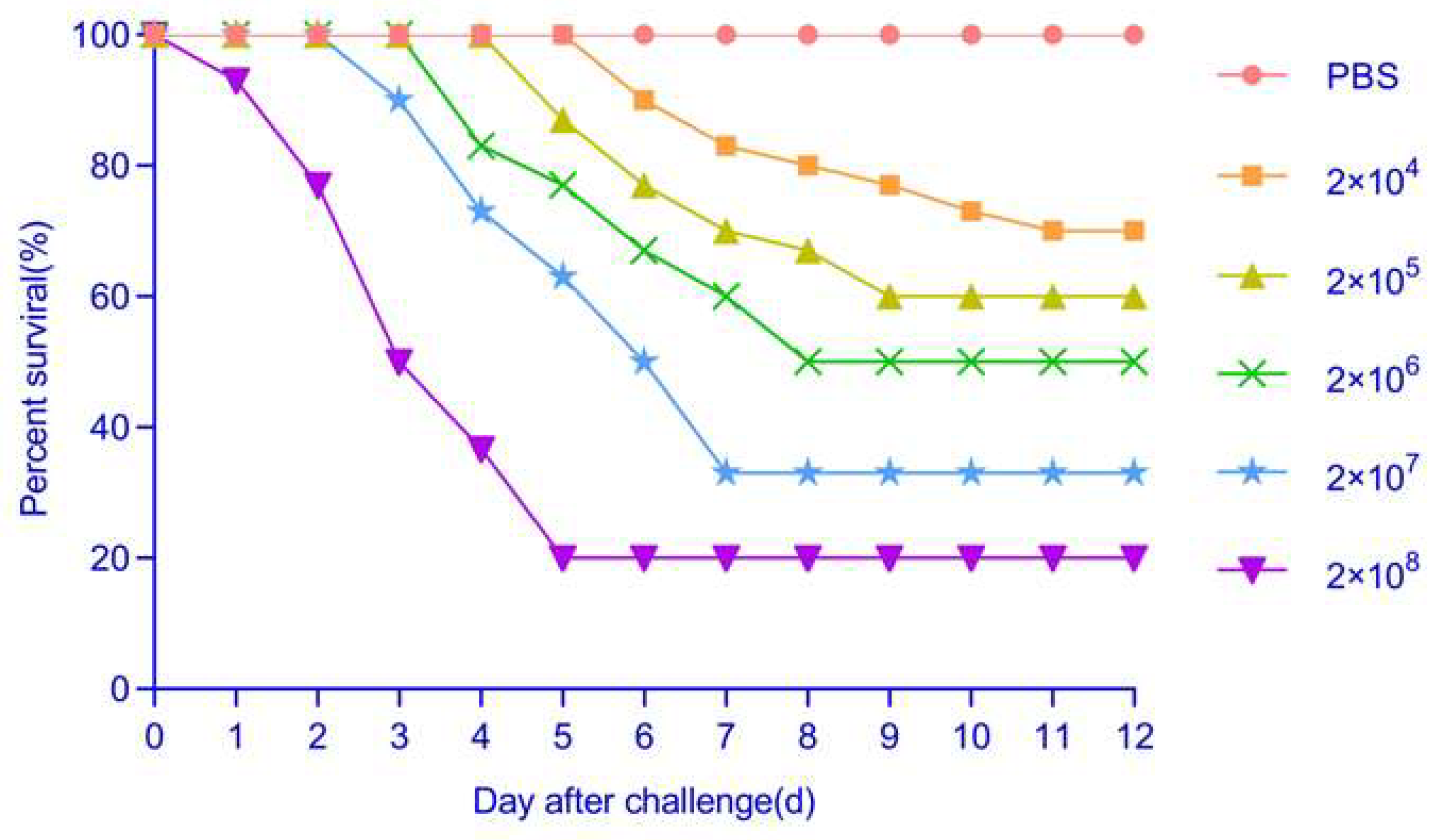
The comparison of the serum biochemical parameters between diseased and healthy bronze gudgeon is shown in Figure 5. Diseased bronze gudgeon had significantly elevated AST (148 g/L) activity and TG (4.1 g/L) concentration compared with those of the healthy controls (p < 0.01) (Figure 5B,D). Conversely, the concentrations of TP (24.2 g/L), ALB (5.0 g/L), T-CHO (3.8 g/L), and GLU (1.5 g/L) were notably reduced in the infected bronze gudgeon compared with those in the healthy specimens (Figure 5C,D). There was no significant difference in the ALT (7.3 g/L) and ALP (22.7 g/L) activities between the two groups (Figure 5B).

3.10. Pathogenicity

The challenge of healthy bronze gudgeon with purified isolate WH10 resulted in hemorrhagic signs reminiscent of those observed in naturally infected fish at around 4 days post-infection (dpi). Within 12 dpi, mortality approached 80%. The pathogenicity test demonstrated mortality in all groups of fish exposed to varying concentrations of bacteria. However, 80% survival was recorded among individuals in the experimental group receiving 2 × 10⁸ CFU/mL fish weight. Conversely, no sign was recorded in the mock-infected fish. According to the experimental results, the LD50 of WH10 was determined as 1.36 × 10⁶ CFU/mL (Figure 6). Samples from challenged and dead fish tested positive for WH10 by PCR. These results unequivocally confirmed that WH10, isolated from infected bronze gudgeon, was the etiological agent of the disease observed in bronze gudgeon farms.

4. Discussion

Aeromonas veronii, a motile, rod-shaped, and Gram-negative bacterium, belongs to the Aeromonadaceae family. It exhibits a broad host range and has the capacity to induce zoonotic motile Aeromonas septicemia (MAS) in humans, terrestrial animals, and various aquatic species [18,19]. Aeromonas veronii infections are particularly lethal to aquatic animals, frequently leading to significant outbreaks and high mortality rates in aquaculture [20]. Reports indicate that Aeromonas outbreaks resulted in a 10% loss in fish farm production in 2017 [21]. Among pathogenic Aeromonas species, such as Aeromonas hydrophila, Aeromonas caviae, Aeromonas sobria, and Aeromonas veronii, the incidence of Aeromonas veronii infections has notably increased in recent years, leading to frequent isolation of these pathogens from diseased fish. In recent years, Aeromonas veronii has been documented in Nile tilapia (Oreochromis niloticus) [22], Chinese Soft-Shelled Turtle (Trionyx sinensis) [12], channel catfish (Ictalurus punctatus) [23], crucian carp (Carassius auratus) [24], Rohu (Labeo rohita) [25], and rainbow trout (Oncorhynchus mykiss) [26]. The clinical manifestations of infected fish often include systemic hemorrhagic septicemia and cutaneous ulcers. In our study, the clinical signs observed in diseased bronze gudgeon comprised hemorrhages on the body surface and fins, along with ulcerated skin lesions. Visceral tissue, including the liver and spleen, exhibited hemorrhagic lesions, which were similar to those commonly observed in fish species infected with Aeromonas veronii. In addition, the pathogenicity test showed that the LD50 was 1.36 × 10⁶ CFU/mL per fish weight, indicating that WH10 exhibited strong virulence towards bronze gudgeon. Furthermore, Aeromonas veronii was successfully detected and isolated from bronze gudgeon artificially infected with WH10. Together, our results indicated that Aeromonas veronii WH10 was the pathogenic bacterium responsible for the observed disease in bronze gudgeon.
Hematology has been used frequently to monitor the health status and immunological responses of aquaculture species [27,28]. Monocytes and neutrophils, serving as the main phagocytic cells in fish, are integral components of the non-specific defense system [29]. In our study, the serum neutrophil and monocyte percentage of Aeromonas veronii-infected bronze gudgeon were increased significantly. This finding might indicate enhanced phagocytosis in the infected bronze gudgeon, which is related to biological responsiveness to environmental parameters and individual health status [30]. Similar alterations in blood cell proportions post-bacterial infection were observed in fancy carp (Cyprinus carpio) infected with Aeromonas veronii [31] and in Vibrio metschnikovii-infected hybrid sturgeon [32]. However, in Aeromonas hydrophila-infected tilapia, numbers of circulating neutrophils decreased, together with augmented accumulation of granulocytes at the inflammatory site [33]. These results differed from those reported herein, which might be related to the fish species and the pathogen under evaluation. These contrasting results might also reflect the fact that a high number of neutrophils in circulation is caused by their delayed apoptosis or impaired clearance from the infection site, thereby contributing to the prolonged secretion of pro-inflammatory mediators, e.g., bacterial components, interleukin-8, or granulocyte macrophage colony-stimulating factor [33]. Lymphocytes are crucial for the body’s cellular immune response, and lower lymphocyte levels are indicative of compromised immune function. Herein, serum lymphocyte levels were decreased significantly in infected bronze gudgeon, indicating a weakened immune system.
Additionally, in fish, the liver is an important organ for protein synthesis, and impairment of hepatic cells leads to a decline in protein production [34]. Serum ALB and TP serve as markers of the immune response and hepatocyte injury, respectively. The observed reduction in serum ALB and TP concentrations in diseased bronze gudgeon signified hepatocyte damage and compromised liver function. Notably, AST is predominantly localized in the cytoplasm of hepatocytes, and its release into the bloodstream occurs exclusively during liver inflammation, necrosis, or intoxication [35]. In this study, diseased bronze gudgeon exhibited a notable increase in serum AST activity (p < 0.01), indicating potential hepatic damage in these individuals. Furthermore, a decrease in T-CHO was observed in the serum of infected bronze gudgeon, suggestive of malnutrition, likely caused by food refusal and starvation during the experimental phase. Pathological alterations in fish kidney, including changes in renal tubules and impaired glomerular filtration function, negatively impact the physiological metabolism of the affected fish [36]. Our findings revealed decreased GLU concentration in diseased fish relative to the control group, suggesting disturbances in glomerular filtration and reabsorption functions after infection with WH10.
Drug sensitivity testing serves as a crucial step to identify effective disease treatment options [37]. Testing the antibiotic response of WH10 demonstrated its sensitivity to compound sulfamethoxazoles, cefothiophene, doxycycline, and sulfamethoxazole. It exhibited moderate sensitivity to gentamicin, amikacin, neomycin, florfenicol, and enrofloxacin, while displaying resistance to ciprofloxacin. The test results were different from those reported previously for Aeromonas veronii [12,38,39]. This variability in antibiotic sensitivity might be attributed to differences in the strains’ origins or their distinct antibiotic resistance profiles. The resistance of Aeromonas veronii to multiple antibiotics represents a significant health concern for both animals and humans. Improper use of antibiotics in aquaculture and the potential for Aeromonas veronii to acquire resistance genes from other multidrug-resistant pathogens are major factors contributing to the emergence of drug-resistant strains [12]. Therefore, selecting antibiotics based on the results of susceptibility testing is crucial for the effective treatment of bacterial diseases.
The pathogenicity of Aeromonas spp. is enhanced by their ability to produce various virulence factors, including proteases, hemolysin, enterotoxins, and toxins in aquatic environments. These virulence factors contribute to the bacteria’s ability to adhere to, invade, and destroy host cells, thereby overcoming the host’s immune response [40]. Assessing the pathogenicity and virulence of a bacterial pathogen according to the presence of virulence factors is essential [41]. In this study, we investigated virulence-related factors associated with Aeromonas species infection, including alt, aha, act, lip, ast, ascV, exu, LuxS, aerA, and hlyA. The ast, alt, and act genes encode cytotoxic enterotoxins, representing critical virulence determinants for Aeromonads, which are potent foodborne pathogens. Knockout mutants of the act gene showed significantly reduced virulence [42]. Herein, Aeromonas veronii WH10 contained act and alt genes, and exhibited a beta hemolysis reaction on blood agar. Additionally, virulence genes encoding secreted proteins and toxins, such as the lip gene, and the gene encoding the quorum sensing signal molecule AI-2-LuxS, are major pathogenic factors of Aeromonas veronii [43,44], contributing to bacterial biofilm production and virulence [45,46]. Aeromonas veronii WH10 contained lip and LuxS genes, suggesting that its high pathogenicity was caused by a combination of multiple virulence-related factor. Our results suggested that an Aeromonas veronii detection method, and even a multivalent vaccine, could be effectively constructed based on its mutual virulence genes.

5. Conclusions

This study identified Aeromonas veronii strain WH10 as the agent that caused an emerging disease in farmed bronze gudgeon in China. This strain was tentatively named WH10 based on its morphology, phylogeny, clinical signs, pathology, and the results of the pathogen challenge. The isolation and identification of WH10 from bronze gudgeon expands our understanding of the host spectrum of Aeromonas veronii. Additionally, this study lays the foundation to develop diagnostic methods for bronze gudgeon disease and to investigate the invasion, transmission mechanisms, and potential therapeutic interventions for related pathogens.

Author Contributions

Methodology, W.L., M.L., N.J., Y.L., J.J., X.L. and Y.F.; Software, M.L., M.X. and J.J.; Validation, M.X., N.J. and Y.F.; Formal analysis, W.L., Y.M. and Y.F.; Investigation, Y.Z.; Resources, X.L., Y.Z.; Data curation, M.L.; Writing—original draft, W.L.; Visualization, X.L.; Supervision, X.L. and Y.F.; Project administration, W.L. and Y.F.; Funding acquisition, Y.F. All authors have read and agreed to the published version of the manuscript.

Funding

This work was supported by the Central Public-interest Scientific Institution Basal Research Fund, CAFS (No. 2023TD46), National Freshwater Aquatic Germplasm Resource Center (FGRC18537).

Institutional Review Board Statement

The study adhered to the guidelines outlined in the Guide for the Care and Use of Laboratory Animals Monitoring Committee of Hubei Province, China. The Committee on the Ethics of Animal Experiments at the Yangtze River Fisheries Research Institute, Chinese Academy of Fishery Sciences approved the protocol (ID number: YFI 2023-liuwenzhi-1124). To minimize their suffering, the bronze gudgeon were euthanized for 20–30 min using 0.1% MS-222 before their tissues were collected.

Informed Consent Statement

Not applicable.

Data Availability Statement

The original contributions presented in the study are included in the article, further inquiries can be directed to the corresponding author/s.

Acknowledgments

We would like to thank Xiaowei Hu, Jiale Zhai, and Mengmeng Chen for their technical help.

Conflicts of Interest

The authors declare that they have no competing interests.

References

  1. Yang, S.R.; Gao, X.; Li, M.Z.; Ma, B.S.; Liu, H.Z. Interannual variations of the fish assemblage in the transitional zone of the Three Gorges Reservoir: Persistence and stability. Environ. Biol. Fishes 2012, 93, 295–304. [Google Scholar] [CrossRef]
  2. Liu, H.Y.; Xiong, F.; Duan, X.B.; Liu, S.P.; Chen, D.Q. Estimating population parameters and abundance of Coreius heterodon in Jiangjin section of the upper Yangtze River. J. Nat. Resour. 2016, 31, 1420–1428. (In Chinese) [Google Scholar]
  3. Chen, B.Y.; Luo, H.H.; Yang, Q.R.; Huang, W.; Han, H.L. The eco-hydrological demand research on Coreius heterodon reproduction in the upper Yangtze River reserve. Appl. Mech. Mater. 2014, 641, 226–231. [Google Scholar] [CrossRef]
  4. Li, T.; Mo, K.L.; Wang, J.; Chen, Q.W.; Zhang, J.Y.; Zeng, C.J.; Zhang, H.; Yang, P.S. Mismatch between critical and accumulated temperature following river damming impacts fish spawning. Sci. Total Environ. 2021, 756, 144052. [Google Scholar] [CrossRef] [PubMed]
  5. Zhou, Y.J.; Wang, G.Q.; Wu, H.L.; Chen, P.; Li, D.F.; Jin, Z.W.; Guo, C.; Ren, S.; Gao, Y. Changes in flow and sediment transport caused by cascade hydropower in the upper reaches of Yangtze River and their influence on spawning of Coreius heterodon. Catena 2024, 235, 107622. [Google Scholar] [CrossRef]
  6. Xu, D.D.; Li, P.; Zhang, Y.G.; Peng, Z.G. Comparative study of the complete mitochondrial genomes of the bronze gudgeon (Coreius heterodon) and largemouth bronze gudgeon (Coreius guichenoti). Mitochondrial DNA 2013, 24, 189–190. [Google Scholar] [CrossRef]
  7. Cai, S.W.; Ni, Z.H.; Li, Y.F.; Shen, Z.W.; Xiong, Z.T.; Zhang, Y.; Zhou, Y.T. Metals in the tissues of two fish species from the rare and endemic fish nature reserve in the upper reaches of the Yangtze River, China. Bull. Environ. Contam. Toxicol. 2012, 88, 922–927. [Google Scholar] [CrossRef]
  8. Luo, Y.P.; Huang, Q.D.; Zhang, Y.R.; Liu, S.T.; Wang, W. Comparison of the body proximate compositions of juvenile bronze gudgeon (Coreius heterodon) and largemouth bronze gudgeon (C. guichenoti) in the upstream region of the Yangtze River. SpringerPlus 2013, 2, 75. [Google Scholar] [CrossRef]
  9. Hickman-Brenner, F.W.; Macdonald, K.L.; Steigerwalt, A.G.; Fanning, G.R.; Brenner, D.J.; Farmer, J.J. Aeromonas veronii, a new ornithine decarboxylase-positive species that may cause diarrhea. J. Clin. Microbiol. 1987, 25, 900–906. [Google Scholar] [CrossRef]
  10. Liu, W.Z.; Zhang, Y.C.; Ma, J.; Jiang, N.; Fan, Y.D.; Zhou, Y.; Cain, K.; Yi, M.S.; Jia, K.T.; Wen, H.; et al. Determination of a novel parvovirus pathogen associated with massive mortality in adult tilapia. PLoS Pathog. 2020, 16, e1008765. [Google Scholar] [CrossRef]
  11. Arroyo, E.; Enríquez, L.; Sánchez, A.; Ovalle, M.; Olivas, A. Scanning electron microscopy of bacteria Tetrasphaera duodecadis. Scanning 2014, 36, 547–550. [Google Scholar] [CrossRef]
  12. Hu, X.W.; Xiao, Z.D.; Li, B.; Xue, M.Y.; Jiang, N.; Fan, Y.D.; Chen, P.; Qi, F.; Kong, X.H.; Zhou, Y. Isolation, identification, and characterization of Aeromonas veronii from Chinese soft-shelled turtle (Trionyx sinensis). Microorganisms 2023, 11, 1304. [Google Scholar] [CrossRef] [PubMed]
  13. Xu, Y.X.; Zhang, H.P.; Xu, G.Y.; Muhamamda, I.; Dong, W.L.; Wang, Y.M.; Kong, L.C.; Ma, H.X. Virulence and drug resistance of Aeromonas veronii isolated from shellfish. Thai J Vet Med. 2021, 51, 21–27. [Google Scholar] [CrossRef]
  14. Yuan, G.L.; Zhu, L.; Jiang, X.Y.; Zhang, J.; Pei, C.; Zhao, X.L.; Li, L.; Kong, X.H. Diagnosis of co-infection with white spot syndrome virus and Aeromonas veronii in red swamp crayfish Procambarus clarkii. Aquaculture 2021, 532, 736010. [Google Scholar] [CrossRef]
  15. Li, T.; Raza, S.H.A.; Yang, B.T.; Sun, Y.F.; Wang, G.Q.; Sun, W.W.; Qian, A.D.; Wang, C.F.; Kang, Y.H.; Shan, X.F. Aeromonas veronii infection in commercial freshwater fish: A potential threat to public health. Animals 2020, 10, 608. [Google Scholar] [CrossRef]
  16. Wang, B.T.; Mao, C.; Feng, J.; Li, Y.; Hu, J.M.; Jiang, B.; Gu, Q.H.; Su, Y.L. A First report of Aeromonas veronii infection of the sea bass, Lateolabrax maculatus in China. Front. Vet Sci. 2021, 7, 600587. [Google Scholar] [CrossRef]
  17. Reed, L.J. A simple method of estimating fifty percent endpoints. Am. J. Epidemiol. 1938, 27, 493–497. [Google Scholar] [CrossRef]
  18. Rahman, M.; Colque-Navarro, P.; Kuhn, I.; Huys, G.; Swings, J.; Mollby, R. Identification and characterization of pathogenic Aeromonas veronii biovar sobria associated with epizootic ulcerative syndrome in fish in Bangladesh. Appl. Environ. Microb. 2002, 68, 650–655. [Google Scholar] [CrossRef]
  19. Gui, M.; Wu, R.; Liu, L.; Wang, S.; Zhang, L.; Li, P. Effects of quorum quenching by AHL lactonase on AHLs, protease, motility and proteome patterns in Aeromonas veronii LP-11. Int. J. Food Microbiol. 2017, 252, 61–68. [Google Scholar] [CrossRef]
  20. Fernández-Bravo, A.; Figueras, M.J. An Update on the genus Aeromonas: Taxonomy, epidemiology, and pathogenicity. Microorganisms 2020, 8, 129. [Google Scholar] [CrossRef]
  21. Abd-El-Malek, A.M. Incidence and virulence characteristics of Aeromonas spp. in fish. Vet World. 2017, 10, 34–37. [Google Scholar] [CrossRef] [PubMed]
  22. Dos Santos, S.B.; Alarcon, M.F.; Ballaben, A.S.; Harakava, R.; Galetti, R.; Guimarães, M.C.; Natori, M.M.; Takahashi, L.S.; Ildefonso, R.; Rozas-Serri, M. First report of Aeromonas veronii as an emerging bacterial pathogen of farmed Nile Tilapia (Oreochromis niloticus) in Brazil. Pathogens 2023, 8, 1020. [Google Scholar] [CrossRef]
  23. Haitham, H.M.; Eric, P. Winter kill in intensively stocked channel catfish (Ictalurus punctatus): Coinfection with Aeromonas veronii, Streptococcus parauberis and Shewanella putrefaciens. J. Fish Dis. 2018, 9, 1339–1347. [Google Scholar]
  24. Chen, F.; Sun, J.F.; Han, Z.R.; Yang, X.J.; Xian, J.A.; Lv, A.J.; Hu, X.C.; Shi, H.Y. Isolation, identification and characteristics of Aeromonas veronii from diseased crucian carp (Carassius auratus gibelio). Front. Microbiol. 2019, 10, 2742. [Google Scholar] [CrossRef] [PubMed]
  25. Behera, B.K.; Parida, S.N.; Kumar, V.; Swain, H.S.; Parida, P.K.; Bisai, K.; Dhar, S.; Das, B.K. Aeromonas veronii is a lethal pathogen isolated from gut of infected Labeo rohita: Molecular insight to understand the bacterial virulence and its induced host immunity. Pathogens 2023, 12, 598. [Google Scholar] [CrossRef]
  26. Reyes-Rodríguez, N.E.; Salgado-Miranda, C.; Flores-Valle, I.T.; González-Gómez, M.; Soriano-Vargas, E.; Peláez-Acero, A.; Vega-Sánchez, V. Molecular identification and virulence potential of the genus Aeromonas isolated from wild rainbow trout (Oncorhynchus mykiss) in Mexico. J. Food Prot. 2019, 10, 1706–1713. [Google Scholar] [CrossRef]
  27. Cannon, M.S.; Mollenhauer, H.H.; Eurell, T.E.; Lewis, D.H.; Cannon, A.M.; Tompkins, C. An ultrastructural study of the leucocytes of the channel fish, Ictalurus punctatus. J. Morphol. 1980, 164, 1–23. [Google Scholar] [CrossRef]
  28. Zarejabad, A.M.; Jalali, M.A.; Sudagar, M.; Pouralimotlagh, S. Hematology of great sturgeon (Huso huso Linnaeus, 1758) juvenile exposed to brackish water environment. Fish Physiol. Biochem. 2010, 36, 655–659. [Google Scholar] [CrossRef]
  29. Dalmo, R.A.; Ingebrigtsen, K.; Bøgwald, J. Non-specific defence mechanisms in fish, with particular reference to the reticuloendothelial system (RES). J. Fish Dis. 1997, 20, 241–273. [Google Scholar] [CrossRef]
  30. Kühlwein, H.; Merrifield, D.L.; Rawling, M.D.; Foey, A.D.; Davies, S.J. Effects of dietary β-(1,3) (1,6)-D-glucan supplementation on growth performance, intestinal morphology and haemato-immunological profile of mirror carp (Cyprinus carpio L.). J. Anim. Physiol. Anim. Nutr. 2014, 98, 279–289. [Google Scholar] [CrossRef] [PubMed]
  31. Rattanachan, S.; Bunnajirakul, S.; Punyadarsaniya, D. Effect of pre-supplementation with Pleurotus sajor-caju crude extracts on body weight and consequence responses of leukocytes and immune organs in fancy carp following inoculation with Aeromonas veronii. Vet. World 2020, 13, 1010–1016. [Google Scholar] [CrossRef] [PubMed]
  32. Xiao, Z.D.; Li, X.D.; Xue, M.Y.; Zhang, M.W.; Liu, W.; Fan, Y.D.; Chen, X.H.; Chu, Z.P.; Gong, F.L.; Zeng, L.B.; et al. Vibrio metschnikovii, a potential pathogen in freshwater-cultured hybrid sturgeon. Animals 2022, 12, 1101. [Google Scholar] [CrossRef]
  33. Charlie-Silva, I.; Klein, A.; Gomes, J.M.M.; Prado, E.J.R.; Moraes, A.C.; Eto, S.F.; Fernandes, D.C.; Fagliari, J.J.; Junior, J.D.C.; Lima, C.; et al. Acute-phase proteins during inflammatory reaction by bacterial infection: Fish-model. Sci. Rep. 2019, 9, 4776. [Google Scholar] [CrossRef]
  34. Javed, M.; Ahmad, M.I.; Usmani, N.; Ahmad, M. Multiple biomarker responses (serum biochemistry, oxidative stress, genotoxicity and histopathology) in Channa punctatus exposed to heavy metal loaded waste. Sci. Rep. 2017, 7, 1675. [Google Scholar] [CrossRef] [PubMed]
  35. van der Oost, R.; Beyer, J.; Vermeulen, N.P.E. Fish bioaccumulation and biomarkers in environmental risk assessment: A review. Environ. Toxicol. Phar. 2003, 13, 57–149. [Google Scholar] [CrossRef] [PubMed]
  36. Bukha, K.K.; Mahgiubi, S.A.M.; Elbahi, A.M.; Sharif, E.A.; Showehdi, M.L.; Ahmed, M.O.; Kammon, A.M.; Abouzeed, Y.M. Pathological lesions associated with Vibrio infection in Atlantic horse mackerel (Trachurus trachurus L.; 1758) from the western coast of Tripoli, Libya. Open Vet. J. 2023, 13, 327–336. [Google Scholar]
  37. Smith, P. Accuracy, precision and meaning of antimicrobial agent susceptibility testing of bacteria associated with fish diseases. Aquaculture 2001, 196, 253–266. [Google Scholar] [CrossRef]
  38. Shameena, S.S.; Kumar, K.; Kumar, S.; Rathore, G. Characteristics of Aeromonas veronii biovars isolated from infected freshwater goldfish (Carassius auratus). Aquaculture 2020, 518, 734819. [Google Scholar] [CrossRef]
  39. Jagoda, S.S.; Wijewardana, T.G.; Arulkanthan, A.; Igarashi, Y.; Tan, E.; Kinoshita, S.; Watabe, S.; Asakawa, S. Characterization and antimicrobial susceptibility of motile aeromonads isolated from freshwater ornamental fish showing signs of septicaemia. Dis. Aquat.Org. 2014, 109, 127–137. [Google Scholar] [CrossRef]
  40. Majeed, S.; De Silva, L.A.D.S.; Kumarage, P.M.; Heo, G.J. Occurrence of potential virulence determinants in Aeromonas spp. isolated from different aquatic environments. J. Appl. Microbiol. 2023, 134, lxad031. [Google Scholar] [CrossRef]
  41. Zhu, M.; Wang, X.R.; Li, J.; Li, G.Y.; Liu, Z.P.; Mo, Z.L. Identification and virulence properties of Aeromonas veronii Bv. Sobria isolates causing an ulcerative syndrome of loach Misgurnus anguillicaudatus. J. Fish Dis. 2016, 39, 777–781. [Google Scholar] [CrossRef] [PubMed]
  42. Sha, J.; Kozlova, E.V.; Chopra, A. Role of various enterotoxins in Aeromonas hydrophila-induced gastroenteritis: Generation of enterotoxin gene-deficient mutants and evaluation of their enterotoxic activity. Infect. Immun. 2002, 70, 1924–1935. [Google Scholar] [CrossRef]
  43. Chen, X.; Schauder, S.; Potier, N.; Van Dorsselaer, A.; Pelczer, I.; Bassler, B.L.; Hughson, F.M. Structural identification of a bacterial quorum-sensing signal containing boron. Nature 2002, 415, 545–549. [Google Scholar] [CrossRef] [PubMed]
  44. Chandrathna, H.P.S.U.; Nikapitiya, C.; Dananjaya, S.H.S.; Wijerathne, C.U.B.; Wimalasena, S.H.M.P.; Kwun, H.J.; Heo, G.J.; Lee, J.; De Zoysa, M. Outcome of co-infection with opportunistic and multidrug resistant Aeromonas hydrophila and A. veronii in zebrafish: Identification, characterization, pathogenicity and immune responses. Fish Shellfish Immunol. 2018, 80, 573–581. [Google Scholar] [CrossRef] [PubMed]
  45. Chung, W.O.; Park, Y.; Lamont, R.J.; McNab, R.; Barbieri, B.; Demuth, D.R. Signaling system in Porphyromonas gingivalis based on a LuxS protein. J. Bacteriol. 2001, 183, 3903–3909. [Google Scholar] [CrossRef]
  46. Frias, J.; Olle, E.; Alsina, M. Periodontal pathogens produce quorum sensing signal molecules. Infect. Immun. 2001, 69, 3431–3434. [Google Scholar] [CrossRef]
Figure 1. Clinical manifestations of infected bronze gudgeon. (A) The infected bronze gudgeon showed hemorrhages on both body surface (black arrow) and liver (blue arrow). (B) Ulcerated skin was evident on the surface of the affected bronze gudgeon (white arrow). (C) Bleeding points were observed on the fin plates (black arrow). (D) The spleen of the diseased bronze gudgeon appeared dark and enlarged (navy blue arrowheads).
Animals 14 02440 g001
Figure 2. Pathological analysis of the diseased bronze gudgeon. (A) Healthy liver. (B) Significant vacuolation (white arrow), along with liver blood sinuses exhibiting swelling and hemorrhaging, was observed in the diseased liver tissue (red arrow and blue triangular arrowheads). (C) Healthy kidney. (D) Infected kidney tissues exhibited edema in the renal tubules (white arrow) and lymphocyte infiltration in and around the affected areas (blue triangular arrowheads). (E) Healthy spleen. (F) The spleen displayed a spectrum of extensive necrosis and vacuolation (white arrow) and extended melanomacrophage center (blue triangular arrowhead).
Animals 14 02440 g002
Figure 3. Scanning electron microscopy and Gram staining of Aeromonas veronii (WH10). (A) Gram staining of isolate WH10 (scale bar: 10 µm); (B) Scanning electron micrograph of isolate WH10 (scale bar: 1.0 µm).
Animals 14 02440 g003
Figure 4. Phylogenetic analysis and PCR detection of virulence genes of isolate WH10. (A) 16S rRNA-based phylogenetic analysis using sequences from Aeromonas veronii, Aeromonas hydrophila, Escherichia coli, Escherichia fergusonii, and Staphylococcus equorum. Red arrow is the strain isolated in our study. (B) Agarose gel electrophoresis of virulence genes from the WH10 strain. M: DNA ladder (DL2000 bp); 1st lane: alt; 2nd lane: aha; 3rd lane: act; 4th lane: lip; 5th lane: ast; 6th lane: ascV; 7th lane: exu; 8th lane: LuxS; 9th lane: aerA; 10th lane: hlyA.
Animals 14 02440 g004
Figure 5. Differential leukocyte counts and biochemical evaluations of serum from bronze gudgeons infected with isolate WH10. (A) The percentage changes in differential leukocyte counts in the affected group compared to the control group. (B) Serum biochemical evaluations of affected bronze gudgeon in showed significantly higher AST, but no significant difference in ALP activity and ALT concentration compared with those in the serum of healthy fish. (C) Significantly lower TP and ALB concentrations in affected fish serum compared with those in the serum of healthy fish. (D) T-CHO and GLU concentrations were significantly lower in the serum of affected fish lower than in that of healthy fish. ** p < 0.01. ns: not significant.
Animals 14 02440 g005
Figure 6. Pathogenicity of different concentrations of the WH10 strain solution toward bronze gudgeon.
Animals 14 02440 g006
Table 1. Primer sequences used for PCR amplification of target genes of Aeromonas veronii.
Gene NamePrimer NamePrimers (5′-3″)References
16S rRNA16S rRNA-FAGAGTTTGATCATGGCTCAG[12]
16S rRNA-RTACGGTTACCTTGTTACGACTT
ascVascV-FAGCAGATGAGTATCGACGG[13]
ascV-RAGGCATTCTCCTGTACCAG
hlyAhlyA-FGGCCGGTGGCCCGAAGATACGGG[14]
hlyA-RGGCGGCGCCGGACGAGACGGGG
aerAaerA-FCAAGAACAAGTTCAAGTGGCCA[13]
aerA-RACGAAGGTGTGGTTCCAGT
LuxSLuxS-FGATCCTCTCCGAGGCGTGG[14]
LuxS-RAGGCTTTTCAGCTTCTCTTCC
actact-FTCTCCATGCTTCCCTTCCACT[13]
act-RAACTGACATCGGCCTTGAACTC
altalt-FTGACCCAGTCCTGGCACGGC[13]
alt-RGGTGATCGATCACCACCAGC
astast-FTCTCCATGCTTCCCTTCCACT[13]
ast-RGTGTAGGGATTGAAGAAGCCG
liplip-FCACCTGGT(T/G)CCGCTCAAG[15]
lip-RGTACCGAACCAGTCGGAGAA
exuexu-FAGACATGCACAACCTCTTCC[16]
exu-RGATTGGTATTGCCTTGCAAG
ahaaha-FGGCTATTGCTATCCCGGCTCTGTT[15]
aha-RCGGTCCACTCGTCGTCCATCTTG
Table 2. Biochemical identification of the WH10 strain.
Reaction ItemResult * Reaction ItemResult *
A1Negative controlNE1GelatinB
A2DextrinPE2Glycyl-L-ProlineB
A3D-MaltoseBE3L-AlanineB
A4D-TrehaloseBE4L-ArginineB
A5D-CellobioseBE5L-Aspartic AcidB
A6GentiobioseBE6L-Glutamic AcidB
A7SucrosePE7L-HistidineB
A8D-TuranoseBE8L-Pyroglutamic AcidB
A9StachyoseBE9L-SerineB
A10Positive controlPE10LincomycinN
A11Acidic PH PH6PE11Guanidine HClB
A12Acidic PH PH5NE12Niaproof 4P
B1D-RaffinoseBF1PectinB
B2α-D-LactoseBF2D-Galacturonic AcidB
B3D-MelibioseBF3L-Galactonic Acid LactoneB
B4β-Methyl-D-GlucosideBF4D-Galactonic AcidP
B5D-SalicinBF5D-Glucuronic AcidN
B6N-Acetyl-D-GlucosaminePF6GlucuronamideN
B7N-Acetyl-β-MannosamineBF7Mucic AcidB
B8N-Acetyl-D-GalactosamineNF8Quinic AcidB
B9N-Acetyl NeuraminicBF9D-Saccharic AcidB
B101% NaClPF10VancomycinP
B114% NaClNF11Tetrazolium VioletN
B128% NaClNF12Tetrazolium BlueP
C1α-D-GlucoseBG1P-Hydroxy-Phenylacetic AcidN
C2D-MannoseBG2Methyl PyruvateB
C3D-FructoseBG3D-Lactic Acid Methyl EsterB
C4D-GalactoseBG4Lactic AcidB
C53-Methyl GlucoseNG5Citric AcidB
C6D-DucroseNG6α-Keto-Glutaric AcidB
C7L-FucoseBG7D-Malic AcidN
C8L-RhamnoseBG8L-Malic AcidP
C9InosineNG9Bromo-Succinic-AcidB
C101% Sodium LactateBG10Nalidixic AcidP
C11Fusidic AcidNG11Lithium ChlorideB
C12D-SerineBG12Potassium TelluriteN
D1D-SorbitolBH1Tween 40P
D2D-MannitolBH2γ-Amino-Butyric-AcidB
D3D-ArabitolNH3α-Hydroxy- Butyric-AcidB
D4Myo-InositolBH4β--Hydroxy-D, L Butyric-AcidB
D5GlycerolBH5α-Keto-Butyric AcidN
D6D-Glucose-6-po4PH6Acetoacetic AcidB
D7D-Fructose-6-po4PH7Propionic AcidN
D8D-Aspartic acidNH8Acetic AcidB
D9D-SerineBH9Formic AcidN
D10TroleandomycinNH10AztreonamB
D11Rifamycin SVPH11Sodium ButyrateN
D12MinocyclineNH12Sodium BromateN
* Abbreviations: P, positive; N, negative; B, borderline.
Table 3. Antibiotic susceptibility analysis of the WH10 strain.
DrugsContent (μg/Piece)Inhibition Zone (mm)Sensitivity
Compound Sulfamethoxazoles23.7529S
Cefothiophene3024S
Doxycycline3019S
Sulfamethoxazole30016S
Gentamicin1013I
Amikacin3014I
Neomycin3014I
Florfenicol3015I
Enrofloxacin1013I
Ciprofloxacin58R
Notes: “S” = Susceptible (diameter > 15 mm); “I” = Intermediate (10 mm < diameter ≤ 15 mm); “R” = Resistant (diameter ≤ 10 mm).
Disclaimer/Publisher’s Note: The statements, opinions and data contained in all publications are solely those of the individual author(s) and contributor(s) and not of MDPI and/or the editor(s). MDPI and/or the editor(s) disclaim responsibility for any injury to people or property resulting from any ideas, methods, instructions or products referred to in the content.

