Article

Antimicrobial Susceptibility of Commensal Escherichia coli from Pig Fecal Samples and Enhanced Sensitivity for Direct Detection of the blaCTX-M Gene by Nested PCR

by
Nutchaba Suchanta
1,2,
Naeem Ullah
1,
Pitak Santanirand
3,
Nutthee Am-In
4 and
Nuntaree Chaichanawongsaroj
1,*
1
Center of Excellence for Innovative Diagnosis of Antimicrobial Resistance, Department of Transfusion Medicine and Clinical Microbiology, Faculty of Allied Health Sciences, Chulalongkorn University, Pathumwan, Bangkok 10330, Thailand
2
Program of Molecular Sciences in Medical Microbiology and Immunology, Department of Transfusion Medicine and Clinical Microbiology, Faculty of Allied Health Sciences, Chulalongkorn University, Pathumwan, Bangkok 10330, Thailand
3
Department of Pathology, Faculty of Medicine Ramathibodi Hospital, Mahidol University, Bangkok 10400, Thailand
4
Department of Obstetrics Gynaecology and Reproduction, Faculty of Veterinary Science, Chulalongkorn University, Pathumwan, Bangkok 10330, Thailand
*
Author to whom correspondence should be addressed.
Animals 2024, 14(18), 2630; https://doi.org/10.3390/ani14182630
Submission received: 7 August 2024 / Revised: 7 September 2024 / Accepted: 9 September 2024 / Published: 10 September 2024
(This article belongs to the Special Issue Reducing Antibiotics in Pig Farming – Multilevel Approach)

Abstract

:

Simple Summary

The dissemination of antimicrobial resistance (AMR) genes in economic animals affects food safety in our life cycle. The pig gut microbiome can be a reservoir of antimicrobial-resistant bacteria. As a result of the comparison of antimicrobial susceptibility profiles and the existence of ESBL genes, we found no difference between antibiotic- and nonantibiotic-using farms. We suggest a basal level of ESBL E. coli persistence on pig farms, which may not depend on antibiotic usage. Direct detection of antimicrobial-resistant genes from pig fecal samples reduced cumbersome bacterial culture processes. The enhanced sensitivity of the nPCR technique facilitates the surveillance of AMR genes, leading to effective control.

Abstract

The commensal Escherichia coli in the gut of pigs is a major reservoir of antimicrobial resistance and can result in possible transmission to humans through the food chain. Direct detection of E. coli from fecal samples is challenging and can be used as a bioindicator of antimicrobial resistance. This study aimed to compare the antimicrobial susceptibility profiles in commensal E. coli from antibiotic- and nonantibiotic-using pig farms and developed the direct detection of ESBL genes in pig fecal samples using nested PCR (nPCR) and multiplex PCR (mPCR) techniques. All direct genotypic results were validated with the results of PCR sequencing of isolated E. coli colonies. The ESBL-producing E. coli were found in 98.6% (145 isolates) and 96.6% (144 isolates) of antibiotic-using and nonantibiotic-using farms, respectively, predominantly CTX-M-55. The nPCR decreased the limit of detection (LOD) from sPCR about 100 times, and the lower LODs of 102, 101, and 1 CFU/mL were reached after incubating samples in an enrichment medium for 2, 4, and 8 h, respectively. The mPCR, sPCR, and nPCR techniques showed sensitivities of 30.15%, 69.85%, and 91.91%, respectively, compared to PCR sequencing. The stability and recycling of ESBL genes were independent of antibiotic usage in commensal E. coli originating in pig farms.

1. Introduction

The inappropriate and high usage of antibiotics in humans and animals has driven the world to the edge of a post-antibiotic era, in which no antibiotic can be used for infectious disease treatment. The dissemination of drug-resistant bacteria has become an important global public health crisis in the One Health system due to horizontal gene transfer in agriculture, livestock, and the environment. The extended-spectrum beta-lactamase (ESBL)-producing bacteria are a World Health Organization (WHO) critical priority concern. The unnecessary use or overuse/misuse of antibiotics in food-animal production, particularly in pigs, leads to the colonization and continuous spreading of drug resistance, ineffective and prolonged treatment, and economic loss [1]. Consequently, establishing a control point to limit the transmission of drug-resistant pathogens is a serious priority. Extended-spectrum beta-lactamase (ESBL)-producing E. coli are a major critical commensal antibiotic-resistance source in food animals. The broad activities of ESBL enzymes, which destroy ampicillin, cephalosporins, and aztreonam, are responsible for several genes, such as blaCTX-M, blaTEM, blaOXA, and blaSHV [2]. The predominant ESBL gene in pigs and pork in many countries, including Thailand, was CTX-M (ranging from 91 to 98%), with different CTX-M subtypes among different areas [3]. A higher incidence of ESBL-producing E. coli has been reported on farms with higher antibiotic usage than those without [4]. However, the rising prevalence of ESBL-producing Enterobacteriaceae has been reported in animals without antibiotic usage [5]. Hence, ESBL-producing E. coli in fecal samples could be used as an indicator for antimicrobial resistance tracking.
Phenotypic antimicrobial susceptibility testing (AST) based on Clinical and Laboratory Standards Institute (CLSI) guidelines is a conventional routine method, while genotypic detection is beneficial for epidemiological purposes. Nowadays, both systems are still based on bacterial culture, which is a cumbersome, time-consuming process that limits the widespread adoption of AMR monitoring [6]. Direct detection from samples or clinical specimens is an ideal detection assay, as it is a “sample in and answer out” method. However, the bottleneck and troublesome process is DNA extraction, especially from fecal specimens, due to a lot of fiber and interference substances, reducing the efficiency of nucleic acid extraction and amplification [7,8]. Nested Polymerase Chain Reaction (nPCR) using two sets of primers and second rounds of PCR amplification could enhance specificity and sensitivity in several pathogenic detections such as feline leukemia virus (FeLV) [9], Borrelia burgdorferi [10], and malaria [11].
This study aimed to compare the antimicrobial susceptibility profiles and the presence of ESBL genes between antibiotic-using and nonantibiotic-using farms. The direct detection of blaCTX-M in pig fecal samples using nPCR, multiplex PCR (mPCR), and singleplex PCR (sPCR) techniques were validated with the PCR sequencing results of E. coli isolates from the same samples. The persistence of antimicrobial resistance in the intestinal tracts of pigs highlighted the importance of effective detection and control of the spread in the food chain.

2. Materials and Methods

2.1. Study Design and Farm Selection

A cross-sectional study was conducted in pig-fattening farms (500–650 animals per farm) located in Thailand’s central and northeastern parts in 2021–2022. Five antibiotic-using (Code: PIG, A1–3, and D1) and five nonantibiotic-using (Code: S1, S2, and ND1–3) farms were randomly selected. The research proposal was approved by the Institutional Animal Care and Use Committee of Chulalongkorn University (Animal Use Protocol 2031065).

2.2. Collection of Fecal Samples

About 30 fecal samples were randomly collected on each farm from healthy pigs in the last week before slaughter. Two hundred and ninety-six samples were kept in Cary Blair medium and transported on ice to the laboratory for analysis within 24–48 h.

2.3. Bacterial Culture, Identification, and Antibiotic Susceptibility Testing

All rectal swabs were suspended in 0.5 mL of 0.85% NaCl and cultured on MacConkey agar (Oxoid, Basingstoke, UK). A cefotaxime disk (30 µg) was incubated on agar at 37 °C for 18–24 h. Colonies that grew around the disk were selected and identified using MALDI-TOF mass spectrometry (Bruker, Karlsruhe, Germany). All E. coli isolates were subjected to antimicrobial susceptibility testing using an automated Sensititre ARIS HiQ system (Thermo Scientific, Waltham, MA, USA). Minimum inhibitory concentrations were determined for the following antibiotics: amikacin, gentamicin, netilmicin, ampicillin, amoxicillin/clavulanic acid, ampicillin/sulbactam, piperacillin/tazobactam, cefuroxime, cefoxitin, ceftriaxone, cefotaxime, ceftazidime, cefepime, ertapenem, doripenem, imipenem, meropenem, ciprofloxacin, levofloxacin, and trimethoprim/sulfamethoxazole. The combination drug method using ceftazidime/ceftazidime–clavulanate and cefotaxime/cefotaxime–clavulanate was also added in the susceptibility panel to confirm ESBL-producing isolates. The results were interpreted according to the Clinical and Laboratory Standards Institute (CLSI) guideline [12]. E. coli ATCC 25922 was used as quality control. The ESBL-producing-E. coli isolates were preserved as 20% glycerol stocks at −80 °C until use.

2.4. DNA Extraction

All 289 E. coli isolates from 10 farms and 136 pig fecal samples from antibiotic-using farms were subjected to DNA extraction using boiling. Briefly, E. coli isolates or fecal specimens were suspended in 250 µL of Tris-EDTA buffer, vortexed, and heated at 98 °C for 10 min. The suspension was centrifuged at 13,000 rpm for 5 min. The DNA in the supernatant was quantified with a spectrophotometer at 260/280 nm and stored at −20 °C until use.

2.5. Multiplex PCR for Detecting CTX-M, TEM, OXA, and SHV Genes and Characterization of ESBL Variants

All 289 E. coli isolated from 10 farms were screened for the presence of the most encountered ESBL genes: blaCTX-M, blaTEM, blaSHV, and blaOXA, using a multiplex PCR (mPCR). The uspA gene, universal stress protein A, was used as an internal control. The sequences of the four specific primers and the sizes of their PCR products are shown in Table 1. The multiplex PCR reactions were conducted in a final volume of 25 µL, comprising 1× Standard Taq reaction buffer, 0.2 mM dNTPs, 50 ng of DNA template, and 0.625 U of Taq polymerase (New England Biolabs, Wiltshire, UK). The primer concentrations were as follows: 0.6 µM for blaCTX-M, 0.8 μM for blaOXA, 0.12 μM for blaTEM, 0.4 μM for blaSHV, and 0.1 μM for uspA. The mixture of positive control genes, including blaCTX-M, blaTEM, blaOXA, and blaSHV originating from E. coli EC137 and K. pneumoniae KP125, were kindly provided by Prof. Visanu Thamlikitkul, Faculty of Medicine Siriraj Hospital, Mahidol University, Bangkok, Thailand. The amplification steps were accomplished using a thermocycler (Bio-Rad, Hercules, CA, USA) with the following conditions: 94 °C for 5 min, followed by 30 cycles at 94 °C for 1 min, 60 °C for 1 min, and 72 °C for 1 min, with a final extension at 72 °C for 5 min. All PCR amplicons were analyzed by 2% agarose gel electrophoresis. The positive PCR amplicons of a single PCR were subjected to sequencing using specific primers to identify ESBL gene variants. The Basic Local Alignment Search Tool was used to interpret DNA sequences with the GenBank database using BLASTn (https://blast.ncbi.nlm.nih.gov/Blast.cgi) accessed on 8 September 2024. Clustal Omega (https://www.ebi.ac.uk/jdispatcher/msa/clustalo) accessed on 8 September 2024 was used for sequence alignment.

2.6. Direct Detection of the blaCTX-M Gene by sPCR, nPCR, and mPCR from Pigs’ Fecal Samples

All 136 DNA samples, extracted directly from pig fecal samples, were subjected to sPCR, nPCR, and mPCR. The sPCR was performed in a total volume of 25 µL, consisting of 1× Standard Taq reaction buffer, 0.2 mM dNTPs, 0.3 µM of each primer, 0.625 U Taq polymerase (New England Biolabs, Wiltshire, UK), and 50 ng of DNA template. The thermal cycling conditions involved an initial denaturation at 95 °C for 15 min, followed by 30 cycles of denaturation at 95 °C for 30 s, annealing at 58 °C for 30 s, and extension at 72 °C for 2 min, with a final extension at 72 °C for 10 min. One microliter of negative results in the sPCR underwent nPCR by setting a secondary round of PCR with the same primer set at 0.075 µM. At the same time, the mPCR reaction was performed as per the previous condition for E. coli colonies. E. coli harboring the blaCTX-M gene and sterile distilled water were incorporated as positive and negative controls, respectively.

2.7. Specificity of nPCR

The specificity of nPCR for the direct detection of the blaCTX-M gene from pig feces was assessed. Ten milligrams of ESBL-negative fecal samples were suspended in 500 µL of 0.85% NaCl. Individual colonies of E. coli ATCC 25922, Klebsiella pneumoniae ATCC 700603, Acinetobacter baumannii ATCC 19606, Pseudomonas aeruginosa ATCC 27853, Proteus mirabilis ATCC 25933, Staphylococcus aureus ATCC 25923, and Enterococcus faecalis ATCC 29212 were each suspended in 500 µL of 0.85% NaCl. Subsequently, each bacterial suspension was combined with 500 µL of fecal samples, resulting in a final volume of 1 mL. Then, samples were subjected to DNA extraction using the boiling method and subsequently amplified by nPCR.

2.8. Limit of Detection of nPCR at Various Incubation Times of Fecal Samples

Three milligrams of ESBL-negative fecal samples were suspended in 150 µL of 0.85% NaCl. E. coli samples harboring the blaCTX-M gene were serially diluted two-fold in 0.85% NaCl to achieve a final bacterial concentration ranging from 108 to 100 CFU/mL. Eight sample batches were prepared, each consisting of 150 µL of each diluted bacteria, 150 µL of fecal sample, and 200 µL of peptone water. These batches were incubated at 0, 1, 2, 3, 4, 6, and 8 h, with incorporated fecal controls. DNA extraction was carried out using the boiling method and subjected to nPCR. The primary and nPCR LOD were compared at each time point.

2.9. Validation of nPCR for the Direct Detection of the blaCTX-M Gene from Pig Feces

The mPCR, sPCR, and nPCR results from 136 fecal samples were validated against the PCR sequencing outcomes of ESBL-producing-E. coli isolates within the identical specimens. The sensitivity and specificity metrics were calculated using MedCalc software version 23.0.2 (https://www.medcalc.org/) accessed on 8 September 2024. The presence of ESBL genes between antibiotic-using and nonantibiotic-using farms, and among each farm, were analyzed using the Mann–Whitney U test with SPSS 29.0.1. A p-value < 0.05 was considered statistically significant.

3. Results

3.1. Antimicrobial Susceptibility Pattern and ESBL Detection

All 145 and 144 ESBL-producing E. coli isolated from antibiotic- and nonantibiotic-using farms demonstrated 100% non-susceptibility to ampicillin, cefuroxime, ceftriaxone, and cefotaxime. Surprisingly, the nonantibiotic-using farms had higher non-susceptible frequencies than antibiotic-using farms for other β-lactam and fluoroquinolone antibiotics including ampicillin/sulbactam, cefoxitin, ceftazidime, cefepime, ciprofloxacin, and levofloxacin (Figure 1a,b). Notably, no resistance to carbapenems was detected in either antibiotic- or nonantibiotic-using farms.
Of the 147 fecal samples in five antibiotic-using farms, 92.5% (136/147) were ESBL positive, divided into 86.7% (26/30), 93.1% (27/29), 93.3% (28/30), 100% (30/30), and 89.3% (25/28) from PIG, A1, A2, A3, and D1 farms, respectively, (Figure 2a). Among the 149 fecal samples in five nonantibiotic-using farms, 94.6% (141/149) were ESBL positive, divided into 93.3% (28/30), 100% (29/29), 96.7% (29/30), 90% (27/30), and 93.3% (28/30) from S1, S2, ND1, ND2, and ND3 farms, respectively, (Figure 2b). Moreover, at least 40% of ESBL-producing-E. coli isolates in PIG, A1, S1, and S2 farms were multi-drug-resistant strains (Figure S1). Phenotypic screening did not reveal a significant difference in ESBLs between antibiotic- and nonantibiotic-using farms (p-value > 0.05).

3.2. Characterization of ESBL Variants

A total of 289 isolates with an ESBL phenotype from antibiotic- (n = 145) and nonantibiotic-using (n = 144) farms were genotypically characterized for four common ESBL genes, including blaCTX-M, blaTEM, blaOXA, and blaSHV, using PCR sequencing. As is shown in Table 2, the frequency of ESBLs in antibiotic-using farms was 98.6% for blaCTX-M and 53.1% for blaTEM, whereas nonantibiotic-using farms had frequencies of 99.3% for blaCTX-M and 75% for blaTEM. All arise from either the CTX-M variant alone or combined with the TEM variants. The CTX-M-55 gene was the most common, with 85.5% and 95.1% in antibiotic- and nonantibiotic-using farms, respectively. A subtype of CTX-M-14 was greater in antibiotic- (13.1%) than in nonantibiotic-using farms (1.4%). Meanwhile, a few CTX-M-15 genes presented only in one nonantibiotic-using farms. TEM-1b, a non-ESBL variant, was dispersed among all of the farms. Only two and one ESBL isolates from the antibiotic- and nonantibiotic- using farms, respectively, did not carry those common ESBL genes. No significant difference in ESBL variants was revealed among each farm (p-value > 0.05) and between antibiotic- and nonantibiotic-using farms (p-value > 0.05).

3.3. Specificity of nPCR for the Direct Fecal Detection of blaCTX-M

No cross-reaction was observed with the reference bacterial strains, including S. aureus ATCC 25923, E. faecalis ATCC 29212, E. coli ATCC 25922, K. pneumoniae ATCC 700603, A. baumannii ATCC 19606, P. aeruginosa ATCC 27853, and P. mirabilis ATCC 25933 (Figure 3).

3.4. Limit of Detection of nPCR for the Direct Fecal Detection of blaCTX-M

The fecal samples were spiked with E. coli harboring the blaCTX-M gene at concentrations ranging from 108 to 100 CFU/mL. To enhance the detection limit, all samples underwent enrichment in peptone and were cultivated from 0 to 8 h. The LODs of sPCR at 0–8 h ranged from 107 to 102 CFU/mL (Table 3 and Figure S2). All negative results in sPCR were subsequently examined using nPCR. The LOD of sPCR decreased when nPCR and a longer incubation time were applied as follows: from 107 to 105 CFU/mL at 0 h, 105 to 104 CFU/mL at 1 h, 104 to 102 CFU/mL at 2 h and 3 h, 103 to 101 CFU/mL at 4 h and 6 h, and 102 to 1 CFU/mL at 8 h.

3.5. Comparison of the Direct Detection of the blaCTX-M Gene from Pig Feces Using mPCR, sPCR, and nPCR Techniques

All 136 pig fecal samples were screened for four common ESBL genes by mPCR. The results showed the presence of blaCTX-M in 30.2% (41/136) of samples, blaTEM in 82.4% (112/136), blaOXA in 11% (15/136), and uspA in 81.6% (111/136). However, the sPCR of E. coli isolates from the same fecal samples revealed 100% of the blaCTX-M gene. Meanwhile, sPCR and nPCR results from direct fecal detection showed 69.9% and 91.9% positivity for the blaCTX-M gene. The sensitivity for the direct detection of the blaCTX-M gene using the mPCR, sPCR, and nPCR techniques was 30.15%, 69.85%, and 91.91%, respectively (Table 4).

4. Discussion

The acceleration of livestock’s antimicrobial resistance (AMR) genes substantially impacts upon the global economy and food security. The surveillance and control of antimicrobial resistance (AMR) genes are a great priority in the One Health approach [15]. Pigs are one of the reservoirs for AMR, especially ESBL-producing bacteria. The overuse or misuse of antibiotics imposes selective pressure, leading to a steady increase in antibiotic resistance in livestock systems, which is transferred to humans and the environment. However, the incidence of AMR has been noticed in animals without antibiotic usage [5]. Thus, antibiotic use may not be the only factor influencing the stability of AMR genes in farms.
ESBL genes are commonly located on plasmids within transposons or insertion sequences. This arrangement facilitates horizontal gene transfer, enabling related bacteria or even bacteria from different families to acquire antibiotic-resistant genes. The acquisition of these genetic elements can lead to the limited effectiveness of therapy, hindering the ability to combat infections effectively [16]. In our study, the high prevalence of ESBL-producing E. coli with MDR profiles existed in antibiotic- and nonantibiotic-using pig farms, with no significant differences. The CTX-M-55 gene, which is usually found in commensal E. coli, was predominantly found in the E. coli isolated in our study [17,18,19]. A few discordant results of ESBL-producing isolates that did not carry the four common ESBL genes in this study might be due to other uncommon ESBL variants or antibiotic-resistant mechanisms [20]. The CTX-M-55 gene was most detected in pork samples from Bangkok retail markets and pig cecum samples in Thailand [16]. CTX-M-14 and CTX-M-15, clinically associated variants, were observed in low frequencies in antibiotic- and nonantibiotic-using farms. Similarly, clinical CTX-M variants were recently found in pig farms [3]. In Mecklenburg–Western Pomerania, comparable ESBLs were found on conventional and organic pig farms, with CTX-M variants being more common [21]. Although low or no use of antibiotics in pig farms reduces AMR in E. coli, a basal level of MDR E. coli persists on farms due to the transmission and recycling of specific clones within distinct pig groups [22]. The presence of ESBL strains in nonantibiotic-using farms might be due to the farm environment, clonal expansion, and horizontal gene transfer resulting in a shift in phylogenetic groups [23]. The abundance and diversity of ESBL genes may not depend on antibiotic use, and high levels of specific genes are sustainable throughout microbial populations in pig farms. AMR originating in pig farms is widely disseminated in the production cycle. Moreover, AMR from human carriers are the external transmission sources that make the AMR gene dynamics with the clinical strains [24].
Although genotypic detection from isolated colonies yields higher sensitivity and lower detection limits, the limitations lie in the cumbersome process and long incubation time of conventional culture, bacterial identification, and antimicrobial susceptibility testing. Biosafety risks are another issue when dealing with pathogenic organisms contained in specimens. The stool is a problematic specimen for DNA extraction due to different substances derived from foods, cell residues, and host DNA contamination. Moreover, the presence of interferences inhibits amplification, leading to low sensitivity. Fecal specimens present challenges for direct bacterial DNA extraction, which affects their usefulness in detecting fecal pathogens and studying the gut microbiome.
The isolation and purification of DNA from fecal samples are crucial to ensure a high yield and quality of isolated nucleic acids. Especially in gut microbiome studies, the purity of the extracted DNA has substantial effects on downstream library preparation, microbial diversity, and taxonomic profiling [25]. The traditional boiling method is simple and low cost, but the purity and yields are problematic. However, sufficient DNA for successful downstream PCR or other amplification principles has been observed in many studies [26,27,28]. The direct boiling of fecal samples across a wide range of concentrations for gut microbiome studies revealed a similar pattern of bacterial communities and highly consistent frequencies of operational taxonomic units compared to those obtained from most commercial kits [29]. In addition, DNA preparation by boiling is suitable for identifying amplified targets from a large quantity of samples in a limited time [30]. The simple boiling method was compared with five commercial kits, including the QIAamp Stool Mini kit (Qiagen, Germantown, MD, USA), the Bioeasy Fecal Magnetic Isolation Kit, the Bioeasy Animal Column Isolation kit, the TIANamp Stool DNA kit, and the UltaraClean Fecal DNA kit, for subsequent use in fecal microbiome analysis. All methods resulted in genomic DNA degradation, although only Qiagen and TIANamp yielded visible amounts of genomic DNA on gel electrophoresis; PCR amplification results from all extraction methods were not different [29].
Although the mPCR is beneficial for detecting multiple targets, amplification from DNA extracted from stool samples yielded very low sensitivity. The low DNA purity largely influences mPCR rather than sPCR and nPCR reactions. The disadvantages of mPCR are the lack of detection sensitivity for a single target, high amount of initial DNA templates, primer competition, and non-specific amplification products [31]. The nPCR re-amplifies the low abundance of the first PCR products, enhancing sensitivity and specificity [32]. However, a high contamination risk is a major disadvantage due to possible carry-over contamination of primary PCR products. Substantial caution must be exercised by separating PCR areas, using the uracil-N-glycosylase (UNG) system, and cleaning the space, pipettes, and other equipment after each round of PCR. The more prolonged incubation of stool in an enrichment medium also increases the bacterial number to a sufficient LOD. Enrichment and dilution of natural PCR inhibitors in stool samples enhance sensitivity for Shigella spp. and enteroinvasive E. coli detection [33]. Directly detecting pathogens or antimicrobial-resistance genes in specimens by nPCR is advantageous in cases of low favorable culture rates and a tiny number of initial microorganisms.

5. Conclusions

The antimicrobial susceptibility profiles and the presence of ESBL genes did not differ between antibiotic- and nonantibiotic-using farms. CTX-M-55 predominated in commensal E. coli isolated from pigs. Direct detection of ESBL genes from pig fecal samples using nPCR with a longer incubation time in an enrichment medium enhanced the sensitivity. Whole-genome analysis of E. coli isolates from antibiotic- and nonantibiotic-using farms will be studied further to deeply compare all data, especially resistomes, virulence genes, pathotypes, the phylogenetic relationship, and mobile genetic elements, which will be beneficial for the assessment of risk factors and antimicrobial gene persistence on pig farms. Comparison of phenotypic and genotypic antimicrobial-resistant profiles of E. coli isolated from diseased pigs and commensal strains also represents our future challenge.

Supplementary Materials

The following supporting information can be downloaded at: https://www.mdpi.com/article/10.3390/ani14182630/s1, Figure S1: The antimicrobial susceptibility profiles of ESBL-producing E. coli isolated from 10 farms; Figure S2: LOD of sPCR and nPCR on agarose gel for direct blaCTX-M detection.

Author Contributions

N.S.: methodology, validation, formal analysis, investigation, visualization, writing—original draft; N.U.: methodology, investigation, writing—original draft; P.S.: methodology, supervision, validation, writing—review and editing; N.A.-I.: resources, supervision, validation, writing—review and editing. N.C.: conceptualization, validation, data curation, formal analysis, supervision, funding acquisition, writing—review and editing. All authors have read and agreed to the published version of the manuscript.

Funding

This research is supported by the Agricultural Research Development Agency (Public Organization) or “ARDA” (CRP6405030840) and partially supported by the Research Unit of Innovative Diagnosis of Antimicrobial Resistance, Ratchadapisek Sompoch Endowment fund.

Institutional Review Board Statement

The research proposal was exempted by the Institutional Animal Care and Use Committee of Chulalongkorn University (Animal Use Protocol 2031065).

Informed Consent Statement

Not applicable.

Data Availability Statement

The datasets generated during the current study are available in the GenBank NCBI repository, with the accession numbers OR672090–OR672093 and OR680712.

Acknowledgments

We would like to give our gratitude to Pattarin Tangtanatakul for their guidance regarding the GenBank NCBI data repository.

Conflicts of Interest

The authors declare no conflicts of interest. The funders had no role in the study design, data collection and analysis, decision to publish, or preparation of the manuscript.

References

  1. Ding, D.; Wang, B.; Zhang, X.; Zhang, J.; Zhang, H.; Liu, X.; Gao, Z.; Yu, Z. The spread of antibiotic resistance to humans and potential protection strategies. Ecotoxicol. Environ. Saf. 2023, 254, 114734. [Google Scholar] [CrossRef] [PubMed]
  2. Husna, A.; Rahman, M.M.; Badruzzaman, A.T.M.; Sikder, M.H.; Islam, M.R.; Rahman, M.T.; Alam, J.; Ashour, H.M. Extended-Spectrum β-Lactamases (ESBL): Challenges and Opportunities. Biomedicines 2023, 11, 2937. [Google Scholar] [CrossRef] [PubMed]
  3. Kanokudom, S.; Assawakongkarat, T.; Akeda, Y.; Ratthawongjirakul, P.; Chuanchuen, R.; Chaichanawongsaroj, N. Rapid detection of extended spectrum β-lactamase producing Escherichia coli isolated from fresh pork meat and pig cecum samples using multiplex recombinase polymerase amplification and lateral flow strip analysis. PLoS ONE 2021, 16, e0248536. [Google Scholar] [CrossRef] [PubMed]
  4. Hammerum, A.M.; Larsen, J.; Andersen, V.D.; Lester, C.H.; Skovgaard Skytte, T.S.; Hansen, F.; Olsen, S.S.; Mordhorst, H.; Skov, R.L.; Aarestrup, F.M.; et al. Characterization of extended-spectrum β-lactamase (ESBL)-producing Escherichia coli obtained from Danish pigs, pig farmers and their families from farms with high or no consumption of third- or fourth-generation cephalosporins. J. Antimicrob. Chemother. 2014, 69, 2650–2657. [Google Scholar] [CrossRef] [PubMed]
  5. Lee, S.; Mir, R.A.; Park, S.H.; Kim, D.; Kim, H.Y.; Boughton, R.K.; Morris, J.G., Jr.; Jeong, K.C. Prevalence of extended-spectrum β-lactamases in the local farm environment and livestock: Challenges to mitigate antimicrobial resistance. Crit. Rev. Microbiol. 2020, 46, 1–14. [Google Scholar] [CrossRef]
  6. Gajic, I.; Kabic, J.; Kekic, D.; Jovicevic, M.; Milenkovic, M.; Mitic Culafic, D.; Trudic, A.; Ranin, L.; Opavski, N. Antimicrobial Susceptibility Testing: A Comprehensive Review of Currently Used Methods. Antibiotics 2022, 11, 427. [Google Scholar] [CrossRef]
  7. Schrader, C.; Schielke, A.; Ellerbroek, L.; Johne, R. PCR inhibitors—Occurrence, properties and removal. J. Appl. Microbiol. 2012, 113, 1014–1026. [Google Scholar] [CrossRef]
  8. Qiu, E.; Li, Z.; Han, S. Methods for detection of Helicobacter pylori from stool sample: Current options and developments. Braz. J. Microbiol. 2021, 52, 2057–2062. [Google Scholar] [CrossRef]
  9. Arjona, A.; Barquero, N.; Doménech, A.; Tejerizo, G.; Collado, V.M.; Toural, C.; Martín, D.; Gomez-Lucia, E. Evaluation of a novel nested PCR for the routine diagnosis of feline leukemia virus (FeLV) and feline immunodeficiency virus (FIV). J. Feline Med. Surg. 2007, 9, 14–22. [Google Scholar] [CrossRef]
  10. Sroka-Oleksiak, A.; Ufir, K.; Salamon, D.; Bulanda, M.; Gosiewski, T. Nested-PCR real time as alternative molecular tool for detection of Borrelia burgdorferi compared to the classical serological diagnosis of the blood. Med. Dosw. Mikrobiol. 2016, 68, 47–56. [Google Scholar] [PubMed]
  11. Fuehrer, H.P.; Fally, M.A.; Habler, V.E.; Starzengruber, P.; Swoboda, P.; Noedl, H. Novel nested direct PCR technique for malaria diagnosis using filter paper samples. J. Clin. Microbiol. 2011, 49, 1628–1630. [Google Scholar] [CrossRef] [PubMed]
  12. M100-S31; Performance Standards for Antimicrobial Susceptibility Testing. Thirty-One Informational Supplement; Clinical and Laboratory Standards Institute: Wayne, PA, USA, 2021.
  13. Hasman, H.; Mevius, D.; Veldman, K.; Olesen, I.; Aarestrup, F.M. beta-Lactamases among extended-spectrum beta-lactamase (ESBL)-resistant Salmonella from poultry, poultry products and human patients in The Netherlands. J. Antimicrob. Chemother. 2005, 56, 115–121. [Google Scholar] [CrossRef] [PubMed]
  14. Kim, J.; Jeon, S.; Rhie, H.; Lee, B.; Park, M.; Lee, H.; Lee, J.; Kim, S. Rapid Detection of Extended Spectrum β-Lactamase (ESBL) for Enterobacteriaceae by use of a Multiplex PCR-based Method. Infect. Chemother. 2009, 41, 181–184. [Google Scholar] [CrossRef]
  15. Alvarez, L.; Carhuaricra, D.; Palomino-Farfan, J.; Calle, S.; Maturrano, L.; Siuce, J. Genomic Profiling of Multidrug-Resistant Swine Escherichia coli and Clonal Relationship to Human Isolates in Peru. Antibiotics 2023, 12, 1748. [Google Scholar] [CrossRef] [PubMed]
  16. Rodríguez-Beltrán, J.; Sørum, V.; Toll-Riera, M.; de la Vega, C.; Peña-Miller, R.; San Millán, Á. Genetic dominance governs the evolution and spread of mobile genetic elements in bacteria. Proc. Natl. Acad. Sci. USA 2020, 117, 15755–15762. [Google Scholar] [CrossRef]
  17. Pires, J.; Huber, L.; Hickman, R.A.; Dellicour, S.; Lunha, K.; Leangapichart, T.; Jiwakanon, J.; Magnusson, U.; Sunde, M.; Järhult, J.D.; et al. Genome-associations of extended-spectrum ß-lactamase producing (ESBL) or AmpC producing E. coli in small and medium pig farms from Khon Kaen province, Thailand. BMC Microbiol. 2022, 22, 253. [Google Scholar] [CrossRef] [PubMed]
  18. Sudatip, D.; Mostacci, N.; Tiengrim, S.; Thamlikitkul, V.; Chasiri, K.; Kritiyakan, A.; Phanprasit, W.; Thinphovong, C.; Abdallah, R.; Baron, S.A.; et al. The risk of pig and chicken farming for carriage and transmission of Escherichia coli containing extended-spectrum beta-lactamase (ESBL) and mobile colistin resistance (mcr) genes in Thailand. Microb. Genom. 2023, 9, 951. [Google Scholar] [CrossRef]
  19. Lay, K.K.; Jeamsripong, S.; Sunn, K.P.; Angkititrakul, S.; Prathan, R.; Srisanga, S.; Chuanchuen, R. Colistin Resistance and ESBL Production in Salmonella and Escherichia coli from Pigs and Pork in the Thailand, Cambodia, Lao PDR, and Myanmar Border Area. Antibiotics 2021, 10, 657. [Google Scholar] [CrossRef]
  20. Hussain, H.I.; Aqib, A.I.; Seleem, M.N.; Shabbir, M.A.; Hao, H.; Iqbal, Z.; Kulyar, M.F.-E.-A.; Zaheer, T.; Li, K. Genetic basis of molecular mechanisms in β-lactam resistant gram-negative bacteria. Microb. Pathog. 2021, 158, 105040. [Google Scholar] [CrossRef] [PubMed]
  21. Meissner, K.; Sauter-Louis, C.; Heiden, S.E.; Schaufler, K.; Tomaso, H.; Conraths, F.J.; Homeier-Bachmann, T. Extended-Spectrum ß-Lactamase-Producing Escherichia coli in Conventional and Organic Pig Fattening Farms. Microorganisms 2022, 10, 603. [Google Scholar] [CrossRef]
  22. Storey, N.; Cawthraw, S.; Turner, O.; Rambaldi, M.; Lemma, F.; Horton, R.; Randall, L.; Duggett, N.A.; AbuOun, M.; Martelli, F.; et al. Use of genomics to explore AMR persistence in an outdoor pig farm with low antimicrobial usage. Microb. Genom. 2022, 8, 782. [Google Scholar] [CrossRef] [PubMed]
  23. Van Hoek, A.H.A.M.; Veenman, C.; Florijn, A.; Huijbers, P.M.C.; Graat, E.A.M.; de Greeff, S.; Dierikx, C.M.; van Duijkeren, E. Longitudinal study of ESBL Escherichia coli carriage on an organic broiler farm. J. Antimicrob. Chemother. 2018, 73, 3298–3304. [Google Scholar] [CrossRef] [PubMed]
  24. Pollock, J.; Muwonge, A.; Hutchings, M.R.; Mainda, G.; Bronsvoort, B.M.; Gally, D.L.; Corbishley, A. Resistance to change: AMR gene dynamics on a commercial pig farm with high antimicrobial usage. Sci. Rep. 2020, 10, 1708. [Google Scholar] [CrossRef] [PubMed]
  25. Fidler, G.; Tolnai, E.; Stagel, A.; Remenyik, J.; Stundl, L.; Gal, F.; Biro, S.; Paholcsek, M. Tendentious effects of automated and manual metagenomic DNA purification protocols on broiler gut microbiome taxonomic profiling. Sci. Rep. 2020, 10, 3419. [Google Scholar] [CrossRef] [PubMed]
  26. Dimitrakopoulou, M.-E.; Stavrou, V.; Kotsalou, C.; Vantarakis, A. Boiling Extraction Method VS Commercial Kits for Bacterial DNA Isolation from Food Samples. J. Food Sci. Nutr. Res. 2020, 3, 311–319. [Google Scholar] [CrossRef]
  27. Yamagishi, J.; Sato, Y.; Shinozaki, N.; Ye, B.; Tsuboi, A.; Nagasaki, M.; Yamashita, R. Comparison of Boiling and Robotics Automation Method in DNA Extraction for Metagenomic Sequencing of Human Oral Microbes. PLoS ONE 2016, 11, e0154389. [Google Scholar] [CrossRef]
  28. Queipo-Ortuño, M.I.; De Dios Colmenero, J.; Macias, M.; Bravo, M.J.; Morata, P. Preparation of bacterial DNA template by boiling and effect of immunoglobulin G as an inhibitor in real-time PCR for serum samples from patients with brucellosis. Clin. Vaccine Immunol. 2008, 15, 293–296. [Google Scholar] [CrossRef]
  29. Peng, X.; Yu, K.Q.; Deng, G.H.; Jiang, Y.X.; Wang, Y.; Zhang, G.X.; Zhou, H.W. Comparison of direct boiling method with commercial kits for extracting fecal microbiome DNA by Illumina sequencing of 16S rRNA tags. J. Microbiol. Methods 2013, 95, 455–462. [Google Scholar] [CrossRef]
  30. Shin, S.K.; Lee, Y.; Kwon, H.; Rhee, J.S.; Kim, J.K. Validation of Direct Boiling Method for Simple and Efficient Genomic DNA Extraction and PCR-based Macroalgal Species Determination. J. Phycol. 2021, 57, 1368–1372. [Google Scholar] [CrossRef]
  31. Ribeiro de Souza da Cunha, M.d.L. Molecular Biology in Microbiological Analysis. In Reference Module in Food Science; Elsevier: Amsterdam, The Netherlands, 2019. [Google Scholar]
  32. Green, M.R.; Sambrook, J. Nested Polymerase Chain Reaction (PCR). Cold Spring Harb. Protoc. 2019, 2019, pdb-prot095182. [Google Scholar] [CrossRef]
  33. Dutta, S.; Chatterjee, A.; Dutta, P.; Rajendran, K.; Roy, S.; Pramanik, K.C.; Bhattacharya, S.K. Sensitivity and performance characteristics of a direct PCR with stool samples in comparison to conventional techniques for diagnosis of Shigella and enteroinvasive Escherichia coli infection in children with acute diarrhoea in Calcutta, India. J. Med. Microbiol. 2001, 50, 667–674. [Google Scholar] [CrossRef] [PubMed]
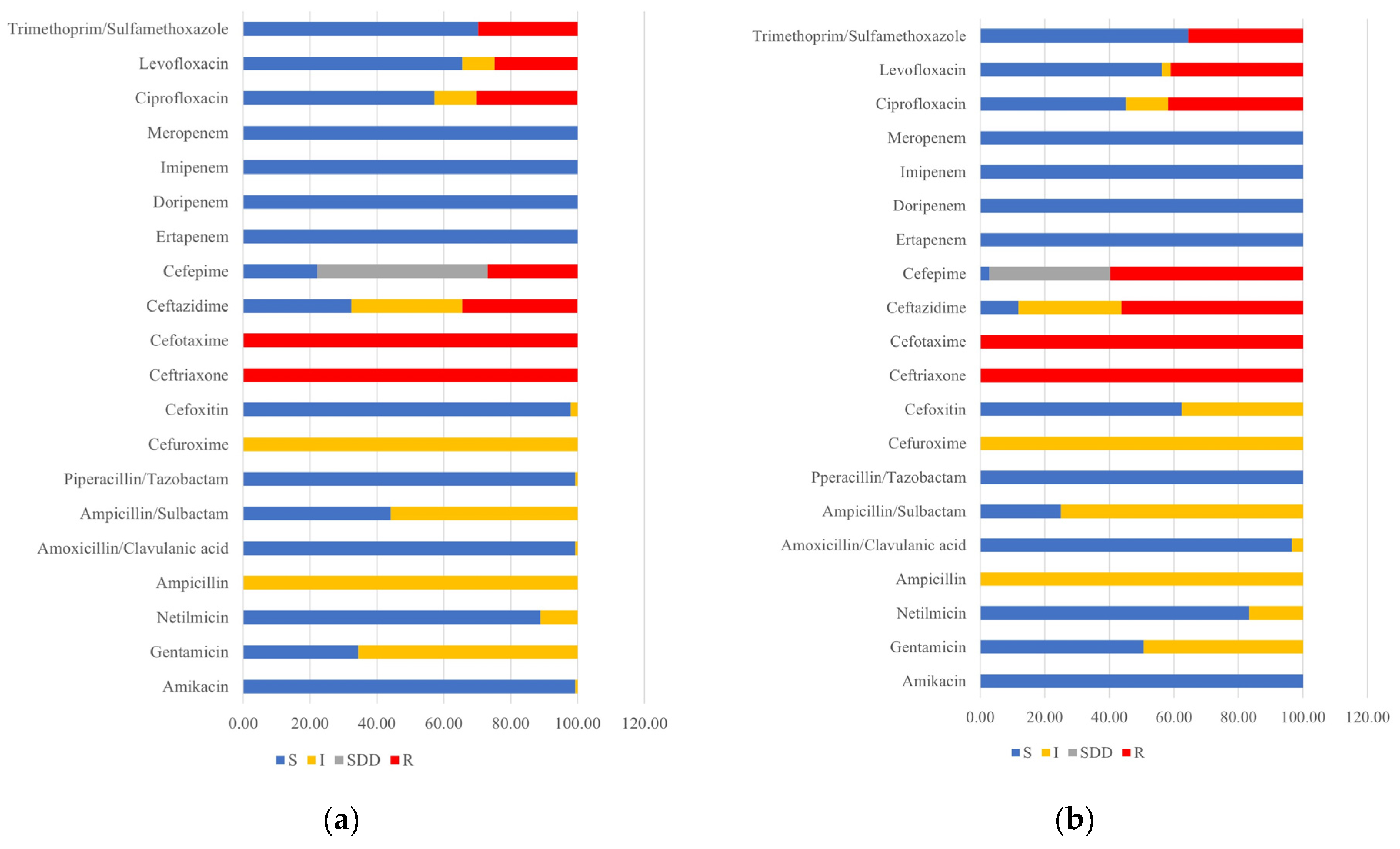
Figure 1. Percentage antimicrobial susceptibility profiles for (a) E. coli isolated from antibiotic-using farms and (b) E. coli isolated from nonantibiotic-using farms (S: susceptible, I: intermediate, SDD: susceptible-dose dependent, and R: resistant).
Animals 14 02630 g001
Figure 2. Phenotypic detection of ESBL-producing-E. coli isolates in (a) antibiotic-using and (b) nonantibiotic-using pig farms.
Animals 14 02630 g002
Figure 3. Specificity of nPCR for direct blaCTX-M detection. Lane M: 100 bp DNA marker (Bio-helix, Taiwan), Lane C−: negative control, Lane 3: Fecal control, Lanes 4–10: ATCC bacterial strain controls, Lane C+: positive control.
Animals 14 02630 g003
Table 1. Primer sequence and amplicon sizes.
Target GenesPrimer Sequence (5′-3′)Amplicon Size (bp)References
blaCTX-MF-ATGTGCAGYACCAGTAARGTKATGGC593[13]
R-TGGGTRAARTARGTSACCAGAAYCAGCGG
blaTEMF-AGTGCTGCCATAACCATGAGTG431[14]
R-CTGACTCCCC GTCGTGTAGATA
blaOXAF-ATTATCTACAGCAGCGCCAGTG296
R-TGCATCCACGTCTTTGGTG
blaSHVF-GATGAACGCTTTCCCATGATG214
R-CGCTGTTATCGCTCATGGTAA
uspAF-AATGCAGGCTACCCAATCAC162[3]
R-GGTGTTGATCAGCTGACGTG
Table 2. Percentage detection of CTX-M and TEM variant genes in antibiotic- and nonantibiotic-using farms. Statistically significant differences are shown according to the degree of significance.
ESBL VariantsAntibiotic-Using FarmsNonantibiotic-Using Farms
No (%)No (%)
PIG
(n = 28)
A1
(n = 30)
A2
(n = 29)
A3
(n = 32)
D1
(n = 26)
Total
(n = 145)
p-Value *S1
(n = 28)
S2
(n = 29)
ND1
(n = 32)
ND2
(n = 27)
ND3
(n = 28)
Total
(n = 144)
p-Value *
CTX-M-55, TEM-1b12 (42.9)9 (30)-20 (62.5)11 (42.3)52 (35.9)0.40628 (100)29 (100)12 (37.5)12 (44.4)27 (96.4)108 (75)0.406
CTX-M-5513 (46.4)6 (20)20 (69)10 (31.3)15 (57.7)64 (44.1)0.406--19 (59.4)9 (33.3)-28 (19.4)0.406
CTX-M-55, TEM-176--8 (27.6)--8 (5.5)0.406------1.000
CTX-M-55, CTX-M-14------1.000----1 (3.6)1 (0.7)0.406
CTX-M-14, TEM-1b1 (3.6)11 (36.7)1 (3.4)2 (6.3)-15 (10.3)0.406------1.000
CTX-M-142 (7.1)2 (6.7)---4 (2.8)0.406--1 (3.1)1 (3.7)-2 (1.4)0.406
CTX-M-15------1.000---5 (18.5)-5 (3.5)0.406
TEM-1b-2 (6.7)---2 (1.4)0.406------1.000
* Mann–Whitney U test with significance at p-value < 0.05.
Table 3. LOD of sPCR and nPCR for direct detection of blaCTX-M in pig feces from 0 to 8 h.
CFU/mLMethodsIncubation Time
0 h1 h2 h3 h4 h6 h8 h
108sPCR+++++++
nPCR+++++++
107sPCR+w++++++
nPCR+++++++
106sPCR++++++
nPCR+++++++
105sPCR+w+++++
nPCR+w++++++
104sPCR+w+w+++
nPCR+w+++++
103sPCR+w++
nPCR+w++++
102sPCR+w+w
nPCR+w+w+++
101sPCR
nPCR+w+w+
100sPCR
nPCR+w
Plus (+) sign represents the presence of the blaCTX-M amplicon; the minus (−) sign represents the absence of the blaCTX-M amplicon; the weak plus (+w) sign represents the presence of the faint blaCTX-M amplicon.
Table 4. Comparison among mPCR, sPCR, and nPCR for direct detection of the blaCTX-M gene from pig fecal samples.
PCR from E. coli IsolatesDirect Detection from Pig Fecal Samples
mPCRsPCRnPCR
+Total+Total+Total
blaCTX-M +41041950951250125
blaCTX-M950954104111011
Total136013613601361360136
Sensitivity: 30.15% CI 95% [22.58–38.60%]
PPV: 100% CI 95% [91.40–100.00%]
Sensitivity: 69.85% CI 95% [61.40–77.42%] PPV: 100% CI 95% [96.19–100.00%]Sensitivity: 91.91% CI 95% [85.99–95.89%] PPV: 100% CI 95% [97.09–100.00%]
Disclaimer/Publisher’s Note: The statements, opinions and data contained in all publications are solely those of the individual author(s) and contributor(s) and not of MDPI and/or the editor(s). MDPI and/or the editor(s) disclaim responsibility for any injury to people or property resulting from any ideas, methods, instructions or products referred to in the content.