Article

Prevalence and Genetic Variation Investigation of the Pseudorabies Virus in Southwest China

1
College of Veterinary Medicine, Southwest University, Chongqing 400715, China
2
Sichuan Boce Testing Technology Co., Ltd., Chengdu 610023, China
3
Chongqing Academy of Animal Science, Chongqing 408599, China
4
National Center of Technology Innovation for Pigs, Chongqing 402460, China
*
Authors to whom correspondence should be addressed.
These authors contributed equally to this work.
Animals 2024, 14(21), 3103; https://doi.org/10.3390/ani14213103
Submission received: 7 October 2024 / Revised: 13 October 2024 / Accepted: 25 October 2024 / Published: 28 October 2024
(This article belongs to the Special Issue Emerging Infectious Diseases and Their Impact on Animal Health)

Simple Summary

Pseudorabies, a severe acute infectious disease caused by the pseudorabies virus (PRV), is a significant threat to the swine industry, leading to substantial economic losses. This study aimed to assess the prevalence of PRV in southwestern China between 2022 and 2024 to control further spread. Initially, we observed a notably high seropositive rate on a single pig farm, prompting a broader investigation across the region. Fortunately, the overall prevalence of PRV was found to be relatively low, aligning with the patterns observed in other parts of China. We also identified two strains of PRV in tissues of dead pigs. However, the genetic analysis of various PRV genes indicated that the Chinese classical strains remain prevalent, and there is potential for genetic recombination. Consequently, the ongoing surveillance of PRV is essential for an effective disease control and management within the swine industry.

Abstract

In 2022, a significant PRV outbreak in a southwestern China pig farm led to a high incidence of sow abortion. A serological analysis using gE antigen-based ELISA revealed a high prevalence (69.30%) of PRV gE antibodies among the affected pigs, with a significant variation across different pig populations (1.11–76.12%). We collected additional 5552 pig serum samples and 580 pig cerebrospinal fluid (CSF) samples from various pig farms in Southwest China between 2022 and 2024. The seropositive rates for PRV gE antibodies ranged from 2.36% and 8.65% in the serum samples, while the positive detection rates for the PRV gE gene in the cerebrospinal fluid samples, as determined by PCR, were between 1.06% and 2.36%. The PCR analysis and sequencing of the PRV gB, gC, gE, and TK genes from eight randomly selected samples identified two distinct strains, CQ1 and CQ2. CQ1’s gC gene showed similarity to the vaccine strain Bartha, while the other genes aligned with Chinese classical strains, suggesting its potential genetic recombination. CQ2 aligned with the Chinese classical strain SC. Although the overall PRV infection in Southwest China’s pig farms is relatively low, occasional outbreaks with high positivity rates are observed. These findings highlight the necessity for increased surveillance and stringent control measures to safeguard the swine industry.

1. Introduction

Pseudorabies (PR), also known as Aujeszky’s disease (AD), is caused by PRV, a significant pathogen of the Alphaherpesvirinae subfamily within the Herpesviridae family. PRV is an enveloped linear double-stranded DNA virus, with a total length of roughly 145 kb, and is recognized for its neurotropic and lymphotropic properties [1,2]. This acute infectious disease can lead to neurological disorders in susceptible animals, with pigs identified as the primary natural reservoir [3]. Moreover, PRV has the capacity to infect a broad range of mammals, including but not limited to mice, cats, foxes, dogs, wolves, and cattle [4,5,6,7,8]. Of particular concern, PRV has been detected and isolated from the cerebrospinal fluid samples of humans [9,10]. The affected patients exhibited respiratory dysfunction and acute neurological symptoms [11,12], indicating the potential for zoonotic transmission. The likelihood of PRV jumping from animals to humans, along with its propensity for mutation and its capacity to co-infect with other pathogens, heightens the complexity of developing effective disease prevention strategies [13].
Clinically, PRV’s impact is age-dependent, with piglets succumbing to severe and debilitating central nervous system disorders post-infection, often proving fatal, with a mortality of 100% [14,15]. In contrast, fattening pigs exhibit respiratory symptoms, while pregnant sows are prone to reproductive disorders and abortion [16]. PRV was first detected in the United States and has since spread to Canada, China, and several other European countries, according to WOAH reports from 2018 to 2024 [17,18]. Many provinces across China have documented the PRV disease, which resulted in substantial economic losses to the swine industry, such as mortality in piglets, abortions in sows, and stunted growth in fattening pigs [14,19]. The introduction of Hungary’s Bartha-K61 gE-deleted live vaccine during the late 1970s marked a significant milestone in the prevention and control of PRV [20]. This vaccine, along with gE ELISA serological testing, has played a significant role in managing PRV infections [21]. From the 1990s to late 2011, over 80% of pigs in China were vaccinated with Bartha-K61 [22], significantly enhancing the country’s ability to prevent and control PRV [20]. However, since 2011, the disease has re-emerged in vaccinated farms, posing a renewed and substantial threat to the pig industry in China [23]. The effectiveness of the Bartha-K61 vaccine against PRV has been notable, but the resurgence of the virus emphasizes the need for continuous research in vaccine development. It also highlights the importance of using serological testing for ongoing disease surveillance to track viral adaptations.
The gE and TK genes of PRV are pivotal in its pathogenicity, with their encoded proteins playing essential roles in disease progression [24,25]. The use of the gE-deleted vaccine is now widespread, making the gE gene a diagnostic marker to distinguish between pigs naturally infected with PRV and those that are vaccinated [26]. Additionally, the gB and gC glycoproteins are recognized as key immunogenic proteins that can induce neutralizing antibodies and activate cellular immune responses. The genetic study of these four proteins is fundamental for understanding PRV’s evolution and holds promise for the development of subunit vaccines [27]. Recent research has discovered that the wild PRV strain has the potential to recombine with the gB and gC genes of the Bartha-K61 vaccine strain, resulting in the emergence of novel recombinant strains with heightened virulence [28]. This recombination event could undermine the effectiveness of conventional vaccinations and challenge the cross-immunity they are designed to provide [29]. The spread of these new PRV variants has been rapid and has caused significant economic losses to China’s swine industry.
Consequently, from 2022 to 2024, we carried out a longitudinal study by collecting samples from a range of pig farms in Southwest China to monitor the prevalence and dissemination of PRV. Concurrently, we conducted a genetic analysis to understand the partial genetic evolution of the virus. The main goal of this study was to enhance our understanding of PRV’s molecular epidemiology, which will be instrumental in developing effective prevention and control strategies tailored to the region.

2. Materials and Methods

2.1. Sample Collection

In 2022, we conducted a study on a pig farm in Southwest China that had experienced a severe sow abortion incident. We collected 469 serum samples from pigs across different age groups and 6 CSF samples from deceased pigs. To further understand the prevalence of PRV in Southwest China, we expanded our collection to include 3757 blood samples and 283 CSF samples from a diverse range of pig farms across the region over the same year. Continuing our surveillance, between 2023 and 2024, we conducted a comprehensive collection of 1795 serum samples and 297 lumbosacral site puncture CSF samples [30] from pig farms in the region for PRV testing. All the large-scale farms under surveillance adhered to a farrow-to-finish strategy and maintained a regular immunization schedule with the Bartha-K61 live vaccine. Importantly, the collection of samples was carefully timed, taking place within 1–2 days after the initial clinical signs of disease were observed by local veterinarians.

2.2. Detection of PRV gE Antibody by ELISA

The serum samples were centrifuged at 13,400× g for 10 min to pellet the cells and debris, allowing for the collection of the supernatant for PRV gE-based ELISA analysis. The ELISA was performed in strict accordance with the manufacturer’s instructions for the PRV gE ELISA antibody detection kit (Cat No. MM22801, Manman, Shanghai, China).

2.3. Detection of PRV Antigen via PCR

All CSF samples were subjected to viral DNA genome extraction using a viral genomic DNA/RNA kit (Cat No. DP304-02, Tiangen, Beijing, China). Subsequently, specific detection primers for PRV-gE were used to amplify and identify the presence of the wild-type PRV pathogen in the samples by detecting the expected amplicon of approximately 250 bp. The amplification was carried out using Takara LA Taq® polymerase with GC Buffer (Cat No. RR02AG, Takara Bio, Beijing, China) to ensure the accuracy of the amplification.

2.4. Partial Genome Sequencing

To amplify PRV’s key genes, including gB, gC, gE, and TK, we designed specific primers with the Oligo 6.0 software, aligned with the gene sequence of the PRV reference strain (GenBank No. KT809429.1) (Table 1). The gB gene was divided into three segments: gB1, gB2, and gB3. Additional primer pairs targeting gE2, gB1, gB2, gB3, gC, and TK were used to amplify the genomic regions encoding these proteins. After amplification, fragments were recovered using a commercial kit from Tiangen (Cat No. DP204-03, Tiangen, Beijing, China), and the purified DNA was ligated into the pMD-19T vector (Cat No. 6013, Takara Bio, Beijing, China). This was followed by the transformation and identification of the bacterial suspension. Once confirmed, bacterial liquid samples were collected and submitted to Sangon Biotech (Shanghai, China) for sequencing [31].

2.5. Phylogenetic and Genomic Analysis

For the genetic analysis of the identified PRV strains, we utilized the SeqMan software within the DNASTAR platform (7.1.0.44) to assemble the sequences. These sequences were then compared with the gB, gC, gE, and TK genes of the PRV reference strains obtained from GenBank, as detailed in Table 2, using the Megalign software (7.1.0.44). To explore the genetic evolution, the MEGA11 software was utilized to construct a phylogenetic tree. The tree was generated using the neighbor-joining method, validated with a bootstrap value of 1000 replicates, and the p-distance substitution model was applied to illustrate the genetic relationships among the strains.

3. Results

3.1. Prevalence Analysis of PRV in Southwest China’s Pig Populations

In a pig farm experiencing sow abortions, we conducted an ELISA-based analysis on 469 serum samples. We found that 325 samples tested positive for PRV gE antibodies, yielding an overall positivity rate of 69.30%. Notably, the infection rate varied among different pig populations: breeding pigs had the lowest positive rate at 1.11% (1/9); suckling piglets at 53.03% (35/66); nursery pigs at 54.35% (25/46); nursing sows at 75.00% (60/80); and pregnant sows exhibited the highest rate of 76.12% (204/268), as detailed in Table 3. Additionally, the PCR-based analysis revealed that three out of six CSF samples were positive for the PRV gE gene, resulting in an antigen positivity rate of 50%.
To extend our investigation to additional pig farms across Southwest China, we conducted a detailed analysis of samples collected in 2022, comprising 3757 serum samples and 283 CSF samples. We identified 209 serum samples positive for PRV gE antibodies, resulting in a positivity rate of 5.56%. Moreover, three tissue samples tested positive for PRV gE antigen, yielding a positivity rate of 1.06%. Our study was further expanded to encompass the years of 2023 and 2024, during which we gathered serum and CSF samples from a variety of pig farms in the region. In 2023, 115 out of 1330 serum samples were positive for gE antibodies, with a positivity rate of 8.65%. Additionally, 7 out of 297 CSF samples were positive for gE antigen, with a positivity rate of 2.36%. In 2024, we observed a seropositivity rate of 2.36%, with 11 out of 465 serum samples testing positive (Table 4).

3.2. PCR Detection of PRV Antigen

Utilizing the specific gE1 primers, we performed PCR amplification to detect PRV antigen. The PCR yielded distinct bands of the anticipated 250 bp size in certain samples, confirming the presence of PRV gE gene and infection with the PRV strain (Figure 1). Following this, we conducted a comprehensive PCR amplification of the gB, gC, gE, and TK genes using a selection of samples. Eight samples were randomly chosen for this analysis. Notably, seven samples exhibited identical sequences and were collectively designated as the CQ1 strain. In contrast, the eighth sample showed a unique sequence, distinguishing it as a separate strain, named CQ2. All sequences were provided in the Supplementary Materials.

3.3. Variation Analysis of gB Gene of Epidemic PRV Strains

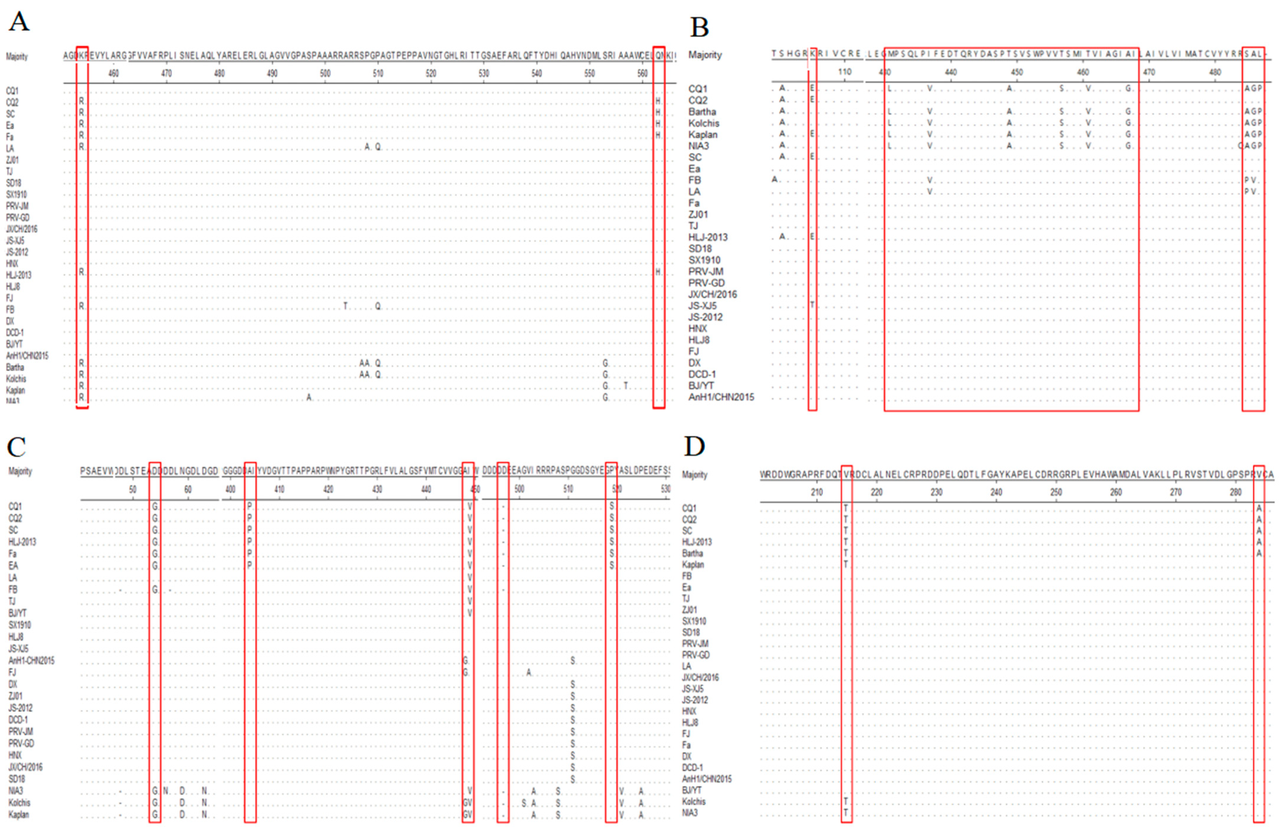
The gB gene sequence of PRV CQ1 strain exhibited nucleotide homology ranging from 98.2% to 99.9% when compared to both Chinese and international reference strains (Table 2). The gB gene of CQ2 strain showed a complete nucleotide identity with the SC strain, a Chinese classic strain isolated in 1986 [32]. A comparative analysis revealed several distinct mutations in the gB gene of the CQ1 strain relative to the SC strain. Specifically, a nucleotide substitution at position 1361 from guanine (G) to adenine (A) resulted in an amino acid change from Arg to Lys. Additionally, a substitution at position 1689 from G to thymine (T) led to a change from His to Glu. The remaining variable amino acid sites in the CQ1 strain’s gB gene were found to be identical to those of the SC strain (Figure 2A).
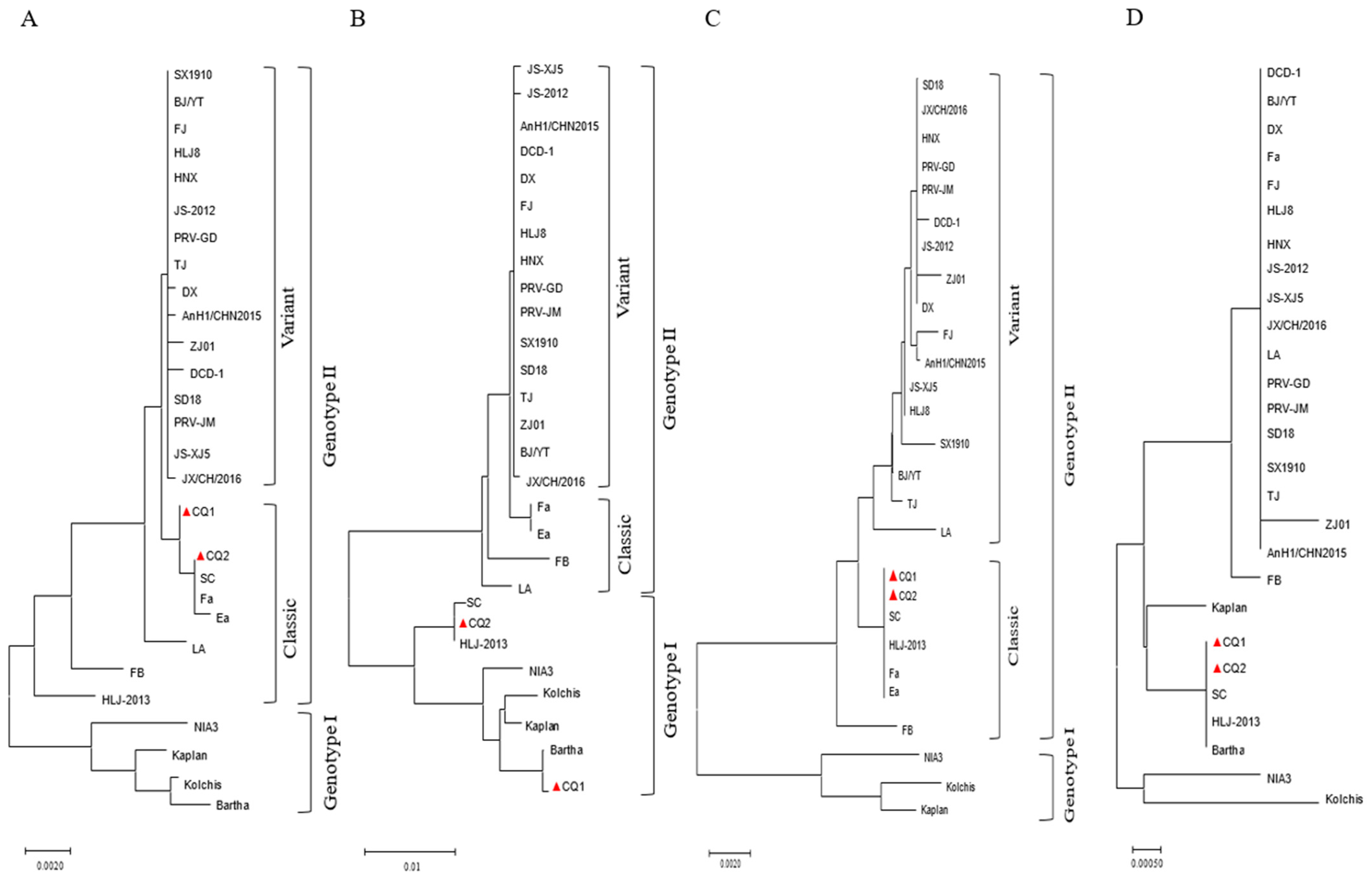
The phylogenetic tree analysis based on the gB gene categorized Chinese strains as genotype II, whereas international strains like Bartha and Kaplan were classified under genotype I (Figure 3A). Within genotype II of PRV, two distinct sub-branches were identified, which lack genetic interrelation. The CQ1 strain was found in one of these sub-branches of genotype II and showed a close genetic affinity with the CQ2 strain and other classical Chinese strains, such as SC, Fa, and Ea. These latter strains were isolated in China between the 1980s and 1990s [22,33]. Conversely, the other subcategory of genotype II encompassed Chinese variant strains such as TJ and JS-2012 that emerged after 2011 [34,35].

3.4. Variation Analysis of gC Gene of Epidemic PRV Strains

The gC gene sequence of the CQ1 strain exhibited a nucleotide homology ranging from 95.4% to 99.9% when compared to the reference strains (Table 2). Notably, the gC gene of the CQ1 strain exhibited the highest genetic similarity with the Bartha strain. Specifically, only a nucleotide change at position 316 from A to G resulted in an amino acid substitution from Lys to Glu. In contrast, the gC gene of the CQ2 strain showed complete nucleotide identity with that of the SC strain. Furthermore, the C-terminus of CQ1’s gC gene is the primary distinction from the CQ2 strain, with nine distinct amino acid differences. These include substitutions such as 431Met to Leu, 437Ile to Val, 449Thr to Ala, 457Thr to Ser, 460Ile to Val, 467Ala to Gly, and 485Ser-Ala-Leu to Arg-Gly-Pro (Figure 2B).
The phylogenetic tree analysis based on the gC gene categorized the PRV strains into two genotypes: genotype I and genotype II. The Chinese strains of PRV were grouped into genotype II, whereas genotype I was primarily composed of international strains (Figure 3B). Interestingly, within genotype I, the CQ1 and CQ2 strains were situated on separate sub-branches. The CQ1 strain was closely related to the Bartha strain on the same sub-branch. In contrast, the CQ2 strain was part of a sub-branch that also included the SC strain and HLJ-2013 strain, which has been circulating since 2013 [36].

3.5. Variation Analysis of gE Gene of Epidemic PRV Strains

The gE gene sequences of CQ1 and CQ2 strains were found to be identical, showing 100% similarity with the Chinese classic strains SC and Ea. A comparison of the nucleotide sequence with other Chinese and international reference strains revealed a high degree of similarity, ranging from 97.4% to 100%. In contrast to the CQ1 and CQ2 strains, the Chinese variant strains contained several mutations. Specifically, a nucleotide change at position 161 from G to A resulted in an amino acid transition from Gly to Asp. Additionally, a base alteration at position 1210 from C to G led to an amino acid substitution from Pro to Ala. At position 1345, a change from G to A substituted Val with Ile. An insertion of the nucleotide sequence CGA at positions 1491–1493 introduced a new amino acid Asp at position 497. Furthermore, a nucleotide change at position 1555 from T to C resulted in the substitution of the amino acid Ser with Pro (Figure 2C).
The phylogenetic tree analysis based on the gE gene delineated the PRV strains into two distinct genotypes: genotype I and genotype II. Genotype I predominantly comprised international strains such as Kaplan and Kolchis, while genotype II was primarily composed of Chinese strains (Figure 3C). This categorization is similar to the genetic evolution tree based on the gB gene. Within genotype II of PRV, there were two distinct sub-branches. The CQ1 and CQ2 strains were found to cluster together with the classical Chinese strains SC, Fa, Ea, and HLJ-2013. This group of strains formed one of the sub-branches. The other sub-branch mainly encompassed Chinese variant strains such as TJ, JS-2012, and others.

3.6. Variation Analysis of TK Gene of Epidemic PRV Strains

The TK gene sequences of the CQ1 and CQ2 strains were identical, showing 100% homology with the SC and HLJ-2013 strains, as well as with the Bartha strain. However, they did not share this level of homology with other classical Chinese strains, such as the Fa strain. The TK gene of the CQ1 and CQ2 strains exhibited nucleotide homology ranging from 99.5% to 100% with other reference strains, including both Chinese and international strains. A detailed comparative analysis with Chinese variant strains identified specific variations within the CQ1 and CQ2 strains. Notably, at positions 643 and 644, the GT nucleotide sequence was replaced by AC, leading to an amino acid change from Val to Thr. Additionally, at position 851, the replacement with C for T resulted in the substitution of Ala with Thr (Figure 2D).
The TK gene of PRV was more conserved than the gE, gB, and gC genes, making it more challenging to cluster based on the phylogenetic tree. For this reason, the TK genotype was not indicated in Figure 3D. Furthermore, the phylogenetic tree analysis confirmed a close relationship between the CQ1 and CQ2 strains and the Bartha and SC strains, indicating a strong evolutionary link (Figure 3D).

4. Discussion

4.1. Prevalence and Economic Impact of PRV Infections in Southwest China: Comparative Analysis and the Imperative for Enhanced Surveillance

Since the second half of 2011, PRV variant strains have been spreading throughout China’s pig farms, resulting in massive economic losses for the nation’s swine industry [29,37,38]. In 2022, a significant sow abortion event occurred at a large-scale pig farm in Chongqing, China. Serological analysis revealed a remarkably high average prevalence rate of PRV on the farm, reaching 69.30%. Particularly, pregnant sows exhibited the highest infection rate. However, the latest data on the seroprevalence of PRV gE antibodies at the individual animal level in China showed a relatively lower rate of 12.36% [39]. Despite this, the high positivity rate observed in our study has raised serious concerns regarding the potential for a widespread PRV epidemic across Southwest China.
To validate this perspective, we conducted an extensive collection of serum and CSF samples from large-scale pig farms across Southwest China from 2022 to 2024. Our research indicates that, in this region, the overall positive rate of PRV has been generally at a low level. This finding is corroborated by previous studies. A study by Sun Y et al. [40], conducted across 27 provinces in China from 2012 to 2017, reported an average positive rate of 8.27% among the samples tested. Similarly, Zhou H et al. [41] identified a 16.3% positive rate for PRV gE antibodies in serum samples from large-scale pig farms in Heilongjiang Province, Northeast China, during the period from 2013 to 2018. Additionally, Lin Y et al. [42] found a 23.55% incidence of positive cases in vaccinated pig farms in Hunan Province, Central China, from 2016 to 2020. These studies collectively indicate that the prevalence of PRV in Southwest China is comparable to the national average, providing a broader context for understanding the regional epidemiology of PRV infection.
Our research shows that PRV infections, leading to abortions at local pig farms, are still prevalent despite vaccinations. The virus might enter farms through various routes, including infected pigs, contaminated equipment, vehicles, or wildlife [17,43]. It might also be introduced by workers’ clothing or footwear [44]. Once inside, this virus can quickly spread in the close quarters of a pig farm, especially where biosecurity is lax. This underscores the importance of stringent surveillance and biosecurity to prevent PRV outbreaks. The differing infection rates likely result from varying farm management practices and control strategies.

4.2. Molecular Characterization of PRV Strains in Southwest China: Implications for Vaccine Efficacy and Disease Management

Previous studies have highlighted the diversity of PRV strains across China [14,41,42,45]. Our survey reinforces this notion, indicating that the prevalent outbreak in Southwest China is largely attributed to the Chinese classical strains classified under genotype II. Through conducting a sequence alignment analysis of the gB, gC, gE, and TK genes from the field samples, we identified two distinct PRV strains, CQ1 and CQ2. Notably, the gE and gB genes of CQ1 resemble to the domestic classic strain SC, whereas its gC gene displays a high degree of similarity to the Bartha strain. This genetic pattern suggests the potential occurrence of recombination events during the PRV pandemic, as reported by multiply research groups [46,47,48]. Although these four genes’ sequences of the CQ2 strain were found to be identical to those of the SC strain, current PRV vaccines, which are effective against classical strains, have been found lacking in preventing infections in local pig farms. This suggests that the existing vaccines may not be fully effective against the new recombinant forms of PRV. These findings underscore the need for a re-evaluation of current vaccination strategies and the development of more comprehensive and adaptive vaccines capable of addressing the evolving genetic landscape of PRV.
The further sequencing analysis of the CQ1 strain has revealed mutations within the gB and gC genes. The gB gene, which encodes an envelope glycoprotein crucial for PRV fusion with host cell membrane during infection [49], has mutations similar to those in Chinese variant strains. These genetic variations in the gB gene are known to influence the immunogenicity of PRV [50], suggesting that they might contribute to the increased virulence of PRV and its ability to evade the immune defenses induced by the vaccine strain Bartha [51]. The gC facilitates the initial attachment of PRV to the cell surface [52]. The gC gene of the CQ1 strain exhibits 99.9% similarity to that of the Bartha strain, with only minor nucleotide discrepancy. This high degree of genetic similarity, along with the mutations in the gB gene, suggests that the CQ1 strain might be a recombinant of both classical and vaccine strains, aligning with the previous reports [53,54]. This hypothesis is further supported by the resemblance of its TK, gB, and gE genes to those of the SC strain. In contrast, the gB, gC, gE, and TK genes of CQ2 exhibit complete homology with the corresponding genes of the SC strain, which was prevalent in the 1980s and 1990s [55]. However, it is challenging to conclusively determine whether the CQ2 strain is the original SC strain due to limitations in sample size, which precluded obtaining the full-length sequence. This constraint prevents a comprehensive genetic analysis and necessitates further investigation. Our findings suggest a rich and intricate evolutionary history of PRV strains, which could have profound effects on the efficacy of current vaccines and virus containment strategies. While the high genetic similarity between CQ2 and the historical SC strain is intriguing, the complete genetic profile and evolutionary path of CQ2 still need to be delineated.
Our survey indicates that the SC strain is not the only variant present in Southwest China, as other wild strains are also circulating. This finding adds to the already complex epidemiological scenario of PRV, emphasizing the critical need for sustained and vigilant surveillance by local pig farmers. Such vigilance is essential for gathering insights that will guide the development of effective disease management strategies tailored to the dynamic genetic landscape of PRV in the region.

5. Conclusions

Our findings suggest that the prevalence of PRV in the region is relatively low, with the circulating strains identified as the classical strains. However, serological data indicate that, despite extensive vaccination efforts on large-scale farms with the current PRV vaccine, these farms remain vulnerable to wild PRV infections. This implies that the existing PRV vaccine may exhibit limited efficacy against the circulating epidemic strains. In light of these findings, there is an urgent need to develop novel vaccines specifically targeting the epidemic strains. Additionally, it is crucial to strengthen PRV surveillance on pig farms, enforce epidemic prevention measures, and work systematically toward the local eradication of PRV through ongoing serological monitoring and detection.

Supplementary Materials

The following supporting information can be downloaded at: https://www.mdpi.com/article/10.3390/ani14213103/s1. The sequences of this study were deposited in GenBank with the accession numbers PQ232108-PQ232115. The sequences will be released to public databases when the data or accession numbers appear in print. The sequence data are supplied in the Supplementary Files.

Author Contributions

Conceptualization, J.W. and Y.W.; Methodology, J.W., J.Z. (Juan Zhang), X.W. and R.Y.; Software, J.W. and Y.L.; Validation, J.Z. (Juan Zhang), X.W. and N.Y.; Formal Analysis, J.W. and J.Z. (Juan Zhang); Investigation, J.W., J.Z. (Juan Zhang), J.Z. (Jun Zhou), R.Y., M.J. and N.Y.; Data Curation, J.W.; Writing—Original Draft Preparation, J.W.; Writing—Review and Editing, Y.W., J.Z. (Junhai Zhu) and L.F.; Visualization, J.Z. (Jun Zhou); Supervision, N.Y., L.Z. and Y.W.; Project Administration, Y.W.; Funding Acquisition, L.F. All authors have read and agreed to the published version of the manuscript.

Funding

This study was supported by Grants from the National Center of Technology Innovation for Pigs (Grant no: NCTIP-XD/C09), the Natural Science Foundation of Chongqing (Grant no. CSTB2024NSCQ-MSX0467), and the Fundamental Research Funds for the Central Universities (SWU-KR22036).

Institutional Review Board Statement

The study was conducted in accordance with the Experimental Animal Center of Southwest University. Approval number SWU_LAC-2022100222. The study was conducted in accordance with the local legislation and institutional requirements.

Informed Consent Statement

Not applicable.

Data Availability Statement

The raw data supporting the conclusions of this article will be made available by the authors upon request.

Acknowledgments

We thank the members of our laboratory and staffs in the pig farm for their assistance in the research.

Conflicts of Interest

Author Jun Zhou was employed by the company Sichuan Boce Testing Technology Co., Ltd. The remaining authors declare that the research was conducted in the absence of any commercial or financial relationships that could be construed as a potential conflict of interest.

References

  1. Zheng, H.H.; Fu, P.F.; Chen, H.Y.; Wang, Z.Y. Pseudorabies Virus: From Pathogenesis to Prevention Strategies. Viruses 2022, 14, 1638. [Google Scholar] [CrossRef] [PubMed]
  2. Bo, Z.; Zhu, J.; Guo, M.; Zhang, C.; Cao, Y.; Zhang, X.; Wu, Y. Gallocatechin Gallate Inhibits the Replication of Pseudorabies Virus via Suppressing the Entry and Release Stages in Its Replication Cycle. Vet. Sci. 2023, 10, 189. [Google Scholar] [CrossRef] [PubMed]
  3. Lu, J.J.; Yuan, W.Z.; Zhu, Y.P.; Hou, S.H.; Wang, X.J. Latent pseudorabies virus infection in medulla oblongata from quarantined pigs. Transbound. Emerg. Dis. 2021, 68, 543–551. [Google Scholar] [CrossRef] [PubMed]
  4. Hagemoser, W.A.; Kluge, J.P.; Hill, H.T. Studies on the pathogenesis of pseudorabies in domestic cats following oral inoculation. Can. J. Comp. Med. 1980, 44, 192–202. [Google Scholar]
  5. Jin, H.L.; Gao, S.M.; Liu, Y.; Zhang, S.F.; Hu, R.L. Pseudorabies in farmed foxes fed pig offal in Shandong province, China. Arch. Virol. 2016, 161, 445–448. [Google Scholar] [CrossRef]
  6. Zhang, L.; Zhong, C.; Wang, J.; Lu, Z.; Liu, L.; Yang, W.; Lyu, Y. Pathogenesis of natural and experimental Pseudorabies virus infections in dogs. Virol. J. 2015, 12, 44. [Google Scholar] [CrossRef]
  7. Lian, K.; Zhang, M.; Zhou, L.; Song, Y.; Wang, G.; Wang, S. First report of a pseudorabies-virus-infected wolf (Canis lupus) in China. Arch. Virol. 2020, 165, 459–462. [Google Scholar] [CrossRef]
  8. McCracken, R.M.; McFerran, J.B.; Dow, C. The neural spread of pseudorabies virus in calves. J. Gen. Virol. 1973, 20, 17–28. [Google Scholar] [CrossRef]
  9. Liu, Q.; Wang, X.; Xie, C.; Ding, S.; Yang, H.; Guo, S.; Li, J.; Qin, L.; Ban, F.; Wang, D.; et al. A Novel Human Acute Encephalitis Caused by Pseudorabies Virus Variant Strain. Clin. Infect. Dis. 2021, 73, e3690–e3700. [Google Scholar] [CrossRef]
  10. Ai, J.W.; Weng, S.S.; Cheng, Q.; Cui, P.; Li, Y.J.; Wu, H.L.; Zhu, Y.M.; Xu, B.; Zhang, W.H. Human Endophthalmitis Caused By Pseudorabies Virus Infection, China, 2017. Emerg. Infect. Dis. 2018, 24, 1087–1090. [Google Scholar] [CrossRef]
  11. Yang, X.; Guan, H.; Li, C.; Li, Y.; Wang, S.; Zhao, X.; Zhao, Y.; Liu, Y. Characteristics of human encephalitis caused by pseudorabies virus: A case series study. Int. J. Infect. Dis. 2019, 87, 92–99. [Google Scholar] [CrossRef] [PubMed]
  12. Yang, H.; Han, H.; Wang, H.; Cui, Y.; Liu, H.; Ding, S. A Case of Human Viral Encephalitis Caused by Pseudorabies Virus Infection in China. Front. Neurol. 2019, 10, 534. [Google Scholar] [CrossRef] [PubMed]
  13. Liu, X.; Zhou, Y.; Luo, Y.; Chen, Y. Effects of gE/gI deletions on the miRNA expression of PRV-infected PK-15 cells. Virus Genes 2020, 56, 461–471. [Google Scholar] [CrossRef]
  14. Zheng, H.H.; Jin, Y.; Hou, C.Y.; Li, X.S.; Zhao, L.; Wang, Z.Y.; Chen, H.Y. Seroprevalence investigation and genetic analysis of pseudorabies virus within pig populations in Henan province of China during 2018–2019. Infect. Genet. Evol. 2021, 92, 104835. [Google Scholar] [CrossRef]
  15. Xiang, S.; Zhou, Z.; Hu, X.; Li, Y.; Zhang, C.; Wang, J.; Li, X.; Tan, F.; Tian, K. Complete Genome Sequence of a Variant Pseudorabies Virus Strain Isolated in Central China. Genome Announc. 2016, 4, e00149-16. [Google Scholar] [CrossRef]
  16. Yao, J.; Li, J.; Gao, L.; He, Y.; Xie, J.; Zhu, P.; Zhang, Y.; Zhang, X.; Duan, L.; Yang, S.; et al. Epidemiological Investigation and Genetic Analysis of Pseudorabies Virus in Yunnan Province of China from 2017 to 2021. Viruses 2022, 14, 895. [Google Scholar] [CrossRef] [PubMed]
  17. Liu, Q.; Kuang, Y.; Li, Y.; Guo, H.; Zhou, C.; Guo, S.; Tan, C.; Wu, B.; Chen, H.; Wang, X. The Epidemiology and Variation in Pseudorabies Virus: A Continuing Challenge to Pigs and Humans. Viruses 2022, 14, 1463. [Google Scholar] [CrossRef]
  18. OIE. OIE World Animal Health Information System. Available online: https://wahis.oie.int/#/dashboards/ (accessed on 30 September 2024).
  19. Sun, Y.; Luo, Y.; Wang, C.H.; Yuan, J.; Li, N.; Song, K.; Qiu, H.J. Control of swine pseudorabies in China: Opportunities and limitations. Vet. Microbiol. 2016, 183, 119–124. [Google Scholar] [CrossRef]
  20. Bo, Z.; Li, X. A Review of Pseudorabies Virus Variants: Genomics, Vaccination, Transmission, and Zoonotic Potential. Viruses 2022, 14, 1003. [Google Scholar] [CrossRef]
  21. Yu, X.; Zhou, Z.; Hu, D.; Zhang, Q.; Han, T.; Li, X.; Gu, X.; Yuan, L.; Zhang, S.; Wang, B.; et al. Pathogenic pseudorabies virus, China, 2012. Emerg. Infect. Dis. 2014, 20, 102–104. [Google Scholar] [CrossRef]
  22. Tong, G.; Chen, H. Pseudorabies epidemic status and control measures in China. Chin. J. Vet. Sci. 1999, 19, 1–2. [Google Scholar]
  23. An, T.Q.; Peng, J.M.; Tian, Z.J.; Zhao, H.Y.; Li, N.; Liu, Y.M.; Chen, J.Z.; Leng, C.L.; Sun, Y.; Chang, D.; et al. Pseudorabies virus variant in Bartha-K61-vaccinated pigs, China, 2012. Emerg. Infect. Dis. 2013, 19, 1749–1755. [Google Scholar] [CrossRef] [PubMed]
  24. Mulder, W.A.; Jacobs, L.; Priem, J.; Kok, G.L.; Wagenaar, F.; Kimman, T.G.; Pol, J.M. Glycoprotein gE-negative pseudorabies virus has a reduced capability to infect second- and third-order neurons of the olfactory and trigeminal routes in the porcine central nervous system. J. Gen. Virol. 1994, 75 Pt 11, 3095–3106. [Google Scholar] [CrossRef]
  25. Ferrari, M.; Mettenleiter, T.C.; Romanelli, M.G.; Cabassi, E.; Corradi, A.; Dal Mas, N.; Silini, R. A comparative study of pseudorabies virus (PRV) strains with defects in thymidine kinase and glycoprotein genes. J. Comp. Pathol. 2000, 123, 152–163. [Google Scholar] [CrossRef]
  26. Li, J.; Fang, K.; Rong, Z.; Li, X.; Ren, X.; Ma, H.; Chen, H.; Li, X.; Qian, P. Comparison of gE/gI- and TK/gE/gI-Gene-Deleted Pseudorabies Virus Vaccines Mediated by CRISPR/Cas9 and Cre/Lox Systems. Viruses 2020, 12, 369. [Google Scholar] [CrossRef]
  27. Freuling, C.M.; Muller, T.F.; Mettenleiter, T.C. Vaccines against pseudorabies virus (PrV). Vet. Microbiol. 2017, 206, 3–9. [Google Scholar] [CrossRef]
  28. He, W.; Auclert, L.Z.; Zhai, X.; Wong, G.; Zhang, C.; Zhu, H.; Xing, G.; Wang, S.; He, W.; Li, K.; et al. Interspecies Transmission, Genetic Diversity, and Evolutionary Dynamics of Pseudorabies Virus. J. Infect. Dis. 2019, 219, 1705–1715. [Google Scholar] [CrossRef]
  29. Ma, Z.; Han, Z.; Liu, Z.; Meng, F.; Wang, H.; Cao, L.; Li, Y.; Jiao, Q.; Liu, S.; Liu, M. Epidemiological investigation of porcine pseudorabies virus and its coinfection rate in Shandong Province in China from 2015 to 2018. J. Vet. Sci. 2020, 21, e36. [Google Scholar] [CrossRef] [PubMed]
  30. Ballesteros, C.; Pouliot, M.; Froment, R.; Maghezzi, M.S.; St-Jean, C.; Li, C.; Paquette, D.; Authier, S. Cerebrospinal Fluid Characterization in Cynomolgus Monkeys, Beagle Dogs, and Gottingen Minipigs. Int. J. Toxicol. 2020, 39, 124–130. [Google Scholar] [CrossRef]
  31. Song, C.; Ye, H.; Zhang, X.; Zhang, Y.; Li, Y.; Yao, J.; Gao, L.; Wang, S.; Yu, Y.; Shu, X. Isolation and Characterization of Yunnan Variants of the Pseudorabies Virus and Their Pathogenicity in Rats. Viruses 2024, 16, 233. [Google Scholar] [CrossRef]
  32. Ye, C.; Guo, J.C.; Gao, J.C.; Wang, T.Y.; Zhao, K.; Chang, X.B.; Wang, Q.; Peng, J.M.; Tian, Z.J.; Cai, X.H.; et al. Genomic analyses reveal that partial sequence of an earlier pseudorabies virus in China is originated from a Bartha-vaccine-like strain. Virology 2016, 491, 56–63. [Google Scholar] [CrossRef] [PubMed]
  33. Hong, W.; Xiao, S.; Zhou, R.; Fang, L.; He, Q.; Wu, B.; Zhou, F.; Chen, H. Protection induced by intramuscular immunization with DNA vaccines of pseudorabies in mice, rabbits and piglets. Vaccine 2002, 20, 1205–1214. [Google Scholar] [CrossRef] [PubMed]
  34. Wang, C.H.; Yuan, J.; Qin, H.Y.; Luo, Y.; Cong, X.; Li, Y.; Chen, J.; Li, S.; Sun, Y.; Qiu, H.J. A novel gE-deleted pseudorabies virus (PRV) provides rapid and complete protection from lethal challenge with the PRV variant emerging in Bartha-K61-vaccinated swine population in China. Vaccine 2014, 32, 3379–3385. [Google Scholar] [CrossRef] [PubMed]
  35. Tong, W.; Liu, F.; Zheng, H.; Liang, C.; Zhou, Y.J.; Jiang, Y.F.; Shan, T.L.; Gao, F.; Li, G.X.; Tong, G.Z. Emergence of a Pseudorabies virus variant with increased virulence to piglets. Vet. Microbiol. 2015, 181, 236–240. [Google Scholar] [CrossRef] [PubMed]
  36. Liu, H.; Shi, Z.; Liu, C.; Wang, P.; Wang, M.; Wang, S.; Liu, Z.; Wei, L.; Sun, Z.; He, X.; et al. Implication of the Identification of an Earlier Pseudorabies Virus (PRV) Strain HLJ-2013 to the Evolution of Chinese PRVs. Front. Microbiol. 2020, 11, 612474. [Google Scholar] [CrossRef]
  37. Zhang, L.; Ren, W.; Chi, J.; Lu, C.; Li, X.; Li, C.; Jiang, S.; Tian, X.; Li, F.; Wang, L.; et al. Epidemiology of Porcine Pseudorabies from 2010 to 2018 in Tianjin, China. Viral Immunol. 2021, 34, 714–721. [Google Scholar] [CrossRef]
  38. Gu, Z.; Hou, C.; Sun, H.; Yang, W.; Dong, J.; Bai, J.; Jiang, P. Emergence of highly virulent pseudorabies virus in southern China. Can. J. Vet. Res. 2015, 79, 221–228. [Google Scholar]
  39. Gao, W.; Jiang, X.; Hu, Z.; Wang, Q.; Shi, Y.; Tian, X.; Qiao, M.; Zhang, J.; Li, Y.; Li, X. Epidemiological investigation, determination of related factors, and spatial-temporal cluster analysis of wild type pseudorabies virus seroprevalence in China during 2022. Front. Vet. Sci. 2023, 10, 1298434. [Google Scholar] [CrossRef]
  40. Sun, Y.; Liang, W.; Liu, Q.; Zhao, T.; Zhu, H.; Hua, L.; Peng, Z.; Tang, X.; Stratton, C.W.; Zhou, D.; et al. Epidemiological and genetic characteristics of swine pseudorabies virus in mainland China between 2012 and 2017. PeerJ 2018, 6, e5785. [Google Scholar] [CrossRef]
  41. Zhou, H.; Pan, Y.; Liu, M.; Han, Z. Prevalence of Porcine Pseudorabies Virus and Its Coinfection Rate in Heilongjiang Province in China from 2013 to 2018. Viral Immunol. 2020, 33, 550–554. [Google Scholar] [CrossRef]
  42. Lin, Y.; Tan, L.; Wang, C.; He, S.; Fang, L.; Wang, Z.; Zhong, Y.; Zhang, K.; Liu, D.; Yang, Q.; et al. Serological Investigation and Genetic Characteristics of Pseudorabies Virus in Hunan Province of China From 2016 to 2020. Front. Vet. Sci. 2021, 8, 762326. [Google Scholar] [CrossRef] [PubMed]
  43. Liu, A.; Xue, T.; Zhao, X.; Zou, J.; Pu, H.; Hu, X.; Tian, Z. Pseudorabies Virus Associations in Wild Animals: Review of Potential Reservoirs for Cross-Host Transmission. Viruses 2022, 14, 2254. [Google Scholar] [CrossRef]
  44. Li, X.D.; Fu, S.H.; Chen, L.Y.; Li, F.; Deng, J.H.; Lu, X.C.; Wang, H.Y.; Tian, K.G. Detection of Pseudorabies Virus Antibodies in Human Encephalitis Cases. Biomed. Environ. Sci. 2020, 33, 444–447. [Google Scholar] [CrossRef]
  45. Gu, J.; Hu, D.; Peng, T.; Wang, Y.; Ma, Z.; Liu, Z.; Meng, F.; Shang, Y.; Liu, S.; Xiao, Y. Epidemiological investigation of pseudorabies in Shandong Province from 2013 to 2016. Transbound. Emerg. Dis. 2018, 65, 890–898. [Google Scholar] [CrossRef] [PubMed]
  46. Bo, Z.; Miao, Y.; Xi, R.; Gao, X.; Miao, D.; Chen, H.; Jung, Y.S.; Qian, Y.; Dai, J. Emergence of a novel pathogenic recombinant virus from Bartha vaccine and variant pseudorabies virus in China. Transbound. Emerg. Dis. 2021, 68, 1454–1464. [Google Scholar] [CrossRef] [PubMed]
  47. Jiang, L.; Cheng, J.; Pan, H.; Yang, F.; Zhu, X.; Wu, J.; Pan, H.; Yan, P.; Zhou, J.; Gao, Q.; et al. Analysis of the recombination and evolution of the new type mutant pseudorabies virus XJ5 in China. BMC Genom. 2024, 25, 752. [Google Scholar] [CrossRef] [PubMed]
  48. Huang, J.; Tang, W.; Wang, X.; Zhao, J.; Peng, K.; Sun, X.; Li, S.; Kuang, S.; Zhu, L.; Zhou, Y.; et al. The Genetic Characterization of a Novel Natural Recombinant Pseudorabies Virus in China. Viruses 2022, 14, 978. [Google Scholar] [CrossRef]
  49. Vallbracht, M.; Brun, D.; Tassinari, M.; Vaney, M.C.; Pehau-Arnaudet, G.; Guardado-Calvo, P.; Haouz, A.; Klupp, B.G.; Mettenleiter, T.C.; Rey, F.A.; et al. Structure-Function Dissection of Pseudorabies Virus Glycoprotein B Fusion Loops. J. Virol. 2018, 92, e01203-17. [Google Scholar] [CrossRef]
  50. Yu, Z.Q.; Tong, W.; Zheng, H.; Li, L.W.; Li, G.X.; Gao, F.; Wang, T.; Liang, C.; Ye, C.; Wu, J.Q.; et al. Variations in glycoprotein B contribute to immunogenic difference between PRV variant JS-2012 and Bartha-K61. Vet. Microbiol. 2017, 208, 97–105. [Google Scholar] [CrossRef]
  51. Hu, R.M.; Zhou, Q.; Song, W.B.; Sun, E.C.; Zhang, M.M.; He, Q.G.; Chen, H.C.; Wu, B.; Liu, Z.F. Novel pseudorabies virus variant with defects in TK, gE and gI protects growing pigs against lethal challenge. Vaccine 2015, 33, 5733–5740. [Google Scholar] [CrossRef]
  52. Mettenleiter, T.C. Immunobiology of pseudorabies (Aujeszky’s disease). Vet. Immunol. Immunopathol. 1996, 54, 221–229. [Google Scholar] [CrossRef] [PubMed]
  53. Ye, C.; Zhang, Q.Z.; Tian, Z.J.; Zheng, H.; Zhao, K.; Liu, F.; Guo, J.C.; Tong, W.; Jiang, C.G.; Wang, S.J.; et al. Genomic characterization of emergent pseudorabies virus in China reveals marked sequence divergence: Evidence for the existence of two major genotypes. Virology 2015, 483, 32–43. [Google Scholar] [CrossRef] [PubMed]
  54. Ren, J.; Wang, H.; Zhou, L.; Ge, X.; Guo, X.; Han, J.; Yang, H. Glycoproteins C and D of PRV Strain HB1201 Contribute Individually to the Escape From Bartha-K61 Vaccine-Induced Immunity. Front. Microbiol. 2020, 11, 323. [Google Scholar] [CrossRef] [PubMed]
  55. Yuan, Q.; Li, Z.; Nan, X.; Wu, Y.; Li, Y. Isolation and identification of pseudorabies virus. Chin. J. Prev. Vet. Med. 1987, 3, 10–11. [Google Scholar]
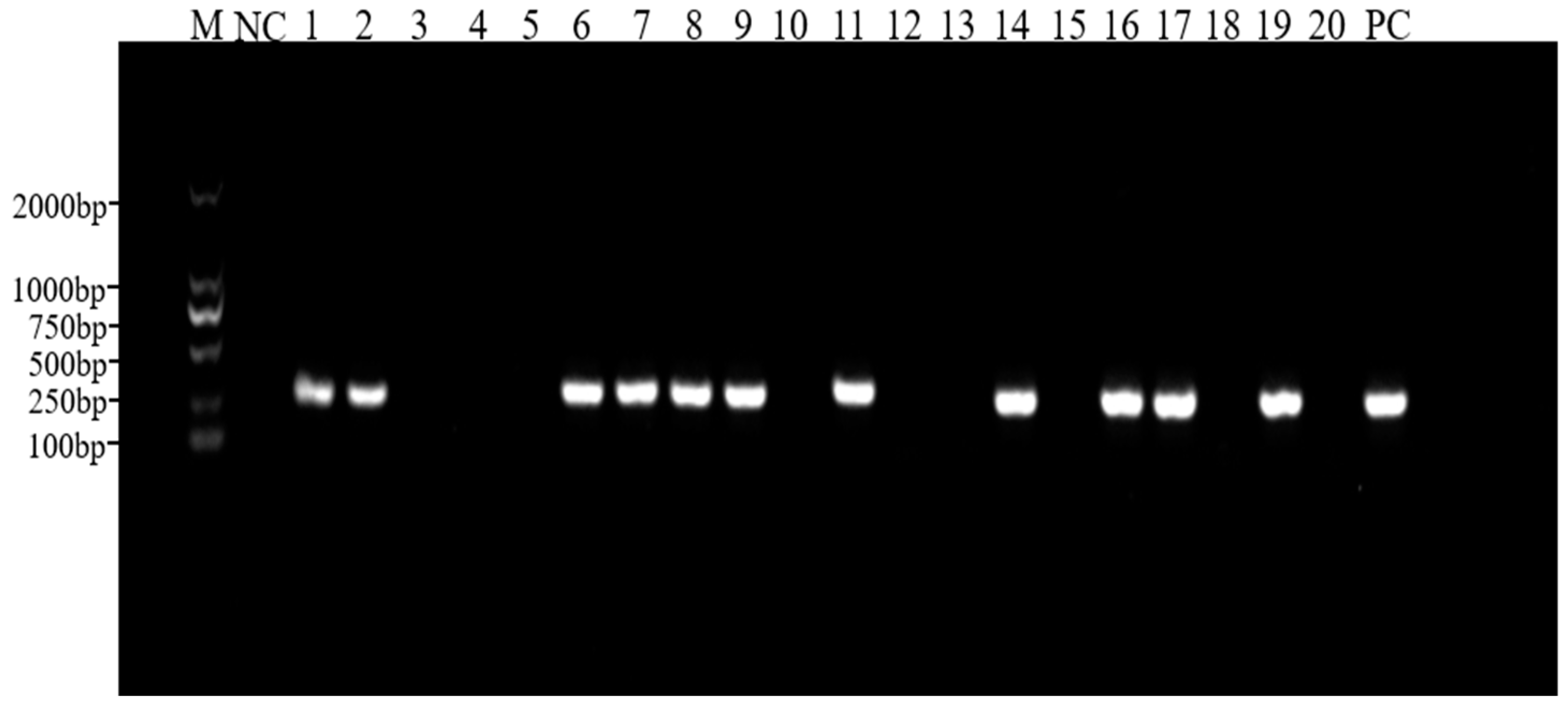
Figure 1. PCR amplification of the PRV gE gene in clinical samples. M, DNA ladder; NC, negative control; Lanes 1–20, selected representative samples; PC, positive control.
Animals 14 03103 g001
Figure 2. Amino acid sequence alignments of PRV gB (A), gC (B), gE (C), and TK (D). The alignment sequence is from Table 2, and the red box indicates the mutation base.
Animals 14 03103 g002
Figure 3. Phylogenetic tree of the gB nucleotide sequence (A), gC nucleotide sequence (B), gE nucleotide sequence (C), and TK nucleotide sequence (D). Nucleotide substitution model: p-distance; 1000 bootstrap replicates. Scale bars indicate nucleotide substitutions per site. The red triangle is the virus identified in the present study.
Animals 14 03103 g003
Table 1. Primers used for PCR and sequencing. Primer names indicate approximate binding positions in the PRV genome.
PrimerSequence (5′-3′)Amplicon Size (bp)Primer Function
gE1-FGCGGACGCACATGCTCTCTC250Detection of gE gene
gE1-RCGGTCACGCCATAGTTGGGT
gE2-FCGTCCCCCAGCCCAAGAT2048Cloning PRV gE gene
gE2-RGTCCCTTGGGGGCCAGCA
gB1-FAGACGTGCGATCAACGGCAT1146Segmented cloning of PRV gB gene
gB1-RAACAAGGACCGCACCCTGTG
gB2-FACCCGCCGCCCAGCTTAAAG1346
gB2-RCGTCTCCAAGGCCGAGTACG
gB3-FGCAGGCCGTAGAAGGGGGAC1126
gB3-RCGGCTTCTACCGCTTCCAGA
gC-FCGTTTCCTGATTCACGCCCAC1917Cloning PRV gC gene
gC-RGCACCATCGACGCCAGCTC
TK-FGCGCACCCCGAGGTTGACTT1255Cloning PRV TK gene
TK-RGACGGGCACGGCAAACTTT
Table 2. PRV nucleotide sequences for phylogenetic analysis and the detection of PRV strains’ (CQ1 and CQ2) nucleotide (nt) and amino acid (aa) sequence identities. All sequences were obtained from the NCBI database.
CQ1 Nucleotide (nt) and Amino Acid (aa) Sequence Identity%CQ2 Nucleotide (nt) and Amino Acid (aa) Sequence Identity%
gBgCgETKgBgCgETK
NoGenBankStrainCollection DataRegionntaantaantaantaantaantaantaantaa
1MK618718.1AnH1/CHN20152015China99.999.595.692.799.199.099.799.199.899.395.593.299.199.099.799.1
2JF797217.1Bartha1961Hungary98.295.799.999.6//100.099.797.896.298.998.0//100.099.7
3KC981239.1BJ/YT2013China99.999.695.692.799.699.599.799.199.899.595.593.299.699.599.799.1
4OL639029.1DCD-12017China99.899.595.692.799.199.099.799.199.799.395.593.299.199.099.799.1
5MZ063026.1DX2012China99.999.595.692.799.599.199.799.199.899.395.593.299.599.199.799.1
6KU315430.1Ea1990China99.999.595.692.5100.0100.099.599.199.999.895.593.0100.0100.099.599.1
7ON005002.1FB1986China99.297.995.492.198.999.199.698.898.998.094.891.898.999.199.698.8
8KM189913.1Fa2001China99.999.795.692.5100.0100.099.799.1100.0100.095.293.0100.0100.099.799.1
9MW286330.1FJ2019China99.999.695.692.799.499.099.799.199.899.595.593.299.499.099.799.1
10KT824771.1HLJ82013China99.999.695.692.799.599.399.799.199.899.595.593.299.599.399.799.1
11MK080279.1HLJ-20132013China99.197.097.498.1100.0100.0100.099.798.897.799.9100.0100.0100.0100.099.7
12KM189912.1HNX2012China99.999.695.692.799.599.199.799.199.899.595.593.099.599.199.799.1
13KP257591.1JS-20122012China99.999.695.692.599.599.199.799.199.899.595.493.299.599.199.799.1
14OP512542.1JS-XJ52015China99.999.695.692.799.599.399.799.199.899.595.493.299.599.399.799.1
15MK806387.1JX/CH/20162016China99.999.595.692.599.399.199.799.199.899.395.493.099.399.199.799.1
16JF797218.1Kaplan1987Hungary98.596.297.999.297.596.299.899.498.096.698.597.397.596.299.899.4
17KT983811.1Kolchis2010Greece98.496.097.598.197.496.299.598.898.096.498.196.397.496.299.598.8
18KU552118.1LA1997China99.698.895.793.199.399.599.799.199.698.993.992.099.399.599.799.1
19KU900059.1NIA31973Belgium98.496.697.297.597.595.899.699.198.396.997.795.797.595.899.699.1
20OK338076.1PRV-GD2021China99.999.695.692.799.599.199.799.199.899.595.593.299.599.199.799.1
21OK338077.1PRV-JM2021China99.999.695.692.799.599.199.799.199.899.595.593.299.599.199.799.1
22KT809429.1SC1986China99.999.797.598.1100.0100.0100.099.7100.0100.0100.0100.0100.0100.0100.0100.0
23MT949536.1SD182020China99.999.695.692.799.398.899.799.199.899.595.593.299.398.899.799.1
24OL606749.1SX19102022China99.999.295.692.799.599.199.799.199.799.395.593.299.599.199.799.1
25KJ789182.1TJ2012China99.999.695.692.799.599.399.799.199.899.595.593.299.599.399.799.1
26KM061380.1ZJ012012China99.899.595.692.798.598.499.699.199.799.395.593.298.598.499.699.1
Table 3. Seroprevalence of PRV gE antibody positive rates among pigs of various ages.
Swine HerdPRV gE Antibodies
Number of DetectionsPositive NumberPositive Rate (%)
breeding pig911.11%
suckling pig663553.03%
nursery pig462554.35%
nursing sow806075.00%
pregnant sow26820476.12%
total46932569.30%
Table 4. Seroprevalence and antigen detection rates of PRV gE in Southwest China from 2022 to 2024.
YearPRV gE AntibodiesPRV gE Antigens
Number of DetectionsPositive NumberPositive Rate (%)Number of DetectionsPositive NumberPositive Rate (%)
202237572095.56%28331.06%
202313301158.65%29772.36%
2024465112.36%///
Disclaimer/Publisher’s Note: The statements, opinions and data contained in all publications are solely those of the individual author(s) and contributor(s) and not of MDPI and/or the editor(s). MDPI and/or the editor(s) disclaim responsibility for any injury to people or property resulting from any ideas, methods, instructions or products referred to in the content.