Article

Canine Distemper Virus in Sardinia, Italy: Detection and Phylogenetic Analysis in Foxes †

by
Elisabetta Coradduzza
1,*,‡,
Fiori Mariangela Stefania
1,‡,
Davide Pintus
1,
Luca Ferretti
2,
Alice Ledda
3,
Gian Simone Chessa
1,
Angela Maria Rocchigiani
1,
Giada Lostia
1,
Renata Rossi
1,
Maria Giovanna Cancedda
1,
Simona Macciocu
1,
Marcella Cherchi
1,
Daniele Denurra
1,
Antonio Pintore
1,
Roberto Bechere
1,
Flavia Pudda
4,
Marco Muzzeddu
4,
Maria Antonietta Dettori
1,
Angelo Ruiu
1,
Paolo Briguglio
5,
Ciriaco Ligios
1 and
Giantonella Puggioni
1
add Show full author list remove Hide full author list
1
Istituto Zooprofilattico Sperimentale della Sardegna, 07100 Sassari, Italy
2
Pandemic Sciences Institute, Big Data Institute, Li Ka Shing Centre for Health Information and Discovery, Nuffield Department for Medicine, University of Oxford, Oxford OX1 2JD, UK
3
UK Health Security Agency, Colindale, London NW9 5EQ, UK
4
Centro di Recupero della Fauna Selvatica Ferita (C.A.R.F.S), Bonassai, Agenzia Forestas Sardegna, 07100 Olmedo, Italy
5
Clinica Veterinaria Duemari, 09170 Oristano, Italy
*
Author to whom correspondence should be addressed.
Presented at the Conference “77 Sisvet Congress”, Parma, Italy, 12–14 June 2024.
These authors contributed equally to this work.
Animals 2024, 14(21), 3134; https://doi.org/10.3390/ani14213134
Submission received: 2 September 2024 / Revised: 17 October 2024 / Accepted: 28 October 2024 / Published: 31 October 2024

Simple Summary

Canine distemper is a contagious infectious disease caused by the canine distemper virus (CDV). The disease affects carnivores and is present in different geographical areas. This work aimed to characterize CDV strains infecting foxes circulating in Sardinia (Italy) from 2014 to 2023, using a multidisciplinary approach. Our sequences, similar to those of central Italy, form a group very close to the European group of wildlife sequences, which leads us to propose it as exclusive to Sardinia (Sardinia-Wildlife). This study enriches knowledge of the strains responsible for natural CDV infection in this species and of the surveillance of their spread in the territory itself. It is important to continue monitoring the distribution of the virus and typing the strains to better understand the dynamics of virus circulation among wild and domestic carnivores.

Abstract

Canine distemper is a contagious infectious disease caused by the canine distemper virus (CDV). The disease affects carnivores and is present in different geographical areas. This work aimed to characterize CDV strains infecting foxes in Sardinia (Italy) from 2014 to 2023, through clinical, anatomic–histopathological, and phylogenetic analysis. Neurological, respiratory, and gastrointestinal clinical signs, including fever, depression, ocular and nasal muco-purulent discharge, and diarrhea, were reported. Necropsy and histopathological examination revealed interstitial pneumonia and mild, diffuse, non-purulent meningoencephalitis with multifocal, moderate to severe demyelination, respectively. Immunohistochemical analysis detected specific CDV positivity in lung and brain sections. For the phylogenetic analyses, a dataset with the sequences of the gene coding for the hemagglutinin (H gene) obtained from fox strains was set up, including a canine strain sequence to assess possible differences. The genetic sequences of the fox strains clustered within an exclusively Sardinian clade. This clade is a sister clade of sequences recently analyzed from central Italy, with a mean genetic divergence of more than 5%. Together, these clades form a sister clade of the Central Europe-Wildlife lineage, with a mean genetic divergence of more than 5% between these clades and the Central European lineage. Sardinian sequences belong to a newly proposed “Sardinia-Wildlife” lineage of CDV. The circulation of this lineage in the investigated foxes and dogs suggests the transmission of the virus between wild and domestic animals. Central Italian viruses may also constitute a new separate CDV lineage.

1. Introduction

Canine distemper virus (CDV) is the etiological agent of a highly prevalent viral infectious disease of carnivores, which could seriously lead to a threat to the conservation of the affected species worldwide [1,2]. The virus affects domestic and wild animals, particularly carnivores [2], threatening some endangered species [3,4]. CDV outbreaks have occurred over the past few years in canids, mustelids, felids, ursids, and even non-human primates [5,6,7,8,9,10,11,12].
This disease, known in dogs as early as 1760, is classified as highly contagious and febrile [13]. CDV has a high tropism for epithelial, lymphoid, and nervous system cells, causing a systemic infection that involves the respiratory, digestive, urinary, lymphatic, skin, skeletal, and central nervous system [14]. This virus, isolated in 1905 by Henri Carrè [15], is a member of the genus Morbillivirus [16] with a non-segmented single-negative-stranded RNA (ssRNA) [14]. The genome is about 16 KB in length and its structure includes six transcription units (ORFs) encoding for six structural proteins (two glycoproteins: hemagglutinin (H) and fusion (F) proteins; one envelope-associated matrix (M) protein; two transcriptase-associated proteins: phosphoprotein (P) and large polymerase (L) protein; and one nucleocapsid (N) protein [17,18]), separated by intergenic untranslated regions (UTRs) [16]. In particular, hemagglutinin (H) is an integral membrane glycoprotein that facilitates the virus binding to the host cell membrane and, compared with other morbilliviruses, it has been shown to have higher heterogeneity [19] permitting clustering viral strains of CDV into at least nine lineages according to their geographical distribution: America 1 (NA1), America 2 (NA2), Europe 1/South America 1 (EU1/SA1), Europe 2/Europe-Wildlife (EU2), Europe 3/Arctic-like (EU3), Asia 1 (AS1), Asia 2 (AS2), South Africa (ZA), and South America 2 (SA2) [16,18,20]. In Italy, EU1/SA1 (found in both domestic dogs and wild carnivores) is particularly prevalent in the north [19,21,22,23], while in the center and south, the most representative lineages are EU2 (rarely found in domestic dogs) and EU3 (found mainly in Italian dogs) [5,24,25]. Within the Europe/South America-1 lineage, two distinct subclades have been identified in northern Italy in recent years [5,19,26]: clade a, which originates from ancient epidemic waves in both domestic and wild carnivores, and clade b, which consists of CDV strains most likely imported from the Balkan region.
The Sardinian fox (Vulpes vulpes ichnusae) is endemic in Sardinia and is anthropized; it is found in forest and scrubland environments, agro-systems, and human settlements [27]. Several phylogeny studies relate the viral sequences of distemper strains from domestic dogs and wild carnivores, suggesting horizontal transmission between species through aerosols and exudates [24,25,26,27,28,29,30]. CDV does not always appear to have a particular impact on foxes [19]: long-term persistence of CDV can occur for decades, sometimes without fatal consequences [8,31] because animals with robust immune responses can recover by developing lifelong immunity to reinfection. However, they play an important role as reservoirs for highly susceptible species. Early detection in wildlife species is therefore particularly important to allow the typing of circulating CDV strains.
This work aimed to characterize phylogenetic distemper strains infecting foxes in Sardinia, Italy, from 2014 to 2023, i.e., [1,12,19]. With this objective, we sequenced the H gene of all CDV virus strains isolated from infected foxes, i.e., [26,32]. In addition to the phylogenetic analyses, histological and immunohistochemical examinations were performed in foxes to better characterize the pathological features of the viral strains affecting this species.

2. Materials and Methods

2.1. Animals and Sampling

A total of 42 animals were examined: 41 foxes (Figure 1) and 1 dog. The addition of the canine sequence (sample already analyzed in our laboratory during routine analysis) aimed to understand possible connections from a phylogenetic point of view with fox sequences. All animals underwent necropsy examination and, for each one, either the brain, lung, spleen, or eyelid were sampled. Then, the most representative sample from a molecular point of view was selected for sequencing (Table 1). Where there were no limitations such as post-mortem alterations or progressed decomposition of the cadavers, the brain and the lung were collected for histological and histochemical exams.

2.2. Pathology and Immunohistochemistry

Collected organs were fixed in 10% formalin and embedded in paraffin for histological evaluation. According to routine laboratory protocols, 4 μm thick sections were stained using the ST Infinity Haematoxylin & Eosin Staining System (Leica Biosystems, Richmond, IL, USA) and then examined using a light microscope (from 50× to 400× of magnification). Furthermore, on selected brain and lung tissue sections, immunohistochemistry (IHC) was performed using a CDV-specific primary antibody (Bio-Rad monoclonal mouse anti-canine distemper virus antibody, clone DV2-12, MCA 1893, Hercules, CA, USA). Moreover, IHC was performed on selected brain and lung sections for different clusters of differentiation markers (CD immunophenotyping) of inflammatory cells. Primary antibodies were rabbit monoclonal CD3 (Clone 7.2.38, DAKO Agilent, Santa Clara, CA, USA), mouse monoclonal CDacy79 (Clone HM57, DAKO Agilent, Santa Clara, CA, USA), monoclonal mouse CD163 (Clone EDHu-1, Bi-Rad, Hercules, CA, USA). Working solutions were 1:1500 for CD3, 1:750 for CDacy79, and 1:800 for CD163.
Sections were deparaffinized and rehydrated through several passages using alcohol and then thoroughly rinsed in water. Heat-induced antigen retrieval was performed in target citrate buffer pH 6.0 (Thermo Fisher Scientific, Waltham, MA, USA) or in target retrieval solution pH 9.0 (Dako Agilent, Santa Clara, CA, USA) to expose antigen epitopes. To quench endogenous peroxidase activity, the slides were immersed in 0.3% H2O2 for 35 min at room temperature and then incubated overnight at 4 °C using the primary antibodies as previously described. Finally, immunoreactivity was detected via the biotin–avidin–peroxidase method using 3,3′-diaminobenzidine as a chromogen. Later, slides were counterstained with Mayer’s hematoxylin and mounted by cover slides. In each IHC run, adequate positive and negative control tissues were included. Moreover, brain, lung, conjunctival mucus, and blood in EDTA were subjected to biomolecular investigation to diagnose the presence of CDV by RT-PCR amplification. The affected foxes that showed gastrointestinal signs were also subjected to molecular investigation using PCR for Parvovirus [33], Nested RTPCR for Coronavirus [34], and Real-Time PCR for pathogenic Leptospira ssp. [35].

2.3. Nucleic Acid Extraction, RT-PCR Amplification, and Sequencing

First, a highly conserved portion of the gene that encodes for the nucleoprotein (N gene, of 287 bp) was chosen to perform a screening RT-PCR suitable for detecting all CDV strains [36]. Subsequently, we amplified the H gene, using three PCRs to cover the entire sequence (sequenced samples in Table 1 and primer set in Table 2). The amplification covered areas from the F gene’s end part to the L gene’s beginning part, which flanks the H gene in the sequence.
RT-PCR was performed using the Superscript One-Step RT-PCR Kit with Platinum Taq (Thermo Fisher Scientific, Waltham, MA, USA), according to the manufacturer’s directions.
The PCR products were purified using the CleanSweep PCR Purification kit (Thermo Fisher Scientific) and sequenced for both forward and reverse strands (using the same primers used for PCR) using a Sanger Sequencing 3500 Series Genetic Analyzers Terminator 3.1 apparatus (Applied Biosystems, Waltham, MA, USA).

2.4. Sequence and Phylogenetic Analyses

Sequences were aligned using BioEdit 7.2.5 [37] and MEGA 7.0 [38] softwares. A maximum likelihood optimized search was employed for the phylogenetic reconstruction of Sardinian and international sequences using IQ-TREE version 1.6.12 [39] with a GTR + Gamma evolutionary model with standard parameters and ultrafast bootstrap approximation. In addition, Bayesian reconstruction of the dated phylogeny of Sardinian sequences was performed using BEAST version 1.10.4 [40] with a Skygrid relaxed-clock model with annual resolution 2003–2023 and 100 million steps for the MCMC chain. Molecular distances for nucleotide and amino acid sequences were computed using R version 4.3.2 and the ape package version 5.7 with default parameters.

3. Results

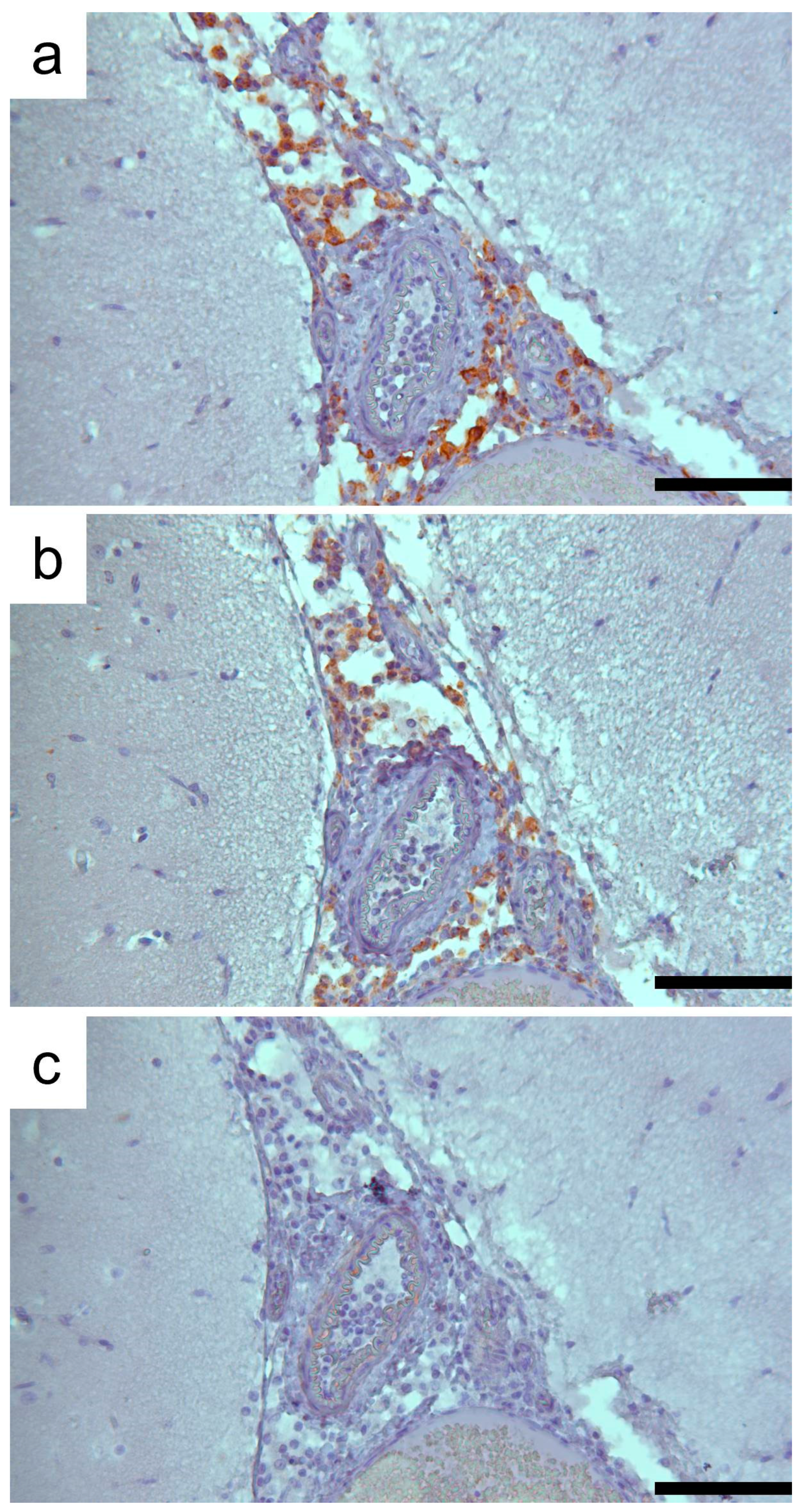
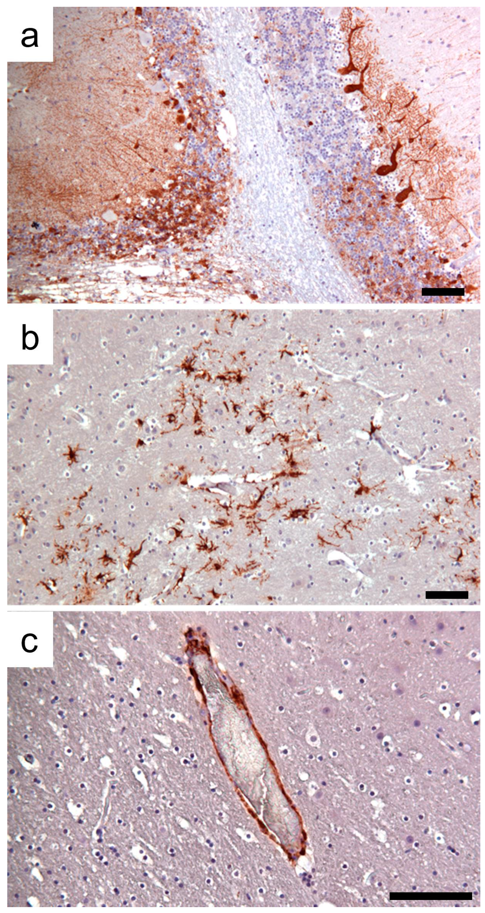
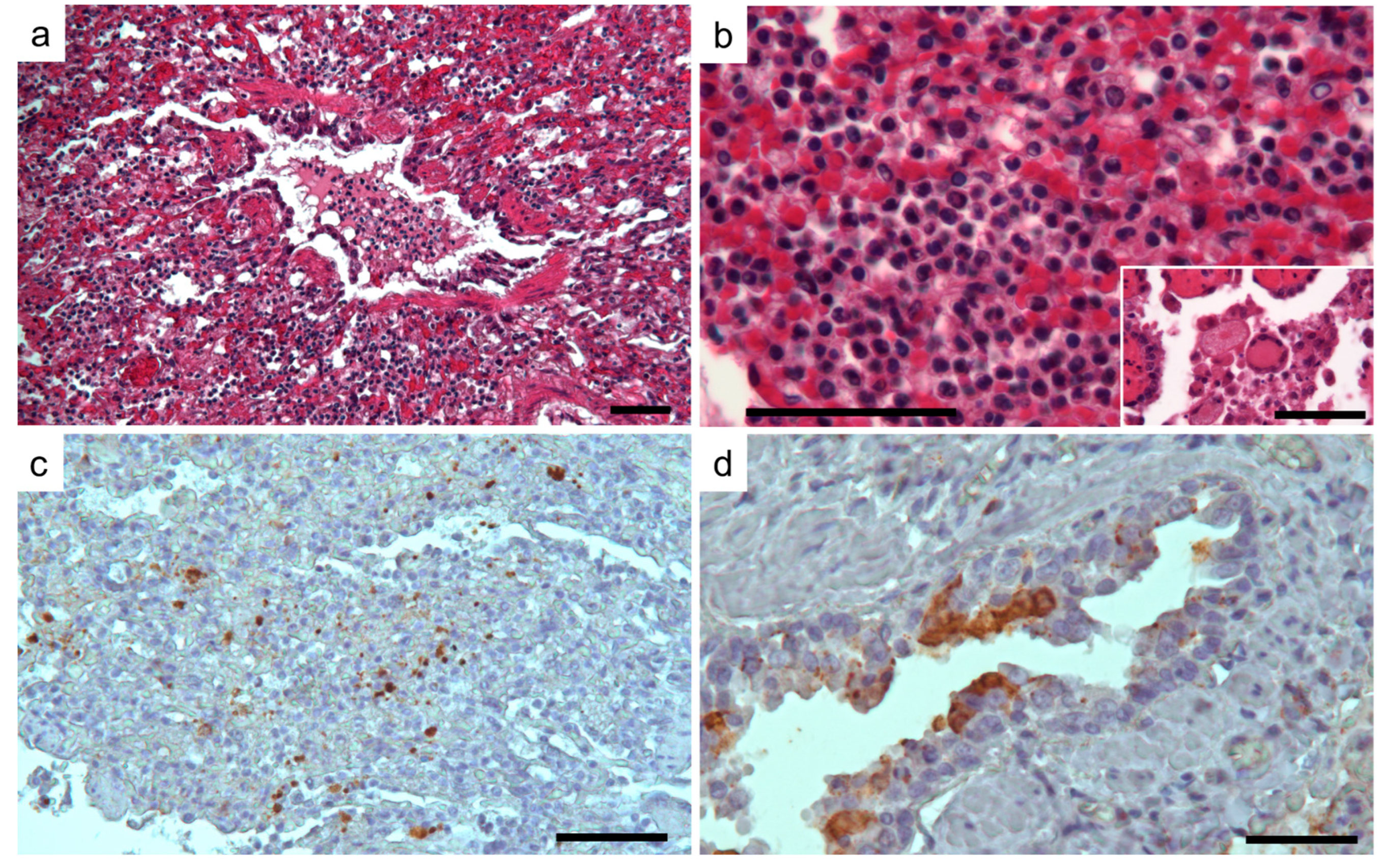
3.1. Clinic, Pathology, and Immunohistochemistry

Sick animals showed sensory depression and ataxia, as well as blindness or severe vision alteration in both eyes, and some of them had hematochezia. All foxes died within 2 to 48 h in the recovery center. Gross examination revealed severe chronic wasting, muco-purulent conjunctivitis, and nasal discharge (Figure 2). Congestion and/or hemorrhage in the lung sometimes associated with sero-hemorrhagic fluid in the pericardium and mild congestion in the vessels of the cerebral cortex were observed.
Histologically, the brain lesions were characterized by moderate to severe demyelination, and the diffuse mild presence of mononuclear inflammatory cells involving the meninges. In-depth examination revealed focal to multifocal vacuolation (demyelination) of the white matter and an increased number of reactive astrocytes, macrophages, and microglial cells, sometimes organized in nodular patterns. A marked spongy state with small or large vacuoles was observed in the demyelinated lesion areas (Figure 3a,b). Occasionally, necrotic neurons with shrunken and eosinophilic cytoplasm and lymphoid cells around blood vessels were found. No viral nuclear inclusion bodies were observed. Lesions were observed in the gray and white matter of serial brain sections but were more severe in the cerebellum (Figure 3c). In the cerebellar sections, the loss of Purkinje cells was segmentally lost, and the progressive loss of myelin characterized by vacuolization of the white matter was evident. The mononuclear infiltrate cells observed in the meninges (Figure 3d) were immunohistochemically characterized mainly by CD163+ macrophages and CD3+ T lymphocytes (Figure 4), whereas CD79+ cells were few or absent.
Neurons and a limited number of glial cells from the cerebral cortex showed strong and specific CDV immunolabeling. Moreover, CDV-positive staining was associated with the endothelial cells of numerous vessels in the cerebral parenchyma and meninges. Confirming the severity of lesions determined in the brain sections, high immunoreactivity for CDV was revealed in the cerebellum section, associated with the molecular and granular layer, and in the body and axons of the Purkinje cells (Figure 5). Histologically, in the lung, pulmonary lesions were characterized by diffuse and severe interstitial pneumonia. As confirmed by CD immunophenotyping, an infiltrate of macrophages (CD163+) and lesser numbers of T lymphocytes (CD3+) associated with edema and erythrocytes enlarged the alveolar septa. Very few neutrophils were also observed. Blood vessels were observed in some pulmonary sections occluded by inflammatory cells and debris. Syncytial giant cells were also observed. Finally, hypertrophy of smooth muscle cells around the airways and blood was determined in some of the examined lungs (Figure 6a,b).
Immunohistochemical staining of the lung highlighted the presence of the CDV antigen in macrophages and lymphocytes, as well as in the epithelial cells of the bronchi and bronchioles (Figure 6c,d).

3.2. Nucleic Acid Extraction, PCR Amplification

In all subjects, it was possible to detect the presence of viral RNA by RT-PCR of the N gene. No other etiological agents were revealed in the samples. The positivity was confirmed by direct sequencing of the amplified H gene.

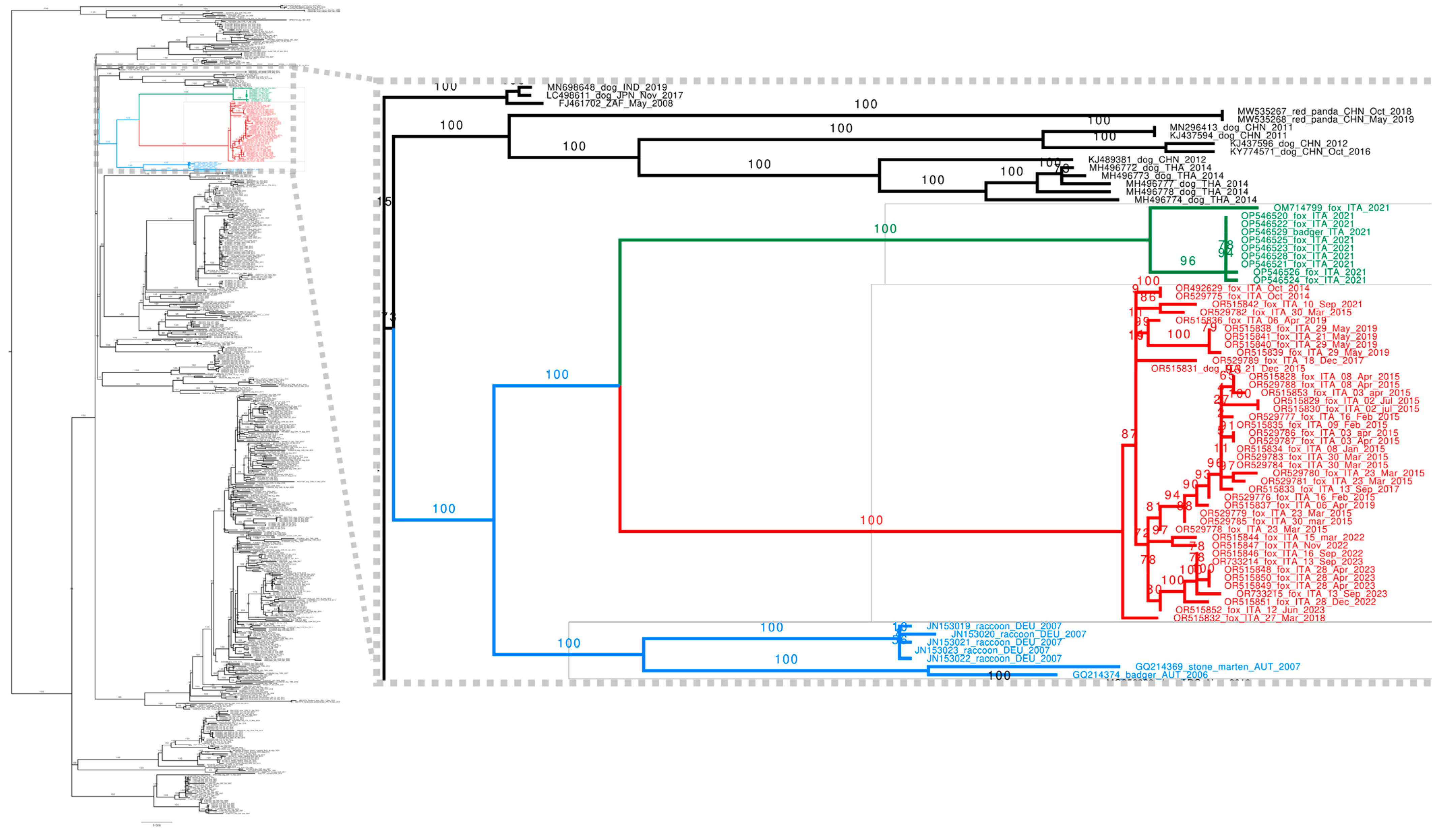
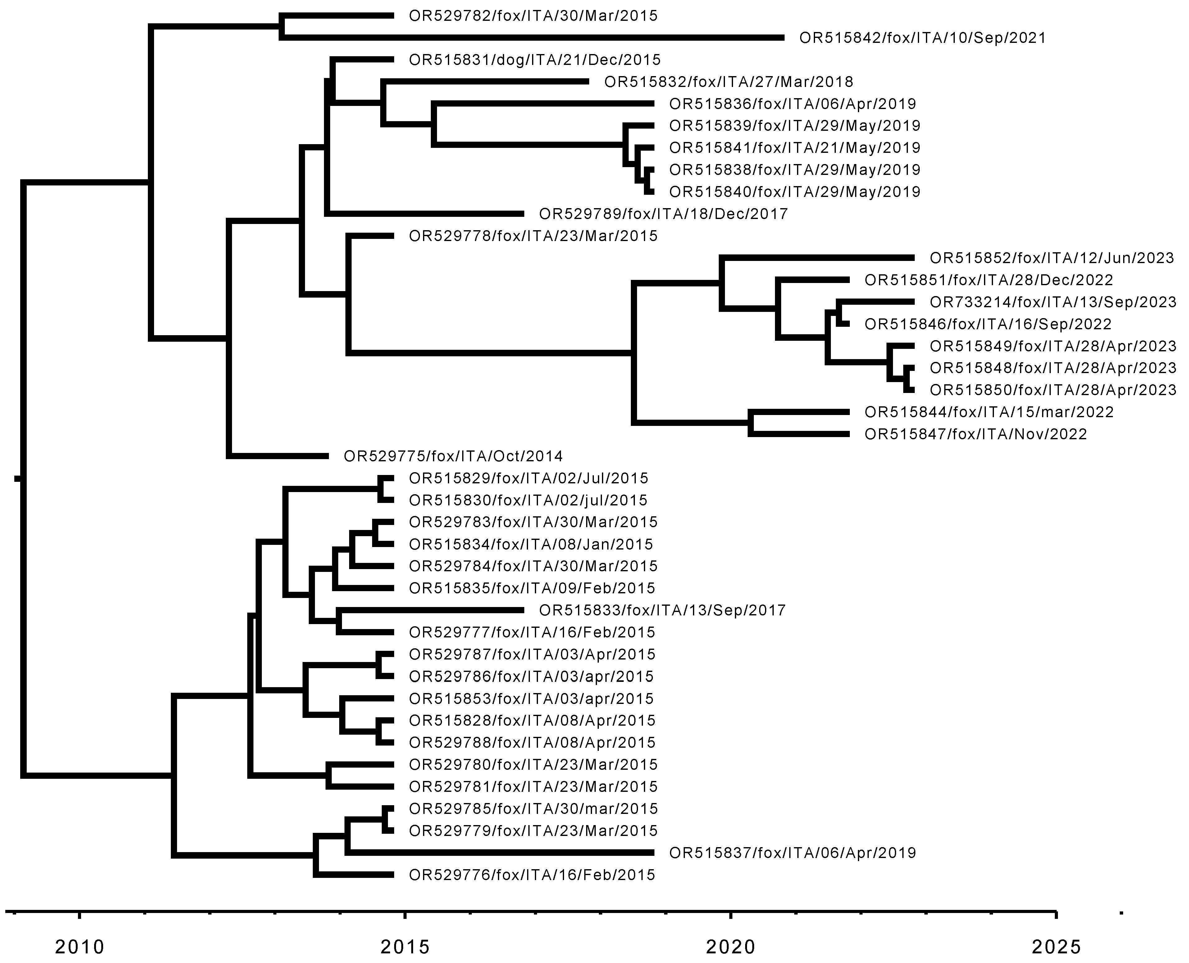
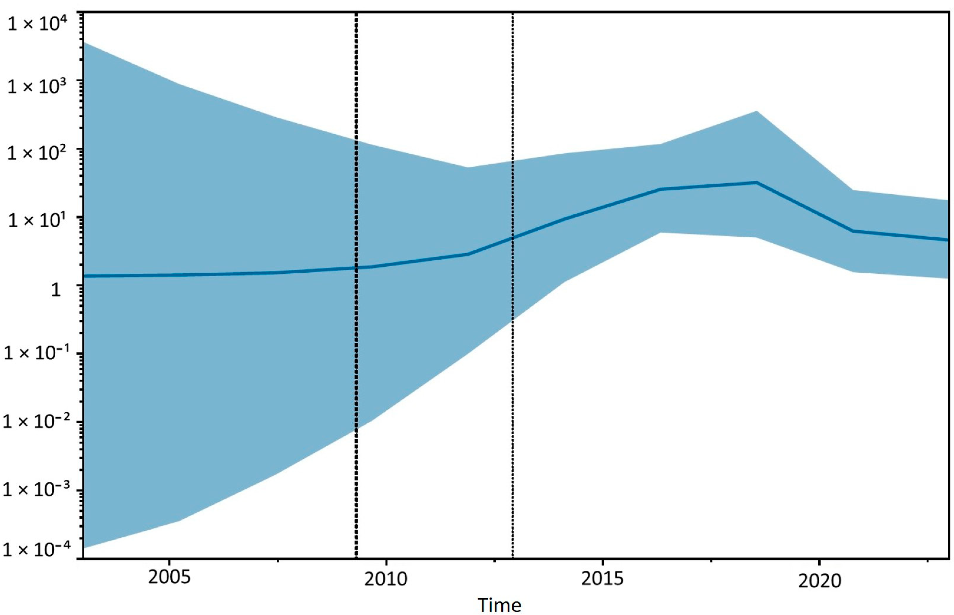
3.3. Sequencing and Phylogenetic Analyses

As can be seen from the reconstruction of the global CDV phylogeny (Figure 7), all our Sardinian sequences from foxes belong to a single clade. Only one other sequence from the dog sample, sequenced in this work, is in the same clade, specifically in the middle, suggesting a transmission involving a host jump from foxes to dogs [31,41].
As shown in Figure 8, the Bayesian reconstruction of the time-calibrated Sardinian phylogeny points to a more recent common ancestor of all sampled Sardinian sequences circulating in 2008 (mean 2008.6, median 2009.3, 95% 95% HPD interval 2002.7–2012.9), suggesting that the lineage currently circulating in Sardinia was introduced to the island a few years before the time when the first sequenced samples were obtained, i.e., 2014 (Figure 9).
A Bayesian reconstruction of the effective population size (Figure 10) suggests that the circulation of CDV in the Sardinian epidemic may have gradually increased in the first decade since its introduction, possibly followed by a slight decrease in recent years.
The maximum likelihood phylogenetic reconstruction places this Sardinian clade close to the Europe-Wildlife lineage (Figure 7). However, it is significantly divergent from published sequences. The closest sister clade contains samples from different mammals from central Italy (foxes and badgers) [25,26], and the two clades are significantly divergent (5.0% at nucleotide level, 4.7% at amino acid level, Table 3). This suggests that these two clades represent two different lineages. Given the geographical proximity, it is plausible that the Sardinian clade may have originated from viruses from the continental part of Italy introduced into the island.
Together, the Sardinian and other Italian sequences represent a sister clade of the published Central European sequences classified as belonging to the Europe-Wildlife lineage. Note that these sister clades are very different from sequences belonging to the Europe-Wildlife lineage from Germany and Austria: the nucleotide divergence is 5.0% and 5.2% for Sardinian and Italian sequences, respectively, and the amino acid divergence is 5.5% and 5.3% (Table 3). This divergence is comparable to the divergence between different CDV lineages [20]. Most likely, Sardinian sequences belong to a novel “Sardinia-Wildlife” lineage.

4. Discussion

The phylogeny, based on the H gene, clusters the Italian strains into three lineages: Europe/South America-1, Europe-Wildlife, and Arctic-like [5,24,25,42]. In northern Italy, two distinct subclades within the Europe/South America-1 lineage have been identified in recent years [5,19,26]: clade a, which originates from ancient epidemic waves in both domestic and wild carnivores, and clade b, which consists of CDV strains most likely imported from the Balkan region. These data show the crossover between domestic and wild carnivores for strains capable of jumping species and the diverse behavior of virus strains, some of which remain specific to the original host. However, to date, the knowledge and understanding of how the virus spreads in southern Italy, both in domestic dogs and wildlife animals, is still very poor and limited [5,20,43,44,45]. Intending to contribute to the current knowledge on the spread of canine distemper virus in Italy, this study provides a molecular characterization of CDV strains based on the H gene sequence obtained from viral samples isolated from foxes in Sardinia and a comparison of them with currently available CDV sequences.
As previously reported, the central nervous system and lungs were two of the main CDV target organs in carnivores [14]. The RT-PCR associated with the immunohistochemical results revealed that, with a high probability, CDV was the etiological agent of the neurological signs in the enrolled foxes in this study. The pulmonary histological lesions and immunohistochemical detection of CDV in the mucosa of the bronchial tree support the potential viral excretion through nasal discharge.
The Sardinian sequences are similar among them but diverge by about 5% from other known CDV sequences, both at the nucleotide and amino acid levels. This divergence is similar to the divergence between different CDV lineages, such as Asia-4 and the recently discovered Asia-6 [46], suggesting a significant degree of isolation among viral populations in Sardinia and elsewhere (Italy and Central Europe). Given their geographical isolation and the exhaustive sampling of viruses circulating on the island, at least in the unique wild carnivore species present, these Sardinian sequences represent a novel “Sardinia-Wildlife” lineage circulating in Sardinia.
The sister clade of Sardinia-Wildlife contains only sequences sampled from wild mammals from central Italy [26], which appear to be representative of the sequences currently circulating in central Italy, given that they include many samples from the Romagna region on the northern side but also a similar sample from the Abruzzo region on the southern side of central Italy [47], about 300 km away from each other. These sequences have about 5% divergence at the nucleotide and amino acid levels from both Sardinia-Wildlife and other Europe-Wildlife sequences from Central Europe. Given their similarity and divergence compared to other CDV sequences, we suggest that central Italian sequences may represent another potential “Italy-Wildlife” lineage, even though the incompleteness and unrepresentativeness of the geographical sampling in central and northern Italy limits the validity of this claim. A more extensive and exhaustive sampling of intermediate geographical regions as a wider number of sequences is needed to properly define the phylogenetic relation between these Italian viruses and Europe-Wildlife.
To gain a better understanding of the evolutionary dynamics of CDV strains in foxes and dogs by studying their amino acid sequence, we evaluated in particular the receptor binding sites 530 and 549 of hemagglutinin, which McCarthy in 2007 [48] had already hypothesized to play a functional role in determining cell tropism since the spread of the virus in non-dog hosts is most associated with amino acid substitutions at these sites. At site 530, dog strains usually present amino acid G or E, whereas amino acid substitutions N, R, D, or S are associated with non-dog hosts. All our strains, including the dog, present the amino acid N at this site, leading to the hypothesis that fox and dog strains have undergone common evolution. This hypothesis would also be confirmed by the 549 binding site in the SLAM binding region, which presents the amino acid Y (typical of canine strains and found in all our sequences) in all of them.

5. Conclusions

This study shows the dynamics of CDV infection within the fox population in Sardinia, enriching our knowledge of the strains responsible for natural CDV infection in this species and of the surveillance of their spread in the territory. Since the number of positive cases in foxes is quite worrying, leading to high exposure to the virus in environments where other susceptible species circulate, it is important to continue monitoring the distribution of the virus and typing the strains to contain its spread and to better understand the dynamic of the virus’s circulation among wild and domestic carnivores.

Author Contributions

Conceptualization, E.C., F.M.S., L.F., A.L. and D.P.; methodology, E.C., G.S.C., F.M.S., L.F., D.P., A.L., A.P., D.D., M.G.C., S.M., F.P., M.M., P.B., M.A.D., R.R. and R.B.; software, E.C., F.M.S., L.F. and A.L.; validation, E.C., F.M.S., L.F. and A.L.; formal analysis, E.C., F.M.S., L.F. and D.P.; investigation, E.C., G.S.C., F.M.S., L.F., A.M.R., D.P., A.L., M.G.C., S.M., A.P., A.R., D.D., M.C., F.P., M.M. and P.B.; resources, E.C.; data curation, E.C., F.M.S., L.F., A.L., D.P. and G.L.; writing—original draft preparation, E.C., L.F., A.L. and D.P.; writing—review and editing, E.C., L.F., A.L., G.P. and C.L.; visualization, E.C., L.F., G.L. and A.L.; supervision, E.C.; project administration, E.C.; funding acquisition, E.C. and G.P. All authors have read and agreed to the published version of the manuscript.

Funding

This study was supported by the research ordinary funds of the Experimental Zooprophylactic Institute of Sardinia, 07100 Sassari, Italy.

Institutional Review Board Statement

Ethical review and approval were waived for this study as the sample collection procedures were performed as part of a national/regional program for the surveillance of infectious diseases in wild animals of the Experimental Zooprophylactic Institute of Sardinia, in accordance with the 2002 ministerial circular recalling the decrees of law 270/93 and 503/70. All research activities in this study complied with institutional and national guidelines.

Informed Consent Statement

Verbal informed consent was obtained from the participants. The rationale for utilizing verbal consent is that our institute is an Italian public experimental institute, institutionally authorized for this type of examination. Owners, in private clinics, are informed that their animal’s corpse will be sent to the institute for further examinations and do not sign a consent form but only give verbal authorization.

Data Availability Statement

The sequences of the CDV hemagglutinin gene obtained during the present study are available in the GenBank nucleotide sequence database under/with the following accession numbers: OR492629; OR529775; OR529776; OR529777; OR529778; OR529779; OR529780; OR529781; OR529782; OR529783; OR529784; OR529785; OR529786; OR529787; OR529788; OR515828; OR515829; OR515830; OR515831; OR515832; OR515833; OR529789; OR515834; OR515835; OR515836; OR515837; OR515838; OR515839; OR515840; OR515841; OR515842; OR515844; OR515846; OR515847; OR515848; OR515849; OR515850; OR515851; OR515852; OR515853; OR733214; OR733215.

Conflicts of Interest

The authors declare no conflicts of interest.

References

  1. Echeverry-Bonilla, D.F.; Buriticá-Gaviria, E.F.; Orjuela-Acosta, D.; Chinchilla-Cardenas, D.J.; Ruiz-Saenz, J. The First Report and Phylogenetic Analysis of Canine Distemper Virus in Cerdocyon thous from Colombia. Viruses 2022, 14, 1947. [Google Scholar] [CrossRef] [PubMed]
  2. Martinez-Gutierrez, M.; Ruiz-Saenz, J. Diversity of susceptible hosts in canine distemper virus infection: A systematic review and data synthesis. BMC Vet. Res. 2016, 12, 78. [Google Scholar] [CrossRef] [PubMed]
  3. Gilbert, M.; Soutyrina, S.V.; Seryodkin, I.V. Canine distemper virus as a threat to wild tigers in Russia and across their range. Integr. Zool. 2015, 10, 329–343. [Google Scholar] [CrossRef] [PubMed]
  4. Williams, E.S.; Thorne, E.T.; Appel, M.J.; Belitsky, D.W. Canine distemper in black-footed ferrets (Mustela nigripes) from Wyoming. J. Wildl. Dis. 1988, 24, 385–398. [Google Scholar] [CrossRef]
  5. Balboni, A.; Savini, F.; Scagliarini, A.; Berti, E.; Naldi, M.; Urbani, L.; Fontana, M.C.; Carra, E.; Gibelli, L.R.M.; Gobbo, F.; et al. Natural distemper infection in stone martens (Martes foina): From infection to neutralizing antibodies. Res. Vet. Sci. 2021, 138, 196–200. [Google Scholar] [CrossRef]
  6. Kennedy, J.M.; Earle, J.A.; Omar, S.; Abdullah, H.A.; Nielsen, O.; Roelke-Parker, M.E.; Cosby, S.L. Canine and phocine distemper viruses: Global spread and genetic basis of jumping species barriers. Viruses 2019, 11, 944. [Google Scholar] [CrossRef]
  7. Loots, A.K.; Mitchell, E.; Dalton, D.L.; Kotzé, A.; Venter, E.H. Advances in canine distemper virus pathogenesis research: A wildlife perspective. J. Gen. Virol. 2017, 98, 311–321. [Google Scholar] [CrossRef]
  8. Origgi, F.C.; Plattet, P.; Sattler, U.; Robert, N.; Casaubon, J.; Mavrot, F.; Pewsner, M.; Wu, N.; Giovannini, S.; Oevermann, A.; et al. Emergence of canine distemper virus strains with modified molecular signature and enhanced neuronal tropism leading to high mortality in wild carnivores. Vet. Pathol. 2012, 49, 913–929. [Google Scholar] [CrossRef]
  9. Pope, J.P.; Miller, D.L.; Riley, M.C.; Anis, E.; Wilkes, R.P. Characterization of a novel canine distemper virus causing disease in wildlife. J. Vet. Diagn. 2016, 28, 506–513. [Google Scholar] [CrossRef]
  10. Sakai, K.; Nagata, N.; Ami, Y.; Seki, F.; Suzaki, Y.; Iwata-Yoshikawa, N.; Suzuki, T.; Fukushi, S.; Mizutani, T.; Yoshikawa, T.; et al. Lethal canine distemper virus outbreak in cynomolgus monkeys in Japan in 2008. J. Virol 2013, 87, 1105–1111. [Google Scholar] [CrossRef]
  11. Sun, Z.; Li, A.; Ye, H.; Shi, Y.; Hu, Z.; Zeng, L. Natural infection with canine distemper virus in hand-feeding Rhesus monkeys in China. Vet. Microbiol. 2010, 141, 374–378. [Google Scholar] [CrossRef] [PubMed]
  12. Oleaga, A.; Blanco Vázquez, C.; Royo, L.J.; Doria Barral, T.; Bonnaire, D.; Armenteros, J.A.; Rabanal, B.; Gortázar, C.; Balseiro, A. Canine distemper virus in wildlife in south-western Europe. Transbound. Emerg. Dis. 2022, 69, e473–e485. [Google Scholar] [CrossRef] [PubMed]
  13. Blancou, J. Dog distemper: Imported into Europe from South America? Hist. Med. Vet. 2004, 29, 35–41. [Google Scholar] [PubMed]
  14. Rendon-Marin, S.; da Fontoura Budaszewski, R.; Canal, C.W.; Ruiz-Saenz, J. Tropism and molecular pathogenesis of canine distemper virus. Virol. J. 2019, 16, 30. [Google Scholar] [CrossRef]
  15. Carré, H. Sur la maladie des jeunes chiens. C. r. Acad. Sci. 1905, 140, 1489–1491. [Google Scholar]
  16. Panzera, Y.; Sarute, N.; Iraola, G.; Hernandez, M.; Perez, R. Molecular phylogeography of canine distemper virus: Geographic origin and global spreading. Mol. Phylogenet Evol. 2015, 92, 147–154. [Google Scholar] [CrossRef]
  17. Ndiana, L.A.; Lanave, G.; Desario, C.; Odigie, A.E.; Madubuike, K.G.; Lucente, M.S.; Ezeifeka, C.A.; Patruno, G.; Lorusso, E.; Elia, G.; et al. Detection of Selected Canine Viruses in Nigerian Free-Ranging Dogs Traded for Meat Consumption. Animals 2023, 13, 1119. [Google Scholar] [CrossRef]
  18. von Messling, V.; Zimmer, G.; Herrler, G.; Haas, L.; Cattaneo, R. The hemagglutinin of canine distemper virus determines tropism and cytopathogenicity. J. Virol. 2001, 75, 6418–6427. [Google Scholar] [CrossRef]
  19. Trogu, T.; Canziani, S.; Salvato, S.; Bianchi, A.; Bertoletti, I.; Gibelli, L.R.; Alborali, G.L.; Barbieri, I.; Gaffuri, A.; Sala, G.; et al. Canine Distemper Outbreaks in Wild Carnivores in Northern Italy. Viruses 2021, 13, 99. [Google Scholar] [CrossRef]
  20. Mira, F.; Purpari, G.; Di Bella, S.; Vicari, D.; Schirò, G.; Di Marco, P.; Macaluso, G.; Battilani, M.; Guercio, A. Update on canine distemper virus (CDV) strains of Arctic-like lineage detected in dogs in Italy. Vet. Ital. 2018, 54, 225–236. [Google Scholar]
  21. Monne, I.; Fusaro, A.; Valastro, V.; Citterio, C.; Dalla Pozza, M.; Obber, F.; Trevisiol, K.; Cova, M.; De Benedictis, P.; Bregoli, M.; et al. Distinct CDV genotype causing a major epidemic in Alpine wildlife. Vet. Microbiol. 2011, 150, 63–69. [Google Scholar] [CrossRef] [PubMed]
  22. Lorusso, A.; Savini, G. Old diseases for new nightmares: Distemper strikes back in Italy. Vet. Ital. 2014, 50, 151–154. [Google Scholar] [PubMed]
  23. Bianco, A.; Zecchin, B.; Fusaro, A.; Schivo, A.; Ormelli, S.; Bregoli, M.; Citterio, C.V.; Obber, F.; Dellamaria, D.; Trevisiol, K.; et al. Two waves of canine distemper virus showing different spatio-temporal dynamics in Alpine wildlife (2006–2018). Infect. Genet. Evol. 2020, 84, 104359. [Google Scholar] [CrossRef] [PubMed]
  24. Di Sabatino, D.; Lorusso, A.; Di Francesco, C.E.; Gentile, L.; Di Pirro, V.; Bellacicco, A.L.; Giovannini, A.; Di Francesco, G.; Marruchella, G.; Marsilio, F.; et al. Arctic lineage-canine distemper virus as a cause of death in Apennine wolves (Canis lupus) in Italy. PLoS ONE 2014, 9, e82356. [Google Scholar] [CrossRef] [PubMed]
  25. Di Sabatino, D.; Di Francesco, G.; Zaccaria, G.; Malatesta, D.; Brugnola, L.; Marcacci, M.; Portanti, O.; De Massis, F.; Savini, G.; Teodori, L.; et al. Lethal distemper in badgers (Meles meles) following epidemic in dogs and wolves. Infect. Genet. Evol. 2016, 46, 130–137. [Google Scholar] [CrossRef]
  26. Trogu, T.; Castelli, A.; Canziani, S.; Tolini, C.; Carrera, M.; Sozzi, E.; Lelli, D.; Tosi, G.; Fiorentini, L.; Di Donato, A.; et al. Detection and Molecular Characterization of Canine Distemper Virus in Wildlife from Northern Italy. Pathogens 2022, 11, 1557. [Google Scholar] [CrossRef]
  27. SardegnaForeste. Available online: https://www.sardegnaforeste.it/fauna/volpe (accessed on 12 February 2024).
  28. Frölich, K.; Czupalla, O.; Haas, L.; Hentschke, J.; Dedek, J.; Fickel, J. Epizootiological investigations of canine distemper virus in free-ranging carnivores from Germany. Vet. Microbiol. 2000, 74, 283–292. [Google Scholar] [CrossRef]
  29. Müller, A.; Silva, E.; Santos, N.; Thompson, G. Domestic dog origin of canine distemper virus in free-ranging wolves in Portugal as revealed by hemagglutinin gene characterization. J. Wildl. Dis. 2011, 47, 725–729. [Google Scholar] [CrossRef]
  30. Beineke, A.; Baumgärtner, W.; Wohlsein, P. Cross-species transmission of canine distemper virus—An update. One Health 2015, 1, 49–59. [Google Scholar] [CrossRef]
  31. Nouvellet, P.; Donnelly, C.A.; De Nardi, M.; Rhodes, C.J.; De Benedictis, P.; Citterio, C.; Obber, F.; Lorenzetto, M.; Dalla Pozza, M.; Cauchemez, S.; et al. Rabies and canine distemper virus epidemics in the red fox population of northern Italy (2006–2010). PLoS ONE 2013, 8, e61588. [Google Scholar] [CrossRef]
  32. Liao, P.; Guo, L.; Wen, Y.; Yang, Y.; Cheng, S. Phylogenetic features of hemagglutin gene in canine distemper virus strains from different genetic lineages. Int. J. Clin. Exp. Med. 2015, 8, 6607–6612. [Google Scholar]
  33. Senda, M.; Parrish, C.R.; Harasawa, R.; Gamoh, K.; Muramatsu, M.; Hirayama, N.; Itoh, O. Detection by PCR of wild-type canine parvovirus which contaminates dog vaccines. J. Clin. Microbiol. 1995, 33, 110–113. [Google Scholar] [CrossRef]
  34. Drzewnioková, P.; Festa, F.; Panzarin, V.; Lelli, D.; Moreno, A.; Zecchin, B.M.; De Benedictis, P.; Leopardi, S. Best Molecular Tools to Investigate Coronavirus Diversity in Mammals: A Comparison. Viruses 2021, 13, 1975. [Google Scholar] [CrossRef]
  35. Stoddard, R.A.; Gee, J.E.; Wilkins, P.P.; McCaustland, K.; Hoffmaster, A.R. Detection of pathogenic Leptospira spp. Through TaqMan polymerase chain reaction targeting LipL32 gene. Diagn. Microbiol. Infect. Dis. 2009, 64, 247–255. [Google Scholar] [CrossRef]
  36. Frisk, A.L.; König, M.; Moritz, A.; Baumgärtner, W. Detection of Canine Distemper Virus Nucleoprotein RNA by Reverse Transcription-PCR Using Serum, Whole Blood, and Cerebrospinal Fluid from Dogs with Distemper. J. Clin. Microbiol. 1999, 37, 3634–3643. [Google Scholar] [CrossRef]
  37. Hall, T.A. BioEdit: A user-friendly biological sequence alignment editor and analysis program for Windows 95/98/NT. Nucl. Acids. Symp. Ser. 1999, 41, 95–98. [Google Scholar]
  38. Kumar, S.; Stecher, G.; Tamura, K. MEGA7: Molecular Evolutionary Genetics Analysis version 7.0 for bigger datasets. Mol. Biol. Evol. 2016, 33, 1870–1874. [Google Scholar] [CrossRef]
  39. Nguyen, L.T.; Schmidt, H.A.; von Haeseler, A.; Minh, B.Q. IQ-TREE: A fast and effective stochastic algorithm for estimating maximum likelihood phylogenies. Mol. Biol. Evol. 2015, 32, 268–274. [Google Scholar] [CrossRef]
  40. Suchard, M.A.; Lemey, P.; Baele, G.; Ayres, D.L.; Drummond, A.J.; Rambaut, A. Bayesian phylogenetic and phylodynamic data integration using BEAST 1.10. Virus Evol. 2018, 4, vey016. [Google Scholar] [CrossRef]
  41. Viana, M.; Cleaveland, S.; Matthiopoulos, J.; Halliday, J.; Packer, C.; Craft, M.E.; Hampson, K.; Czupryna, A.; Dobson, A.P.; Dubovi, E.J.; et al. Dynamics of a morbillivirus at the domestic-wildlife interface: Canine distemper virus in domestic dogs and lions. Proc. Natl. Acad. Sci. USA 2015, 112, 1464–1469. [Google Scholar] [CrossRef]
  42. Guercio, A.; Mira, F.; Di Bella, S.; Gucciardi, F.; Lastra, A.; Purpari, G.; Castronovo, C.; Pennisi, M.; Di Marco Lo Presti, V.; Rizzo, M.; et al. Biomolecular Analysis of Canine Distemper Virus Strains in Two Domestic Ferrets (Mustela putorius furo). Vet. Sci. 2023, 10, 375. [Google Scholar] [CrossRef] [PubMed]
  43. Alfano, F.; Lanave, G.; Lucibelli, M.G.; Miletti, G.; D’Alessio, N.; Gallo, A.; Auriemma, C.; Amoroso, M.G.; Lucente, M.S.; De Carlo, E.; et al. Canine Distemper Virus in Autochtonous and Imported Dogs, Southern Italy (2014–2021). Animals 2022, 12, 2852. [Google Scholar] [CrossRef] [PubMed]
  44. Martella, V.; Cirone, F.; Elia, G.; Lorusso, E.; Decaro, N.; Campolo, M.; Desario, C.; Lucente, M.S.; Bellacicco, A.L.; BlixenkroneMøller, M.; et al. Heterogeneity within the hemagglutinin genes of canine distemper virus (CDV) strains detected in Italy. Vet. Microbiol. 2006, 116, 301–309. [Google Scholar] [CrossRef] [PubMed]
  45. Martella, V.; Pratelli, A.; Cirone, F.; Zizzo, N.; Decaro, N.; Tinelli, A.; Foti, M.; Buonavoglia, C. Detection and genetic characterization of canine distemper virus (CDV) from free-ranging red foxes in Italy. Mol. Cell Probes 2002, 16, 77–83. [Google Scholar] [CrossRef] [PubMed]
  46. Wang, R.; Wang, X.; Zhai, J.; Zhang, P.; Irwin, D.M.; Shen, X.; Chen, W.; Shen, Y. A new canine distemper virus lineage identified from red pandas in China. Transbound. Emerg. Dis. 2021, 69, e944–e952. [Google Scholar] [CrossRef]
  47. Ricci, I.; Cersini, A.; Manna, G.; Marcario, G.A.; Conti, R.; Brocherel, G.; Grifoni, G.; Eleni, C.; Scicluna, M.T. A Canine Distemper Virus Retrospective Study Conducted from 2011 to 2019 in Central Italy (Latium and Tuscany Regions). Viruses 2021, 13, 272. [Google Scholar] [CrossRef]
  48. McCarthy, A.J.; Shaw, M.A.; Goodman, S.J. Pathogen evolution and disease emergence in carnivores. Proc. Biol. Sci. 2007, 274, 3165–3174. [Google Scholar] [CrossRef]
Figure 1. Map of the study area showing the locations where fox samples were collected. The numbers indicate how many of the 41 samples examined in this study were collected in that area.
Animals 14 03134 g001
Figure 2. External examination of a fox with CVD. Eye with muco-purulent conjunctivitis.
Animals 14 03134 g002
Figure 3. Photomicrographs of representative histological brain sections of CVD-infected foxes. (a) Cerebral cortex: severe demyelinating encephalitis with vacuolization of the white matter due to myelin sheath edema. (b) Cerebral cortex: nodular gliosis in a moderate demyelinating area of the cortex. (c) Cerebellum: demyelinating areas in the substantia alba of the cerebellum associated with the loss of Purkinje cells and the hypocellular granule cell layer. (d) Cerebral cortex: mild meningeal lymphohistiocytic infiltrates. Hematoxylin and eosin staining. Scale bar 100 µm.
Animals 14 03134 g003
Figure 4. CD-immunophenotyping of the meningeal inflammatory cells. (a) Immunohistochemical labeling of CD163+ macrophages. (b) Immunohistochemical reactivity of CD3+ T lymphocytes. (c) No CD79+ B lymphocytes were revealed. 3-30-diaminobenzidine (DAB) chromogen with Mayer’s hematoxylin counterstain. Scale bar 100 µm.
Animals 14 03134 g004
Figure 5. Photomicrographs of representative CDV immunoreactivity in brain sections. (a) Cerebellum: high immunoreactivity in the granular and molecular layers, and in the few visible Purkinje cells. (b) Intense and specific immunohistochemical CDV expression in neurons. (c) CVD immunohistochemical staining associated with endothelial cells. 3-30-diaminobenzidine (DAB) chromogen with Mayer’s hematoxylin counterstain. Scale bar 100 µm.
Animals 14 03134 g005
Figure 6. Photomicrographs of representative histological lung sections of CVD-infected foxes. (a,b) Two different magnifications of interstitial pneumonia are characterized by debris and inflammatory cells in the bronchiolar lumen and by thickening of the alveolar septa with macrophages, lymphocytes, and erythrocytes predominantly. The syncytial cell is revealed in the insert. Hematoxylin and eosin staining. Scale bar 100 µm. (c,d) Positive immunolabelling is associated with inflammatory cells, resembling macrophages, and in the bronchiolar epithelial cells. 3-30-diaminobenzidine (DAB) chromogen with Mayer’s hematoxylin counterstain.
Animals 14 03134 g006
Figure 7. Maximum likelihood reconstruction of the global CDV phylogeny. Sardinian sequences sampled in this work are shown in red, while those belonging to the European wildlife lineage are in blue and green; the latter color used is for the Italian sequences belonging to the sister clade to the Sardinian one. Branches are annotated by ultrafast bootstrap values (splits are considered reliable if >=95).
Animals 14 03134 g007
Figure 8. Bayesian reconstruction of the time-calibrated phylogeny for all Sardinian CDV sequences studied in this work, obtained from the BEAST posterior as Maximum Clade Credibility tree with median node heights.
Animals 14 03134 g008
Figure 9. Bayesian reconstruction of the posterior distribution for the date of the Most Recent Common Ancestor of the time-calibrated phylogeny for all Sardinian CDV sequences studied in this work. The blue area represents the 95% Highest Posterior Density (HPD) interval.
Animals 14 03134 g009
Figure 10. Bayesian skyGrid reconstruction of the time-dependent effective population size of CDV in Sardinia. The continuous line shows the posterior mean log effective population size, while the blue shadow represents the corresponding 95% HPD. The dotted vertical lines represent the 95% HPD for the date of the root of the tree.
Animals 14 03134 g010
Table 1. Information on hemagglutinin GenBank codes of Sardinian CDV strains detected in the present study.
Strain IDSampleAccession NumberCountryCollection DateHost
Sardinian 1BrainOR492629Italy3 October 2014Fox
Sardinian 2SpleenOR529775Italy3 October 2014Fox
Sardinian 3BrainOR529776Italy16 February 2015Fox
Sardinian 4LungOR529777Italy16 February 2015Fox
Sardinian 5LungOR529778Italy23 March 2015Fox
Sardinian 6BrainOR529779Italy23 March 2015Fox
Sardinian 7BrainOR529780Italy23 March 2015Fox
Sardinian 8LungOR529781Italy23 March 2015Fox
Sardinian 9BrainOR529782Italy30 March 2015Fox
Sardinian 10BrainOR529783Italy30 March 2015Fox
Sardinian 11LungOR529784Italy30 March 2015Fox
Sardinian 12EyelidOR529785Italy30 March 2015Fox
Sardinian 13BrainOR529786Italy3 April 2015Fox
Sardinian 14Brain OR529787Italy3 April 2015Fox
Sardinian 15BrainOR529788Italy8 April 2015Fox
Sardinian 16LungOR515828Italy8 April 2015Fox
Sardinian 17BrainOR515829Italy2 July 2015Fox
Sardinian 18LungOR515830Italy2 July 2015Fox
Sardinian 19BrainOR515831Italy21 December 2015Dog
Sardinian 20BrainOR515832Italy27 March 2018Fox
Sardinian 21BrainOR515833Italy13 September 2017Fox
Sardinian 22BrainOR529789Italy18 December 2017Fox
Sardinian 23BrainOR515834Italy8 January 2015Fox
Sardinian 24LungOR515835Italy9 February 2015Fox
Sardinian 25BrainOR515836Italy6 April 2019Fox
Sardinian 26LungOR515837Italy6 April 2019Fox
Sardinian 27LungOR515838Italy29 May 2019Fox
Sardinian 28BrainOR515839Italy29 May 2019Fox
Sardinian 29EyelidOR515840Italy29 May 2019Fox
Sardinian 30LungOR515841Italy21 May 2019Fox
Sardinian 31SpleenOR515842Italy10 September 2021Fox
Sardinian 33Brain OR515844Italy15 March 2022Fox
Sardinian 35LungOR515846Italy16 September 2022Fox
Sardinian 36BrainOR515847Italy28 November 2022Fox
Sardinian 37BrainOR515848Italy28 April 2023Fox
Sardinian 38LungOR515849Italy28 April 2023Fox
Sardinian 39EyelidOR515850Italy28 April 2023Fox
Sardinian 40BrainOR515851Italy28 December 2022Fox
Sardinian 41BrainOR515852Italy12 June 2023Fox
Sardinian 43EyelidOR515853Italy3 April 2015Fox
Sardinian 44BrainOR733214Italy13 September 2023Fox
Sardinian 45LungOR733215Italy13 September 2023Fox
Table 2. Primer set used to amplify the CDV complete H gene.
Primer SetPrimer SequenceSize ReferencesAnnealing
Temperature (C°)
1st PCR: gF67085′ TCTAACCAGATCCTTGAGAC 3′926 bpThis study56°
1st PCR: gH7633rev5′ TGGTGGGAATATGTCACCTCT 3′
2nd PCR: CDVH 388 for5′ GAATTCGACTTCCGCGATCTCC 3′968 bpThis study50°
2nd PCR: CDVH 1335 rev5′ ATGGTAAGCCATCCGGAGTTC 3′
3rd PCR: GH 8360 for5′ GGTCCGTTATACTGAATGG 3′792 bpThis study53°
3rd PCR: gL 9151 rev5′ AGGAGCTGATAGTTATGTC 3′
Table 3. Mean amino acid and nucleotide divergence between sequences from different clades.
Amino Acid Divergence
SardiniaCentral ItalyCentral Europe
Sardinia 4.73%5.52%
Central Italy 5.34%
Central Europe
Nucleotide Divergence
SardiniaCentral ItalyCentral Europe
Sardinia 5.03%
Central Italy 5.20%
Central Europe5.01%
Disclaimer/Publisher’s Note: The statements, opinions and data contained in all publications are solely those of the individual author(s) and contributor(s) and not of MDPI and/or the editor(s). MDPI and/or the editor(s) disclaim responsibility for any injury to people or property resulting from any ideas, methods, instructions or products referred to in the content.

