


Correlation between Cross-Sectional Anatomy and Computed Tomography of the Normal Six-Banded Armadillo (Euphractus sexcintus) Nasal Cavity and Paranasal Sinuses


 , ,
, ,  ,
,
Abstract
Simple Summary
Abstract
1. Introduction
2. Materials and Methods
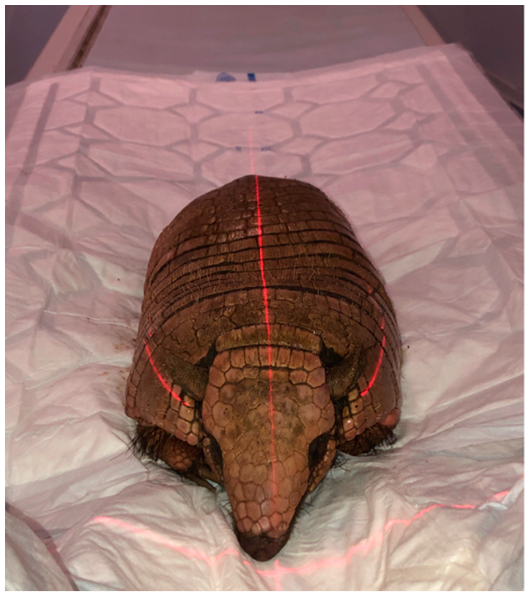
2.1. Animals
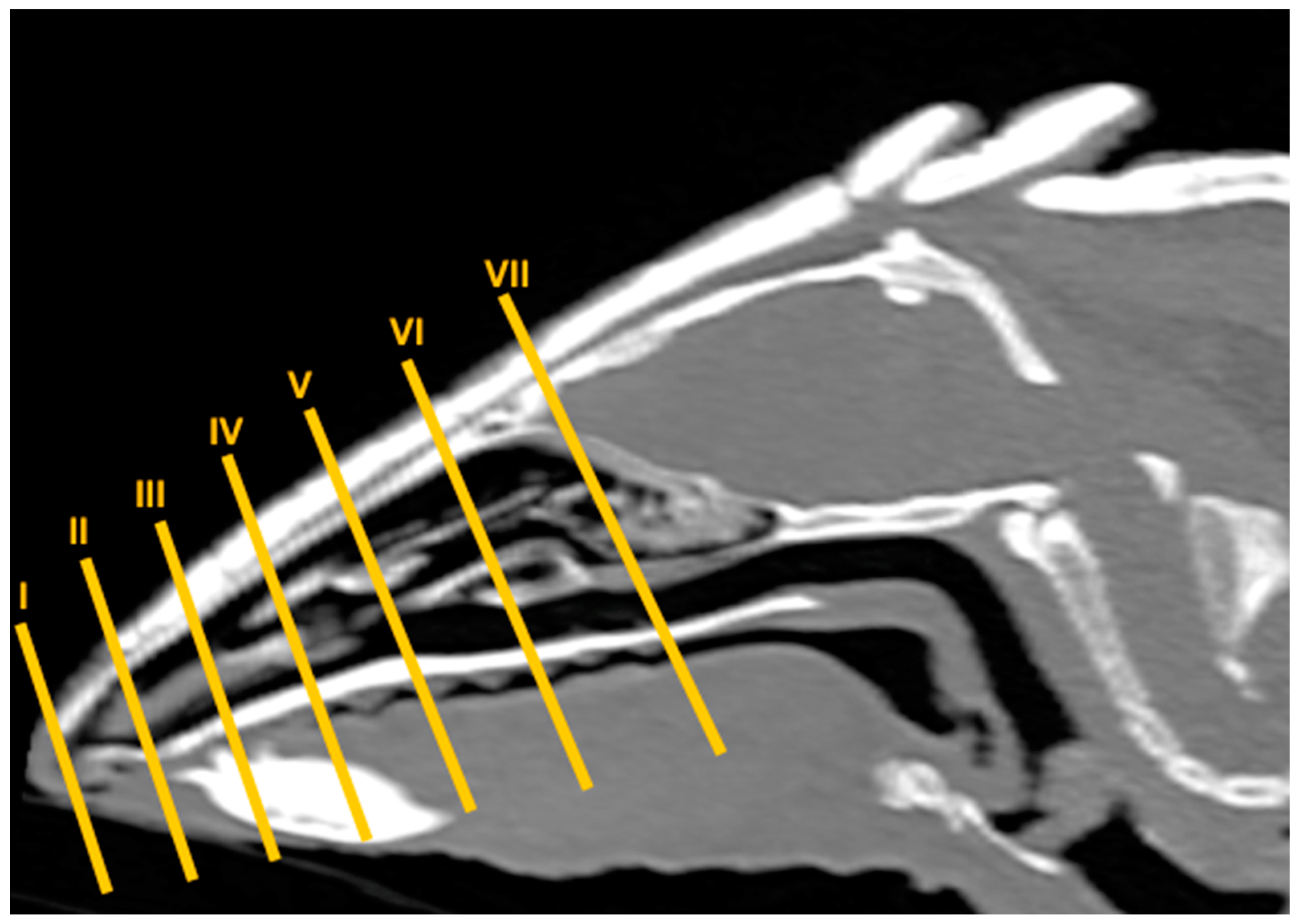
2.2. CT Technique
2.3. Anatomic Evaluation
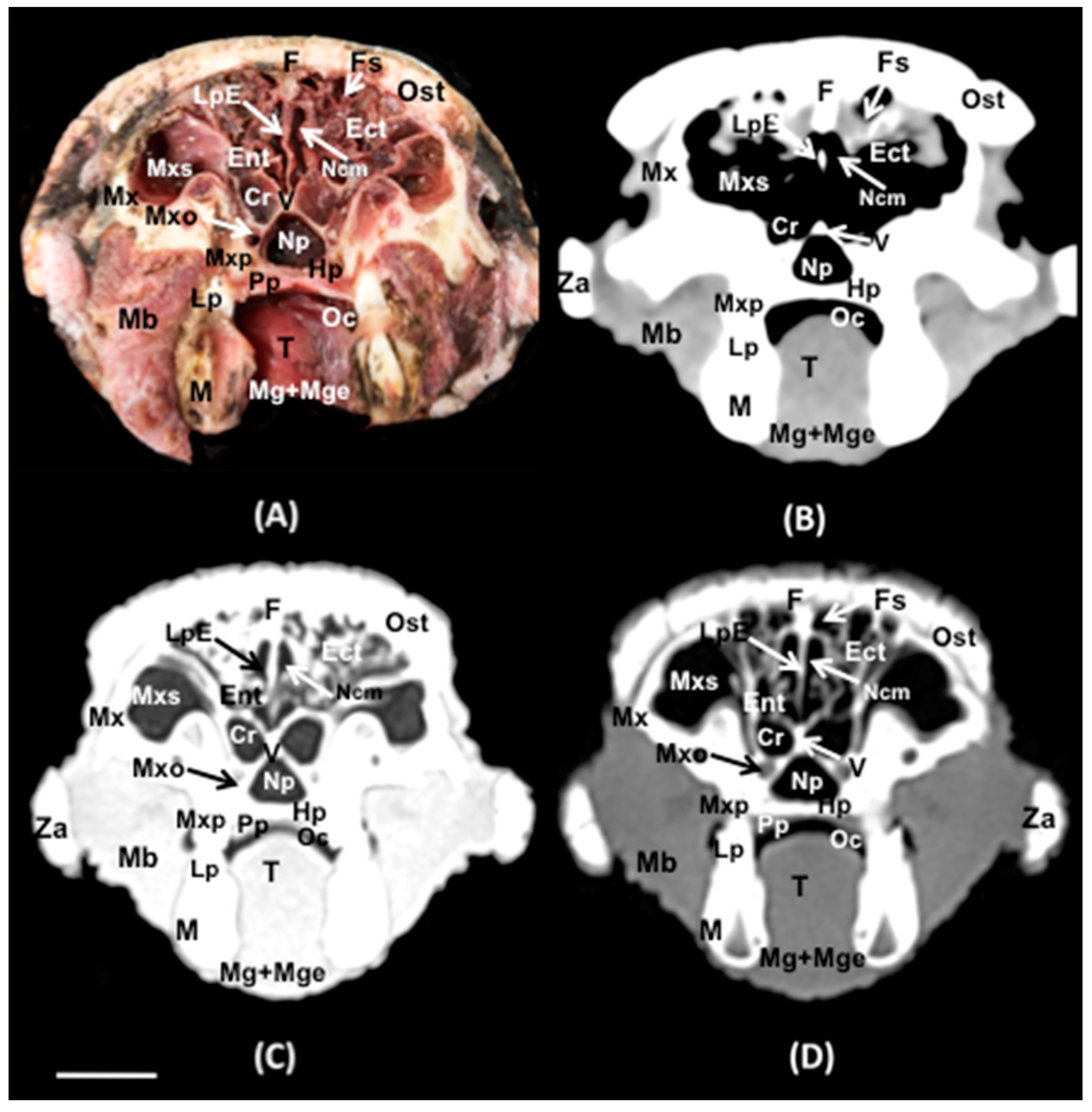
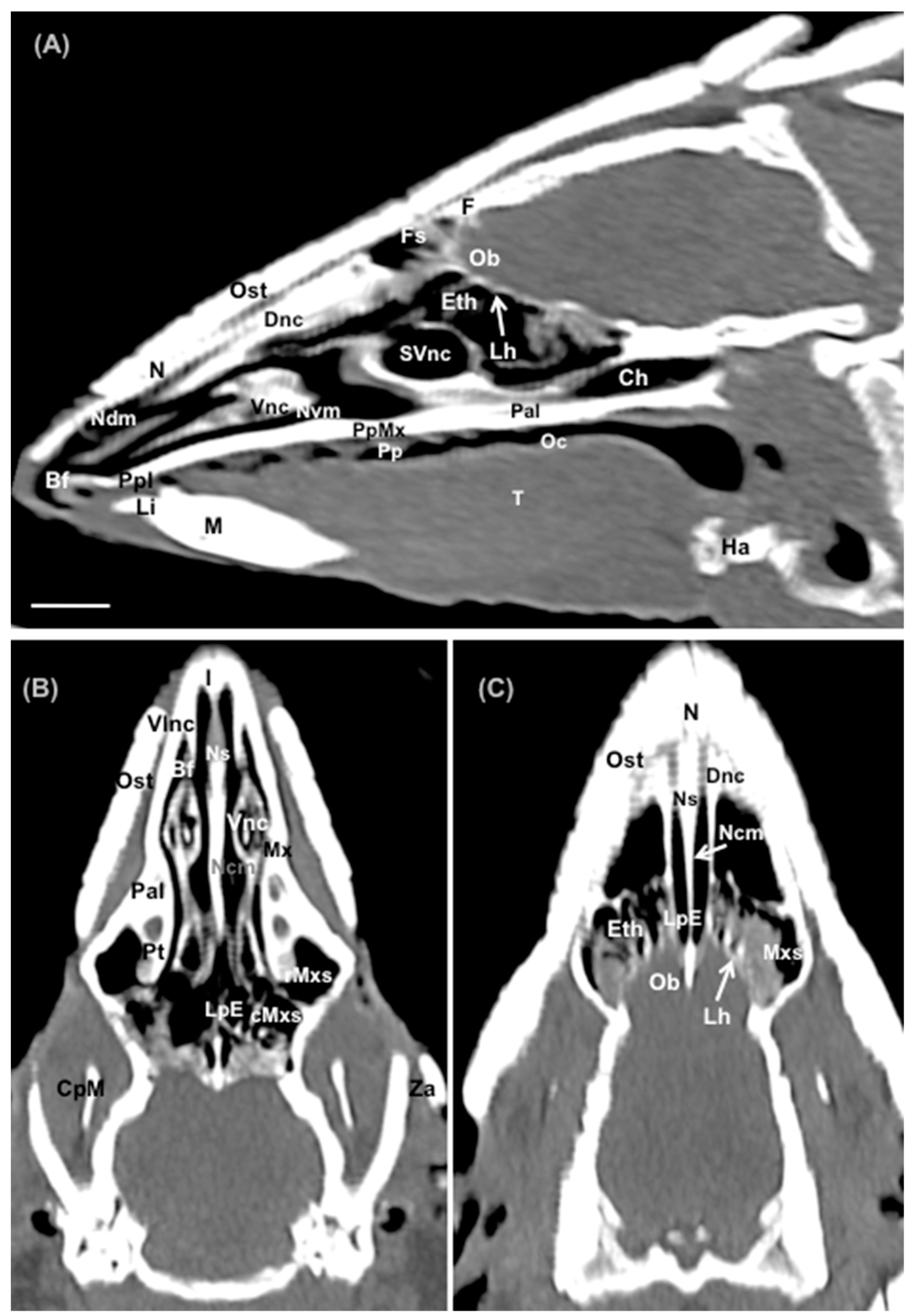
3. Results
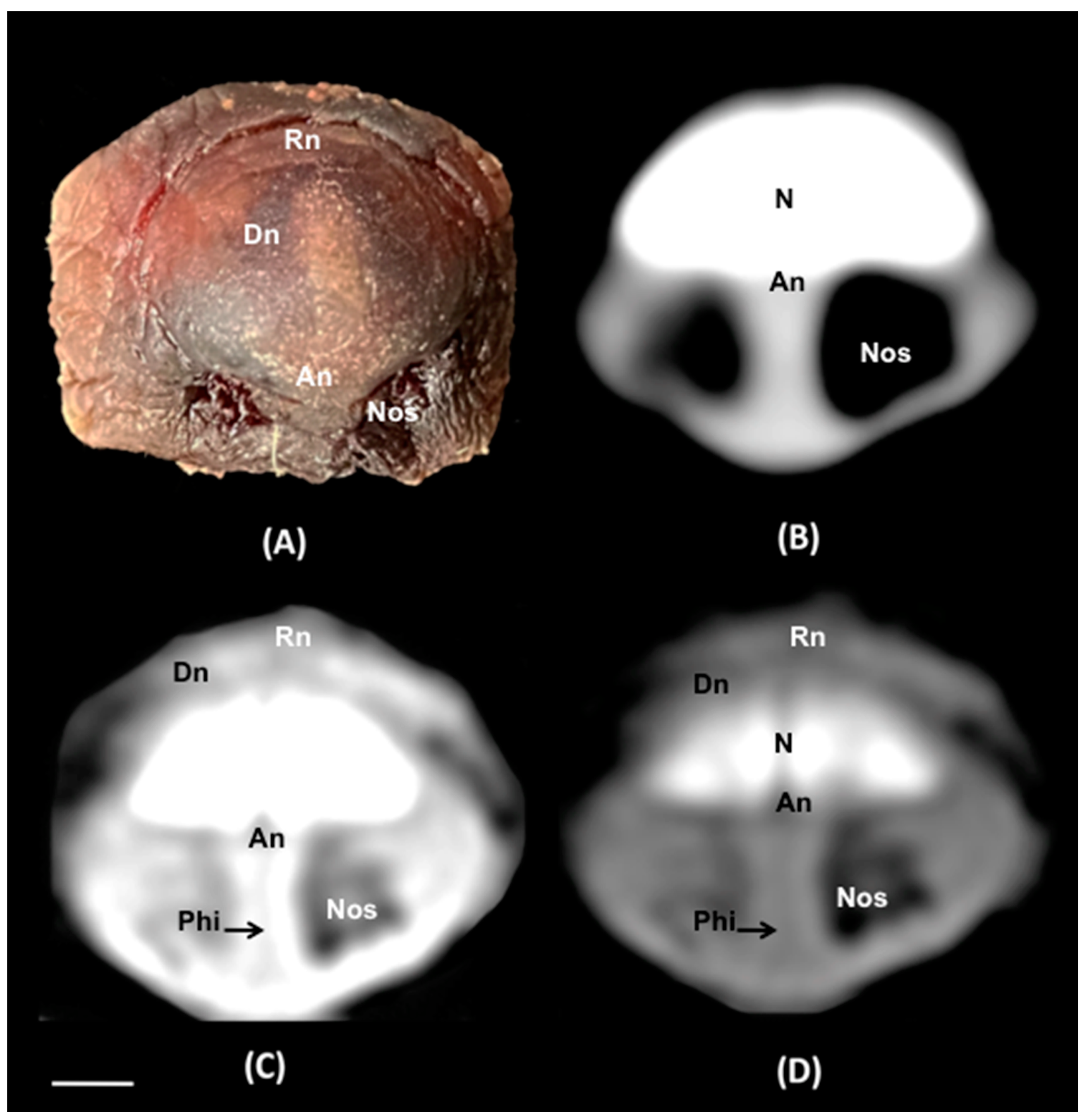
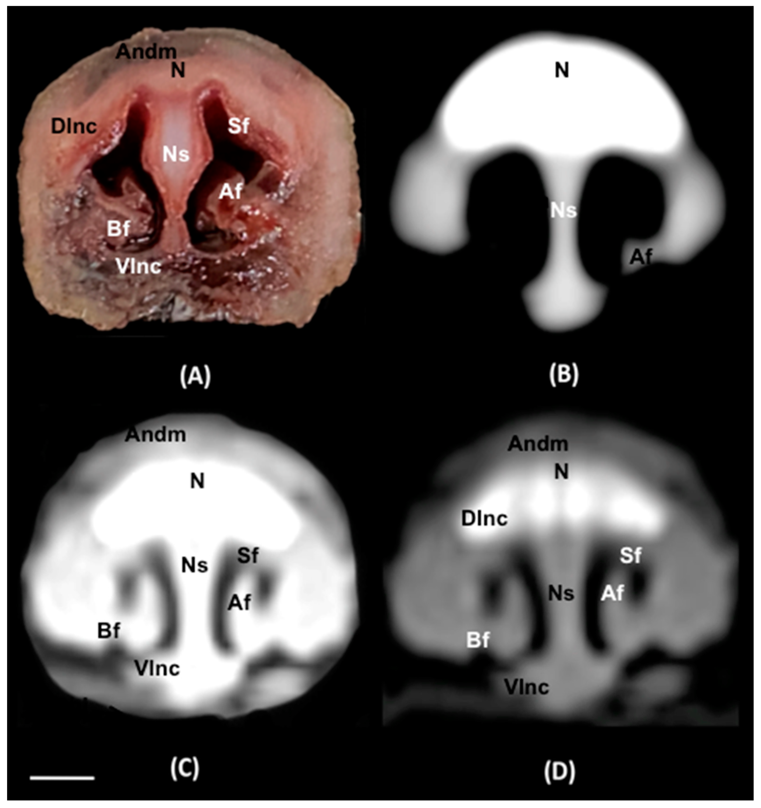
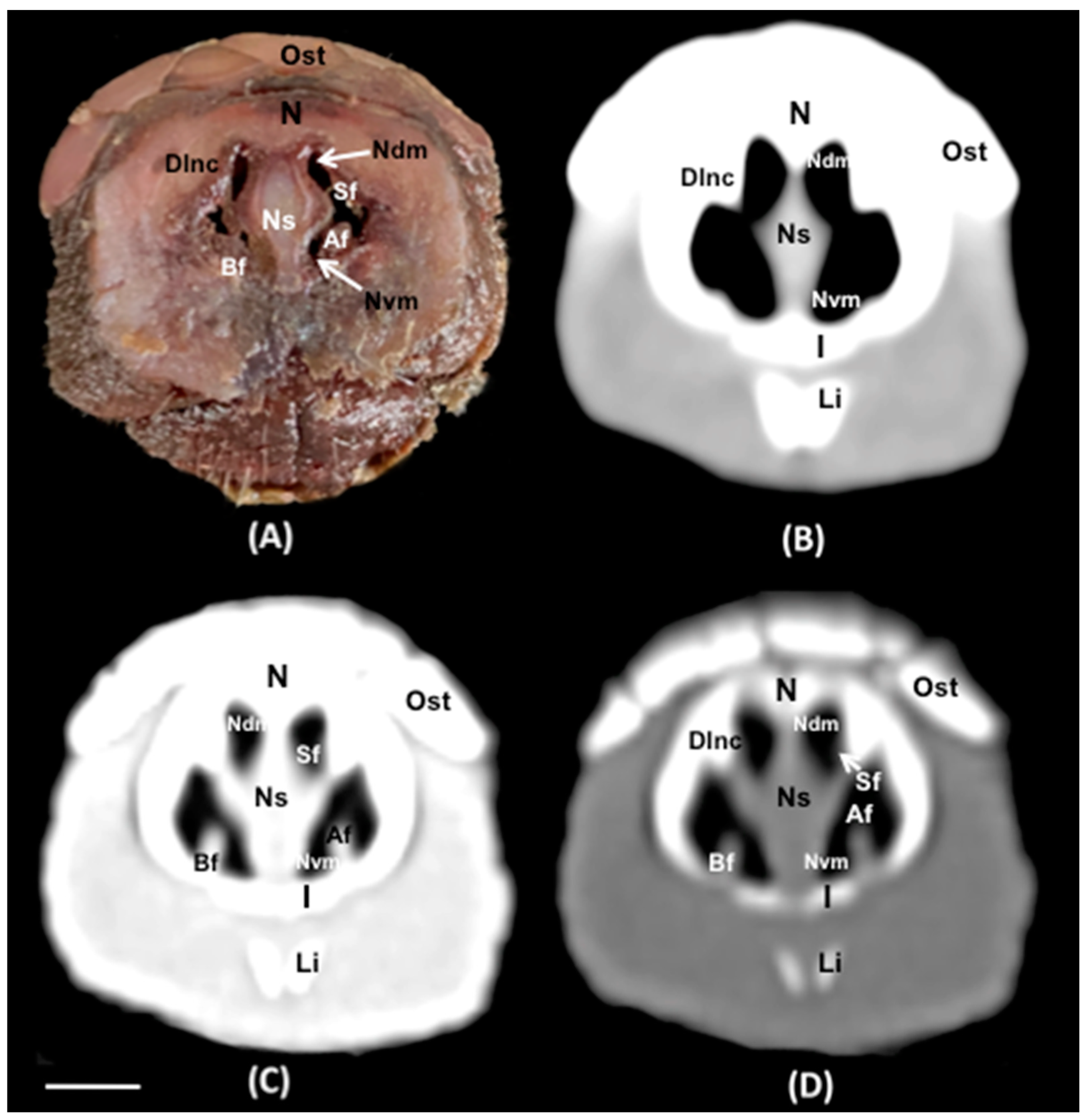
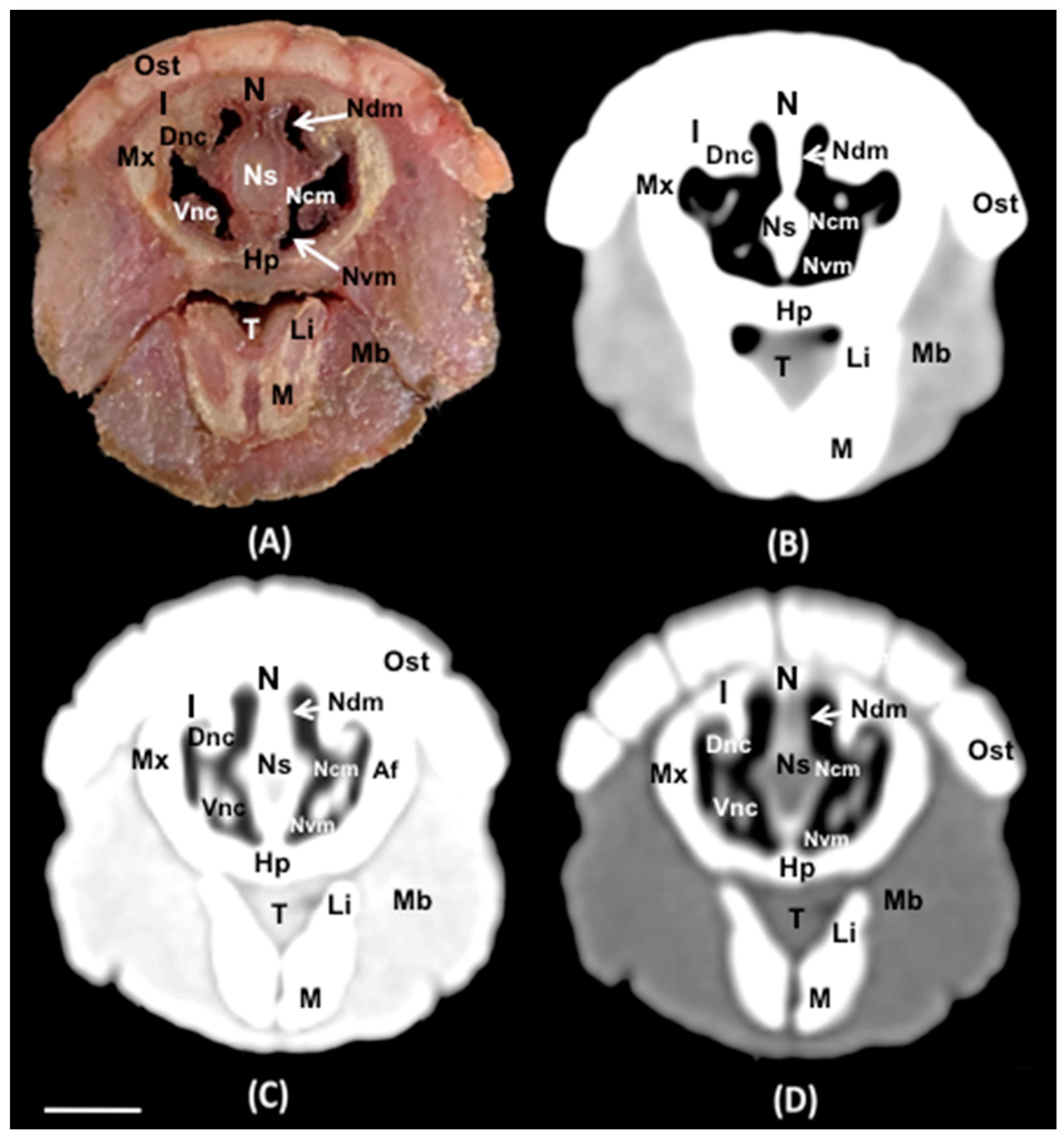
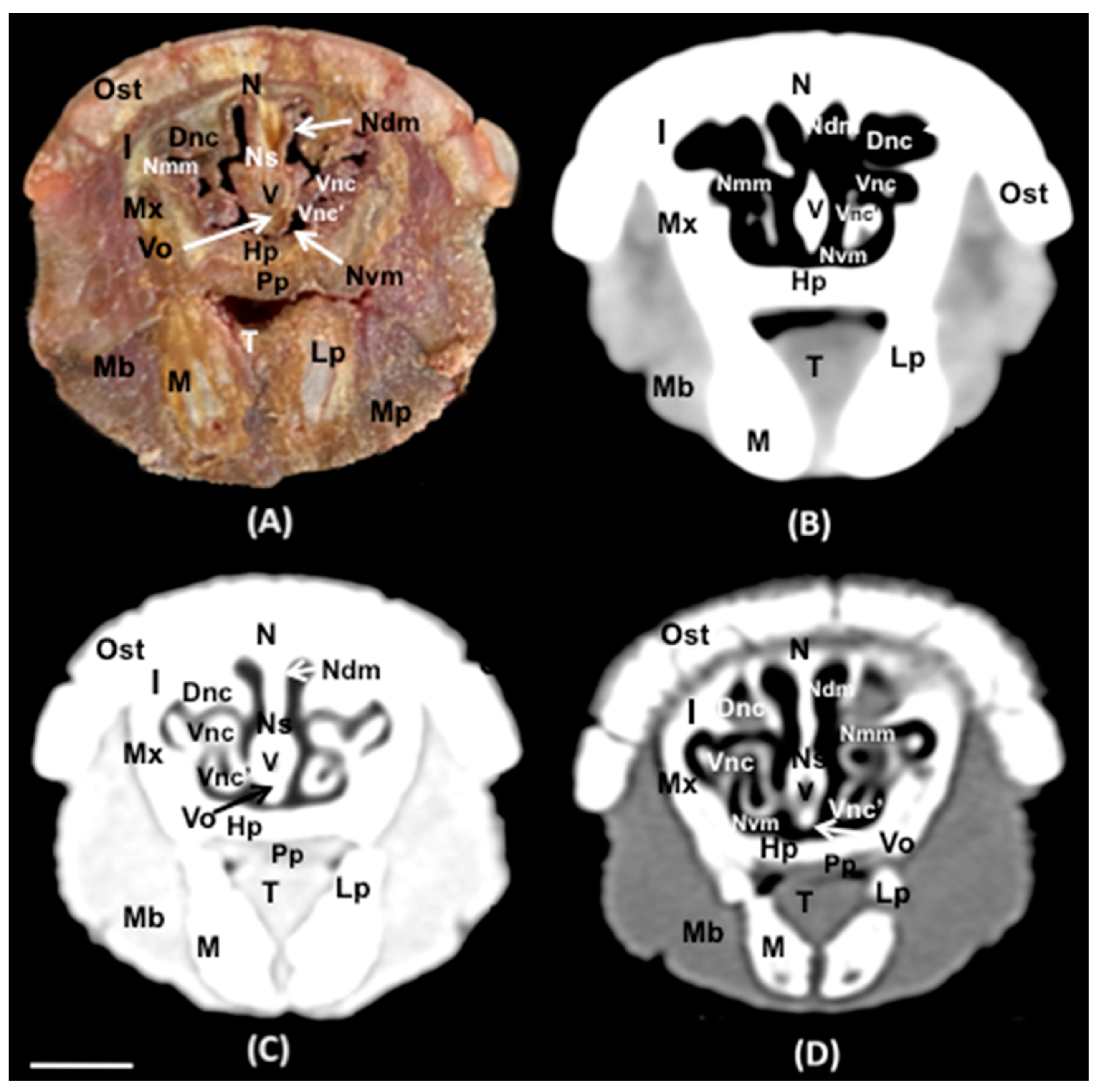
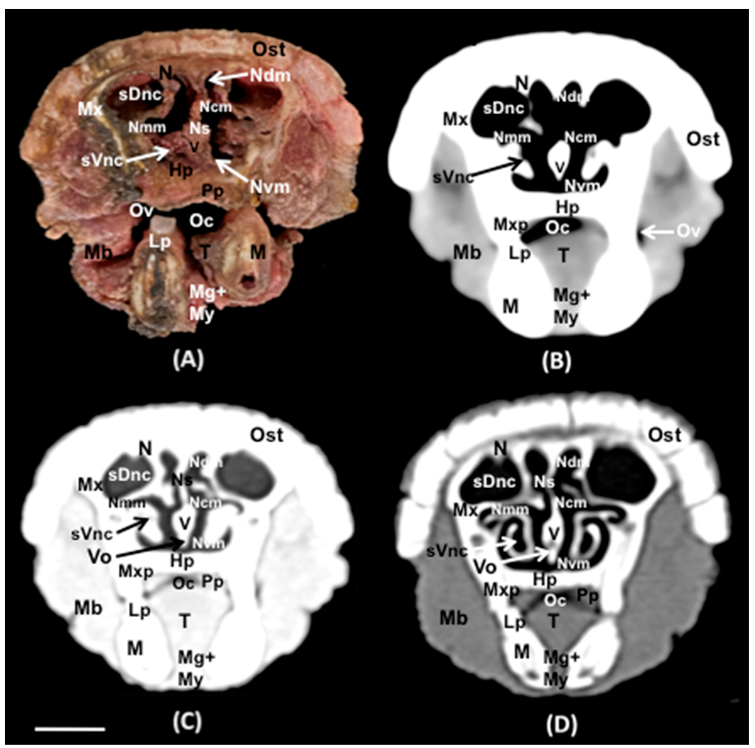
3.1. Anatomical Sections
3.2. Computed Tomography (CT)


4. Discussion
5. Conclusions
Author Contributions
Funding
Institutional Review Board Statement
Informed Consent Statement
Data Availability Statement
Acknowledgments
Conflicts of Interest
References
- Gardner, A.L. Order Cingulata. In Mammal Species of the World: A Taxonomic and Geographic Reference, 3rd ed.; Wilson, D.E., Reeder, D.M., Eds.; Johns Hopkins University Press: Baltimore, MD, USA, 2005; p. 97. ISBN 978-0-8018-8221-0. [Google Scholar]
- Redford, K.H.; Wetzel, R.M. Euphractus sexcinctus. Mamm. Species 1985, 252, 1–4. [Google Scholar] [CrossRef]
- Brittany, B. Euphractus sexcinctus. Available online: https://animaldiversity.org/accounts/Euphractus_sexcinctus/ (accessed on 13 February 2023).
- Schaller, G.B. Mammals and their biomass on a Brazilian ranch. Arq. Zool. 1983, 31, 1–36. [Google Scholar] [CrossRef]
- McNab, B.K. Energetics and the limits to a temperate distribution in armadillos. J. Mammal. 1980, 61, 606–627. [Google Scholar] [CrossRef]
- Alves, L.S.; Midon, M.; Filadelpho, A.L.; Vulcano, L.C.; Knotek, Z. Gross Osteology, Radiographic and Computed Tomographic Morphology of the Axial Skeleton of the Nine-Banded Armadillo (Dasypus novemcinctus). Anat. Histol. Embryol. 2017, 46, 162–177. [Google Scholar] [CrossRef] [PubMed]
- Farrow, C.S. Veterinary Diagnostic Imaging: Birds, Exotic Pets and Wildlife; Mosby Elsevier: St. Louis, MO, USA, 2009. [Google Scholar]
- Lauridsen, H.; Hansen, K.; Wang, T.; Agger, P.; Andersen, J.L.; Knudsen, P.S.; Rasmussen, A.S.; Uhrenholt, L.; Pedersen, M. Inside out: Modern Imaging Techniques to Reveal Animal Anatomy. PLoS ONE 2011, 6, e17879. [Google Scholar] [CrossRef] [PubMed]
- Behroozi, M.; Billings, B.K.; Helluy, X.; Manger, P.R.; Güntürkün, O.; Ströckens, F. Functional MRI in the Nile Crocodile: A New Avenue for Evolutionary Neurobiology. Proc. R. Soc. B Biol. Sci. 2018, 285, 20180178. [Google Scholar] [CrossRef] [PubMed]
- Knipe, M.F. Principles of Neurological Imaging of Exotic Animal Species. Vet. Clin. North Am. Exot. Anim. Pract. 2007, 10, 893–907. [Google Scholar] [CrossRef] [PubMed]
- Banzato, T.; Hellebuyck, T.; Van Caelenberg, A.; Saunders, J.H.; Zotti, A. A Review of Diagnostic Imaging of Snakes and Lizards. Vet. Rec. 2013, 173, 43–49. [Google Scholar] [CrossRef]
- González Rodríguez, E.; Encinoso Quintana, M.; Morales Bordon, D.; Garcés, J.G.; Artiles Nuez, H.; Jaber, J.R. Anatomical Description of Rhinoceros Iguana (Cyclura cornuta cornuta) Head by Computed Tomography, Magnetic Resonance Imaging and Gross-Sections. Animals 2023, 13, 955. [Google Scholar] [CrossRef]
- Morales-Bordon, D.; Encinoso, M.; Arencibia, A.; Jaber, J.R. Cranial Investigations of Crested Porcupine (Hystrix cristata) by Anatomical Cross-Sections and Magnetic Resonance Imaging. Animals 2023, 13, 2551. [Google Scholar] [CrossRef]
- Fumero-Hernández, M.; Encinoso, M.; Melian, A.; Nuez, H.A.; Salman, D.; Jaber, J.R. Cross Sectional Anatomy and Magnetic Resonance Imaging of the Juvenile Atlantic Puffin Head (Aves, Alcidae, Fratercula arctica). Animals 2023, 13, 3434. [Google Scholar] [CrossRef] [PubMed]
- Gaudin, T.J.; Biewener, A.A. The functional morphology of xenarthrous vertebrae in the armadillo Dasypus novemcinctus (Mammalia, Xenarthra). J. Morphol. 1992, 214, 63–81. [Google Scholar] [CrossRef] [PubMed]
- Oliver, J.D.; Jones, K.E.; Pierce, S.E.; Hautier, L. Size and shape regional differentiation during the development of the spine in the nine-banded armadillo (Dasypus novemcinctus). Evol. Dev. 2021, 23, 496–512. [Google Scholar] [CrossRef] [PubMed]
- Alves, L.S.; Babicsak, V.R.; Charlier, M.G.S.; Vulcano, L.C. Radiography, computed tomography and 3D reconstruction of the pelvis in the nine-banded armadillo, Dasypus novemcinctus. In Proceedings of the 40th World Small Animal Veterinary Association Congress, Bangkok, Thailand, 15–18 May 2015; Volume 40, pp. 66–67. [Google Scholar]
- Vizcaıno, S.F.; Milne, N. 2002: Structure and function in armadillo limbs (Mammalia: Xenarthra: Dasypodidae). J. Zool. 2002, 257, 117–127. [Google Scholar] [CrossRef]
- Wible, J.R. Petrosal anatomy of the nine-banded armadillo, Dasypus novemcintus Linnaeus, 1758 (Mammalia, Xenarthra, Dasypodidae). Ann. Carnegie Mus. 2010, 79, 1–28. [Google Scholar] [CrossRef]
- Silva, A.B.; Sousa, M.M.; Silva, J.V.; Oliveira, I.M.; Barbosa, C.M.; Santos, M.; Conde, A.M. Anatomy of the nasal cavity of nine-banded armadillo (Dasypus novemcinctus, Linnaeus, 1758). J. Interdiscip. Biocienc. 2016, 1, 1–4. [Google Scholar] [CrossRef]
- Billet, G.; Hautier, L.; de Thoisy, B.; Delsuc, F. The hidden anatomy of paranasal sinuses reveals biogeographically distinct morphotypes in the nine-banded armadillo (Dasypus novemcinctus). PeerJ 2017, 5, e3593. [Google Scholar] [CrossRef] [PubMed]
- Fonseca, C.M.B.; da Silva, A.B.S.; Cavalcante, M.M.A.S.; de Oliveira, I.M.; Moura, S.M.S.; Cunha, B.M.; Leite, C.M.C.; de Carvalho, M.A.M.; Moura, W.R.A.; Rizzo, M.D.S.; et al. Morphology of laryngeal cartilage of the nine-banded armadillo (Dasypus novemcinctus) Linnaeus, 1758. Microsc. Res. Tech. 2017, 80, 1089–1095. [Google Scholar] [CrossRef] [PubMed]
- Vizcaíno, S.F.; Fariña, R.A.; Bargo, M.S.; De Iuliis, G. Functional and phylogenetic assessment of the masticatory adaptations in Cingulata (Mammalia, Xenarthra). Rev. Asos. Paleontol. Argent. 2004, 41, 651–664. [Google Scholar]
- Alves, L.S.; Oliva, L.R.; Charlier, M.G.S.; Bonatelli, S.P.; Inamassu, L.R.; Vulcano, L.C.; Teixeira, C.R. Fratura em seguimento lombar da coluna vertebral em um tatugalinha (Dasypus novemcinctus). An. Simp. Intern. Diag. Imag. SINDIV 2013, 3, 57–58. [Google Scholar]
- Boyde, A.; Mills, D.; Abba, A.M.; Ezquiaga, M.C. Fleas and lesions in armadillo osteoderms. J. Anat. 2023, 242, 1029–1036. [Google Scholar] [CrossRef] [PubMed]
- Getty, R. The Anatomy of the Domestic Animals, 5th ed.; Macmillan Company of India Limited: West Bengal, India, 2004. [Google Scholar]
- Arencibia, A.; Vázquez, J.M.; Jaber, R.; Gil, F.; Ramiírez, J.A.; Rivero, M.; González, N.; Wisner, E.R. Magnetic resonance imaging and cross-sectional anatomy of the normal equine sinuses and nasal passages. Vet. Radiol. Ultrasound 2000, 41, 313–319. [Google Scholar] [CrossRef] [PubMed]
- Burk, R.L. Computed tomographic anatomy of the canine nasal passages. Vet. Radiol. Ultrasound 1992, 33, 170–176. [Google Scholar] [CrossRef]
- Arencibia, A.; Vazquez, J.M.; Rivero, M.; Latorre, R.; Sandoval, J.A.; Vilar, J.M.; Ramirez, J.A. Computed tomography of normal cranioencephalic structures in two horses. Anat. Histol. Embryol. 2000, 29, 295–299. [Google Scholar] [CrossRef]
- Arencibia, A.; Hidalgo, M.R.; Vázquez, J.M.; Contreras, S.; Ramírez, G.; Orós, J. Sectional Anatomic and Magnetic Resonance Imaging Features of the Head of Juvenile Loggerhead Sea Turtles (Caretta Caretta). Am. J. Vet. Res. 2012, 73, 1119–1127. [Google Scholar] [CrossRef] [PubMed]
- Bercier, M.; Alexander, K.; Gorow, A.; Pye, G.W. Computed tomography and magnetic resonance for the advanced imaging of the normal nasal cavity and paranasal sinuses of the koala (Phascolarctos cinereus). J. Zoo Wildl. Med. 2014, 45, 766–774. [Google Scholar] [CrossRef]
- Van Caelenberg, A.I.; De Rycke, L.M.; Hermans, K.; Verhaert, L.; Van Bree, H.J.; Gielen, I.M. Low-field magnetic resonance imaging and cross-sectional anatomy of the rabbit head. Vet. J. 2011, 188, 83–91. [Google Scholar] [CrossRef] [PubMed]
- Mahdy, M. Correlation between computed tomography, magnetic resonance imaging and cross-sectional anatomy of the head of the guinea pig (Cavia porcellus, Linnaeus 1758). Anat. Histol. Embryol. 2022, 51, 51–61. [Google Scholar] [CrossRef] [PubMed]
- De Rycke, L.M.; Saunders, J.H.; Gielen, I.M.; van Bree, H.J.; Simoens, P.J. Magnetic resonance imaging, computed tomography, and cross-sectional views of the anatomy of normal nasal cavities and paranasal sinuses in mesaticephalic dogs. Am. J. Vet. Res. 2003, 64, 1093–1098. [Google Scholar] [CrossRef] [PubMed]
- Capello, V. Diagnostic Imaging of Dental Disease in Pet Rabbits and Rodents. Vet. Clin. North Am. Exot. Anim. Pract. 2016, 19, 57–82. [Google Scholar] [CrossRef]
- Encinoso, M.; Morales, D.; Déniz, S.; Guerra, J.V.; Jaber, J.R. Computed Tomography and Magnetic Resonance Imaging of a Rhinosinusitis Secondary to a Dental Abscess in a Crested Porcupine (Hystrix cristata). Slov. Vet. Res. 2023, 60, 37–43. [Google Scholar] [CrossRef]


Disclaimer/Publisher’s Note: The statements, opinions and data contained in all publications are solely those of the individual author(s) and contributor(s) and not of MDPI and/or the editor(s). MDPI and/or the editor(s) disclaim responsibility for any injury to people or property resulting from any ideas, methods, instructions or products referred to in the content. |
© 2024 by the authors. Licensee MDPI, Basel, Switzerland. This article is an open access article distributed under the terms and conditions of the Creative Commons Attribution (CC BY) license (https://creativecommons.org/licenses/by/4.0/).
Share and Cite
Jaber, J.R.; Morales Bordon, D.; Arencibia, A.; Corbera, J.A.; Conde-Felipe, M.; Ayala, M.D.; Encinoso, M. Correlation between Cross-Sectional Anatomy and Computed Tomography of the Normal Six-Banded Armadillo (Euphractus sexcintus) Nasal Cavity and Paranasal Sinuses. Animals 2024, 14, 1135. https://doi.org/10.3390/ani14071135
Jaber JR, Morales Bordon D, Arencibia A, Corbera JA, Conde-Felipe M, Ayala MD, Encinoso M. Correlation between Cross-Sectional Anatomy and Computed Tomography of the Normal Six-Banded Armadillo (Euphractus sexcintus) Nasal Cavity and Paranasal Sinuses. Animals. 2024; 14(7):1135. https://doi.org/10.3390/ani14071135
Chicago/Turabian StyleJaber, José Raduan, Daniel Morales Bordon, Alberto Arencibia, Juan Alberto Corbera, Magnolia Conde-Felipe, Maria Dolores Ayala, and Mario Encinoso. 2024. "Correlation between Cross-Sectional Anatomy and Computed Tomography of the Normal Six-Banded Armadillo (Euphractus sexcintus) Nasal Cavity and Paranasal Sinuses" Animals 14, no. 7: 1135. https://doi.org/10.3390/ani14071135
APA StyleJaber, J. R., Morales Bordon, D., Arencibia, A., Corbera, J. A., Conde-Felipe, M., Ayala, M. D., & Encinoso, M. (2024). Correlation between Cross-Sectional Anatomy and Computed Tomography of the Normal Six-Banded Armadillo (Euphractus sexcintus) Nasal Cavity and Paranasal Sinuses. Animals, 14(7), 1135. https://doi.org/10.3390/ani14071135