
Dissecting the Effects of Cephenemyia stimulator on the Olfactory Turbinates and Nasopharynx of Roe Deers (Capreolus capreolus)

 ,
,  , ,
, ,  and
and 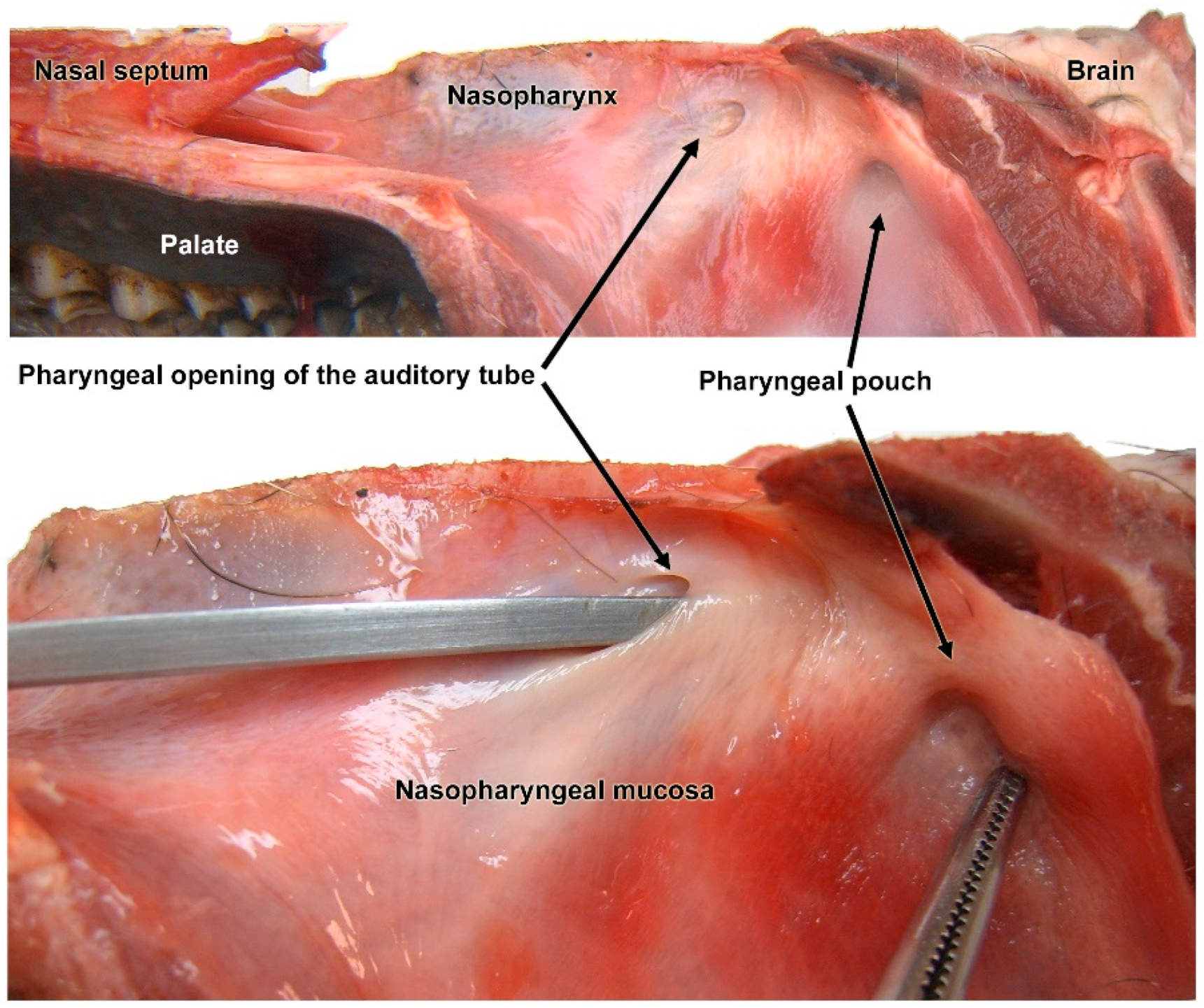
Abstract
:Simple Summary
Abstract
1. Introduction
2. Materials and Methods
2.1. Samples
2.2. Pathological Study
3. Results
3.1. Macroscopic Study (Figure 1 and Figure 2)


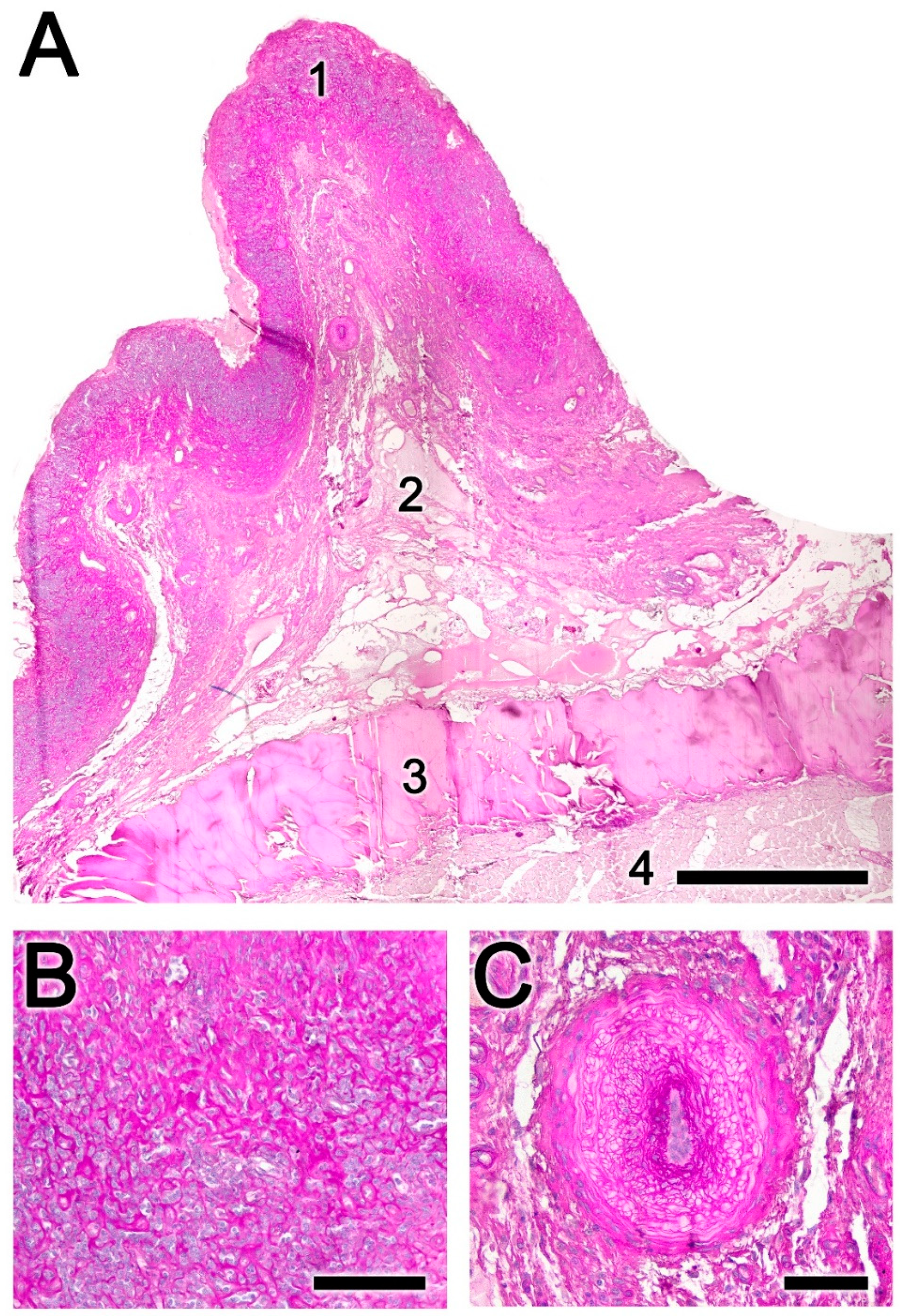
3.2. Histopathology of the Nasopharynx (Figure 3, Figure 4, Figure 5, Figure 6 and Figure 7)


3.3. Histopathology of the Nasal Cavity (Figure 8 and Figure 9)


4. Discussion
5. Conclusions
Author Contributions
Funding
Institutional Review Board Statement
Informed Consent Statement
Data Availability Statement
Acknowledgments
Conflicts of Interest
References
- Sugár, L. The Occurence of Nasal Throat Bot Flies (Oestridae) in Wild Ruminants in Hungary. Parasitol. Hung. 1974, 7, 181–189. [Google Scholar]
- Cogley, T.P. Effects of Cephenemyia spp. (diptera: Oestridae) on the nasopharynx of black-tailed deer (Odocoileus hemionus columbianus). J. Wildl. Dis. 1987, 23, 596–605. [Google Scholar] [CrossRef] [PubMed]
- Sugár, L. Nasopharyngeal Bot Infestation of Wild Ruminants (Oestridosis). In Diseases of Wildlife; Mezőgazdasági Kiadó: Budapest, Hungary, 1978; pp. 156–158. [Google Scholar]
- Tabouret, G.; Vouldoukis, I.; Duranton, C.; Prevot, F.; Bergeaud, J.P.; Dorchies, P.; Mazier, D.; Jacquiet, P. Oestrus ovis (Diptera: Oestridae): Effects of Larval Excretory/Secretory Products on Nitric Oxide Production by Murine RAW 264·7 Macrophages. Parasite Immunol. 2001, 23, 111–119. [Google Scholar] [CrossRef] [PubMed]
- Tabouret, G.; Bret-Bennis, L.; Dorchies, P.; Jacquiet, P. Serine Protease Activity in Excretory–Secretory Products of Oestrus Ovis (Diptera: Oestridae) Larvae. Vet. Parasitol. 2003, 114, 305–314. [Google Scholar] [CrossRef] [PubMed]
- Angulo-Valadez, C.E.; Scholl, P.J.; Cepeda-Palacios, R.; Jacquiet, P.; Dorchies, P. Nasal Bots… a Fascinating World! Vet. Parasitol. 2010, 174, 19–25. [Google Scholar] [CrossRef] [PubMed]
- Kusak, J.; Špičić, S.; Slijepčević, V.; Bosnić, S.; Rajković Janje, R.; Duvnjak, S.; Sindičić, M.; Majnarić, D.; Cvetnić, Ž.; Huber, Đ. Health Status of Red Deer and Roe Deer in Gorski Kotar, Croatia. Vet. Arh. 2012, 82, 59–73. [Google Scholar]
- Žele Vengušt, D.; Kuhar, U.; Jerina, K.; Vengušt, G. Twenty Years of Passive Disease Surveillance of Roe Deer (Capreolus capreolus) in Slovenia. Animals 2021, 11, 407. [Google Scholar] [CrossRef] [PubMed]
- Flis, M.; Rataj, B.; Grela, E.R. Occurrence of Cephenemyia stimulator Larvae in Male Roe Deer (Capreolus capreolus L.) in the Lublin Upland, Poland, and Their Impact on Particular Animal Health Indicators. J. Vet. Res. 2021, 65, 287–292. [Google Scholar] [CrossRef] [PubMed]
- Notario, A.; Castresana, L. Contribution to the Knowledge of Cephenemyia Stimulator Clark, 1815 (Diptera, Oestridae) in Spain. Folia Venatoria 2001, 30–31, 325–326. [Google Scholar]
- Fidalgo, L.E.; López, A.M.; Pérez, J.M.; Martínez, C. Bases Epidemiológicas Para El Control de Cephenemyia Stimulator En Corzos En España; Universidade de Santiago de Compostela & Fundación FEDENCA: Galicia, Spain, 2013. [Google Scholar]
- Király, I.; Egri, B. Epidemiological Characteristics of Cephenemyia stimulator (Clark, 1815) Larval Infestation in European Roe Deer (Capreolus capreolus) in Hungary. Acta Zool. Acad. Sci. Hung. 2007, 53, 271–279. [Google Scholar]
- Ortiz-Leal, I.; Torres, M.V.; Villamayor, P.R.; López-Beceiro, A.; Sanchez-Quinteiro, P. The Vomeronasal Organ of Wild Canids: The Fox (Vulpes Vulpes) as a Model. J. Anat. 2020, 237, 890–906. [Google Scholar] [CrossRef] [PubMed]
- Zumpt, F. Myiasis in Man and Animals in the Old World: A Textbook for Physicians, Veterinarians and Zoologists; Butterworths: London, UK, 1965. [Google Scholar]
- Yao, H.; Liu, M.; Ma, W.; Yue, H.; Su, Z.; Song, R.; Ma, Q.; Li, L.; Wu, Z.; Ma, Y.; et al. Prevalence and Pathology of Cephalopina Titillator Infestation in Camelus Bactrianus from Xinjiang, China. BMC Vet. Res. 2022, 18, 360. [Google Scholar] [CrossRef]
- Shamsi, E.; Radfar, M.H.; Nourollahifard, S.R.; Bamorovat, M.; Nasibi, S.; Fotoohi, S.; Hakimi Parizi, M.; Kheirandish, R. Nasopharyngeal Myiasis Due to Cephalopina Titillator in Southeastern Iran: A Prevalence, Histopathological, and Molecular Assessment. J. Parasit. Dis. 2023, 47, 369–375. [Google Scholar] [CrossRef] [PubMed]
- Azizi, H.; Kojouri, G.; Pirali, Y.; Maghami, M.; Zahirabadi, M.B. Clinical, Pathological and Epidemiological Aspects of Cephalopina Titillator Larval, Exciter in Camelus Dromedaris from the Rafsanjan Region, Iran. Asian Res. J. Agric. 2024, 17, 28–35. [Google Scholar] [CrossRef]
Disclaimer/Publisher’s Note: The statements, opinions and data contained in all publications are solely those of the individual author(s) and contributor(s) and not of MDPI and/or the editor(s). MDPI and/or the editor(s) disclaim responsibility for any injury to people or property resulting from any ideas, methods, instructions or products referred to in the content. |
© 2024 by the authors. Licensee MDPI, Basel, Switzerland. This article is an open access article distributed under the terms and conditions of the Creative Commons Attribution (CC BY) license (https://creativecommons.org/licenses/by/4.0/).
Share and Cite
Ortiz-Leal, I.; Torres, M.V.; López-Beceiro, A.; Sanchez-Quinteiro, P.; Fidalgo, L. Dissecting the Effects of Cephenemyia stimulator on the Olfactory Turbinates and Nasopharynx of Roe Deers (Capreolus capreolus). Animals 2024, 14, 1297. https://doi.org/10.3390/ani14091297
Ortiz-Leal I, Torres MV, López-Beceiro A, Sanchez-Quinteiro P, Fidalgo L. Dissecting the Effects of Cephenemyia stimulator on the Olfactory Turbinates and Nasopharynx of Roe Deers (Capreolus capreolus). Animals. 2024; 14(9):1297. https://doi.org/10.3390/ani14091297
Chicago/Turabian StyleOrtiz-Leal, Irene, Mateo V. Torres, Ana López-Beceiro, Pablo Sanchez-Quinteiro, and Luis Fidalgo. 2024. "Dissecting the Effects of Cephenemyia stimulator on the Olfactory Turbinates and Nasopharynx of Roe Deers (Capreolus capreolus)" Animals 14, no. 9: 1297. https://doi.org/10.3390/ani14091297
APA StyleOrtiz-Leal, I., Torres, M. V., López-Beceiro, A., Sanchez-Quinteiro, P., & Fidalgo, L. (2024). Dissecting the Effects of Cephenemyia stimulator on the Olfactory Turbinates and Nasopharynx of Roe Deers (Capreolus capreolus). Animals, 14(9), 1297. https://doi.org/10.3390/ani14091297