Article

Prenatal Exposure to Persistent Organic Pollutants and Maternal Folic Acid Supplementation: Their Impact on Glucose Homeostasis in Male Rat Descendants

1
School of Nutrition, Faculty of Agricultural and Food Sciences, Université Laval, 2425 rue de l’Agriculture, Quebec City, QC G1V 0A6, Canada
2
Institute of Nutrition and Functional Foods, Université Laval, 2440 Boulevard Hochelaga, Quebec City, QC G1V 0A6, Canada
3
Centre de Recherche en Reproduction, Développement et Santé Intergénérationnelle, Université Laval, 2425 rue de l’Agriculture, Quebec City, QC G1V 0A6, Canada
4
Department of Animal Sciences, Faculty of Agricultural and Food Sciences, Université Laval, 2425 rue de l’Agriculture, Quebec City, QC G1V 0A6, Canada
5
Department of Medicine, Faculty of Medicine, Quebec Heart and Lung Institute Cardiology Group, Université Laval, 2725 Chemin Ste-Foy, Quebec City, QC G1V 4G5, Canada
6
Department of Pharmacology and Therapeutics, Faculty of Medicine, McGill University, 3655 Promenade Sir William Osler Room 1325, Montreal, QC H3G 1Y6, Canada
7
Department of Animal Science, Faculty of Agricultural and Environmental Sciences, McGill University, 21111 Lakeshore Road, Sainte Anne de Bellevue, QC H9X 3V9, Canada
8
Department of Pediatrics, Montreal Children’s Hospital, McGill University Health Centre Research Institute, McGill University, 1001 Boulevard Décarie, Montreal, QC H4A 3J1, Canada
9
Nutrition Research Division, Health Canada, 251 Promenade Sir Frederick Banting, Ottawa, ON K1A 0K9, Canada
*
Author to whom correspondence should be addressed.
Environments 2021, 8(3), 24; https://doi.org/10.3390/environments8030024
Submission received: 6 February 2021 / Revised: 15 March 2021 / Accepted: 15 March 2021 / Published: 22 March 2021
(This article belongs to the Special Issue Effect of Environmental Pollutants on Metabolic Defects)

Abstract

:
Exposure to persistent organic pollutants (POPs) is associated with insulin resistance while folic acid (FA) may offer a protective effect. However, the paternal contribution to metabolic phenotypes in offspring is not well known yet. Hence, we investigated whether maternal exposure to POPs affects glucose homeostasis and whether maternal FA supplementation counteracts POP effects transmitted via male descendants. Sprague–Dawley founder dams (F0) were fed a diet containing 2 or 6 mg/kg of FA and were force-fed with either a POP mixture or corn oil for 9 weeks. Subsequent male descendants did not receive any treatment. Blood glucose, plasma insulin and C-peptide were measured during an oral glucose tolerance test in males aged 90 and 180 days from generation 1 (F1), 2 (F2) and 3 (F3). Prenatal POP exposure increased fasting glucose in 90-day-old F1 males and C-peptide in 90-day-old F2 males. Prenatal FA supplementation decreased C-peptide in 90 and 180-day-old F1 males. In 180-day-old F3 males, FA supplementation counteracted POPs on fasting and postglucose C-peptide, indicating reduced insulin secretion. Prenatal exposure to an environmentally relevant POP mixture caused abnormalities in glucose homeostasis that are transmitted from one generation to the next through the paternal lineage. Prenatal FA supplementation counteracted some of the deleterious effects of POPs on glucose homeostasis.

1. Introduction

According to the World Health Organization (WHO), the incidence of diabetes in the adult population nearly doubled worldwide between 1980 and 2014, increasing from 4.7% to 8.5% [1]. This chronic metabolic disease is characterized by decreased insulin secretion and/or insulin sensitivity and an increase in blood glucose known as glucose intolerance. In the Canadian Inuit population, the prevalence of diabetes rose from 2% in 2001 to 5% in 2012 to 97% of cases were type 2 diabetes [2,3]. This is partly due to a higher rate of obesity and changes in dietary habits. In recent decades, consumption patterns in this population reflect consumption of energy-dense foods with low nutrient density [4] characterized by a higher sugar and fat intakes and a lower intake of micronutrients, including folate [5].
Persistent organic pollutants (POPs) refer to residues derived from industrial chemicals, such as polychlorinated biphenyls (PCBs), organochlorine pesticides (OCPs), and polybrominated diphenyl ethers (PBDEs), that are resistant to biodegradation and have remained in the environment for decades after bans on their use [6,7]. Many POPs have been transported by atmospheric and ocean currents over long distances and are deposited in cold regions, such as the Arctic [8]. POPs are highly lipophilic, thereby bioaccumulating and persisting in adipose tissues of animals and humans [9]. Biomagnification has led to high concentrations of POPs at the top of the Arctic food chain [10]. Inuit people are exposed to high levels of POPs through their traditional diets made up of fish and marine mammals [11,12,13]. Therefore, POP exposure has emerged as an additional factor in the pathogenesis of type 2 diabetes in the Inuit population [14]. Indeed, the endocrine-disruptive potential of some POPs is proposed to perturb glucose homeostasis in humans. Even at low exposures found in serum samples of US population, POPs are associated with higher body mass index and insulin resistance [15]. Serum and total body POP concentrations are positively correlated with fasting blood glucose levels in human adults [16]. A lower level of GLUT4 glucose transporters has been observed in adipose tissues of Vietnam war veterans who were exposed to dioxin via Agent Orange [17].
There is growing evidence that nutrients can modulate gene and epigenetic expression associated with metabolic dysfunctions. For instance, as a donor of one carbon group in one-carbon metabolism, folate plays a key role in de novo nucleotide and methionine synthesis, and as such, impacts DNA integrity and stability [18]. Intake of folic acid (FA), the synthetic form of folate, has been proposed to lower DNA damage related to oxidative stress produced by type 2 diabetes [19]. Using the Agouti Avy mouse model, FA supplementation of dams during pregnancy decreased obesity severity in offspring over three generations [20] and reduced phenotypic abnormalities caused by exposure to an endocrine disrupter, bisphenol A [21]. It is, therefore, possible that FA supplementation has an impact on intergenerational and transgenerational phenotypic expression. Moreover, although most of the studies focus on maternal health, several studies suggest that the paternal environment can also influence the health or disease development status of offspring [22,23,24]. Of particular interest are early symptoms of metabolic syndrome observed in F1 male rats exposed to DDT, an OCP, in utero (during embryonic stages) and in F2 male rats exposed through the germ line [23]. This study showed a transgenerational effect of DDT on F3 male offspring through epigenetic mechanisms [23].
The aim of the present study was therefore to examine whether FA supplementation of the maternal diet in pregnancy can overcome the effects of prenatal exposure to POPs at levels equivalent to those observed in the Northern Quebec Inuit population, and thereby improve glucose homeostasis in male offspring and their male descendants in a rat model at 90 and 180 days old, which are approximately equivalent to young adult and adult men, respectively.

2. Materials and Methods

2.1. Animal Care

Sprague–Dawley rats were purchased from Charles-River Laboratories (St. Constant, QC, Canada) and housed in the animal facilities at the Institute of Nutrition and Functional Foods (INAF, Québec, QC, Canada). The F0 generation consisted of females (n = 24) weighing about 160 g at the age of 5 weeks and males (n = 12) weighing about 220 g at the age of 10 weeks. F0 females were housed two per cage under controlled temperature (22 °C) and relative humidity (50%) with a 12 h day/night cycle and received food and water ad libitum. A 10-day period of adaptation was required for the F0 animals prior to administration of the experimental compounds. During this acclimation, they were fed the AIN-93G purified diet (# 110700 by Dyets, Inc, Bethlehem, PA, USA) [25]. Animal care and all treatment procedures were compliant with the guidelines of the Canadian Council on Animal Care and approved by the Université Laval Animal Research Ethics Committee (certificate # 2015010-2).

2.2. POP Mixture

As described previously [22], the POP mixture (Table 1) was designed to mimic the contaminants found in the adipose tissue of ringed seal blubber [26], a traditional food of the Inuit population. The administered dose corresponded to metabolite levels measured in Nunavik Inuit [22]. Its main components are a mixture of PCBs called Aroclor and congener neat mixture (AccuStandard, New Haven, CT, USA). The chemicals were weighed then dissolved in corn oil to yield a 10× stock solution that was shielded from light at room temperature. The mixture was diluted with corn oil for administration by gavage.

2.3. Diet Formulation

Two purified diets based on the AIN-93G basic diet [25] were purchased from Dyets Inc. (Bethlehem, PA, USA) (Table 2). As described previously [27], the control diet contained 2 mg of FA per kg, which is equivalent to the basic requirement for rodents [25] and corresponds to the dietary reference intake of 0.4 mg per day for human adults [27]. The supplemented diet contained 6 mg of FA per kg, representing three times the minimum recommended intake for rodents and approximates the FA intake that would be consumed by a woman in Canada from fortified foods and prenatal supplements [27]. The control and supplemented diets are hereinafter designated, respectively, as 1× (2 mg FA/kg diet) and 3× (6 mg FA/kg diet).

2.4. Experimental Design

2.4.1. F0 Generation

As shown in Figure 1, F0 Sprague–Dawley founder dams were fed the 1× or 3× diet [28] and received by gavage (3 days/week) either a corn oil control or the POP mixture starting at the age of 7 weeks. The F0 females were weighed three times a week prior to gavage. At the age of 12 weeks, they were mated with untreated F0 males. Except during mating, sires were fed the control diet only. Reproduction plus gestation was 4 weeks long. The experimental treatments thus lasted 9 weeks. All F0 postpartum females were fed the control diet only and the POP mixture treatment was ended. The four experimental groups (n = 6/group) were thus (1) corn oil plus 1× FA (CTRL), (2) POP mixture plus 1× FA (POPs), (3) corn oil plus 3× FA (FA), and (4) POP mixture plus 3× FA (POPs + FA).

2.4.2. F1, F2 and F3 Generations

For each generation, twelve males (n = 12) were kept for reproduction and ten males (n = 10) were used for experiments. F1 males (120 days old (n = 12)) were mated with 10-week-old untreated females fed the 1× FA diet (n = 6) to yield generation F2, of which the males (n = 12) were mated with untreated females fed the 1× FA diet (n = 6) to yield generation F3. F1 male pups were weaned to 1× FA diet at the age of 21 days and maintained on diet for the duration of the study. All subsequent generations were also weaned to and maintained on the 1× FA diet. F1 males were exposed to treatments in utero, F2 were exposed to treatments as germ cells in utero, and F3 were not directly exposed (ancestral exposure). F1 and F2 phenotypic variations were defined as “intergenerational” whereas F3 phenotypic variations were considered to be “transgenerational”. All F1, F2 and F3 males were weighed and sexed at the age of 21 days and coded using a nontoxic marker (Sharpie, AP Seal) on the tail. They were then selected to ensure approximately equal weights within and between groups, housed two per cage for two weeks and then one per cage for food intake and body weight measurements (twice a week). On day 194, F1–F3 males were anesthetized using 3% isoflurane and sacrificed by exsanguination via cardiac puncture.

2.5. Blood Collection

On days 90 and 180 of the F1, F2 and F3 generations, glucose tolerance tests (GTTs) were performed. For GTT assessment, rats were fasted 12 h before gavage with 50% dextrose solution (2 g/kg). Blood samples were obtained from the saphenous vein and blood glucose levels were measured (0, 15, 30, 60, 90 and 120 min after ingestion) using a portable glucometer (Contour, Bayer, Leverkusen, Germany). Plasma was separated by centrifugation (950× g, 10 min) and immediately snap-frozen in liquid nitrogen, then stored at −80 °C until analysis.

2.6. Plasma Biochemistry, Homeostasis Model Assessment of Insulin Resistance (HOMA-IR) and Matsuda Indexes

Plasma insulin and C-peptide concentrations were determined using ELISA kits per manufacturer’s instructions (Mercodia, Uppsala, Sweden). The homeostasis model assessment of insulin resistance (HOMA-IR) and the Matsuda index were used as proxies of insulin sensitivity, respectively, in the fasting state and during the glucose tolerance test [29,30]. HOMA-IR index was calculated after 12h fasting and Matsuda index was calculated using all GTT time points.

2.7. Glucose Tolerance Test

The incremental area under the curve (IAUC) was calculated for glucose, insulin, and C-peptide using the trapezoidal method to evaluate glucose tolerance. The insulin/glucose ratio based on IAUC was used as an additional measurement of β-cell function. The C-peptide/insulin ratio based on IAUC was determined to evaluate insulin clearance.

2.8. Statistical Analysis

Data are expressed as the mean ± standard error of the mean (SEM) for each treatment group. One-way analysis of variance (ANOVA) for a 2 × 2 factorial design using the MIXED procedure (SAS software version 9.4, SAS Institute Inc., Cary, NC, USA) was applied to fasting variables when comparing the four groups. Two-way ANOVA with repeated measurements was applied to data at different time points during the glucose tolerance test. The F0 founder female for each male rat was controlled in the model. When non-normal data were observed, a logarithmic transformation of the data was performed to obtain normality. The main effects of POPs and FA as well as their interaction (POPs*FA) were then determined. Differences were considered significant at p ≤ 0.05. When a significant (p ≤ 0.05) POPs*FA interaction was observed, the Tukey Honest Significant Difference (HSD) test was performed to determine between-group differences.

3. Results and Discussion

3.1. Body Weight and Food Intake

As presented in Table 3, neither POPs nor FA had any effect on body weight, weight gain or food intake of F0 dams or of male offspring throughout all generations, indicating that the metabolic changes observed following the treatments were independent of food intake and body weight.

3.2. Exposure to POPs

In recent years, the impact of environmental pollutants on the development of metabolic disorders such as type 2 diabetes [31,32], insulin resistance [33] and obesity has been investigated in humans [15,34] and animal models [35,36,37]. Male mice fed a diet that approximates a western diet rich in farmed Atlantic salmon fillets contaminated with POPs (OCPs, dioxins, furans, and PCBs), induced a mild increase in blood glucose and a large increase in plasma insulin [35].
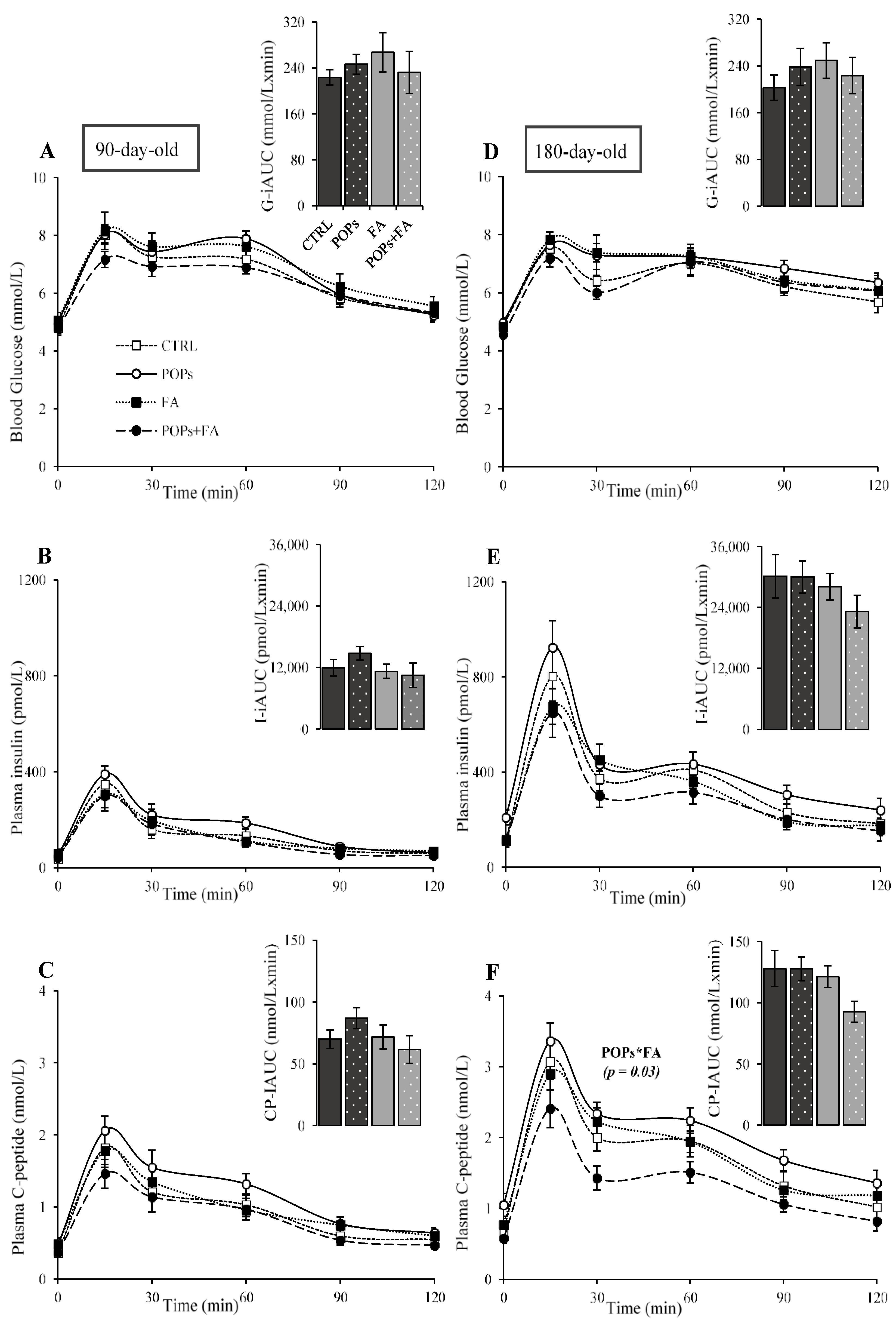
A high-fat diet containing PCBs appears to increase postprandial blood glucose as well as fasting and postprandial plasma insulin in mice [37]. There is little evidence, however, that the effects of prenatal exposure to POPs on glucose homeostasis affect more than one generation of descendants. The present study in rats showed that prenatal exposure to an environmentally relevant POP mixture was associated with increased fasting glucose (p = 0.002) and a tendency for higher fasting insulin (p = 0.08) in 90-day-old F1 males (Figure 2A,B). Although glucose IAUC was lower in these groups than in the other groups (p = 0.03) (Figure 3A insert), this reduction cannot be interpreted as a capacity of prenatal POP exposure to improve glucose tolerance due to the calculation of IAUC, which included their high fasting glycemia. More importantly, in these animals, prenatal exposure to POPs increased the HOMA-IR index, a widely used indicator of insulin resistance (p = 0.01) (Table 4). Thus, the increase in fasting glucose and HOMA-IR suggests higher insulin resistance in 90-day-old F1 males.
In the corresponding F2 male offspring, fasting plasma C-peptide increased (p = 0.05) (Figure 2C) while the Matsuda index decreased (p = 0.03) (Table 5), indicating heightened insulin secretion and suggesting again increased insulin resistance. This is consistent with previous human and in vitro studies showing that POPs (PCBs and OCPs) at low doses can impair insulin secretion through pancreatic β-cell dysfunction [38]. As in male rats aged 90 days (Figure 2A,B), fasting glucose and insulin levels did not change in F2 males aged 180 days (Figure 2D,E).
In F2 males aged 90 and 180 days, no changes in blood glucose, plasma insulin and C-peptide levels (Figure 4A–F) and IAUC (Figure 4A insert to Figure 4F insert) were noted. Finally, ancestral exposure to POPs was also associated with increased insulin secretion as shown by an elevation of C-peptide (Figure 2F) in the fasting state and during the glucose tolerance test (Figure 5F) in F3 male rats aged 180 days. These data indicate that prenatal exposure to our POP mixture may cause insulin resistance, impair fasting glucose, and impact C-peptide level in male rats for at least three generations, suggesting that transient changes in phenotypic expression may underlie some metabolic disorders.

3.3. Folic Acid Supplementation

Our results show that a physiologically relevant supplementation of the F0 maternal diet with FA before and during gestation reduced plasma C-peptide measured in F1 male rats aged 90 days following fasting (p = 0.03) (Figure 2C) and during the glucose tolerance test overall (p = 0.04) (Figure 3C) without modifying blood glucose (Figure 2A and Figure 3A), thus indicating lowered insulin secretion. Moreover, prenatal FA supplementation tended to decrease insulin clearance based on the C-peptide/insulin IAUC (p = 0.06) (Table 4). In F1 male rats aged 180 days, the tendency to lower insulin (p = 0.07) (Figure 2E) supported by the reduced fasting C-peptide level (p = 0.04) (Figure 2F) also indicates less insulin secretion due to FA supplementation of the F0 maternal diet. In these F1 males, it is noteworthy that FA supplementation improved glucose tolerance, as shown by the reduced glucose IAUC (p = 0.03) (Figure 3D insert). These results, in F1 rats aged 90 and 180 days, suggest that modifications of offspring’s phenotype in young adults may represent adaptive responses that predict a similar phenotype in adult life. These observations are consistent with previous reports that FA supplementation might improve insulin secretion in patients being treated for type 2 diabetes [39]. Our results on glucose tolerance and insulin secretion in F1 male rats suggest that the beneficial intergenerational (F0 to F1) effect of FA supplementation only might be due to its role as a methyl donor [40] and hence attributable to direct effects on F1 males in utero. The F0 maternal diet, however, had no effect on glucose homeostasis in F2 and F3 males (Figure 2A–F, Figure 4A–F and Figure 5A–F).

3.4. Exposure to POPs Combined with FA Supplementation

That a maternal dietary FA supplement before and during gestation might counteract the effects of in utero exposure to POPs has previously been suggested by independent demonstrations that show that in utero exposure to POPs can lead to insulin resistance and that prenatal supplementation with FA can improve insulin sensitivity in offspring [41,42]. Our present results further demonstrate that FA supplementation can mitigate the decreased hepatic insulin extraction, based on the C-peptide/insulin IAUC, observed in F1 male rats aged 180 days in association with exposure of F0 dams to POPs (p = 0.01) (Table 4). We observed several other interesting effects of the dietary FA supplementation of POP-exposed F0 dams. In F3 males aged 180 days, ancestral FA supplementation counteracted the high fasting plasma C-peptide due to POPs (p = 0.01) (Figure 2F) and reversed the C-peptide increase noted during the glucose tolerance test (p = 0.03) (Figure 5F), indicating reduced insulin secretion. Indeed, FA appeared to have a protective effect against ancestral POP exposure in F3 males aged 180 days. In addition, FA supplementation restored the lowered Matsuda index in F3 male aged 180 days (p = 0.05) (Table 6), suggesting improved insulin sensitivity overriding the deleterious effect of ancestral exposure to POPs. Together, these results suggest that as a supplement in the F0 dam diet, FA may counteract the reduced insulin sensitivity induced by POPs, leading to paternally mediated transgenerational beneficial effects on insulin secretion phenotype.
The mechanisms by which FA improves glucose tolerance and insulin sensitivity and counteracts the deleterious effects of POPs through the male lineage are still unclear—two mechanisms could be responsible. On one hand, direct in utero exposure might explain the POP-dependent and FA-dependent changes to glucose and insulin dynamics in generation F1. Alonso-Magdalena et al. [43] proposed that these pollutants could act as endocrine disruptors and cause insulin resistance in mice offspring when exposed during gestation through β-cell dysfunction. La Merrill et al. [44] have shown that maternal exposure to DDT during gestation induced glucose intolerance in mice offspring aged 8 months. Additionally, severe maternal undernutrition during gestation can impact glucose metabolism in offspring that persists into adulthood. An early study showed that maternal protein restriction (by 40–50% of normal intake) during gestation and lactation induced β-cell defects and cause hypertension and insulin resistance in offspring [45]. Rando and Simmons [46] attributed deteriorated metabolic phenotypes in offspring to adverse intrauterine environment, uteroplacental dysfunction and reactive oxygen species (ROS) production. Additionally, Buettner et al. [47] suggested that the beneficial effects of FA on glucose homeostasis are associated with adiponectin levels in circulation. Indeed, adiponectin is related to insulin sensitivity through its ability to promote increased fatty acid oxidation in muscle as well as reduced glucose production in the liver and inflammatory processes in endothelial cells, mainly through the activation of 5′ adenosine monophosphate-activated protein kinase (AMPK). On the other hand, since F2 males were exposed through the F1 germline, the mode of action of POPs and FA has been likely different. According to Feng et al. [48], the extent of reprogramming of DNA methylation (via cytosine) in germ lines is substantial and limits the potential for transgenerational harmful epigenetic modifications causing predisposition to chronic diseases. The local changes in cytosine methylation, although not well understood yet, could explain at least in part the absence of effects on glucose and insulin in F2 and F3 males aged 90 and 180 days and the lack of effects on C-peptide in F3 males aged 90 days. In contrast, it is possible that the inheritance of changes in histone marks and miRNAs by paternal sperm due to POPs and FA contributed to epigenetic inheritance on C-peptide levels across generations up to F3 males aged 180 days, showing persistent epigenetic changes in offspring. Indeed, epigenetic alterations that may have occurred from the F1 generation necessarily need to be considered with regard to effects observed in F2 and F3 generations. There is growing evidence that epigenetic modifications occur during spermatogenesis and that these are transmissible from one generation to another [49,50]. Several studies have shown that POPs may interfere with epigenetic marks, including miRNA, DNA and histone methylation [51,52]. Higher concentrations of DDT and DDE in the plasma of the Inuit population have been correlated with overall hypomethylation of the genome [52]. By acting as a methyl donor, FA may play an important role in epigenetic modulation of gene expression [53,54] by opposing DNA hypomethylation. Using a mouse model, it has been shown that supplementing the maternal diet with FA can reverse the DNA hypomethylation effect of bisphenol A on the offspring [21]. In another study also using rodents, it has been demonstrated that methyl donor supplementation can prevent transgenerational transmission of maternal obesity until generation F3 [20]. Recently, our team reported that FA supplementation could palliate POP-induced alterations on sperm miRNA profiles across multiple generations of male rats [55]. The possibility that maternal FA supplementation can reverse any POP-induced epigenetic marks in male offspring suggests that a relatively facile epigenetic therapy could slow or stop the accelerated development of metabolic syndrome in future generations. Further investigation is needed to confirm a link between metabolic disorders induced by prenatal exposure to POPs and male-line epigenetic mechanisms treatable by methyl donors such as FA.
Among the limitations of this study, although proxies of insulin resistance (Matsuda index, HOMA-IR) can be used in rat models when sensitivity and resistance are secondary outcomes, they should be interpreted with caution since they were developed mainly for human studies [56]. Another factor to consider is that our animals were fed a low-fat diet, and nutritional status can modulate the severity of metabolic dysfunctions associated with exposure to POPs [57]. Since high-fat high-sucrose diets are known to exacerbate weight gain and contribute to insulin resistance and dyslipidemia [35]; rats fed such diets might be more susceptible to the toxicity of POPs than those fed a low-fat diet. The diet used in our study, however, does not induce any obesity related confounding factors. In addition, from our results, the age of the males is an important factor to be considered when examining in effects of POPs and FA on glucose homeostasis, as we observed different outcomes in the 90- and 180-day-old male rats, revealing an impact of animal age on the intergenerational and transgenerational metabolic responses.

4. Conclusions

Using a rat model, we aimed to investigate whether dietary FA supplementation mitigates and/or counteracts metabolic disturbances caused by prenatal exposure to a mixture of POPs at levels mimicking those observed in Northern Quebec Inuit population. We focused on paternal transmission by examining glucose homeostasis in three generations of male descendants of females exposed to POPs. To the best of our knowledge, this is the first study showing that exposure of F0 dams to POPs induced deleterious effects inter- and transgenerationally on key parameters of glucose homeostasis in F1, F2 and F3 males via paternal transmission, as evidenced by insulin secretion impairment, likely resulting from deteriorated insulin sensitivity. Our results also show that FA supplementation of the F0 dam diet not only improved glucose homeostasis in F1 males but can also alleviate the negative effects of prenatal exposure to POPs on insulin resistance (Matsuda index) and glucose-stimulated insulin secretion (C-peptide) in F3 males, independently of changes in body weight. In agreement with our hypothesis, FA supplementation of the F0 dam diet before and during gestation partly counteracted the detrimental effects of POP exposure on glucose homeostasis for up to three generations of male descendants. This study also revealed a beneficial effect of FA supplementation on metabolic health regardless of POP exposure. Then, we discussed the influence of the in utero environment on offspring phenotypes, as they directly experienced the gestational environment. Finally, since fathers can contribute to additional epigenetic information in offspring through the germ cell line, we highlighted the significance of the paternal environment in the transmission of health outcomes to the descendants. Further studies are needed to gain better understanding of the mechanisms by which direct and ancestral exposure to POPs leads to modifications of glucose homeostasis and how FA supplementation might counteract such effects.

Author Contributions

J.L.B., J.T., S.K. and A.J.M. contributed to initial conception or design; final design, P.N., M.D., N.L., J.L.B. and H.J.; data acquisition, analysis, or interpretation, P.N., M.D., P.M.H., M.L., N.L., B.M., A.M., J.L.B. and H.J.; drafting the manuscript, P.N.; critically revising the manuscript, P.N., M.D., P.M.H., M.L., P.L.C., B.M., N.L., S.K., J.T., A.J.M., A.M., J.L.B. and H.J. All authors have read and agreed to the published version of the manuscript.

Funding

This research was funded by the Canadian Institutes of Health Research, grant number TE1-138294.

Data Availability Statement

The data presented in this study are available on request from the corresponding author.

Acknowledgments

We thank the technicians’ team, and especially Claudia Toulouse, Jessie Tremblay, and Guylaine Jalbert, from the animal facilities of the Institute of Nutrition and Functional Foods for their expert help with animal experimentations. We thank the students for helping us during the experimental protocol and Stephen Davids for editing the manuscript.

Conflicts of Interest

The authors declare that they do not have any conflict of interest.

References

  1. World Health Organization. Global Report on Diabetes; WHO: Geneva, Switzerland, 2016; ISBN 978-92-4-156525-7. [Google Scholar]
  2. Tait, H. Aboriginal Peoples Survey, 2006: Inuit Health and Social Conditions; Statistics Canada: Ottawa, ON, Canada, 2008; ISBN 978-1-100-11396-8. [Google Scholar]
  3. Wallace, S. Inuit Health: Selected Findings from the 2012 Aboriginal Peoples Survey; Statistics Canada: Ottawa, ON, Canada, 2014; ISBN 978-1-100-22738-2. [Google Scholar]
  4. Hopping, B.N.; Erber, E.; Mead, E.; Sheehy, T.; Roache, C.; Sharma, S. Socioeconomic Indicators and Frequency of Traditional Food, Junk Food, and Fruit and Vegetable Consumption among Inuit Adults in the Canadian Arctic. J. Hum. Nutr. Diet. 2010, 23, 51–58. [Google Scholar] [CrossRef] [PubMed]
  5. Hopping, B.N.; Mead, E.; Erber, E.; Sheehy, C.; Roache, C.; Sharma, S. Dietary Adequacy of Inuit in the Canadian Arctic. J. Hum. Nutr. Diet. 2010, 23, 27–34. [Google Scholar] [CrossRef]
  6. Ritter, L.; Solomon, K.; Forget, J. A Review of Selected Persistent Organic Pollutants DDT-Aldrin-Dieldrin-Endrin-Chlordane Heptachlor-Hexachlorobenzene-Mïrex-Toxaphene Polychlorinated Biphenyls Dioxins and Furans. Available online: https://www.who.int/ipcs/assessment/en/pcs_95_39_2004_05_13.pdf (accessed on 21 January 2021).
  7. Weber, R.; Aliyeva, G.; Vijgen, J. The Need for an Integrated Approach to the Global Challenge of POPs Management. Environ. Sci. Pollut. Res. 2013, 20, 1901–1906. [Google Scholar] [CrossRef] [Green Version]
  8. Barrie, L.A.; Gregor, D.; Hargrave, B.; Lake, R.; Muir, D.; Shearer, R.; Tracey, B.; Bidleman, T. Arctic Contaminants: Sources, Occurrence and Pathways. Sci. Total Environ. 1992, 122, 1–74. [Google Scholar] [CrossRef]
  9. Jones, K.C.; De Voogt, P. Persistent Organic Pollutants (POPs): State of the Science. Environ. Pollut. 1999, 100, 209–221. [Google Scholar] [CrossRef]
  10. Muir, D.C.G.; Wagemann, R.; Hargrave, B.T.; Thomas, D.J.; Peakall, D.B.; Norstrom, R.J. Arctic Marine Ecosystem Contamination. Sci. Total Environ. 1992, 122, 75–134. [Google Scholar] [CrossRef]
  11. Johansen, P.; Muir, D.; Asmund, G.; Riget, F. Human Exposure to Contaminants in the Traditional Greenland Diet. Sci. Total Environ. 2004, 331, 189–206. [Google Scholar] [CrossRef] [PubMed]
  12. Dewailly, E.; Ayotte, P.; Bruneau, S.; Laliberte, C.; Muir, D.C.G.; Norstrom, R.J. Inuit Exposure to Organochlorines through the Aquatic Food Chain in Arctic Quebec. Environ. Health Perspect. 1993, 101, 618–620. [Google Scholar] [CrossRef] [PubMed]
  13. Butler Walker, J.; Seddon, L.; McMullen, E.; Houseman, J.; Tofflemire, K.; Corriveau, A.; Weber, J.P.; Mills, C.; Smith, S.; Van Oostdam, J. Organochlorine Levels in Maternal and Umbilical Cord Blood Plasma in Arctic Canada. Sci. Total Environ. 2003, 302, 27–52. [Google Scholar] [CrossRef]
  14. Singh, K.; Chan, H.M. Persistent Organic Pollutants and Diabetes among Inuit in the Canadian Arctic. Environ. Int. 2017, 101, 183–189. [Google Scholar] [CrossRef]
  15. Lee, D.H.; Steffes, M.W.; Sjödin, A.; Jones, R.S.; Needham, L.L.; Jacobs, D.R. Low Dose Organochlorine Pesticides and Polychlorinated Biphenyls Predict Obesity, Dyslipidemia, and Insulin Resistance among People Free of Diabetes. PLoS ONE 2011, 6, e15977. [Google Scholar] [CrossRef] [Green Version]
  16. Dirinck, E.L.; Dirtu, A.C.; Govindan, M.; Covaci, A.; Van Gaal, L.F.; Jorens, P.G. Exposure to Persistent Organic Pollutants: Relationship with Abnormal Glucose Metabolism and Visceral Adiposity. Diabetes Care 2014, 37, 1951–1958. [Google Scholar] [CrossRef] [Green Version]
  17. Fujiyosh, P.T.; Michalek, J.E.; Matsumura, F. Molecular Epidemiologic Evidence for Diabetogenic Effects of Dioxin Exposure in U.S. Air Force Veterans of the Vietnam War. Environ. Health Perspect. 2006, 114, 1677–1683. [Google Scholar] [CrossRef] [PubMed] [Green Version]
  18. Hoffbrand, A.; Weir, D. The History of Folic Acid. Br. J. Haematol. 2001, 113, 579–589. [Google Scholar] [CrossRef]
  19. Lazalde-Ramos, B.P.; Zamora-Perez, A.L.; Sosa-Macías, M.; Guerrero-Velázquez, C.; Zúñiga-González, G.M. DNA and Oxidative Damages Decrease after Ingestion of Folic Acid in Patients with Type 2 Diabetes. Arch. Med. Res. 2012, 43, 476–481. [Google Scholar] [CrossRef] [PubMed]
  20. Waterland, R.A.; Travisano, M.; Tahiliani, K.G.; Rached, M.T.; Mirza, S. Methyl Donor Supplementation Prevents Transgenerational Amplification of Obesity. Int. J. Obes. 2008, 32, 1373–1379. [Google Scholar] [CrossRef] [PubMed] [Green Version]
  21. Dolinoy, D.C.; Huang, D.; Jirtle, R.L. Maternal Nutrient Supplementation Counteracts Bisphenol A-Induced DNA Hypomethylation in Early Development. Proc. Natl. Acad. Sci. USA 2007, 104, 13056–13061. [Google Scholar] [CrossRef] [Green Version]
  22. Anas, M.-K.I.; Guillemette, C.; Ayotte, P.; Pereg, D.; Giguère, F.; Bailey, J.L. In Utero and Lactational Exposure to an Environmentally Relevant Organochlorine Mixture Disrupts Reproductive Development and Function in Male Rats. Biol. Reprod. 2005, 73, 414–426. [Google Scholar] [CrossRef] [Green Version]
  23. Skinner, M.K.; Manikkam, M.; Tracey, R.; Guerrero-Bosagna, C.; Haque, M.; Nilsson, E.E. Ancestral Dichlorodiphenyltrichloroethane (DDT) Exposure Promotes Epigenetic Transgenerational Inheritance of Obesity. BMC Med. 2013, 11, 228. [Google Scholar] [CrossRef] [Green Version]
  24. Khezri, A.; Lindeman, B.; Krogenaes, A.K.; Berntsen, H.F.; Zimmer, K.E.; Ropstad, E. Maternal Exposure to a Mixture of Persistent Organic Pollutants (POPs) Affects Testis Histology, Epididymal Sperm Count and Induces Sperm DNA Fragmentation in Mice. Toxicol. Appl. Pharmacol. 2017, 329, 301–308. [Google Scholar] [CrossRef]
  25. Reeves, P.G.; Nielsen, F.H.; Fahey, G.C. AIN-93 Purified Diets for Laboratory Rodents: Final Report of the American Institute of Nutrition Ad Hoc Writing Committee on the Reformulation of the AIN-76A Rodent Diet. J. Nutr. 1993, 123, 1939–1951. [Google Scholar] [CrossRef] [PubMed]
  26. Muir, D.; Braune, B.; DeMarch, B.; Norstrom, R.; Wagemann, R.; Lockhart, L.; Hargrave, B.; Bright, D.; Addison, R.; Payne, J.; et al. Spatial and Temporal Trends and Effects of Contaminants in the Canadian Arctic Marine Ecosystem: A Review. Sci. Total Environ. 1999, 230, 83–144. [Google Scholar] [CrossRef]
  27. Swayne, B.G.; Kawata, A.; Behan, N.A.; Williams, A.; Wade, M.G.; MacFarlane, A.J.; Yauk, C.L. Investigating the Effects of Dietary Folic Acid on Sperm Count, DNA Damage and Mutation in Balb/c Mice. Mutat. Res. Fundam. Mol. Mech. Mutagen. 2012, 737, 1–7. [Google Scholar] [CrossRef] [PubMed]
  28. Swayne, B.G.; Behan, N.A.; Williams, A.; Stover, P.J.; Yauk, C.L.; MacFarlane, A.J. Supplemental Dietary Folic Acid Has No Effect on Chromosome Damage in Erythrocyte Progenitor Cells of Mice. J. Nutr. 2012, 142, 813–817. [Google Scholar] [CrossRef] [Green Version]
  29. Antunes, L.C.; Elkfury, J.L.; Jornada, M.N.; Foletto, K.C.; Bertoluci, M.C. Validation of HOMA-IR in a Model of Insulin-Resistance Induced by a High-Fat Diet in Wistar Rats. Arch. Endocrinol. Metab. 2016, 60, 138–142. [Google Scholar] [CrossRef] [Green Version]
  30. Pacini, G.; Omar, B.; Ahrén, B. Methods and Models for Metabolic Assessment in Mice. J. Diabetes Res. 2013, 2013, 1–8. [Google Scholar] [CrossRef]
  31. Airaksinen, R.; Rantakokko, P.; Eriksson, J.G.; Blomstedt, P.; Kajantie, E.; Kiviranta, H. Association between Type 2 Diabetes and Exposure to Persistent Organic Pollutants. Diabetes Care 2011, 34, 1972–1979. [Google Scholar] [CrossRef] [Green Version]
  32. Ngwa, E.N.; Kengne, A.P.; Tiedeu-Atogho, B.; Mofo-Mato, E.P.; Sobngwi, E. Persistent Organic Pollutants as Risk Factors for Type 2 Diabetes. Diabetol. Metab. Syndr. 2015, 7, 41. [Google Scholar] [CrossRef] [Green Version]
  33. Ruzzin, J.; Petersen, R.; Meugnier, E.; Madsen, L.; Lock, E.J.; Lillefosse, H.; Ma, T.; Pesenti, S.; Sonne, S.B.; Marstrand, T.T.; et al. Persistent Organic Pollutant Exposure Leads to Insulin Resistance Syndrome. Environ. Health Perspect. 2010, 118, 465–471. [Google Scholar] [CrossRef] [PubMed] [Green Version]
  34. Chang, J.W.; Chen, H.L.; Su, H.J.; Lee, C.C. Abdominal Obesity and Insulin Resistance in People Exposed to Moderate-to-High Levels of Dioxin. PLoS ONE 2016, 11, e0145818. [Google Scholar] [CrossRef] [PubMed] [Green Version]
  35. Ibrahim, M.M.; Fjære, E.; Lock, E.J.; Naville, D.; Amlund, H.; Meugnier, E.; Battistoni, B.L.M.; Frøyland, L.; Madsen, L.; Jessen, N.; et al. Chronic Consumption of Farmed Salmon Containing Persistent Organic Pollutants Causes Insulin Resistance and Obesity in Mice. PLoS ONE 2011, 6, e25170. [Google Scholar] [CrossRef]
  36. Howell, G.E.; Mulligan, C.; Meek, E.; Chambers, J.E. Effect of Chronic p,p’-Dichlorodiphenyldichloroethylene (DDE) Exposure on High Fat Diet-Induced Alterations in Glucose and Lipid Metabolism in Male C57BL/6H Mice. Toxicology 2015, 328, 112–122. [Google Scholar] [CrossRef]
  37. Gray, S.L.; Shaw, A.C.; Gagne, A.X.; Chan, H.M. Chronic Exposure to PCBs (Aroclor 1254) Exacerbates Obesity-Induced Insulin Resistance and Hyperinsulinemia in Mice. J. Toxicol. Environ. Health 2013, 76, 701–715. [Google Scholar] [CrossRef] [PubMed]
  38. Lee, Y.M.; Ha, C.M.; Kim, S.A.; Thoudam, T.; Yoon, Y.R.; Kim, D.J.; Kim, H.C.; Moon, H.B.; Park, S.; Lee, I.K.; et al. Low-Dose Persistent Organic Pollutants Impair Insulin Secretory Function of Pancreatic b-Cells: Human and In Vitro Evidence. Diabetes 2017, 66, 2669–2680. [Google Scholar] [CrossRef] [Green Version]
  39. Gargari, B.P.; Aghamohammadi, V.; Aliasgharzadeh, A. Effect of Folic Acid Supplementation on Biochemical Indices in Overweight and Obese Men with Type 2 Diabetes. Diabetes Res. Clin. Pract. 2011, 94, 33–38. [Google Scholar] [CrossRef] [PubMed]
  40. Finer, S.; Saravanan, P.; Hitman, G.; Yajnik, C. The Role of the One-Carbon Cycle in the Developmental Origins of Type 2 Diabetes and Obesity. Diabet. Med. 2014, 31, 263–272. [Google Scholar] [CrossRef] [PubMed]
  41. Song, Y.; Yang, L. Transgenerational Pancreatic Impairment with Igf2/H19 Epigenetic Alteration Induced by p,p’-DDE Exposure in Early Life. Toxicol. Lett. 2017, 280, 222–231. [Google Scholar] [CrossRef] [PubMed]
  42. Cuthbert, C.E.; Foster, J.E.; Ramdath, D.D. A Maternal High-Fat, High-Sucrose Diet Alters Insulin Sensitivity and Expression of Insulin Signalling and Lipid Metabolism Genes and Proteins in Male Rat Offspring: Effect of Folic Acid Supplementation. Br. J. Nutr. 2017, 118, 580–588. [Google Scholar] [CrossRef] [Green Version]
  43. Alonso-Magdalena, P.; Quesada, I.; Nadal, A. Endocrine Disruptors in the Etiology of Type 2 Diabetes Mellitus. Nat. Rev. Endocrinol. 2011, 7, 346–353. [Google Scholar] [CrossRef] [PubMed]
  44. La Merrill, M.; Karey, E.; Moshier, E.; Lindtner, C.; La Frano, M.R.; Newman, J.W.; Buettner, C. Perinatal Exposure of Mice to the Pesticide DDT Impairs Energy Expenditure and Metabolism in Adult Female Offspring. PLoS ONE 2014, 9, e103337. [Google Scholar] [CrossRef] [PubMed] [Green Version]
  45. Dahri, S.; Snoeck, A.; Reusens-Billen, B.; Remacle, C.; Hoet, J.J. Islet Function in Offspring of Mothers on Low-Protein Diet during Gestation. Diabetes 1991, 40, 115–120. [Google Scholar] [CrossRef]
  46. Rando, O.J.; Simmons, R.A. I’m Eating for Two: Parental Dietary Effects on Offspring Metabolism. Cell 2015, 161, 93–105. [Google Scholar] [CrossRef] [PubMed] [Green Version]
  47. Buettner, R.; Bettermann, I.; Hechtl, C.; Gäbele, E.; Hellerbrand, C.; Schölmerich, J.; Bollheimer, L.C. Dietary Folic Acid Activates AMPK and Improves Insulin Resistance and Hepatic Inflammation in Dietary Rodent Models of the Metabolic Syndrome. Horm. Metab. Res. 2010, 42, 769–774. [Google Scholar] [CrossRef]
  48. Feng, S.; Jacobsen, S.E.; Reik, W. Epigenetic Reprogramming in Plant and Animal Development. Science 2010, 330, 622–627. [Google Scholar] [CrossRef] [PubMed] [Green Version]
  49. Belleau, P.; Deschênes, A.; Scott-Boyer, M.P.; Lambrot, R.; Dalvai, M.; Kimmins, S.; Bailey, J.; Droit, A. Inferring and Modeling Inheritance of Differentially Methylated Changes across Multiple Generations. Nucleic Acids Res. 2018, 46, e85. [Google Scholar] [CrossRef] [PubMed] [Green Version]
  50. Siklenka, K.; Erkek, S.; Godmann, M.; Lambrot, R.; McGraw, S.; Lafleur, C.; Cohen, T.; Xia, J.; Suderman, M.; Hallett, M.; et al. Disruption of Histone Methylation in Developing Sperm Impairs Offspring Health Transgenerationally. Science 2015, 350, aab2006. [Google Scholar] [CrossRef]
  51. Consales, C.; Toft, G.; Leter, G.; Bonde, J.P.E.; Uccelli, R.; Pacchierotti, F.; Eleuteri, P.; Jönsson, B.A.G.; Giwercman, A.; Pedersen, H.S.; et al. Exposure to Persistent Organic Pollutants and Sperm DNA Methylation Changes in Arctic and European Populations. Environ. Mol. Mutagen. 2016, 57, 200–209. [Google Scholar] [CrossRef]
  52. Rusiecki, J.A.; Baccarelli, A.; Bollati, V.; Tarantini, L.; Moore, L.E.; Bonefeld-Jorgensen, E.C. Global DNA Hypomethylation Is Associated with High Serum-Persistent Organic Pollutants in Greenlandic Inuit. Environ. Health Perspect. 2008, 116, 1547–1552. [Google Scholar] [CrossRef]
  53. Kandi, V.; Vadakedath, S. Effect of DNA Methylation in Various Diseases and the Probable Protective Role of Nutrition: A Mini-Review. Cureus 2015, 7, e309. [Google Scholar] [CrossRef] [Green Version]
  54. Lambrot, R.; Xu, C.; Saint-Phar, S.; Chountalos, G.; Cohen, T.; Paquet, M.; Suderman, M.; Hallett, M.; Kimmins, S. Low Paternal Dietary Folate Alters the Mouse Sperm Epigenome and Is Associated with Negative Pregnancy Outcomes. Nat. Commun. 2013, 4, 2889. [Google Scholar] [CrossRef]
  55. Herst, P.M.; Dalvai, M.; Lessard, M.; Charest, P.L.; Navarro, P.; Joly-Beauparlant, C.; Droit, A.; Trasler, J.M.; Kimmins, S.; MacFarlane, A.J.; et al. Folic Acid Supplementation Reduces Multigenerational Sperm MiRNA Perturbation Induced by in Utero Environmental Contaminant Exposure. Environ. Epigenetics 2019, 5, 1–10. [Google Scholar] [CrossRef] [PubMed]
  56. Muniyappa, R.; Chen, H.; Muzumdar, R.H.; Einstein, F.H.; Yan, X.; Yue, L.Q.; Barzilai, N.; Quon, M.J. Comparison between Surrogate Indexes of Insulin Sensitivity/Resistance and Hyperinsulinemic Euglycemic Clamp Estimates in Rats. Am. J. Physiol. Endocrinol. Metab. 2009, 297, E1023–E1029. [Google Scholar] [CrossRef] [PubMed]
  57. Petriello, M.C.; Newsome, B.J.; Dziubla, T.D.; Hilt, J.Z.; Bhattacharyya, D.; Hennig, B. Modulation of Persistent Organic Pollutant Toxicity through Nutritional Intervention: Emerging Opportunities in Biomedicine and Environmental Remediation. Sci. Total Environ. 2014, 491–492, 11–16. [Google Scholar] [CrossRef] [PubMed] [Green Version]
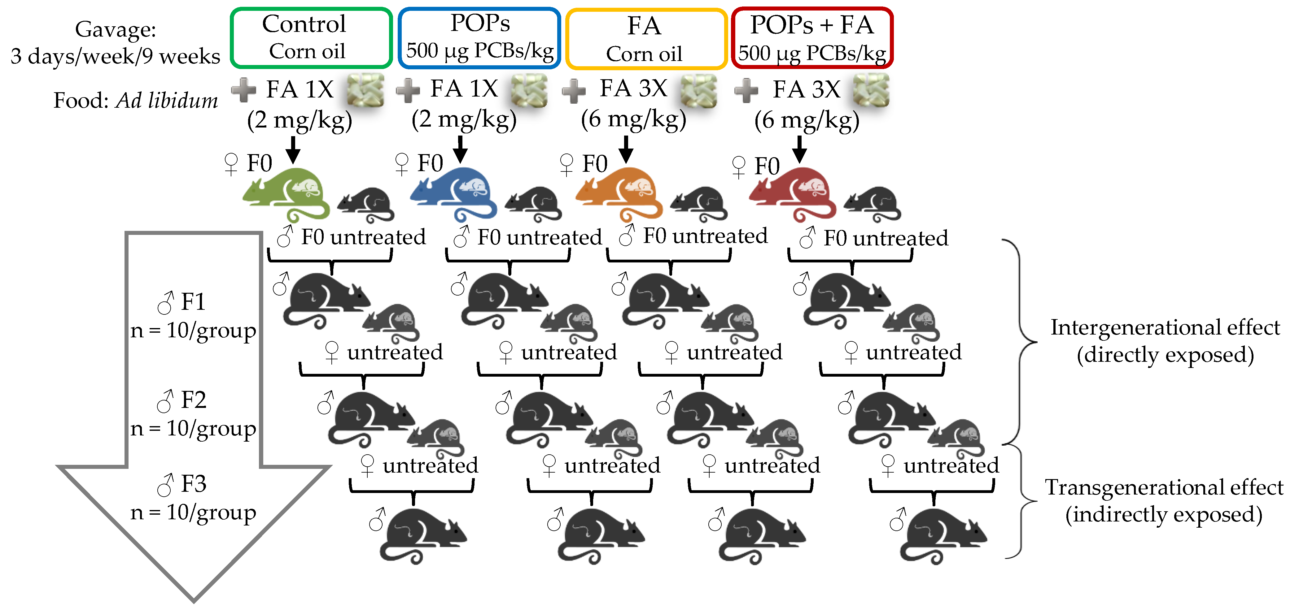
Figure 1. Experimental design. Four treatment groups of Sprague–Dawley F0 founder dams were fed ad libitum with a diet containing 2 (1×) (CTRL) or 6 mg/kg diet (3×) of FA and received by gavage (3 days/week) either corn oil (CTRL) or the POP mixture. Treatments started before reproduction with untreated males and were administered until parturition (9 weeks in total). All F0 postpartum founder dams and subsequent generations were fed the control diet 1× ad libitum only and the POP mixture treatment was ended. Then, F1 males were mated with untreated females to yield generation F2 and so on until F3 lineage. Since F1 males were exposed to treatments in utero and F2 were exposed as germ cells in utero, F1 and F2 phenotypic variations were defined as “intergenerational”. F3 generation was not directly exposed (ancestral exposure), so F3 phenotypic variations were defined as “transgenerational”.
Environments 08 00024 g001
Figure 2. Impacts of POP exposure and/or folic acid (FA) supplementation of F0 female rats on fasting blood glucose, plasma insulin and C-peptide in F1, F2 and F3 male rats aged 90 days (AC) and 180 days (DF). Results are expressed as mean ± SEM (n = 9 or 10). Analysis of variance (ANOVA) for a 2 × 2 factorial experiment was used to calculate main effects of POPs and FA and the POPs*FA interaction. A Tukey post hoc test was used when an interaction was significant. Bars with differing letters are significantly different (p ≤ 0.05).
Environments 08 00024 g002
Figure 3. Impact of POP exposure and/or FA supplementation of F0 female rats on blood glucose, plasma insulin and C-peptide during a glucose tolerance test in F1 male rats aged 90 days (AC) and 180 days (DF). The histogram shows incremental area under the curve (IAUC) values. Results are expressed as mean ± SEM (n = 7 to 10). Two-way ANOVA with repeated measurements was used to compare time points within groups. ANOVA for a 2 × 2 factorial experiment was used to analyze the POP and FA main effects and the POPs*FA interaction.
Environments 08 00024 g003
Figure 4. Impact of POP exposure and/or FA supplementation of F0 female rats on blood glucose, plasma insulin and C-peptide during a glucose tolerance test in F2 male rats aged 90 days (AC) and 180 days (DF). The histogram shows IAUC values. Results are expressed as mean ± SEM (n = 9 or 10). Two-way ANOVA with repeated measurements was used to compare time points within groups. ANOVA for a 2 × 2 factorial experiment was used to analyze the POP and FA main effects and the POPs*FA interaction.
Environments 08 00024 g004
Figure 5. Impact of POP exposure and/or FA supplementation of F0 female rats on blood glucose, plasma insulin and C-peptide during a glucose tolerance test in F3 male rats aged 90 days (AC) and 180 days (DF). The histogram shows IAUC values. Results are expressed as mean ± SEM (n = 9 or 10). Two-way ANOVA with repeated measurements was used to compare time points within groups. ANOVA for a 2 × 2 factorial experiment was used to analyze the POPs and FA main effects and the POPs*FA interaction.
Environments 08 00024 g005
Table 1. Composition of the persistent organic pollutant (POP) mixture [22].
CompoundCAS Number Origin 1Quantity in the Mix (%)Administered Dose (µg/kg of Body Weight)
Aroclor and congener neat mix 257-74-9AccuStandard32.4500
Technical grade chlordane72-55-9AccuStandard21.4330.3
p,p’-dichlorodiphenyldichloroethylene (p,p’-DDE)50-29-3Sigma-Aldrich19.3297.8
p,p’-dichlorodiphenyltrichloroethane (p,p’-DDT)8001-35-2SigmaAldrich6.8104.9
Technical grade toxaphene319-84-6AccuStandard6.5100.0
α-hexachlorocyclohexane (α-HCH)309-00-2Sigma-Aldrich6.295.7
Aldrin60-57-1Sigma-Aldrich2.538.6
Dieldrin95-94-3Sigma-Aldrich2.132.4
1,2,4,5-tetrachlorobenzene 72-54-8Sigma-Aldrich0.913.9
p, p’-dichlorodiphenyldichloroethane (p, p’-DDD)319-85-7Sigma-Aldrich0.57.7
β-hexachlorocyclohexane (β-HCH)118-74-1Sigma-Aldrich0.46.2
Hexachlorobenzene2385-85-5AccuStandard0.46.2
Mirex58-89-9Sigma-Aldrich0.23.1
Lindane608-93-5Sigma-Aldrich0.23.1
Pentachlorobenzene57-74-9Sigma-Aldrich0.23.1
1 AccuStandard Inc. (New Haven, CY, USA); Sigma-Aldrich Inc. (St. Louis, MI, USA); 2 PCB mixture: Aroclor 1260 (58.9% of total), Aroclor 1254 (39.3% of total), 2,4,4′-trichlorobiphenyl (PCB 28, 1% of total), 2,2′,4,4′-tetrachlorobiphenyl (PCB 47, 0.8% of total), 3,3′,4,4′,5-pentachlorobiphenyl (PCB 126, 0.02% of total), and 3,3′,4,4′-tetrachlorobiphenyl (PCB 77, 0.004% of total).
Table 2. Composition of the diets.
Ingredients1× Diet 1 (g/kg of Diet)3× Diet 2 (g/kg of Diet)
Casein 3200200
L-Cystine33
Sucrose100100
Cornstarch397.486396.286
Dyetrose 4132132
Soybean oil7070
t-butylhydroquinone0.0140.014
Cellulose5050
Mineral Mix #2100253535
Vitamin Mix #310025100
Vitamin Mix #317761 (no folate)010
Folic acid premix (5 mg/g)01.2
Choline bitartrate2.52.5
Total10001000
1 DYET #110700, AIN-93G purified rodent diet with 2 mg/kg FA. 2 DYET #117819GI, Modified AIN-93G purified rodent diet with 6 mg/kg FA. 3 Casein high-nitrogen for diet and sterile casein for 3× diet. 4 Food-grade depolymerized cornstarch.
Table 3. Weight gain and food intake of F0 rat dams; body weight of F1, F2, F3 male rats at the ages of 21, 90 and 180 days, total weight gain and food intake.
CTRLPOPsFAPOPs+FA
F0
Weight gain 1 (g)73 ± 673 ± 665 ± 884 ± 8
Food intake (g/day)14 ± 0.215 ± 115 ± 0.314 ± 0.9
F1
Weight on day 21 (g) 51 ± 252 ± 148 ± 251 ± 0.6
Weight on day 90 (g)386 ± 9400 ± 10384 ± 9387 ± 12
Weight on day 180 2 (g)493 ± 12521 ± 14499 ± 12493 ± 18
Total weight gain 2,3 (g)441 ± 39469 ± 43451 ± 36442 ± 53
Food intake 2 (g/day)18 ± 0.319 ± 0.418 ± 0.419 ± 0.6
F2
Weight on day 21 (g) 51 ± 250 ± 250 ± 151 ± 0.9
Weight on day 90 (g)410 ± 11408 ± 11415 ± 11408 ± 13
Weight on day 180 2 (g)538 ± 15522 ± 15544 ± 15520 ± 18
Total weight gain 2,3 (g)486 ± 44472 ± 44494 ± 49470 ± 58
Food intake 2 (g/day)20 ± 0.619 ± 0.519 ± 0.619 ± 0.6
F3
Weight on day 21 (g) 52 ± 253 ± 252 ± 153 ± 2
Weight on day 90 (g)411 ± 7419 ± 8403 ± 8401 ± 9
Weight on day 180 2 (g)537 ± 12547 ± 15526 ± 11512 ± 17
Total weight gain 2,3 (g)485 ± 37494 ± 46474 ± 33461 ± 50
Food intake 2 (g/day)19 ± 0.320 ± 0.320 ± 0.619 ± 0.6
Results are expressed as mean ± standard error of the mean (SEM), n = 10. No significant differences were observed. 1 From the age of 7 weeks to 16 weeks (based on postpartum weight). 2 n = 9 or 10. 3 Difference between weights on days 180 and 21.
Table 4. Estimates of insulin sensitivity, insulin clearance (hepatic extraction) and β-cell function in F1 male rats.
CTRLPOPsFAPOPs+FAPOPsFAPOPs*FA
90-day-oldp values
Insulin/glucose IAUC 1 (pmol/mmol)51 ± 1374 ± 2662 ± 1351 ± 12nsnsns
C-peptide/insulin IAUC 2 (nmol/pmol) × 10−35.3 ± 0.35.4 ± 0.94.2 ± 0.44.3 ± 0.5nsns
HOMA-IR index1.4 ± 0.32.0 ± 0.40.9 ± 0.11.8 ± 0.5*nsns
Matsuda index 39.9 ± 2.28.1 ± 1.511.7 ± 2.210.8 ± 1.9nsnsns
180-day-oldp values
Insulin/glucose IAUC 4 (pmol/mmol)135 ± 13183 ± 67176 ± 24126 ± 21nsnsns
C-peptide/insulin IAUC 5 (nmol/pmol) × 10−34.4 ab ± 0.33.6 c ± 0.33.9 bc ± 0.24.8 a ± 0.3nsns*
HOMA-IR index 44.1 ± 0.84.8 ± 1.32.3 ± 0.22.4 ± 0.3nsnsns
Matsuda index 43.6 ± 0.64.6 ± 1.34.5 ± 0.34.9 ± 0.5nsnsns
Results are expressed as mean ± SEM (n = 10). ns: nonsignificant; ‡ 0.05 < p ≤ 0.06; † p ≤ 0.05; * p ≤ 0.01. The Tukey post hoc test was used when an interaction was significant; different letters indicate significant difference (p ≤ 0.05). 1 n = 6 to 8. 2 n = 4 to 8. 3 n = 8 or 9. 4 n = 9 or 10. 5 n = 8 to 10.
Table 5. Estimates of insulin sensitivity, insulin clearance (hepatic extraction) and β-cell function in F2 male rats.
CTRLPOPsFAPOPs+FAPOPsFAPOPs*FA
90-day-oldp values
Insulin/glucose IAUC (pmol/mmol)64 ± 9123 ± 1765 ± 1283 ± 25nsnsns
C-peptide/insulin IAUC (nmol/pmol) × 10−35.0 ± 0.34.6 ± 0.35.4 ± 0.64.9 ± 0.5nsnsns
HOMA-IR index1.0 ± 0.11.6 ± 0.21.3 ± 0.21.3 ± 0.1nsnsns
Matsuda index9.3 ± 0.96.1 ± 0.59.0 ± 1.37.9 ± 0.5nsns
180-day-oldp values
Insulin/glucose IAUC 1 (pmol/mmol)141 ± 31150 ± 23152 ± 41138 ± 30nsnsns
C-peptide/insulin IAUC 1 (nmol/pmol) × 10−34.9 ± 0.44.4 ± 0.24.6 ± 0.34.6 ± 0.5nsnsns
HOMA-IR index 13.3 ± 0.83.9 ± 0.63.7 ± 0.93.5 ± 1nsnsns
Matsuda index 14.7 ± 0.83.9 ± 0.65.6 ± 1.35.2 ± 1.0nsnsns
Results are expressed as mean ± SEM (n = 10). ns: nonsignificant; ‡ 0.05 < p ≤ 0.06; † p ≤ 0.05; * p ≤ 0.01.1 n = 9 or 10.
Table 6. Estimates of insulin sensitivity, insulin clearance (hepatic extraction) and β-cell function in F3 male rats.
CTRLPOPsFAPOPs+FAPOPsFAPOPs*FA
90-day-oldp values
Insulin/glucose IAUC (pmol/mmol)56 ± 962 ± 650 ± 1065 ± 17nsnsns
C-peptide/insulin IAUC (nmol/pmol) × 10−36.3 ± 0.56.0 ± 0.66.7 ± 0.77.2 ± 1.1nsnsns
HOMA-IR index1.1 ± 0.21.6 ± 0.32.1 ± 0.91.4 ± 0.4nsnsns
Matsuda index10.5 ± 1.47.3 ± 0.59.3 ± 1.512.3 ± 2.1nsnsns
180-day-oldp values
Insulin/glucose IAUC (pmol/mmol)154 ± 23139 ± 20143 ± 31122 ± 20nsnsns
C-peptide/insulin IAUC (nmol/pmol) × 10−34.4 ± 0.34.5 ± 0.34.5 ± 0.44.3 ± 0.3nsnsns
HOMA-IR index3.8 ± 0.86.7 ± 1.03.6 ± 0.63.3 ± 0.8nsnsns
Matsuda index3.9 a ± 0.62.4 b ± 0.34.1 a ± 0.84.9 a ± 0.8nsns
Results are expressed as mean ± SEM (n = 9 or 10). ns: nonsignificant; † p ≤ 0.05; * p ≤ 0.01. The Tukey post hoc test was used when an interaction was significant; different letters indicate significant differences (p ≤ 0.05).
Publisher’s Note: MDPI stays neutral with regard to jurisdictional claims in published maps and institutional affiliations.