Article

Feasibility of Using Intraoperative Neuromonitoring in the Prophylaxis of Dysesthesia in Transforaminal Endoscopic Discectomies of the Lumbar Spine

by
Paulo Sérgio Teixeira de Carvalho
1,
Max Rogério Freitas Ramos
2,
Alcy Caio da Silva Meireles
3,
Alexandre Peixoto
4,
Paulo de Carvalho, Jr.
5,
Jorge Felipe Ramírez León
6,
Anthony Yeung
7 and
Kai-Uwe Lewandrowski
1,6,8,*
1
The Federal University of the State of Rio de Janeiro UNIRIO, Pain and Spine Minimally Invasive Surgery Service at Gaffrée Guinle University Hospital HUGG, Tijuca, Rio de Janeiro 20270-004 RJ, Brazil
2
Federal University of the Rio de Janeiro State UNIRIO, Orthopedic Clinics at Gaffrée Guinle University Hospital HUGG, Tijuca, Rio de Janeiro 20270-004 RJ, Brazil
3
The Meireles Neurology Institute, Bonsucesso - Rio de Janeiro 20230-240 RJ, Brazil
4
Federal University of the Rio de Janeiro State UNIRIO, 775 – Maracanã, Rio de Janeiro 20270-004 RJ, Brazil
5
KRH Nordstadt Krankenhaus Hospital, 30167 Hanover, Germany
6
Fundación Universitaria Sanitas, Clínica Reina Sofía-Clínica Colsanitas, Centro de Columna-Cirugía Mínima Invasiva, Bogotá 104-76, D.C., Colombia
7
Department of Neurosurgery Albuquerque, University of New Mexico School of Medicine, New Mexico Associate, Desert Institute for Spine Care, Phoenix, AZ 85020, USA
8
Center for Advanced Spine Care of Southern Arizona and Surgical Institute of Tucson, AZ 85712, USA
*
Author to whom correspondence should be addressed.
Brain Sci. 2020, 10(8), 522; https://doi.org/10.3390/brainsci10080522
Submission received: 20 July 2020 / Revised: 30 July 2020 / Accepted: 3 August 2020 / Published: 5 August 2020
(This article belongs to the Special Issue Degenerative Spinal Disease)

Abstract

:
(1) Background: Postoperative nerve root injury with dysesthesia is the most frequent sequela following lumbar endoscopic transforaminal discectomy. At times, it may be accompanied by transient and rarely by permanent motor weakness. The authors hypothesized that direct compression of the exiting nerve root and its dorsal root ganglion (DRG) by manipulating the working cannula or endoscopic instruments may play a role. (2) Objective: To assess whether intraoperative neurophysiological monitoring can help prevent nerve root injury by identifying neurophysiological events during the initial placement of the endoscopic working cannula and the directly visualized video endoscopic procedure. (3) Methods: The authors performed a retrospective chart review of 65 (35 female and 30 male) patients who underwent transforaminal endoscopic decompression for failed non-operative treatment of lumbar disc herniation from 2012 to 2020. The patients’ age ranged from 22 to 86 years, with an average of 51.75 years. Patients in the experimental group (32 patients) had intraoperative neurophysiological monitoring recordings using sensory evoked (SSEP), and transcranial motor evoked potentials (TCEP), those in the control group (32 patients) did not. The SSEP and TCMEP data were analyzed and correlated to the postoperative course, including dysesthesia and clinical outcomes using modified Macnab criteria, Oswestry disability index (ODI), visual analog scale (VAS) for leg and back pain. (4) Results: The surgical levels were L4/L5 in 44.6%, L5/S1 in 23.1%, and L3/L4 in 9.2%. Of the 65 patients, 56.9% (37/65) had surgery on the left, 36.9% (24/65) on the right, and the remaining 6.2% (4/65) underwent bilateral decompression. Postoperative dysesthesia occurred in 2 patients in the experimental and six patients in the control group. In the experimental neuromonitoring group, there was electrodiagnostic evidence of compression of the exiting nerve root’s DRG in 24 (72.7%) of the 32 patients after initial transforaminal placement of the working cannula. A 5% or more decrease and a 50% or more decrease in amplitude of SSEPs and TCEPs recordings of the exiting nerve root were resolved by repositioning the working cannula or by pausing the root manipulation until recovery to baseline, which typically occurred within an average of 1.15 min. In 15 of the 24 patients with such latency and amplitude changes, a foraminoplasty was performed before advancing the endoscopic working cannula via the transforaminal approach into the neuroforamen to avoid an impeding nerve root injury and postoperative dysesthesia. (5) Conclusion: Neuromonitoring enabled the intraoperative diagnosis of DRG compression during the initial transforaminal placement of the endoscopic working cannula. Future studies with more statistical power will have to investigate whether employing neuromonitoring to avoid intraoperative compression of the exiting nerve root is predictive of lower postoperative dysesthesia rates in patients undergoing videoendoscopic transforaminal discectomy.

1. Introduction

Videoendoscopy for the treatment of lumbar herniated discs is now common place with favorable clinical results comparable to microdiscectomy and a low rate of complication rate [1,2]. The transforaminal approach is frequently applied to the endoscopic treatment of herniated discs mainly at the L3/L4 and L4/L5 level. The approach is also feasible at L5/S1 but may be technically more demanding because of the configuration of the iliac wing, sacralization of the L5 vertebral body, or because of degenerative vertical collapse of the spine [3,4]. Placing the working cannula may be harder at this transitional level due to steeper attack angles making injury to the exiting L5 nerve root more likely. Regardless of the level, additional risks for nerve root injury due to increasing surgery time and more aggressive manipulation may arise if the surgeon is attempting to access a highly stenotic neuroforamen during the endoscopic decompression procedure. Compression of the dorsal root ganglion of the exiting nerve root may occur due to manipulation of the beveled endoscopic working cannula leading to temporary ischemia and postoperative dysesthesias in a percentage considerable number of patients [5].
Neuromonitoring could aid in mitigating these risk particularly if surgeons perform the lumbar transforaminal endoscopic surgery under general anesthesia [6]. Therefore, the authors decided to retrospectively study the result obtained by using neuromonitoring during transforaminal videoendoscopy in patients who were treated for herniated discs. While the three senior key opinion leader (KOL) surgeons (JFRL, ATY, and KUL) of this article have performed over 18,000 endoscopic spine surgeries between them [7,8,9,10,11,12,13,14,15,16,17,18] under monitored local anesthesia care (MAC) and sedation [12,18,19] with minimal complications [1,20] and utilizing the patients’ direct intraoperative feedback to treat validated pain generators [11,15,19,21,22], they recognize that neuromonitoring in some countries and in some clinical settings is the standard of care [23,24,25,26,27,28,29,30,31,32] in spite of added cost [33]. In some instances, it may even be a matter of necessity if MAC protocols for spine surgery are not supported by the local anesthesia teams. Therefore, the authors wanted to study the feasibility of identifying intraoperative neuromonitoring events that could be predictive of postoperative dysesthesias with the transforaminal endoscopic decompression procedure [34]. If feasible, surgical protocols aimed at lowering the incidence or perhaps even preventing injury to the exiting nerve root DRG could be developed for those surgeons who are unable to perform the surgery under local anesthesia with sedation [35].

2. Material and Method

2.1. Patients

The charts, imaging studies, and neuromonitoring examinations of 65 patients who underwent transforaminal endoscopic discectomy from 2012 to 2020 were retrospectively reviewed. The surgeries were performed by the first and second author. There were 35 female (53.8%) and 30 (46.2%) male patients. There were 35 (53.8%) females and another 30 (46.2%) male patients. The average age was 51.75 years ranging from 22 to 86 years with a standard deviation (SDV) of 14.916 and followed normal distribution (Figure 1 and Figure 2). Patients in the experimental group (32 patients) had intraoperative neurophysiological monitoring recordings using sensory evoked (SSEP), and transcranial motor evoked potentials (TCEP), those in the control group (32 patients) did not. Both groups were age and gender matched, and similar rates of comorbidities, and surgical level distribution (see below). All patients in this consecutive case series provided informed consent and Institutional Review Board approval was obtained (CEIFUS 106-19). Written informed consent was obtained from the patient for publication of this report and any accompanying images.

2.2. Inclusion/Exclusion Criteria

Only patients presenting with radicular pain with a herniated disc confirmed by magnetic resonance imaging (MRI) or computed tomography (CT) were selected for this study. Patients with unmanageable radicular pain unresponsive to a minimum of 12 weeks of medical and interventional conservative care, with a positive Lasègue’s tension sign, and minimal low back pain were included. A normal preoperative electroneuromyographic study was another prerequisite for study inclusion. Patients with instability, deformity, or any electrodiagnostic abnormality were excluded from the study. Exclusion of patients from the study was prompted by a concurrent diagnosis of infection, tumor, or metastatic disease, or any electrodiagnostic evidence of chronic demyelination, or deinnervation in the dermatomes innervated by the affected surgical nerve roots. Exclusion was also prompted if patients displayed any of the following radiographic parameters.

2.3. Preoperative Radiographic Evaluation

Radiographic stenosis parameters were also evaluated. These included the posterior intervertebral disc and foraminal height [36]. Crossectional imaging showing 15 mm or less for the height of the neuroforamen, 3 mm or less measured as posterior intervertebral disc height, or the width of the neuroforamen was recorded as abnormal [36]. As previously published and validated, diagnostic selective nerve root blocks were used to determine the symptomatic painful level. This protocol was highly relevant in choosing the surgical level(s) in patients with multilevel disease [37,38,39,40,41,42,43,44]. Exclusion of patients from the study was prompted by a concurrent diagnosis of overt spondylolisthesis with more than 3 mm of translational motion on dynamic extension/flexion views. If patients were suspected of having claudication or mechanical back pain symptoms due to severe central stenosis, <100 mm2 at the surgical level or from facet arthropathy, they were typically also excluded from the study.

2.4. Anesthesia and Neuromonitoring

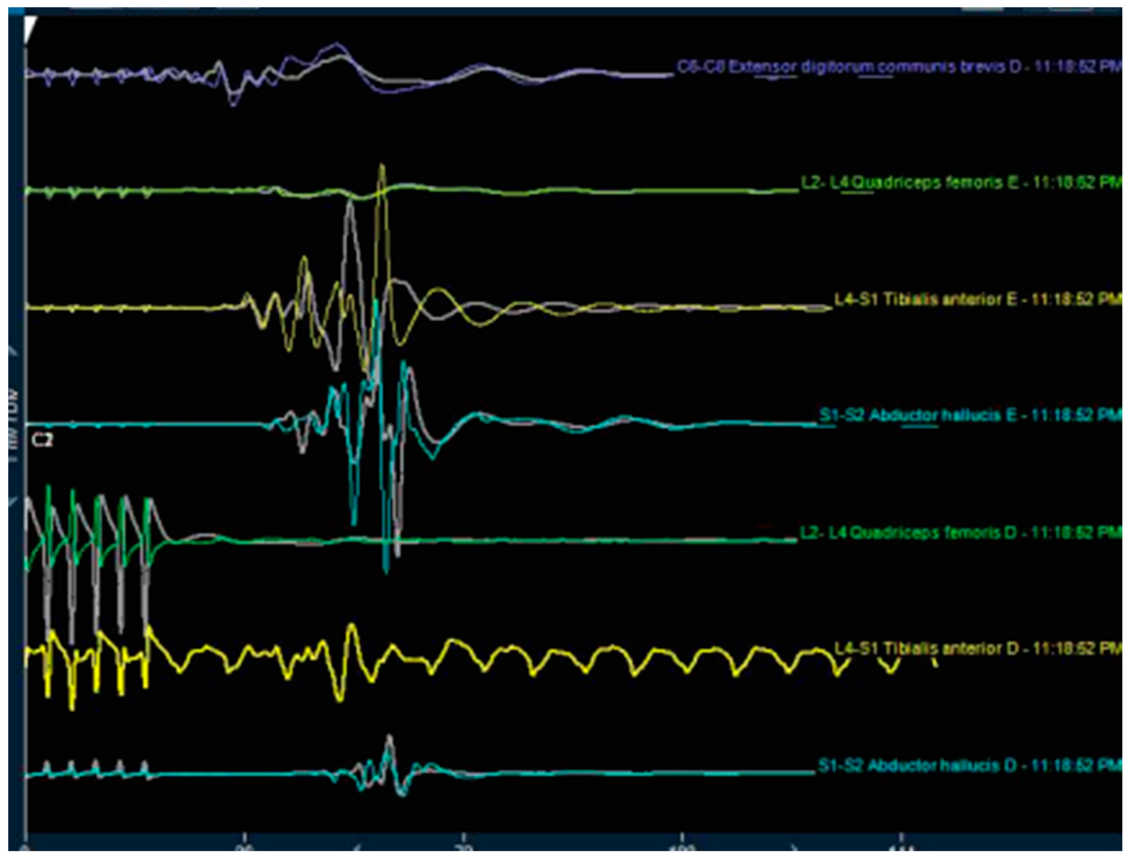
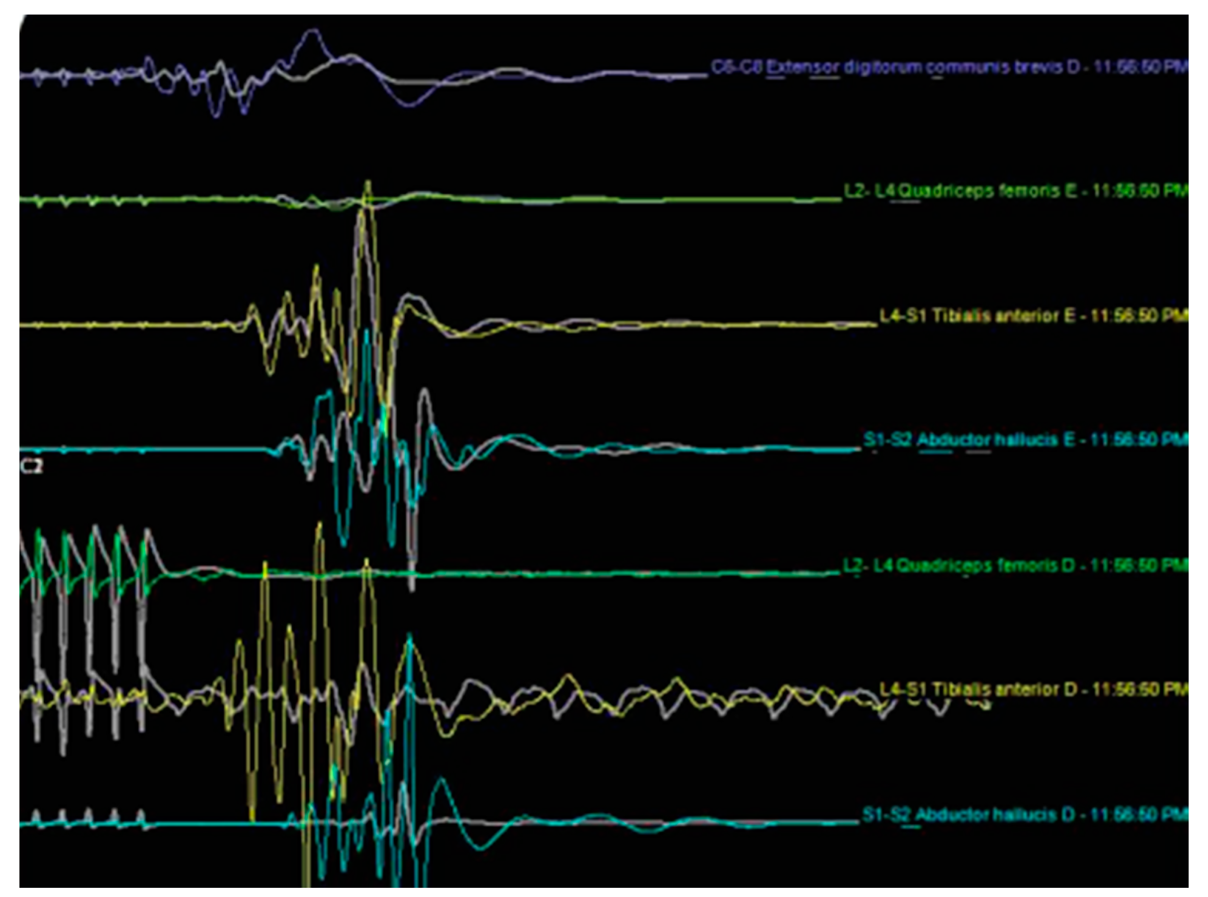
All patients underwent general anesthesia before the placement of the neuromonitoring probes in the lower limbs. Leads were placed in dermatomes corresponding to the roots exiting from the surgical level. The dermatome innervated by the level above the surgical segment was also monitored as a control. The neurophysiological monitoring was performed by recording using sensory evoked (SSEP), and transcranial motor evoked potentials (TCEP). Intra- and postoperative wave recordings were analyzed for changes in the frequency, amplitude, and latency (Figure 3 and Figure 4). A 5% increase or more in the SSEP latency or a 50% reduction in the amplitude was considered intraoperative evidence of a significant neuromonitoring event, suggesting injury to the DRG of the exiting nerve root. For the TCMEP, these parameters were a similar increase in latency, a decrease in the number of phases, and a 50% decrease in the amplitude. Conversely, a 50% increase in the SSEP and TCMEP amplitude, or a 10% latency decrease, was considered a significant improvement of the neuromonitoring parameters. The SSEP and TCMEP voltage improvements were recorded. Adverse neuromonitoring exiting nerve root compression events were analyzed and correlated to postoperative course including dysesthesia and outcome. In the case of a neuromonitoring event presumably due to compression or ischemia of the exiting nerve root, the surgery described below was paused, and the working cannula was withdrawn and then repositioned. The operation was resumed when neuromonitoring response caused by the surgical manipulation had returned at minimum to baseline. Foraminoplasty was performed in those patients where SSEP and TCMEP recordings did not return to baseline or were observed repeatedly with attempts to reposition the working cannula.

2.5. Endoscopic Discectomy Procedure

The surgeries were performed by the first and second author employing the transforaminal “outside-in” technique with the patient in prone position [45]. Serial dilation was employed to place the working cannula. Since patients with bony foraminal stenosis were excluded from the study, a routine foraminoplasty was not necessary. If bleeding occurred, a radiofrequency probe (Elliquence® Baldwin, NY) was used for coagulation [46]. The endoscopic decompression procedure was directly visualized throughout the surgery. The location of the herniated disc and the presence of any other anatomical anomalies leading to inflammation or tethering the nerve roots bordering the triangular safe zone at the surgical level were recorded as the authors thought that they potentially could increase the risk of nerve root injury and postoperative development of irritation of the dorsal root ganglion (DRG). Fluoroscopic surveillance images were occasionally taken for orientation and verification of the decompression.

2.6. Clinical Follow-Up and Primary Outcome Measures

Primary clinical outcome measures were reductions in the visual-analog scales (VAS) [47] for leg (VAS-LEG) and back pain (VAS-BACK) ranging from no pain (0) to worst pain (10) and the Oswestry disability index ODI. The ODI is a ten-item composite instrument assessing pain intensity, personal care, and function including walking, lifting, personal care, sitting, standing, sleeping, social interaction, and traveling [48,49]. Each ODI item is scored from 0 (no impairment) to 5 (worst impairment). The individual scores are summed and then multiplied by two to obtain the ODI index ranging from 0 to 100. In addition, patients were evaluated with use of the modified Macnab criteria as [50,51]. Postoperatively, patients were seen in follow-up for reevaluation at 6 weeks and then at 3, 12, and 24 months. Any clinical evidence of new onset of dysethetic leg pain due to DRG irritation was recorded.

2.7. Postoperative Rehabilitation

Most patients did not require postoperative rehabilitation and supportive care requirements. Study patients treated for any acute onset of dysesthetic leg pain after an initial postoperative period of good pain relief with nonsteroidal anti-inflammatories, gabapentin, and transforaminal epidural steroid injections (TESI) pain syndromes were counted as having an irritation of the dorsal root ganglion (DRG). For the purpose of this study analysis, successful postoperative administration of 1% lidocaine-containing TESI with therapeutic pain relief was considered proof of a DRG irritation due to intraoperative nerve root injury.

2.8. Statistical Analysis

For the clinical outcome analysis, descriptive statistics (mean and standard deviation), cross-tabulation statistics of sensitivity, specificity, and measures of association were computed for two-way tables using IBM SPSS Statistics software, Chicago, IL, Version 26.0. The Pearson χ2 and the likelihood-ratio χ2 tests were used as statistical measures of association between dysesthesia, SSEP, TCMEP neuromonitoring events, and clinical outcome measures. The confidence intervals for the likelihood ratios were calculated using the “log method” according to Liberati and Altman et al. [52].

3. Results

Sixty-five study patients in total had surgery at 65 levels. There were 32 patients in the neuromonitoring and 33 patients in the control group. The average postoperative follow-up was 20.55 months, ranging from 12 to 30 months, with a SDV of 7.537 months. Most patients underwent surgery on the left side with 56.9% (37/65), whereas 36.9% (24/65) had surgery on the right, and the remaining 6.2% (4/65) of patients underwent bilateral surgery. As expected, the most common surgical segments were L4/L5 in 44.6%, L5/S1 in 23.1%, and L3/L4 in 9.2% of patients, respectively. The surgical level distribution is shown in Table 1. Eight (12.3%) of the 65 patients developed a postoperative dysesthesia from compression of the exiting nerve root and injury to its DRG during initial endoscopic working cannula placement. Crosstabulation of dysesthesia versus dichotomized age of less than 41 (p = 0.248) and greater than 41 years (p = 0.330) did not shown any statistically significant impact of age on the dysesthesia rate on Chi-square testing.
Final clinical outcomes were favorable, with 51 (78.5%) patients reporting excellent and the remaining 14 (21.5%) patients indicating good Macnab outcomes. At final follow-up, 75% (6 patients) of dysesthesia patient had excellent and the remaining 25% (2 patients) had good Macnab outcomes. At the final follow-up, the ODI improved from 31.71 ± 16.17 preoperatively to 19.02 ± 8.96 postoperatively (p < 0.0001). The VAS leg score reduced from 8.86 ± 0.93 before thee endoscopic decompression to 1.15 ± to 1.27 at the final follow-up (p < 0.0001). The VAS back score reductions were more modest from preoperative 4.92 ± 1.27 to postoperative 3.2 ± 0.775, respectively (p < 0.0001). The results of the paired-T testing with 95% confidence interval numbers are summarized in Table 2 and Table 3. The mean ODI reduction was 12.69 ± 13.12, on par with reported MCID (Minimal Clinically Important Difference) reductions reported for the transforaminal endoscopic decompression procedure. The mean VAS leg score reduction was 7.71 ± 1.9, being much larger than the reported MCID with the procedure. The mean VAS back reduction of 1.72 ± 1.21 did fall short of MCID numbers reported for this outcome tool with the transforaminal endoscopic procedure.
There were also statistically significant (p < 0.0001) improvements of the mean SSEP and TCMEP voltage measurements as a result of the transforaminal endoscopic decompression procedure (Figure 1). There were 24 (72.7%) of the 33 patients who underwent SSEP and TCMEP neuromonitoring during the transforaminal endoscopy, which showed a signal response due to compression of the DRG of the exiting nerve root during the initial position of the working cannula. In 14 (58.3%) of these 24 patients, withdrawing, waiting, and repositioning the endoscopic working cannula did not eliminate the intraoperative compression injury visibly demonstrated by the neuromonitoring. Hence, these patients underwent a foraminoplasty, which allowed the surgeon to advance the working cannula into the neuroforamen without visible irritation of the exiting nerve root’s DRG. The average waiting time until SSEP and TCMEP signals had normalized to prior baseline after irritation of the DRG of the exiting nerve root during the initial placement of the working cannular was 1.15 min. While the dysesthesia rate in the neuromonitoring (experimental) group was one-third of that observed in the control group, the authors’ study did not have sufficient statistical power to demonstrate the benefit with statistical significance (Pearson Chi-square = 0.557; p = 0.456).

4. Discussion

One of the most common problems after transforaminal endoscopic decompression is dysesthesia due to compression of the dorsal root ganglion by the working cannula and its manipulation during its initial placement and the discectomy or foraminoplasty procedure. The senior author reported the incidence of this unavoidable sequala at 12.45% based on a study in 1839 patients with foraminal stenosis being a statistically significant risk factor [1]. The last three authors of this article have performed the endoscopic transforaminal decompression under local anesthesia using MAC protocols to be able to use patient feedback during surgery while treating the predominant pain generator [11,22]. Their reported dysesthesia rate was lower [1,8,10] than reported by other surgeons in another multicenter study with seven participating surgeons and sites [53]. They reported the incidence of DRG irritation to average 21.5%, ranging from 5% to 41.2% in patients with excellent and good Macnab outcomes [53]. DRG irritation rates were independent of surgical level but highly dependent on surgeon skill level.
While the course of postoperative DRG irritation following a transforaminal endoscopic discectomy is benign and typically self-limiting with a reduced physical activity program, supportive medical and interventional care measures such as gabapentin, pregabalin, or a transforaminal epidural steroid injection (TESI), the condition can be quite annoying to patients until it resolves typically 2 to 4 weeks postoperatively [53]. A preoperative education program warning patients of the possibility of burning leg pain developing in the dermatome of the exiting nerve root of the surgical level some 5 to 10 days following the transforaminal endoscopic discectomy after an initial pain-free interval. Patients should also be instructed to avoid narcotic pain medication as they are not an effective treatment for this condition and that follow up with their surgeon in the office setting is more appropriate than seeking help in the emergency room where inconsequential imaging studies ultimately do not change management are ordered. The readmission rate to a hospital in the immediate postoperative was reported to 0.87% and compared favorably to the readmission rates reported in traditional microdiscectomy (4.1% to 5.8%) [20]. Yeung et al. reported the incidence of postoperative dysesthesia at 9.7% in his series of 176 patients [10,12]. Nellensteijn et al. corroborated these findings in his systematic review carried out in 2010 and concluded that comorbidities might impact the dysesthesia rate [54].
Hence, attempts to minimize the dysesthesia rate following an expertly executed transforaminal endoscopic decompression are highly relevant to the patients’ perception of the endoscopic procedure. Patient satisfaction may decrease when a bothersome dysesthesia sets in after an initial 5 to 10 days postoperative interval of pain relief. The authors investigated the feasibility of employing intraoperative neuromonitoring during the initial endoscopic working cannula position to avoid incidental compression of the exiting nerve root at the surgical level. That part of the procedure is typically not directly visualized. The authors found that neuromonitoring may detect such compression of the exiting nerve root and resolve it with the reposition of the working cannula during the initial steps of the endoscopic procedure. The detected SSEP and EMG neuromonitoring events with decrease amplitude and latency resolved with withdrawing and repositioning the working cannula and by waiting until these SSEPs and EMG signals had returned to baseline waves. Typically, such waiting times were on the order of one to two minutes but on average 1.15 min. Occasionally, the surgeon had to pause for up to three minutes before being able to commence the percutaneous endoscopic decompression procedure. Only in those patients where the SSEP and EMG signals did not return to baseline, the surgeons performed a foraminoplasty involving resection of the tip of the superior articular process with the intent of diminishing the presumed nerve root compression associated with advancing the endoscopic working cannula into Kambin’s triangle—the safe zone formed by the exiting and traversing nerve root and the pedicle of the inferior vertebral body. This has been corroborated by Jun-Song-Yang et al. who stated that foraminoplasty could be essential and necessary to facilitate the approach to a foraminal hernia, avoiding the manipulation and compression of the dorsal root ganglion and the symptomatic compressed nerve roots. The latter are typically inflamed, irritable, and susceptible to additional injury. Therefore, using the information obtained from the intraoperative neuromonitoring has the potential to decrease the incidence of neurological dysfunction associated with the transforaminal approach [55]. In this study, the intraoperative execution of this protocol has notably reduced the dysesthesia rate by one third in comparison to the rate observed in the control group, where the authors performed the same surgery without intraoperative monitoring.
While the application of neuromonitoring to improve outcomes with the outpatient transforaminal endoscopic decompression procedure is uncommon to current routine clinical practice with surgeries being done under MAC local anesthesia, it is frequently employed in traditional open and other forms of minimally invasive spinal surgery in the United States and the world over. From 2008 to 2014, the number of neuromonitored spine cases increased from 31,762 to 125,835, signifying a 296% increase over one decade [56]. Its benefit in reducing neurological morbidity has been demonstrated during spinal cord decompression and deformity surgery [30,33,34,56]. The benefits far outweigh the risk of routine neuromonitoring since complications with it are extremely rare [57]. Nonetheless, the added cost has been recognized [58,59], questioning its routine use during low-risk lumbar decompressions [58]. However, the authors stipulated that its use would lower surgical risk and improve clinical outcomes with the transforaminal endoscopic discectomy when the procedure is performed under general anesthesia. This hypothesis seems to be corroborated by the findings of this study. While the authors have no direct explanation as to why the observed dysesthesia rate was lower with concurrent application of neuromonitoring in the experimental versus the control group, the most plausible one is that the ability to assess in real-time intraoperatively whether their surgical access creates additional compression and to then act on that information by changing the course of the transforaminal endoscopic operation is likely the most relevant reason. As a result, the risk of injury to the exiting nerve root on top of what may have been caused by the herniated disc already may be lower. Not knowing the exact mechanism(s) of provoking postoperative dysesthesia is also one of the most significant limitations of the authors’ study besides a small sample size in both the experimental neuromonitoring and the control groups. The authors had no way to determine whether the risk for postoperative dysesthesia was solely determined by the surgical compression of the exiting nerve root, or whether there were any confounding factors, such as the size and location of the disc herniation, the duration of symptoms, extent of root ischemia, and duration of the neural element compression. Additionally, comorbidities, such as neuropathy, metabolic disease, including diabetes or renal disease, or age may also have played a role. Unfortunately, the statistical power of this feasibility study was insufficient to determine whether postoperative dysesthesia is more frequent in older than in younger patients. However, such an association appears likely since older patient suffer from more advanced degenerative changes of the facet joint complex, which may contribute to foraminal stenosis. More rigorous manipulation of the exiting nerve during stenosis decompression may increase the dysesthesia rate.
The implication is that the use of intraoperative neuromonitoring when applied during transforaminal endoscopic decompressions under general anesthesia may be appropriate and can lower the dysesthesia rates with the endoscopic decompression procedure. Future studies should go beyond the scope of this feasibility study in regards to the number of patient as we were unable to demonstrate a statistically significant benefit. Moreover, a more detailed subgroup analysis should be performed to validate further the conclusion of this retrospective study carried on the cases of five surgeons from four countries. Surgeon training and skill level may certainly also impact the dysesthesia rate, and again the authors had no reasonable way to quantify that to facilitate extrapolation of their experience to other clinical sites.

5. Conclusions

Neuromonitoring enabled the intraoperative detection of root compression of the exiting nerve root by the endoscope access cannula and by surgical manipulation. Its application resulted in a three-fold reduction of postoperative dysesthesia in patients undergoing videoendoscopic transforaminal discectomy under general anesthesia. The combined 65-year clinical experience of the three senior authors shows that neuromonitoring is not mandatory, particularly when the transforaminal endoscopic is done under local anesthesia with direct patient feedback, but may be considered by those surgeons who perform it under general anesthesia.

Author Contributions

Each of the authors has met the criteria for authorship established by the International Committee of Medical Journal Editors (ICMJE). The authors certify to have made the following contributions to this manuscript. All authors (P.S.T.d.C., M.R.F.R., A.C.d.S.M., A.P., P.d.C.J., J.F.R.L., A.Y., and K.U.L.) have made substantial contributions to the conception or design of the work. The majority of the data acquisition, analysis, or interpretation of data for the work was done by P.S.T.d.C., M.R.F.R., A.Y., and K.U.L. The drafting of the manuscript, editing, revising and final approval critical for relevant intellectual content was done primarily by P.S.T.d.C., M.R.F.R., J.F.R.L., A.Y., and K.U.L. All authors agreed to be accountable for all aspects of the work in ensuring that questions related to the accuracy or integrity of any part of the work are appropriately investigated and resolved. The literature search was done by P.S.T.d.C., A.C.d.S.M., A.P., P.d.C.J., J.F.R.L., A.Y., and K.U.L. The entire study’s integrity is guaranteed by Paulo Sérgio Teixeira de Carvalho, Max Rogério Freitas Ramos, and Kai-Uwe Lewandrowski. The clinical work was executed by P.S.T.d.C., M.R.F.R., A.C.d.S.M., A.P. The statistical analysis was done by M.R.F.R. and K.U.L. All authors have read and agreed to the published version of the manuscript.

Funding

This research received no external funding.

Conflicts of Interest

This manuscript is not meant for or intended to push any other agenda other than reporting the clinical outcome data following endoscopic spinal decompression for sciatica-type back and leg pain. The motive for compiling this clinically relevant information is by no means created and/or correlated to directly enrich anyone due to its publication. The authors are accountable for all aspects of the work in ensuring that questions related to the accuracy or integrity of any part of the work are appropriately investigated and resolved. Indirect conflicts of interest (honoraria, consultancies to sponsoring organizations.

Disclaimer

The views expressed in this article represent those of the authors and no other entity or organization.

References

  1. Lewandrowski, K.U. Incidence, Management, and Cost of Complications After Transforaminal Endoscopic Decompression Surgery for Lumbar Foraminal and Lateral Recess Stenosis: A Value Proposition for Outpatient Ambulatory Surgery. Int. J. Spine Surg. 2019, 13, 53–67. [Google Scholar] [CrossRef]
  2. Zhou, C.; Zhang, G.; Panchal, R.R.; Ren, X.; Xiang, H.; Xuexiao, M.; Chen, X.; Tongtong, G.; Hong, W.; Dixson, A.D. Unique Complications of Percutaneous Endoscopic Lumbar Discectomy and Percutaneous Endoscopic Interlaminar Discectomy. Pain Physician 2018, 21, E105–E112. [Google Scholar]
  3. Li, Z.Z.; Hou, S.X.; Shang, W.L.; Cao, G.; Qin, J.; Cai, Y. The strategy and early clinical outcome of full-endoscopic L5/S1 discectomy through interlaminar approach. Clin. Neurol. Neurosurg. 2015, 133, 40–45. [Google Scholar] [CrossRef]
  4. Xu, H.; Liu, X.; Liu, G.; Zhao, J.; Fu, Q.; Xu, B. Learning curve of full-endoscopic technique through interlaminar approach for L5/S1 disk herniations. Cell Biochem. Biophys. 2014, 70, 1069–1074. [Google Scholar] [CrossRef]
  5. Butler, A.J.; Alam, M.; Wiley, K.; Ghasem, A.; Iii, A.J.R.; Wang, J.C. Endoscopic Lumbar Surgery: The State of the Art in 2019. Neurospine 2019, 16, 15–23. [Google Scholar] [CrossRef]
  6. Asch, H.L.; Lewis, P.J.; Moreland, D.B.; Egnatchik, J.G.; Yu, Y.J.; Clabeaux, D.E.; Hyland, A.H. Prospective multiple outcomes study of outpatient lumbar microdiscectomy: should 75 to 80% success rates be the norm? J. Neurosurg. Spine 2002, 96, 34–44. [Google Scholar] [CrossRef]
  7. Lewandrowski, K.-U.; Dowling, Á.; De Carvalho, P.; Calderaro, A.L.; Dos Santos, T.S.; Silva, M.S.D.L.E.; León, J.F.R.; Yeung, A. Indication And Contraindication Of Endoscopic Transforaminal Lumbar Decompression. World Neurosurg. 2020. [Google Scholar] [CrossRef]
  8. Lewandrowski, K.-U.; Ransom, N.A. Five-year clinical outcomes with endoscopic transforaminal outside-in foraminoplasty techniques for symptomatic degenerative conditions of the lumbar spine. J. Spine Surg. 2020, 6, S54–S65. [Google Scholar] [CrossRef]
  9. Tsou, P.M.; Yeung, C.A.; Yeung, A.T. Posterolateral transforaminal selective endoscopic discectomy and thermal annuloplasty for chronic lumbar discogenic pain: a minimal access visualized intradiscal surgical procedure. Spine J. 2004, 4, 564–573. [Google Scholar] [CrossRef]
  10. Yeung, A.; Lewandrowski, K.-U. Five-year clinical outcomes with endoscopic transforaminal foraminoplasty for symptomatic degenerative conditions of the lumbar spine: a comparative study of inside-out versus outside-in techniques. J. Spine Surg. 2020, 6, S66–S83. [Google Scholar] [CrossRef]
  11. Yeung, A.; Lewandrowski, K.-U. Early and staged endoscopic management of common pain generators in the spine. J. Spine Surg. 2020, 6, S1–S5. [Google Scholar] [CrossRef]
  12. Yeung, A.; Roberts, A.; Zhu, L.; Qi, L.; Zhang, J.; Lewandrowski, K.-U. Treatment of Soft Tissue and Bony Spinal Stenosis by a Visualized Endoscopic Transforaminal Technique Under Local Anesthesia. Neurospine 2019, 16, 52–62. [Google Scholar] [CrossRef]
  13. Yeung, A.T. Minimally Invasive Disc Surgery with the Yeung Endoscopic Spine System (YESS). Surg. Technol. Int. 1999, 8, 267–277. [Google Scholar]
  14. Yeung, A.T. The Evolution and Advancement of Endoscopic Foraminal Surgery: One Surgeon’s Experience Incorporating Adjunctive Techologies. SAS J. 2007, 1, 108–117. [Google Scholar] [CrossRef] [Green Version]
  15. Yeung, A.T.; Gore, S. In-vivo Endoscopic Visualization of Patho-anatomy in Symptomatic Degenerative Conditions of the Lumbar Spine II: Intradiscal, Foraminal, and Central Canal Decompression. Surg. Technol. Int. 2011, 21, 299–319. [Google Scholar]
  16. Yeung, A.T.; Tsou, P.M. Posterolateral endoscopic excision for lumbar disc herniation: Surgical technique, outcome, and complications in 307 consecutive cases. Spine 2002, 27. [Google Scholar] [CrossRef]
  17. Yeung, A.T.; Yeung, C.A. Advances in endoscopic disc and spine surgery: foraminal approach. Surg. Technol. Int. 2003, 11, 255–263. [Google Scholar]
  18. Yeung, A.T.; Yeung, C.A. In-vivo endoscopic visualization of patho-anatomy in painful degenerative conditions of the lumbar spine. Surg. Technol. Int. 2006, 15, 243–256. [Google Scholar]
  19. Dowling, Á.; Lewandrowski, K.-U.; Da Silva, F.H.P.; Parra, J.A.A.; Portillo, D.M.; Giménez, Y.C.P. Patient selection protocols for endoscopic transforaminal, interlaminar, and translaminar decompression of lumbar spinal stenosis. J. Spine Surg. 2020, 6, S120–S132. [Google Scholar] [CrossRef]
  20. Lewandrowski, K.-U. Readmissions After Outpatient Transforaminal Decompression for Lumbar Foraminal and Lateral Recess Stenosis. Int. J. Spine Surg. 2018, 12, 342–351. [Google Scholar] [CrossRef] [Green Version]
  21. Bechara, B.P.; Agarwal, V.; Boardman, J.; Perera, S.; Weiner, D.K.; Vo, N.; Kang, J.; Sowa, G.A. Correlation of Pain With Objective Quantification of Magnetic Resonance Images in Older Adults With Chronic Low Back Pain. Spine 2014, 39, 469–475. [Google Scholar] [CrossRef] [PubMed]
  22. Lewandrowski, K.-U. The strategies behind "inside-out" and "outside-in" endoscopy of the lumbar spine: treating the pain generator. J. Spine Surg. 2020, 6, S35–S39. [Google Scholar] [CrossRef] [PubMed]
  23. Kumar, N.G.V.; Vijayaraghavan, G.; Ravikumar, N.; Ding, Y.; Yin, M.L.; Patel, R.S.; Naresh, N.; Hey, H.W.D.; Lau, L.L.; Liu, G. Intraoperative Neuromonitoring (IONM): Is There a Role in Metastatic Spine Tumor Surgery? Spine 2019, 44, E219–E240. [Google Scholar] [CrossRef] [PubMed]
  24. Kim, J.H.; Lenina, S.; Mosley, G.; Meaike, J.; Tran, B.; Kim, J.S.; Cho, S.K. The Efficacy of Intraoperative Neurophysiological Monitoring to Detect Postoperative Neurological Deficits in Transforaminal Lumbar Interbody Fusion Surgery. Oper. Neurosurg. 2018, 16, 71–78. [Google Scholar] [CrossRef]
  25. Kahraman, S.; Göçmen, S.; Gokmen, M.H.A.; Acka, G.; Pusat, S. Intraoperative Neurophysiologic Monitoring for Lumbar Intradural Schwannomas: Does It Affect Clinical Outcome? World Neurosurg. 2019. [Google Scholar] [CrossRef]
  26. Riley, M.; Doan, A.T.; Vogel, R.; Aguirre, A.O.; Pieri, K.S.; Scheid, E.H. Use of motor evoked potentials during lateral lumbar interbody fusion reduces postoperative deficits. Spine J. 2018, 18, 1763–1778. [Google Scholar] [CrossRef]
  27. Barzilai, O.; Lidar, Z.; Constantini, S.; Salame, K.; Bitan-Talmor, Y.; Korn, A. Continuous mapping of the corticospinal tracts in intramedullary spinal cord tumor surgery using an electrified ultrasonic aspirator. J. Neurosurgery Spine 2017, 27, 161–168. [Google Scholar] [CrossRef]
  28. Acharya, S.; Palukuri, N.; Gupta, P.; Kohli, M. Transcranial Motor Evoked Potentials during Spinal Deformity Corrections—Safety, Efficacy, Limitations, and the Role of a Checklist. Front. Surg. 2017, 4. [Google Scholar] [CrossRef] [Green Version]
  29. Polly, D.; Rice, K.; Tamkus, A. What Is the Frequency of Intraoperative Alerts During Pediatric Spinal Deformity Surgery Using Current Neuromonitoring Methodology? A Retrospective Study of 218 Surgical Procedures. Neurodiagnostic J. 2016, 56, 17–31. [Google Scholar] [CrossRef]
  30. Lee, J.M.; Kim, N.H.; Kim, H.S.; Choi, B.K.; Han, I.H. The Applicability of Intraoperative Neuromonitoring in Patients with Preoperative Motor Weakness during Spine Surgery. Korean J. Spine 2016, 13, 9–12. [Google Scholar] [CrossRef] [Green Version]
  31. Zieliński, P.; Furtak, J. Influence of intraoperative neurophysiologic monitoring on the development of surgical dissection techniques. Expert Rev. Med. Devices 2012, 9, 571–575. [Google Scholar] [CrossRef] [PubMed]
  32. Eager, M.; Shimer, A.; Jahangiri, F.R.; Shen, F.; Arlet, V. Intraoperative neurophysiological monitoring (IONM): lessons learned from 32 case events in 2069 spine cases. Am. J. Electroneurodiagnostic Technol. 2011, 51, 247–263. [Google Scholar] [CrossRef] [PubMed]
  33. Jea, A. Editorial. Intraoperative neuromonitoring: gold standard or fool’s gold? Neurosurg. Focus 2017, 43, E9. [Google Scholar] [CrossRef] [PubMed]
  34. Cole, T.; Veeravagu, A.; Zhang, M.; Li, A.; Ratliff, J.K. Intraoperative Neuromonitoring in Single-Level Spinal Procedures. Spine 2014, 39, 1950–1959. [Google Scholar] [CrossRef] [PubMed]
  35. Cho, J.Y.; Lee, H.-Y.; Lee, S.-H. Prevention of Development of Postoperative Dysesthesia in Transforaminal Percutaneous Endoscopic Lumbar Discectomy for Intracanalicular Lumbar Disc Herniation: Floating Retraction Technique. min - Minim. Invasive Neurosurg. 2011, 54, 214–218. [Google Scholar] [CrossRef]
  36. Hasegawa, T.; An, H.S.; Haughton, V.; Nowicki, B.H. Lumbar foraminal stenosis: critical heights of the intervertebral discs and foramina. A cryomicrotome study in cadavera. J. Bone Jt. Surgery-American Vol. 1995, 77, 32–38. [Google Scholar] [CrossRef]
  37. Botwin, K.; Brown, L.A.; Fishman, M.; Rao, S. Fluoroscopically guided caudal epidural steroid injections in degenerative lumbar spine stenosis. Pain Physician 2007, 10, 547–558. [Google Scholar]
  38. El-Khoury, G.Y.; Ehara, S.; Weinstein, J.N.; Montgomery, W.J.; Kathol, M.H. Epidural steroid injection: a procedure ideally performed with fluoroscopic control. Radiology 1988, 168, 554–557. [Google Scholar] [CrossRef]
  39. El-Khoury, G.Y.; Renfrew, D.L. Percutaneous procedures for the diagnosis and treatment of lower back pain: diskography, facet-joint injection, and epidural injection. Am. J. Roentgenol. 1991, 157, 685–691. [Google Scholar] [CrossRef] [Green Version]
  40. Erçalık, T.; Atalay, K.G.; Toprak, C.Ş.; Gündüz, O.H. Outcome measurement in patients with low back pain undergoing epidural steroid injection. Turk. J. Phys. Med. Rehabil. 2019, 65, 154–159. [Google Scholar] [CrossRef]
  41. Lee, I.S.; Kim, S.H.; Lee, J.W.; Hong, S.H.; Choi, J.-Y.; Kang, H.S.; Song, J.-W.; Kwon, A.K. Comparison of the temporary diagnostic relief of transforaminal epidural steroid injection approaches: conventional versus posterolateral technique. Am. J. Neuroradiol. 2007, 28, 204–208. [Google Scholar]
  42. Lee, J.W.; Kim, S.H.; Lee, I.S.; Choi, J.-A.; Choi, J.-Y.; Hong, S.H.; Kang, H.S. Therapeutic Effect and Outcome Predictors of Sciatica Treated Using Transforaminal Epidural Steroid Injection. Am. J. Roentgenol. 2006, 187, 1427–1431. [Google Scholar] [CrossRef] [PubMed]
  43. Lewandrowski, K.-U. Successful outcome after outpatient transforaminal decompression for lumbar foraminal and lateral recess stenosis: The positive predictive value of diagnostic epidural steroid injection. Clin. Neurol. Neurosurg. 2018, 173, 38–45. [Google Scholar] [CrossRef] [PubMed]
  44. MacVicar, J.; King, W.; Landers, M.H.; Bogduk, N. The Effectiveness of Lumbar Transforaminal Injection of Steroids: A Comprehensive Review with Systematic Analysis of the Published Data. Pain Med. 2013, 14, 14–28. [Google Scholar] [CrossRef] [PubMed] [Green Version]
  45. Lewandrowski, K.U. “Outside-in” technique, clinical results, and indications with transforaminal lumbar endoscopic surgery: a retrospective study on 220 patients on applied radiographic classification of foraminal spinal stenosis. Int. J. Spine Surg. 2014, 8, 26. [Google Scholar] [CrossRef] [Green Version]
  46. Beyaz, S.G.; Inanmaz, M.E.; Zengin, E.Ş.; Ülgen, A.M. Combined Use of High Radiofrequency Disk Ablation, Annulus Modulation, and Manual Nucleotomy in a Patient with Extruded Disk Herniation. Pain Pr. 2016, 16, E74–E80. [Google Scholar] [CrossRef] [PubMed]
  47. Huskisson, E.C.; Jones, J.; Scott, P.J. Application Of Visual-Analogue Scales To The Measurement Of Functional Capacity. Rheumatology 1976, 15, 185–187. [Google Scholar] [CrossRef]
  48. Fairbank, J.C.; Pynsent, P.B. The Oswestry Disability Index. Spine 2000, 25, 2940–2952. [Google Scholar] [CrossRef]
  49. Fairbank, J. Use of Oswestry Disability Index (ODI). Spine 1995, 20, 1535–1537. [Google Scholar] [CrossRef]
  50. Macnab, I. Negative disc exploration. An analysis of the causes of nerve-root involvement in sixty-eight patients. J. Bone Joint Surg. Am. 1971, 53, 891–903. [Google Scholar] [CrossRef]
  51. Macnab, I. The surgery of lumbar disc degeneration. Surg. Annu. 1976, 8, 447–480. [Google Scholar] [PubMed]
  52. Liberati, A.; Altman, U.G.; Tetzlaff, J.; Mulrow, C.; Gøtzsche, P.C.; A Ioannidis, J.P.; Clarke, M.; Devereaux, P.; Kleijnen, J.; Moher, D. The PRISMA statement for reporting systematic reviews and meta-analyses of studies that evaluate healthcare interventions: explanation and elaboration. BMJ 2009, 339, b2700. [Google Scholar] [CrossRef] [PubMed] [Green Version]
  53. Lewandrowski, K.-U.; Dowling, Á.; Calderaro, A.L.; Dos Santos, T.S.; Bergamaschi, J.P.M.; León, J.F.R.; Yeung, A. Dysethesia due to irritation of the dorsal root ganglion following lumbar transforaminal endoscopy: analysis of frequency and contributing factors. Clin. Neurol. Neurosurg. 2020, 197, 106073. [Google Scholar] [CrossRef] [PubMed]
  54. Nellensteijn, J.; Ostelo, R.; Bartels, R.; Peul, W.; Van Royen, B.; Van Tulder, M.W. Transforaminal endoscopic surgery for symptomatic lumbar disc herniations: a systematic review of the literature. Eur. Spine J. 2010, 19, 181–204. [Google Scholar] [CrossRef] [PubMed] [Green Version]
  55. Yang, J.-S.; Chu, L.; Chen, C.-M.; Wang, X.-F.; Xie, P.-G.; Deng, R.; Yu, K.; Shi, L.; Zhang, Z.-X.; Rong, L.; et al. Foraminoplasty at the Tip or Base of the Superior Articular Process for Lateral Recess Stenosis in Percutaneous Endoscopic Lumbar Discectomy: A Multicenter, Retrospective, Controlled Study with 2-Year Follow-Up. BioMed Res. Int. 2018, 2018, 1–9. [Google Scholar] [CrossRef] [Green Version]
  56. Laratta, J.L.; Shillingford, J.N.; Ha, A.; Lombardi, J.M.; Reddy, H.P.; Saifi, C.; Ludwig, S.C.; Lehman, R.A.; Lenke, L.G. Utilization of intraoperative neuromonitoring throughout the United States over a recent decade: an analysis of the nationwide inpatient sample. J. Spine Surg. 2018, 4, 211–219. [Google Scholar] [CrossRef]
  57. Sharan, A.; Groff, M.W.; Dailey, A.T.; Ghogawala, Z.; Resnick, D.K.; Watters, W.C.; Mummaneni, P.V.; Choudhri, T.F.; Eck, J.C.; Wang, J.C.; et al. Guideline update for the performance of fusion procedures for degenerative disease of the lumbar spine. Part 15: Electrophysiological monitoring and lumbar fusion. J. Neurosurg. Spine 2014, 21, 102–105. [Google Scholar] [CrossRef] [Green Version]
  58. Krause, K.L.; Ii, B.C.; Obayashi, J.T.; Kawamoto, A.; Than, K.D. Intraoperative neuromonitoring for one-level lumbar discectomies is low yield and cost-ineffective. J. Clin. Neurosci. 2020, 71, 97–100. [Google Scholar] [CrossRef]
  59. Grosland, J.O.; Todd, M.M.; Goldstein, P.A. Neuromonitoring in the ambulatory anesthesia setting: a pro-con discussion. Curr. Opin. Anaesthesiol. 2018, 31, 667–672. [Google Scholar] [CrossRef]
Figure 1. Age distribution of the 65 study patients with the superimposed expected normal distribution (black line). Patient’s age ranged from 22 to 86 years of age and averaged 51.75 years with a standard deviation of 14.916.
Brainsci 10 00522 g001
Figure 2. The quantile-quantile plot of the endoscopy study patients’ age shows normal distribution. The average age was 51.75 ± 14.916 years ranging from 22 to 86 years.
Brainsci 10 00522 g002
Figure 3. Preoperative examination (waves decreased in right anterior tibial and abductor of halux).
Brainsci 10 00522 g003
Figure 4. Postoperative examination (waves with amplitudes recovered in right anterior tibial and abductor of halux).
Brainsci 10 00522 g004
Table 1. Surgical level distribution in patients who underwent transforaminal endoscopy (n = 65).
LevelNumber of PatientsPercentCumulative Percent
L2-L311.51.5
L3-L469.210.8
L3-L546.216.9
L4-L52944.661.5
L4-S11015.476.9
L5-S11523.1100.0
Total65100.0
Table 2. Results of paired T-testing comparing means of preop to postop Oswestry disability index (ODI), visual analog scale (VAS)-leg and back.
MeanStandard DeviationStandard Error Mean95% Confidence Interval tDegree of FreedomSignificance (2-Tailed)
LowerUpper
ODI-Preop–ODI-Postop12.69213.1221.6289.44115.9447.79864<0.0001
VAS-Back Preop–VAS-Back Postop1.7231.2060.1501.4242.02211.52364<0.0001
VAS-Leg Preop–VAS-Leg Postop7.7081.9020.2367.2368.17932.67764<0.0001
Table 3. Results of paired T-testing comparing intra- and postoperative mean voltage changes as a result of the transforaminal endoscopic decompression.
Neuromonitoring ModalityMean Voltage (MV)Number of PatientsStandard DeviationStandard Error Mean
SSEP Intraoperatively11.82331.8450.321
SSEP Postoperativly15.36331.2450.217
TCMEP Intraoperatively10.61331.4130.246
TCMEP Postoperatively17.64330.9620.168
Mean VoltageStandard DeviationStandard Error Mean95% Confidence IntervaltdfSignificance (2-tailed)
LowerUpper
SSEP MV Intra–SSEP MV Postoperatively−3.5452.2230.387−4.334−2.757−9.16132<0.0001
TCMEP MV intra–TCMEP MV Postoperatively−7.0301.3110.228−7.495−6.566−30.81432<0.0001
SSEP—somatosensory evoked potentials; TCMEP—transcranial motor evoked potentials; MV—mean voltage; df—degree of freedom.