


Brain Sci. 2026, 16(5), 535; https://doi.org/10.3390/brainsci16050535 (registering DOI) - 19 May 2026
Abstract
Background: Neural stability, defined as trial-by-trial fluctuations in neural responses to the repetitive sensory input, is an indicator of neural processing stability. The auditory brainstem response (ABR) can provide an electrophysiological measure of neural stability. Findings on neural stability differences between autistic and
[...] Read more.
Background: Neural stability, defined as trial-by-trial fluctuations in neural responses to the repetitive sensory input, is an indicator of neural processing stability. The auditory brainstem response (ABR) can provide an electrophysiological measure of neural stability. Findings on neural stability differences between autistic and neurotypical individuals are inconsistent, potentially due to methodological differences and sample heterogeneity. This study aimed to investigate the relationship between neural stability in the brainstem and autistic traits in a group of children with and without a diagnosis of autism. We examined whether the degree of neural stability differs based on the evoking stimulus and response component analyzed, and whether neural stability relates to parent-reported autistic traits, as measured by the Autism Spectrum Quotient (AQ) and social responsiveness scale-2 (SRS-2). Methods: In total, 41 participants had usable click ABRs and 34 had usable sABRs. Neural stability was quantified using Pearson correlation analyses between binaurally evoked subaverage ABR waveforms. Parent-reported measures of autistic traits were collected. Results: Neural stability differed across ABR components, with the click ABR being significantly more stable than sABR components. Decreased neural stability is significantly related to autistic traits measured by the AQ but not the SRS-2. There was no significant response component by AQ interaction. Conclusions: Neural stability in the auditory brainstem pathway is linked to individual differences in autistic traits measured by the AQ but not the SRS, implying that early sensory processing neural stability may be related to broader features of autistic traits rather than social communication alone.
Full article
(This article belongs to the Special Issue Rethinking Neurodevelopmental Disorders: Beyond One-Size-Fits-All)
►
Show Figures
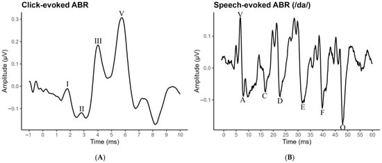
Figure 1


