1. Introduction
Intracranial dermoid cyst is a rare, benign, nonneoplastic tumor-like lesion representing 0.04–0.6% of primary brain tumors [
1]. Even though both dermoid cyst and teratoma can contain hair or bone components, dermoid cyst is considered to arise from ectodermal cells, whereas teratoma is considered to arise from misplaced multipotent germ cells [
2,
3,
4,
5]. Dermoid cysts are different from teratoma, which is classified as a neoplastic tumor [
6] containing all three embryonic germ cell layers [
3]. Dermoid cysts are also distinguishable from epidermoid cysts, which contain keratinaceous debris, solid crystalline cholesterol, but not dermal appendages, even though both are congenital, ectodermal inclusion cysts [
7].
Clinically, seizure (44%) and headache (67%) are common [
5,
8]. Two types of seizures have been described in previous reports. One type involves generalized seizures that are considered to result from exposure to inflammatory contents following rupture of the dermoid cyst [
7,
8,
9,
10,
11,
12]. Generalized seizures concomitant with symptoms of aseptic meningitis are explained by this theory and are thus considered an acute symptomatic seizure. The other type of seizure involves impaired awareness. Many reports have described patients showing not only the type of generalized seizure with symptoms of aseptic meningitis, but also chronic impaired awareness seizures without concomitant aseptic meningitis, which could be considered as epileptic seizures [
7,
8,
9,
10,
11].
We encountered a patient with anti-seizure medication (ASM)-resistant epilepsy with a dermoid cyst located in the temporal region, who never showed symptoms of aseptic meningitis. We hypothesized that the temporal lobe dermoid cyst in combination with other factors such as focal cortical dysplasia (FCD), etc., were causing the epileptic seizure.
2. Case Presentation
2.1. Clinical Course
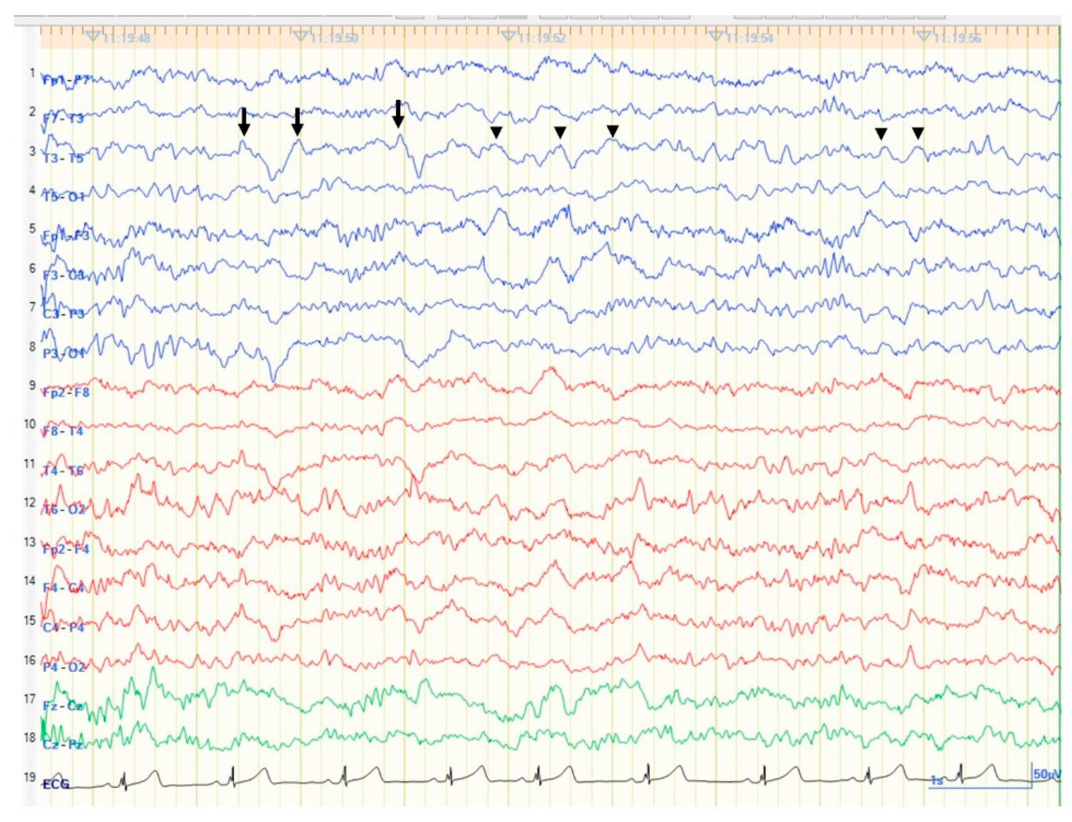
A 17-year-old left-handed girl was referred from a local physician for control of medically intractable epileptic seizures. She had experienced epileptic seizures since the age of 13 years. At that time, she had described a weekly olfactory aura as an indescribable but uncomfortable smell, followed by epigastric sensation. Those seizures often (monthly) led to loss of awareness with mouthing, drooling, and dystonic posturing of the right hand. This impaired awareness seizure generally lasted 1–2 minutes. This impaired awareness seizure rarely proceeded to bilateral tonic-clonic seizures, only once in several years. Electroencephalograms (EEG) showed left temporal focal slowing without epileptiform discharges (
Figure 1). When she underwent long term video EEG for a week, she only reported the olfactory aura without impaired awareness seizure, and showed no EEG changes. Magnetic resonance imaging (MRI) of the brain showed a left temporal lesion. Left temporal lobe epilepsy secondary to the left temporal lesion was diagnosed and levetiracetam was started. The patient remained free from focal-onset impaired awareness seizures and focal-to-bilateral tonic-clonic seizures for three years, but still experienced olfactory auras during this period.
Olfactory auras gradually increased in frequency to daily and also increased in intensity. Focal-onset impaired awareness seizures and focal-to-bilateral tonic-clonic seizures also recurred from the age of 17 years. Lacosamide was added to levetiracetam, but seizures were not controlled. She was therefore referred to our hospital for consideration of the possibility of epilepsy surgery.
No neurological abnormalities were identified other than the epileptic seizures. Laboratory data were within normal ranges, including alpha fetoprotein and human chorionic gonadotropin.
2.2. Neuropsychological Examinations
The Wechsler adult intelligence scale-4th edition showed: total intelligence quotient (IQ) index, 114; verbal comprehension index, 110; perceptual reasoning index, 114; working memory index, 109; and processing speed index 118. The revised Wechsler memory scale showed: auditory memory index, 129; visual memory index, 111; general memory index, 129; attention index, 121; and delayed memory index, 124.
To perform Wada test, we placed the 4-French catheter in the internal carotid artery through a guiding catheter in the right femoral artery. We manually injected diluted propofol (10 mg per 10 mL solution of saline) through the catheter. Scalp EEG was performed simultaneously to observe the diffuse slowing produced by the propofol in the injected hemisphere [
13]. The Wada test showed left-hemisphere language dominance.
2.3. Neuro-Imaging
MRI (
Figure 2) showed a well-delineated lesion in the left temporal lobe. The lesion had two major components. One component was mainly located in the medial temporal region and showed signal heterogeneity on T1-weighted imaging (WI) and hyperintensity on T2-WI. This massive component was displacing the left temporal lobe upwards and the hippocampus backward. This massive component was an extra-axial lesion, because T1-WI and fluid-attenuated inversion recovery (FLAIR) imaging showed a thin, hypointense space between the lesion and temporal lobe structures. The other component was a cystic part at the bottom of the temporal tip. This cystic part existed inside the temporal lobe. However, computed tomography showed a high-density lesion in the temporal tip, continuous with the massive medial temporal lesion. Diffusion-weighted imaging showed a low-intensity lesion with a slight hyperintense area inside the lesion.
2.4. Surgery
Since seizures had remained uncontrolled, she exhibited both right hand dystonic posturing during impaired awareness seizures [
14], and focal slowing in the left temporal region on EEG, we regarded those seizures as associated with the left temporal lesion. We therefore performed left temporal lesionectomy and resection of the tip of the antero-inferior temporal lobe in an awake state. We limited the resection area to the antero-inferior temporal lobe with the lesion because the patient showed high IQ and memory scores. We therefore designed the resection area from the beginning without performing a subdural electrode study in an attempt to preserve these higher brain and memory functions.
The inferior temporal gyrus was slightly firm. We first approached the lesion from the superior side of the firm inferior temporal gyrus. We then approached the massive cystic part, which was encapsulated by a thin, transparent, and fibrous membrane. Inside the capsule, yellowish, scrambled-egg-like creamy parts were found, containing many white, nose-hair-like structures. We completely removed this creamy lesion with its fibrous capsule, which was not adherent to surrounding structures and was easily removable. The cystic part in the anterior temporal tip was also removed with the antero-inferior tip of the temporal lobe. Neither dermal sinus nor bone deficits were seen.
2.5. Histopathology
The massive cystic lesion depicted in the MRI contained keratinaceous debris. Other small cystic structures were identified in the brain parenchyma. These small cystic structures had an epithelial lining of keratinizing stratified squamous epithelium and were seen in the cortex. Some parts of the epithelium of the cysts contained born components. Hairs were seen adjacent to the bone components and also in the brain parenchyma. The keratinized substance was present in a form that was involved in the brain parenchyma, surrounded by glial cell proliferation. Near the keratinized substance, dysmorphic neurons situated in the white matter were indicative of focal cortical dysplasia (FCD) (
Figure 3).
2.6. Postoperative Course
Following excision of the tip of the left temporal lobe, which included the cystic lesion, the patient remained free from daily olfactory auras as well as focal-onset impaired awareness seizures and focal-to-bilateral tonic-clonic seizures for more than 50 days. Post-operative MRI depicted total removal of the lesion with the tip of the left temporal lobe (
Figure 4). Post-operative scalp EEG showed no focal slowing and epileptiform discharges observed, even though a continuous breach rhythm was seen in the left fronto-temporal area.
The patient remained free from daily olfactory auras right after the surgery, but as the follow-up period was short, we considered that freedom from seizures might not have been completely secured for the patient, and long-term follow-up remains important.
3. Discussion
We have reported a case of temporal lobe dermoid cyst concomitant with FCD that caused temporal lobe epilepsy. The dermoid cyst existed in the left temporal fossa, but not in midline structures, and the patient had never experienced any episodes of meningitis or shown any findings of rupture on serial MRI. We therefore considered that the epileptic seizures had not involved rupture of the dermoid cyst. Epileptic seizures were naturally considered to potentially involve the interplay of the FCD and dermoid cysts in the brain parenchyma [
15,
16,
17,
18].
Histopathological examination showed multiple small dermoid cysts in addition to the major dermoid component seen on MRI. FCDs and bone components and hair were found to be involved in the brain parenchyma. In the process of neural maturation, as the timing of neural tube formation is related to formation of both dermoid cysts and FCDs, this complex of abnormal structures in the left temporal lobe might arise at this time [
8,
19,
20].
Other factors that might induce epileptogenicity could include possible foreign body reactions to keratinization of the dermoid cysts, metabolic interactions around the lesion, possible substantial secretions from the dermoid cyst, or micro-rupture that could not be detected on neuroimaging or from clinical symptoms. However, this was only a case study, and more cases need to be evaluated to clarify these factors.
However, the most important point that this case made us carefully consider was that risk factors were not the single cause of epilepsy, since the temporal lobe had both a dermoid cyst and FCD.
Incomplete resection of the dermoid cyst might cause recurrence, and gross total resection is thus expected [
21,
22], but since multiple dermoid cysts were present in the brain parenchyma in addition to the MRI visible lesion in this case, consideration should be given to the risk of recurrence even if gross total removal is performed, because residual dermoid cysts might be left in the brain parenchyma.
In terms of seizure, caution is also warranted not only about acute symptomatic seizures caused by ruptured dermoid cyst, but also about epileptic seizures associated with cysts located in non-midline, supratentorial regions.
For the surgical management of both dermoid cyst and ASM-resistant epilepsy, since invasive monitoring risks rupture of the dermoid cyst, detecting epileptogenicity in the brain to determine the optimal area for resection and simultaneously preserving higher brain function by brain mapping is a tall order. As this was the first case report of dermoid cyst concomitant with FCD, little is known about the exact epileptogenicity. We hope this case summaries provide valuable data. Collecting more data such as intraoperative EEG [
11] in a global, multicenter study is thus warranted in future cases.
4. Conclusions
A patient with medically intractable epilepsy secondary to left temporal lobe dermoid cyst showed multiple intramedullary cysts and focal cortical dysplasia that might have interacted to create epileptogenicity. To our knowledge, this is the first case report of dermoid cyst concomitant with FCD.
Author Contributions
Acquisition of data: K.H., A.F., H.E. and T.O.; analysis and interpretation of data: A.F. and C.I.; surgery: K.H. and A.F.; histopathology: Y.O.; advice regarding the paper: A.F., C.I., H.E. and T.O. All authors have read and agreed to the published version of the manuscript.
Funding
This research received no external funding.
Institutional Review Board Statement
The study was conducted according to the guidelines of the Declaration of Helsinki, and approved by The ethics committee at Seirei Hamamatsu General Hospital.
Informed Consent Statement
Written, informed consent for the publication of this case report was obtained from the patient’s guardian.
Data Availability Statement
The data are not publicly available due to patients’ privacy.
Conflicts of Interest
The authors declare no conflict of interest.
References
- Lunardi, P.; Missori, P. Supratentorial dermoid cysts. J. Neurosurg. 1991, 75, 262–266. [Google Scholar] [CrossRef]
- Geyik, A.M.; Geyik, S.; Erkutlu, I.; Alptekin, M.; Gezgin, I.; Dokur, M. Multicentric Dentigerous Dermoid Cyst with an Unusual Location in the Central Nervous System. Surg. J. 2016, 2, e1–e4. [Google Scholar] [CrossRef] [PubMed] [Green Version]
- Agrawal, M.; Uppin, M.S.; Patibandla, M.R.; Bhattacharjee, S.; Panigrahi, M.K.; Saradhi, V.; Rani, J.Y.; Purohit, A.K.; Challa, S. Teratomas in central nervous system: A clinico-morphological study with review of literature. Neurol. India 2010, 58, 841–846. [Google Scholar] [CrossRef] [Green Version]
- Schijman, E.; Monges, J.; Cragnaz, R. Congenital dermal sinuses, dermoid and epidermoid cysts of the posterior fossa. Childs Nerv. Syst. 1986, 2, 83–89. [Google Scholar] [CrossRef]
- Forbes, K. Osborn, Salzman, Barkovich et al.: Diagnostic Imaging; Springer: Berlin/Heidelberg, Germany, 2012. [Google Scholar]
- Louis, D.N.; Perry, A.; Wesseling, P.; Brat, D.J.; Cree, I.A.; Figarella-Branger, D.; Hawkins, C.; Ng, H.K.; Pfister, S.M.; Reifenberger, G.; et al. The 2021 WHO Classification of Tumors of the Central Nervous System: A summary. Neuro-oncology 2021, 23, 1231–1251. [Google Scholar] [CrossRef] [PubMed]
- Ray, M.J.; Barnett, D.W.; Snipes, G.J.; Layton, K.F.; Opatowsky, M.J. Ruptured intracranial dermoid cyst. Peer-Rev. J. Bayl. Scott White Health 2012, 25, 23–25. [Google Scholar] [CrossRef] [PubMed] [Green Version]
- Shashidhar, A.; Sadashiva, N.; Prabhuraj, A.R.; Narasingha Rao, K.; Tiwari, S.; Saini, J.; Shukla, D.; Devi, B.I. Ruptured intracranial dermoid cysts: A retrospective institutional review. J. Clin. Neurosci. 2019, 67, 172–177. [Google Scholar] [CrossRef] [PubMed]
- Alam, Y.; Mugge, L.A.; Purdy, J.; Mrak, R.E.; Schroeder, J. Long-term Seizure Disorder Caused by a Dermoid Cyst with Catastrophic Developments. Cureus 2018, 10, e3272. [Google Scholar] [CrossRef] [PubMed] [Green Version]
- Shehadi, J.A.; Alorainy, I.A.; Johnston, K.M. Temporal dermoid cyst with a partial dermal sinus tract. Can. J. Neurol. Sci. 1999, 26, 321–324. [Google Scholar] [CrossRef] [Green Version]
- Sugano, H.; Shimizu, H.; Sunaga, S.; Arai, N.; Tamagawa, K. Temporal lobe epilepsy caused by dermoid cyst. Neurol. Med. Chir. 2006, 46, 206–209. [Google Scholar] [CrossRef] [PubMed] [Green Version]
- Bishnoi, I.; Bishnoi, S.; Gahlawat, N.; Bhardwaj, L.; Duggal, G.; Singal, G.; Singh, P. Management of a rare case of intraventricular ruptured dermoid cyst and chemical meningitis. Br. J. Neurosurg. 2018, 1–4. [Google Scholar] [CrossRef]
- Fujimoto, A.; Okanishi, T.; Nishimura, M.; Kanai, S.; Sato, K.; Enoki, H. The Wada test might predict postoperative fine finger motor deficit after hemispherotomy. J. Clin. Neurosci. 2017, 45, 319–323. [Google Scholar] [CrossRef] [PubMed]
- Kotagal, P.; Lüders, H.; Morris, H.H.; Dinner, D.S.; Wyllie, E.; Godoy, J.; Rothner, A.D. Dystonic posturing in complex partial seizures of temporal lobe onset: A new lateralizing sign. Neurology 1989, 39, 196–201. [Google Scholar] [CrossRef] [PubMed]
- Sakakura, K.; Fujimoto, A.; Arai, Y.; Ichikawa, N.; Sato, K.; Baba, S.; Inenaga, C.; Matsumura, A.; Ishikawa, E.; Enoki, H.; et al. Posttraumatic epilepsy may be a state in which underlying epileptogenicity involves focal cortical dysplasia. Epilepsy Behav. 2021, 114, 107352. [Google Scholar] [CrossRef] [PubMed]
- Ikawa, A.; Fujimoto, A.; Arai, Y.; Otsuki, Y.; Nozaki, T.; Baba, S.; Sato, K.; Enoki, H. Case Report: Late-Onset Temporal Lobe Epilepsy Following Subarachnoid Hemorrhage: An Interplay Between Pre-existing Cortical Development Abnormality and Tissue Damage. Front. Neurol. 2021, 12, 599130. [Google Scholar] [CrossRef] [PubMed]
- Itamura, S.; Okanishi, T.; Arai, Y.; Nishimura, M.; Baba, S.; Ichikawa, N.; Hirayama, Y.; Ishihara, N.; Hiraide, T.; Ishigaki, H.; et al. Three Cases of Hemiconvulsion-Hemiplegia-Epilepsy Syndrome with Focal Cortical Dysplasia Type IIId. Front. Neurol. 2019, 10, 1233. [Google Scholar] [CrossRef] [PubMed] [Green Version]
- Sakakura, Y.; Fujimoto, A.; Okanishi, T.; Masuda, Y.; Nishimura, M.; Enoki, H. A case of posterior quadrant disconnection for post-subdural empyema epilepsy-focal cortical dysplasia might be the underlying cause of refractory epilepsy. Interdiscip. Neurosurg. 2018, 14, 50–52. [Google Scholar] [CrossRef]
- Cotter, D.; Honavar, M.; Lovestone, S.; Raymond, L.; Kerwin, R.; Anderton, B.; Everall, I. Disturbance of Notch-1 and Wnt signalling proteins in neuroglial balloon cells and abnormal large neurons in focal cortical dysplasia in human cortex. Acta Neuropathol. 1999, 98, 465–472. [Google Scholar] [CrossRef] [PubMed]
- Sarnat, H.B.; Philippart, M.; Flores-Sarnat, L.; Wei, X.C. Timing in neural maturation: Arrest, delay, precociousness, and temporal determination of malformations. Pediatr. Neurol. 2015, 52, 473–486. [Google Scholar] [CrossRef] [PubMed]
- Perry, A.; Brat, D.J. Practical Surgical Neuropathology: A Diagnostic Approach; Elsevier Health Sciences: Amsterdam, The Netherlands, 2010. [Google Scholar]
- Fanous, A.A.; Gupta, P.; Li, V. Analysis of the growth pattern of a dermoid cyst. J. Neurosurg. Pediatr. 2014, 14, 621–625. [Google Scholar] [CrossRef] [PubMed] [Green Version]
| Publisher’s Note: MDPI stays neutral with regard to jurisdictional claims in published maps and institutional affiliations. |
© 2021 by the authors. Licensee MDPI, Basel, Switzerland. This article is an open access article distributed under the terms and conditions of the Creative Commons Attribution (CC BY) license (https://creativecommons.org/licenses/by/4.0/).


 ,
, 