

Listening to 15 Hz Binaural Beats Enhances the Connectivity of Functional Brain Networks in the Mental Fatigue State—An EEG Study

Abstract
:1. Introduction
2. Materials and Methods
2.1. Participants
2.2. Material
2.2.1. Mental Fatigue-Inducing Task: The Improved Version of TloadDback (Time Load Dual Back)
2.2.2. Scales and Paradigms
Visual Analogue Scale-Fatigue (VAS-f)
Intrinsic Motivation Inventory (IMI)
Other Cognitive Scales and Tasks
2.2.3. Auditory Stimulation
2.2.4. Electroencephalogram (EEG)
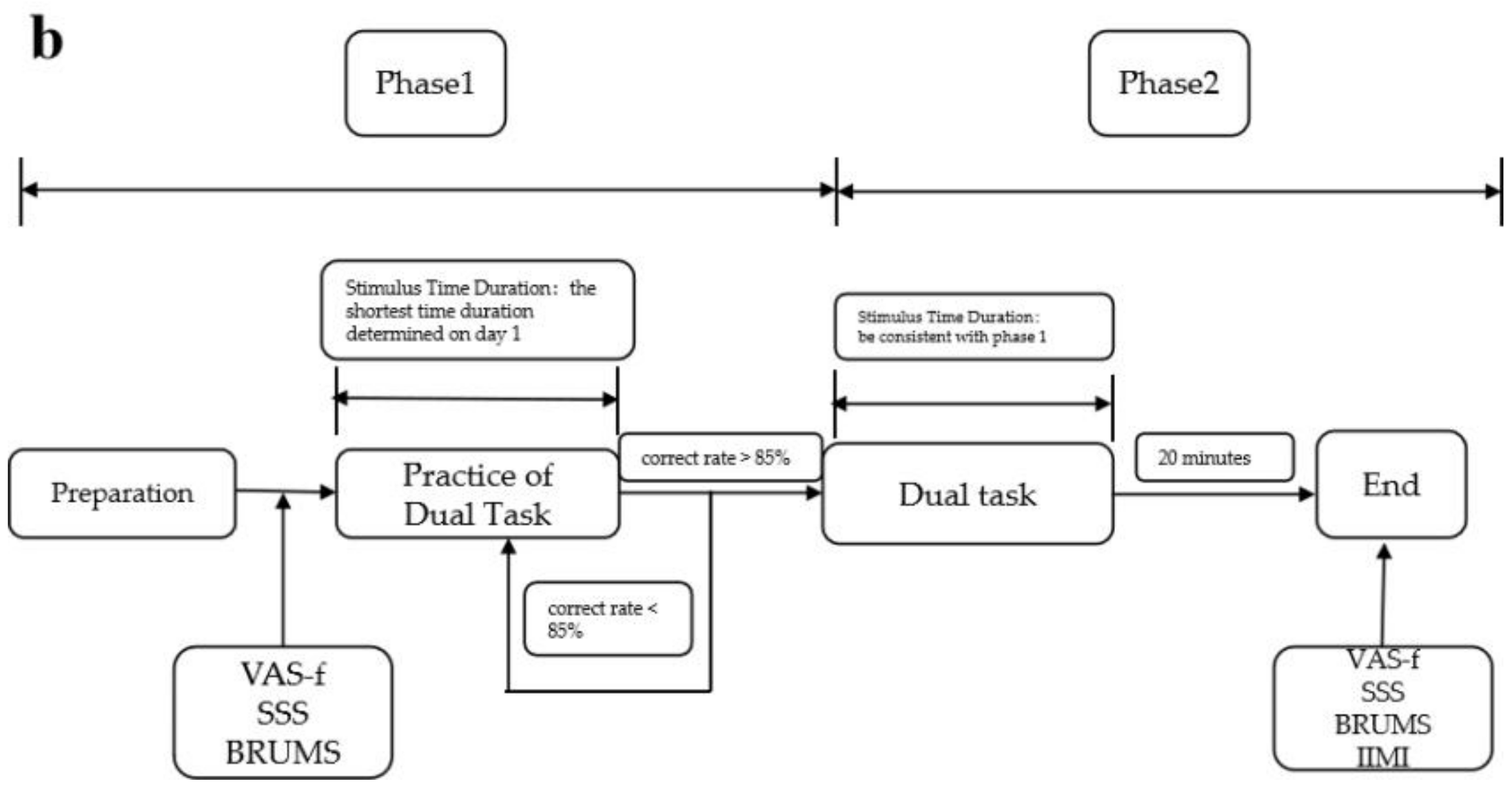
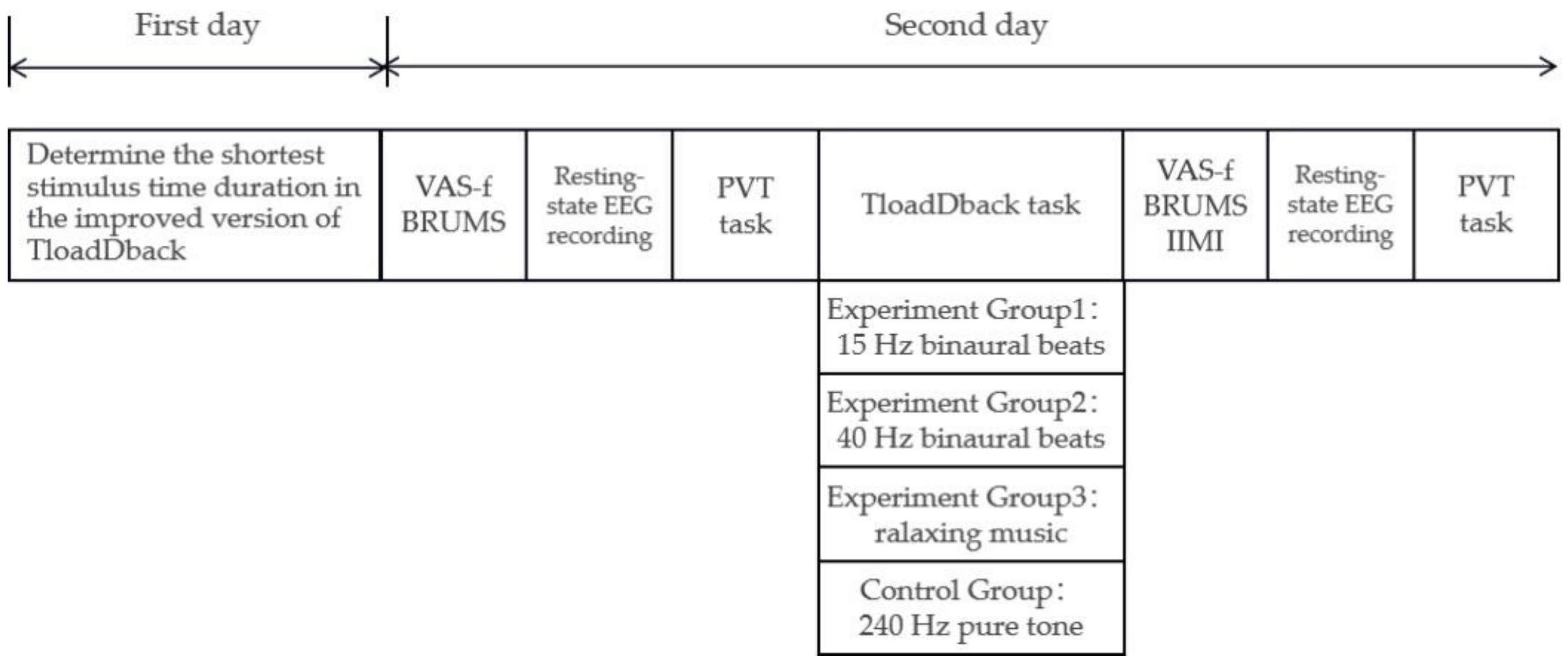
2.2.5. Experimental Procedure
2.2.6. Data Processing of Scales and Tasks
2.2.7. Data Processing of Electroencephalographic
2.2.8. Statistical Analysis
3. Results
3.1. Scales
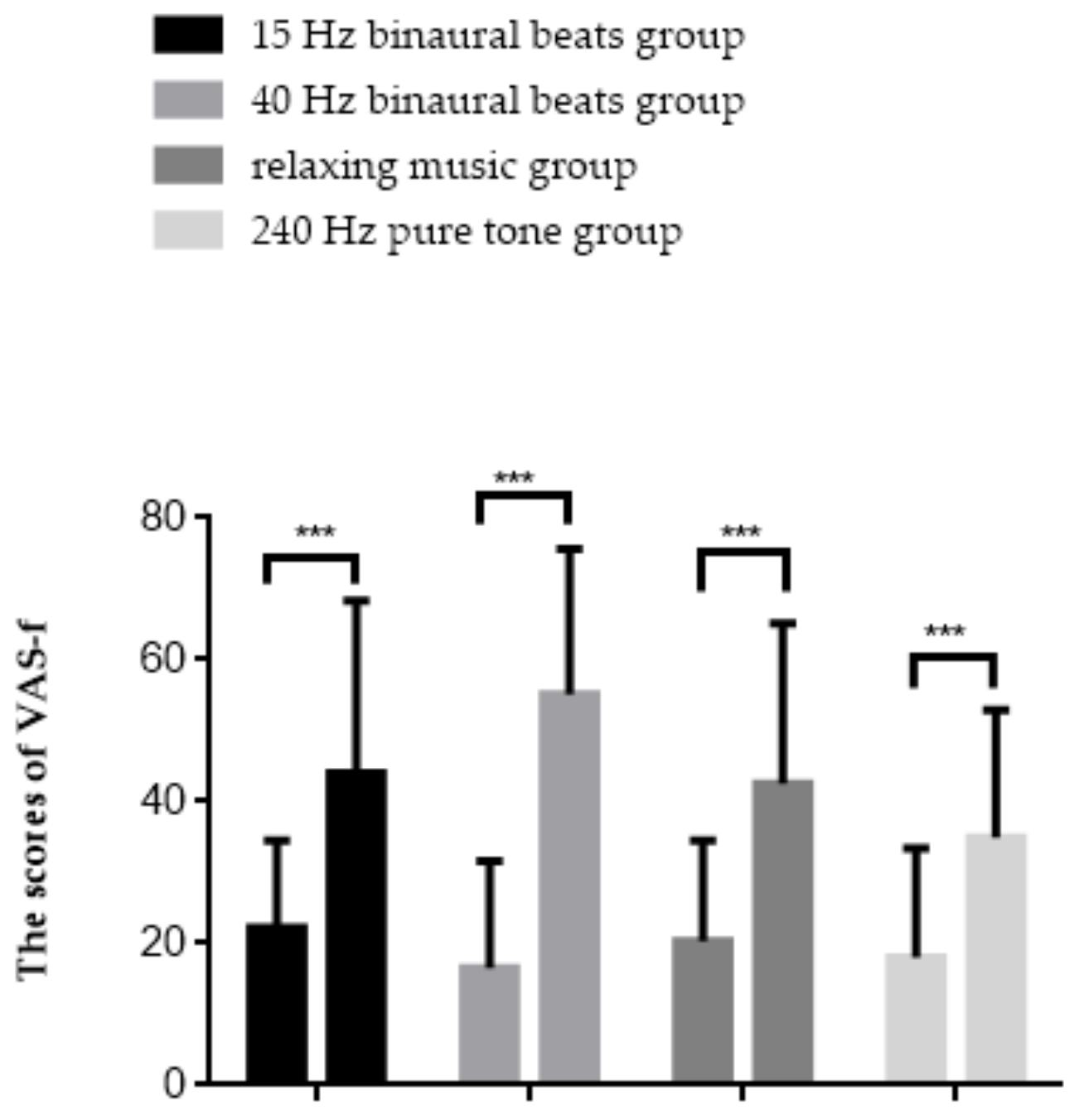
3.1.1. Visual Analogue Scale-Fatigue
3.1.2. BRUMS
3.1.3. “Effort” Subscale in Intrinsic Motivation Scale
3.2. Behavioural Performances
3.2.1. PVT
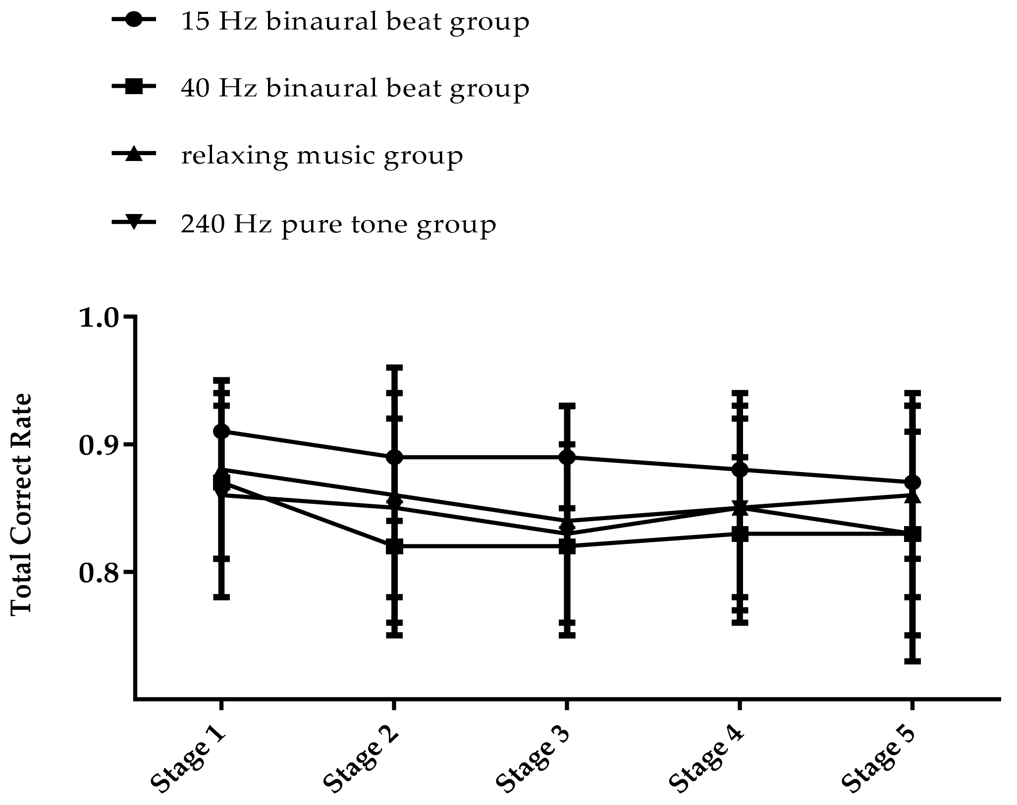
3.2.2. TloadDback
3.3. Electroencephalographic
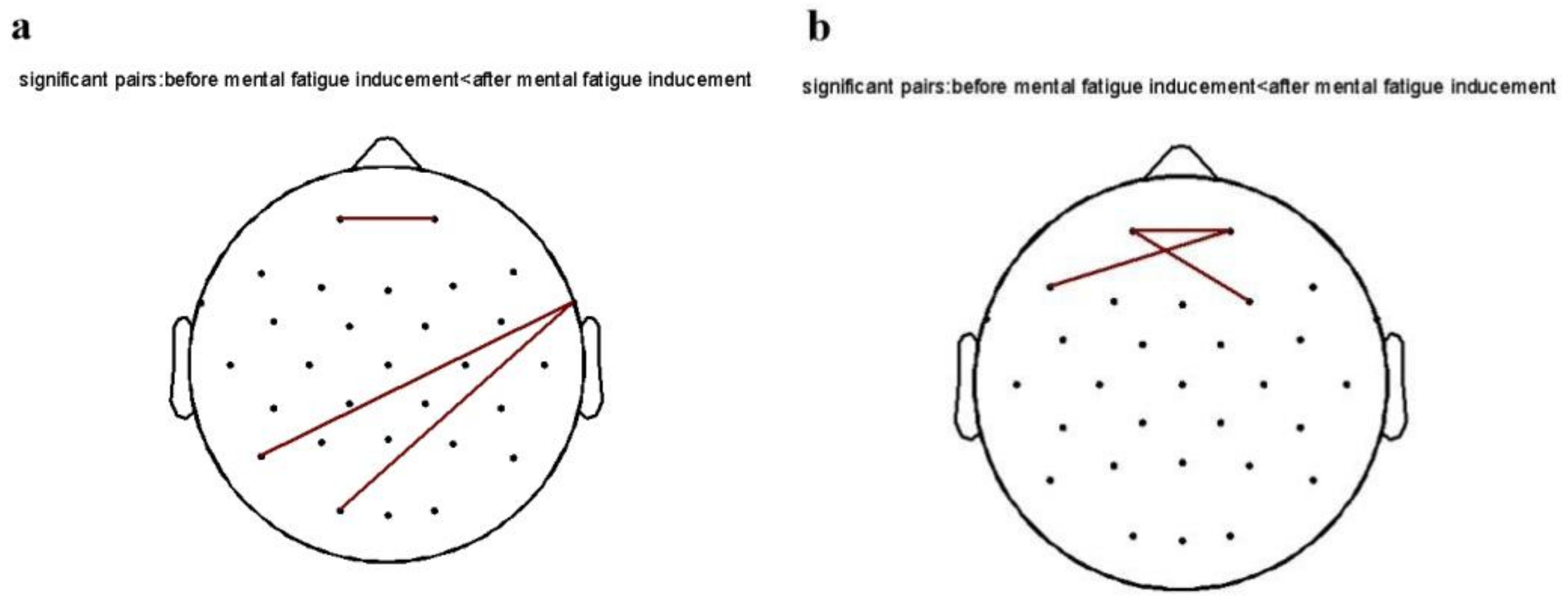
3.3.1. Functional Connectivity
3.3.2. Topological Structure
4. Discussion
5. Limitations
6. Conclusions
Author Contributions
Funding
Institutional Review Board Statement
Informed Consent Statement
Data Availability Statement
Conflicts of Interest
References
- Lal, S.K.; Craig, A. A critical review of the psychophysiology of driver fatigue. Biol. Psychol. 2001, 55, 173–194. [Google Scholar] [PubMed]
- Boksem, M.A.S.; Meijman, T.F.; Lorist, M.M. Effects of mental fatigue on attention: An ERP study. Cogn. Brain Res. 2005, 25, 107–116. [Google Scholar]
- Grillon, C.; Quispe-Escudero, D.; Mathur, A.; Ernst, M. Mental fatigue impairs emotion regulation. Emotion 2015, 15, 383–389. [Google Scholar]
- Liu, Q.; Zhou, R.; Liu, L.; Zhao, X. Effects of 72 hours total sleep deprivation on male astronauts’ executive functions and emotion. Compr Psychiatry 2015, 61, 28–35. [Google Scholar]
- Tian, S.; Wang, Y.; Dong, G.; Pei, W.; Chen, H. Mental Fatigue Estimation Using EEG in a Vigilance Task and Resting States. In Proceedings of the 2018 40th Annual International Conference of the IEEE Engineering in Medicine and Biology Society, Honolulu, HI, USA, 18–21 July 2018. [Google Scholar]
- Mun, S.; Kim, E.; Park, M. Effect of mental fatigue caused by mobile 3D viewing on selective attention: An ERP study. Int. J. Psychophysiol. 2014, 94, 373–381. [Google Scholar]
- Zhang, Y.; Gong, J.; Miao, D.; Zhu, X.; Yang, Y. Subjective evaluation of mental fatigue and characteristics of attention during a driving simulation. Soc. Behav. Personal. Int. J. 2011, 39, 15–20. [Google Scholar]
- Tian, H. Cognitive Fatigue in Memory Task Error of Middle School Students based on Brain Evoked Potential. NeuroQuantology 2018, 16, 669–675. [Google Scholar]
- Killgore, W.D.S.; Balkin, T.J.; Wsensten, N.J. Impaired decision making following 49 h of sleep deprivation. J. Sleep Res. 2006, 15, 7–13. [Google Scholar]
- Hart, R.P.; Buchsbaum, D.G.; Wade, J.B.; Hamer, R.M.; Kwentus, J.A. Effect of sleep deprivation on first-year residents’ response times, memory, and mood. J. Med. Educ. 1987, 62, 940–942. [Google Scholar]
- Hu, X.; Lodewijks, G. Detecting fatigue in car drivers and aircraft pilots by using non-invasive measures: The value of differentiation of sleepiness and mental fatigue. J. Saf. Res. 2020, 72, 173–187. [Google Scholar]
- Liu, X.D.; Jian-Guang, L.I.; Lian, Y.L.; Bai, Y.F.; Liu, J.W. Research on Mental Fatigue of Soldiers in the Arid Desert. Hosp. Adm. J. Chin. People’s Lib. Army 2011, 19, 124–126. [Google Scholar]
- Di Stasi, L.L.; Renner, R.; Catena, A.; Cañas, J.J.; Velichkovsky, B.M.; Pannasch, S. Towards a driver fatigue test based on the saccadic main sequence: A partial validation by subjective report data. Transp. Res. Part C Emerg. Technol. 2012, 21, 122–133. [Google Scholar]
- Iampetch, S.; Punsawad, Y.; Wongsawat, Y. EEG-based mental fatigue prediction for driving application. In Proceedings of the 5th 2012 Biomedical Engineering International Conference, Ubon Ratchathani, Thailand, 5–7 December 2012. [Google Scholar]
- Driskell, J.E.; Mullen, B. The Efficacy of Naps as a Fatigue Countermeasure: A Meta-Analytic Integration. Hum. Factors 2005, 47, 360–377. [Google Scholar] [PubMed]
- Hartzler, B.M. Fatigue on the flight deck: The consequences of sleep loss and the benefits of napping. Accid. Anal. Prev. 2014, 62, 309–318. [Google Scholar] [PubMed]
- Oriyama, S.; Miyakoshi, Y.; Rahman, M.M. The effects of a 120-min nap on sleepiness, fatigue, and performance during 16-hour night shifts: A pilot study. J. Occup. Health 2019, 61, 368–377. [Google Scholar]
- Kubo, T.; Takahashi, M.; Sallinen, M.; Kubo, Y.; Suzumura, H. How are Leisure Activity and Shiftwork Schedule Associated with Recovery from Fatigue in Shiftwork Nurses? Sangyō Eiseigaku Zasshi = J. Occup. Health 2013, 55, 90–102. [Google Scholar]
- Tietzel, A.J.; Lack, L.C. The Short-Term Benefits of Brief and Long Naps Following Nocturnal Sleep Restriction. Sleep 2001, 24, 293–300. [Google Scholar]
- Helton, W.S.; Russell, P.N. Rest Is Still Best: The Role of the Qualitative and Quantitative Load of Interruptions on Vigilance. Hum. Factors J. Hum. Factors Ergon. Soc. 2017, 59, 91. [Google Scholar]
- Chen, X.; Zhang, L.; Yang, D.; Li, C.; An, G.; Wang, J.; Shao, Y.; Fan, R.; Ma, Q. Effects of Caffeine on Event-Related Potentials and Neuropsychological Indices After Sleep Deprivation. Front. Behav. Neurosci. 2020, 14, 108. [Google Scholar]
- Nawrot, P.; Jordan, S.; Eastwood, J.; Rotstein, J.; Hugenholtz, A.; Feeley, M. Effects of caffeine on human health. Food Addit. Contam. 2003, 20, 1–30. [Google Scholar]
- Gupta, A.; Bhushan, B.; Behera, L. Short-term enhancement of cognitive functions and music: A three-channel model. Sci. Rep. 2018, 8, 15528. [Google Scholar] [PubMed]
- Huang, R.H.; Shih, Y.N. Effects of background music on concentration of workers. Work 2011, 38, 383–387. [Google Scholar] [PubMed]
- Shih, Y.N.; Huang, R.H.; Chiang, H.Y. Background music: Effects on attention performance. Work 2012, 42, 573–578. [Google Scholar] [PubMed]
- Heuberger, E.; Ilmberger, J. The influence of essential oils on human vigilance. Nat. Prod. Commun. 2010, 5, 1441–1446. [Google Scholar] [PubMed]
- Rickard, N.S. The Effect of Music on Cognitive Performance: Insight From Neurobiological and Animal Studies. Behav. Cogn. Neurosci. Rev. 2005, 4, 235. [Google Scholar]
- Dean, R.T.; Freya, B.; Emery, S. Acoustic Intensity Causes Perceived Changes in Arousal Levels in Music: An Experimental Investigation. PLoS ONE 2011, 6, e18591. [Google Scholar]
- Al-Shargie, F.; Tariq, U.; Mir, H.; Alawar, H.; Babiloni, F.; Al-Nashash, H. Vigilance Decrement and Enhancement Techniques: A Review. Brain Sci. 2019, 9, 178. [Google Scholar]
- Nittono, H.; Tsuda, A.; Akai, S.; Nakajima, Y. Tempo of background sound and performance speed. Percept. Mot. Ski. 2000, 90, 1122. [Google Scholar]
- Mckel, M.; Strk, T.; Vollert, J.; Rcker, L.; Frei, U. Stress reduction through listening to music: Effects on stress hormones, haemodynamics and psychological state in patients with arterial hypertension and in healthy subjects. Dtsch. Med. Wochenschr. 1995, 120, 745–752. [Google Scholar]
- Saghari, H.; Sheibani, V.; Esmaeilpour, K.; Rehman, N.U. Music Alleviates Learning and Memory Impairments in an Animal Model of Post-Traumatic Stress Disorder. Biointerface Res. Appl. Chem. 2021, 11, 7775–7784. [Google Scholar]
- Bensimon, M.; Amir, D.; Wolf, Y. Drumming through trauma: Music therapy with post-traumatic soldiers. Arts Psychother. 2008, 35, 34–48. [Google Scholar]
- Li, S.; Zhang, L. Light Music Conduces to Releasing Short-term Mental Fatigue of College Students. Chin. J. Sports Med. 2015, 34, 578. [Google Scholar]
- Yokoyama, M.; Oguri, K.; Miyaji, M. Effect of sound pressure levels of music on driver’s drowsiness. In Proceedings of the 15th World Congress on Intelligent Transport Systems and ITS America’s 2008 Annual MeetingITS AmericaERTICOITS JapanTransCore, New York, NY, USA, 16–20 November 2008. [Google Scholar]
- Guo, W.; Ren, J.; Wang, B.; Zhu, Q. Effects of Relaxing Music on Mental Fatigue Induced by a Continuous Performance Task: Behavioral and ERPs Evidence. PLoS ONE 2015, 10, e0136446. [Google Scholar] [CrossRef] [PubMed]
- Schellenberg, E.G. Cognitive Performance After Listening to Music: A Review of the Mozart Effect. Music Health Wellbeing 2012, 324–338. [Google Scholar]
- Jacquet, T.; Poulin-Charronnat, B.; Bard, P.; Perra, J.; Lepers, R. Physical activity and music to counteract mental fatigue. Neuroscience 2021, 478, 75–88. [Google Scholar] [PubMed]
- O’Keeffe, K.; Dean, J.; Hodder, S.; Lloyd, A. Self-selected motivational music enhances physical performance in normoxia and hypoxia in young healthy males. Front. Psychol. 2021, 12, 787496. [Google Scholar]
- Phneah, S.W.; Nisar, H. EEG-based alpha neurofeedback training for mood enhancement. Australas. Phys. Eng. Sci. Med. 2017, 40, 325–336. [Google Scholar]
- Dalton, B.H.; Behm, D.G.; Kibele, A. Effects of sound types and volumes on simulated driving, vigilance tasks and heart rate. Occup. Ergon. 2007, 7, 153–168. [Google Scholar] [CrossRef]
- Oron-Gilad, T.; Ronen, A.; Shinar, D. Alertness maintaining tasks (AMTs) while driving. Accid. Anal. Prev. 2008, 40, 851–860. [Google Scholar] [CrossRef]
- Bhatti, A.M.; Majid, M.; Anwar, S.M.; Khan, B. Human emotion recognition and analysis in response to audio music using brain signals. Comput. Hum. Behav. 2016, 65, 267–275. [Google Scholar] [CrossRef]
- Brodsky, W. The effects of music tempo on simulated driving performance and vehicular control. Transp. Res. Part F Traffic Psychol. Behav. 2001, 4, 219–241. [Google Scholar] [CrossRef]
- Alikonis, C.R.; Warm, J.S.; Matthews, G.; Dember, W.N.; Hitchcock, E.M.; Kellaris, J.J. Vigilance, Workload, and Boredom: Two Competing Models; SAGE Publications: Los Angeles, CA, USA, 2002. [Google Scholar]
- Moyosola, S.M.; Alexandru, M.D.; Nicole, G.; Al Gayar Sarmad Monadel, S.; Pavaloiu, B.; Boiangiu, C. (Eds.) Development of a low-cost and user-friendly neurofeedback tool to treat depression, insomnia, anxiety, pain and ADHD using an Arduino and Android Application. In Proceedings of the 2019 International Conference on Automation, Computational and Technology Management (ICACTM), London, UK, 24–26 April 2019; pp. 493–499. [Google Scholar]
- Dadashi, M.; Birashk, B.; Taremian, F.; Asgarnejad, A.A.; Momtazi, S. Effects of increase in amplitude of occipital alpha & theta brain waves on global functioning level of patients with GAD. Basic Clin. Neurosci. 2015, 6, 14–20. [Google Scholar] [PubMed]
- Oster, G. Auditory beats in the brain. Sci. Am. 1973, 229, 94–103. [Google Scholar] [CrossRef] [PubMed]
- Lane, J.D.; Kasian, S.J.; Owens, J.E.; Marsh, G.R. Binaural auditory beats affect vigilance performance and mood. Physiol. Behav. 1998, 63, 249–252. [Google Scholar] [CrossRef]
- Reedijk, S.A.; Bolders, A.; Hommel, B. The impact of binaural beats on creativity. Front. Hum. Neurosci. 2013, 7, 786. [Google Scholar] [CrossRef]
- Schwarz, D.; Taylor, P. Human auditory steady state responses to binaural and monaural beats. Clin. Neurophysiol. 2005, 116, 658–668. [Google Scholar] [CrossRef]
- Kuwada, S.; Wickesberg, T.Y. Response of cat inferior colliculus neurons to binaural beat stimuli: Possible mechanisms for sound localization. Science 1979, 206, 586–588. [Google Scholar] [CrossRef]
- Beauchene, C.; Abaid, N.; Moran, R.; Diana, R.A.; Leonessa, A. The effect of binaural beats on verbal working memory and cortical connectivity. J. Neural Eng. 2017, 14, 026014. [Google Scholar] [CrossRef] [Green Version]
- Chaieb, L.; Wilpert, E.C.; Reber, T.P.; Fell, J. Differential oscillatory electroencephalogram between attention-deficit/hyperactivity disorder subtypes and tyn and mood states. Front. Psychiatry 2015, 6, 422–429. [Google Scholar]
- Turow, G.; Lane, J.D. Binaural beat stimulation: Altering vigilance and mood states. In Music, Science, and the Rhythmic Brain: Cultural and Clinical Implications; Routledge: London, UK, 2011; pp. 122–136. [Google Scholar]
- Colzato, L.S.; Barone, H.; Sellaro, R.; Hommel, B. More attentional focusing through binaural beats: Evidence from the global–local task. Psychol. Res. 2017, 81, 271–277. [Google Scholar] [CrossRef]
- Padmanabhan, R.; Hildreth, A.J.; Laws, D. A prospective, randomised, controlled study examining binaural beat audio and pre-operative anxiety in patients undergoing general anaesthesia for day case surgery. Anaesthesia 2005, 60, 874–877. [Google Scholar] [CrossRef] [PubMed]
- Ortiz, T.; Martínez, A.M.; Fernández, A.; Maestu, F.; Poch, J. Impact of auditory stimulation at a frequency of 5 Hz in verbal memory. Actas Esp Psiquiatr. 2008, 36, 307–313. [Google Scholar] [PubMed]
- Lopez-Caballero, F.; Escera, C. Binaural Beat: A Failure to Enhance EEG Power and Emotional Arousal. Front. Hum. Neurosci. 2017, 11, 557. [Google Scholar] [CrossRef] [PubMed]
- Goodin, P.; Ciorciari, J.; Baker, K.; Carrey, A.; Harper, M.; Kaufman, J. A high-density EEG investigation into steady state binaural beat stimulation. PLoS ONE 2012, 7, e34789. [Google Scholar]
- Mcintire, L.K.; Mckinley, R.A.; Nelson, J.M.; Goodyear, C. Transcranial direct current stimulation versus caffeine as a fatigue countermeasure. Brain Stimul. 2017, 10, 1070–1078. [Google Scholar] [CrossRef]
- Chen, M.H.; Wylie, G.R.; Sandroff, B.M.; Dacosta-Aguayo, R.; Genova, H.M. Neural mechanisms underlying state mental fatigue in multiple sclerosis: A pilot study. J. Neurol. 2020, 267, 2372–2382. [Google Scholar] [CrossRef]
- Lim, J.; Wu, W.; Wang, J.; Detre, J.A.; Dinges, D.F.; Rao, H. Imaging brain fatigue from sustained mental workload: An ASL perfusion study of the time-on-task effect. NeuroImage 2010, 49, 3426–3435. [Google Scholar] [CrossRef]
- Delorme, A.; Makeig, S. EEGLAB: An open source toolbox for analysis of single-trial EEG dynamics including independent component analysis. J. Neurosci. Methods 2004, 134, 9–21. [Google Scholar] [CrossRef]
- Herlambang, M.B.; Taatgen, N.A.; Cnossen, F. The Role of Motivation as a Factor in Mental Fatigue. Hum. Factors J. Hum. Factors Ergon. Soc. 2019, 61, 1171–1185. [Google Scholar] [CrossRef]
- Linnhoff, S.; Wolter-Weging, J.; Zaehle, T. Objective electrophysiological fatigability markers and their modulation through tDCS. Clin. Neurophysiol. 2021, 132, 1721–1732. [Google Scholar] [CrossRef]
- Sandry, J.; Genova, H.M.; Dobryakova, E.; Deluca, J.; Wylie, G. Subjective Cognitive Fatigue in Multiple Sclerosis Depends on Task Length. Front. Neurol. 2014, 5, 214. [Google Scholar] [CrossRef] [PubMed]
- Brijesh; Kanda, R.K.; Kalra, P. Mental Fatigue Measurement using EEG While Performing General Mechanical Assembly Task. Int. J. Sci. Res. Dev. 2015, 3, 1526–1528. [Google Scholar]
- Koudelková, Z.; Strmiska, M. (Eds.) Introduction to the identification of brain waves based on their frequency. In MATEC Web of Conferences; EDP Sciences: Les Ulis, France, 2018. [Google Scholar]
- Mazaheri, A.; Fassbender, C.; Coffey-Corina, S.; Hartanto, T.A.; Schweitzer, J.B.; Mangun, G.R. Differential oscillatory electroencephalogram between attention-deficit/hyperactivity disorder subtypes and typically developing adolescents. Biol. Psychiatry 2014, 76, 422–429. [Google Scholar] [CrossRef] [PubMed]
- Koudelková, Z.; Strmiska, M.; Jašek, R. Analysis of brain waves according to their frequency. Int. J. Biol. Biomed. Eng. 2018, 12, 202–207. [Google Scholar]
- Bazanova, O.M.; Vernon, D. Interpreting EEG alpha activity. Neurosci. Biobehav. Rev. 2014, 44, 94–110. [Google Scholar] [CrossRef]
- Zhao, C.; Zhao, M.; Yang, Y.; Gao, J.; Rao, N.; Lin, P. The reorganization of human brain networks modulated by driving mental fatigue. IEEE J. Biomed. Health Inform. 2016, 21, 743–755. [Google Scholar] [CrossRef]
- Beauchene, C.; Abaid, N.; Moran, R.; Diana, R.A.; Leonessa, A. The effect of binaural beats on visuospatial working memory and cortical connectivity. PLoS ONE 2016, 11, e0166630. [Google Scholar]
- Hassan, M.; Wendling, F. Electroencephalography source connectivity: Aiming for high resolution of brain networks in time and space. IEEE Signal Process. Mag. 2018, 35, 81–96. [Google Scholar] [CrossRef]
- Hassan, M.; Dufor, O.; Merlet, I.; Berrou, C.; Wendling, F. EEG source connectivity analysis: From dense array recordings to brain networks. PLoS ONE 2014, 9, e105041. [Google Scholar]


| Age | Scores | |||
|---|---|---|---|---|
| PSQI | ESS | MES | ||
| 15 Hz binaural beats group | 23.27 ± 3.90 | 5.13 ± 2.20 | 5.13 ± 2.50 | 51.47 ± 8.25 |
| 40 Hz binaural beats group | 20.80 ± 2.40 | 4.87 ± 2.45 | 4.87 ± 3.09 | 50.73 ± 7.94 |
| relaxing music group | 20.47 ± 2.00 | 3.93 ± 1.28 | 5.53 ± 2.70 | 48.93 ± 4.64 |
| 240 Hz pure tone group | 23.20 ± 3.61 | 3.40 ± 2.77 | 4.73 ± 2.94 | 52.07 ± 2.61 |
| Group | Stage | |||||
|---|---|---|---|---|---|---|
| Stage 1 | Stage 2 | Stage 3 | Stage 4 | Stage 5 | ||
| 1-back task reaction time | 1 | 579.53 ± 67.68 | 581.67 ± 52.18 | 584.16 ± 40.23 | 571.19 ± 48.16 | 561.35 ± 52.65 |
| 2 | 532.78 ± 41.33 | 534.34 ± 59.17 | 535.60 ± 58.64 | 534.44 ± 76.03 | 523.20 ± 86.75 | |
| 3 | 564.35 ± 90.49 | 566.19 ±100.64 | 558.45 ± 116.91 | 542.80 ± 109.73 | 541.86 ± 103.16 | |
| 4 | 583.94 ± 97.55 | 593.53 ± 99.85 | 583.20 ± 119.32 | 569.03 ± 123.58 | 559.99 ± 135.74 | |
| odd-even decision task reaction time | 1 | 562.46 ± 58.99 | 570.77 ± 35.90 | 573.51 ± 48.99 | 567.14 ± 57.96 | 566.34 ± 53.54 |
| 2 | 535.46 ± 43.81 | 544.51 ± 51.61 | 547.42 ± 56.99 | 548.93 ± 53.77 | 545.11 ± 58.98 | |
| 3 | 554.59 ± 71.41 | 556.53 ± 84.34 | 556.10 ± 98.08 | 546.38 ± 92.06 | 550.62 ± 89.08 | |
| 4 | 580.67 ± 98.10 | 589.39 ± 99.59 | 588.89 ± 118.34 | 580.72 ± 123.25 | 573.47 ± 131.10 | |
| accuracy rate | 1 | 0.91 ± 0.04 | 0.89 ± 0.05 | 0.89 ± 0.04 | 0.88 ± 0.05 | 0.87 ± 0.06 |
| 2 | 0.87 ± 0.06 | 0.82 ± 0.07 * | 0.82 ± 0.07 * | 0.83 ± 0.06 * | 0.83 ± 0.08 | |
| 3 | 0.88 ± 0.07 | 0.86 ± 0.10 | 0.84 ± 0.09 ** | 0.85 ± 0.09 | 0.86 ± 0.08 | |
| 4 | 0.86 ± 0.08 | 0.85 ± 0.07 | 0.83 ± 0.07 | 0.85 ± 0.07 | 0.83 ± 0.10 | |
| Before Fatigue Inducement | After Fatigue Inducement | ||
|---|---|---|---|
| 15 Hz binaural beats group | Clustering coefficient | 0.19 ± 0.03 | 0.19 ± 0.02 |
| Average path length | 1.20 ± 0.13 | 1.13 ± 0.08 * | |
| Nodal efficiency | 0.17 ± 0.02 | 0.18 ± 0.01 | |
| 40 Hz binaural beats group | Clustering coefficient | 0.20 ± 0.03 | 0.20 ± 0.03 |
| Average path length | 1.14 ± 0.16 | 1.12 ± 0.15 | |
| Nodal efficiency | 0.18 ± 0.02 | 0.18 ± 0.02 | |
| Relaxing music group | Clustering factor | 0.19 ± 0.04 | 0.18 ± 0.03 |
| Average path length | 1.21 ± 0.25 | 1.24 ± 0.27 | |
| Nodal efficiency | 0.17 ± 0.03 | 0.17 ± 0.03 | |
| 240 Hz pure tone group | Clustering coefficient | 0.18 ± 0.03 | 0.18 ± 0.02 |
| Average path length | 1.19 ± 0.13 | 1.15 ± 0.08 | |
| Nodal efficiency | 0.17 ± 0.02 | 0.18 ± 0.01 |
Publisher’s Note: MDPI stays neutral with regard to jurisdictional claims in published maps and institutional affiliations. |
© 2022 by the authors. Licensee MDPI, Basel, Switzerland. This article is an open access article distributed under the terms and conditions of the Creative Commons Attribution (CC BY) license (https://creativecommons.org/licenses/by/4.0/).
Share and Cite
Wang, X.; Lu, H.; He, Y.; Sun, K.; Feng, T.; Zhu, X. Listening to 15 Hz Binaural Beats Enhances the Connectivity of Functional Brain Networks in the Mental Fatigue State—An EEG Study. Brain Sci. 2022, 12, 1161. https://doi.org/10.3390/brainsci12091161
Wang X, Lu H, He Y, Sun K, Feng T, Zhu X. Listening to 15 Hz Binaural Beats Enhances the Connectivity of Functional Brain Networks in the Mental Fatigue State—An EEG Study. Brain Sciences. 2022; 12(9):1161. https://doi.org/10.3390/brainsci12091161
Chicago/Turabian StyleWang, Xinlu, Hongliang Lu, Yang He, Kewei Sun, Tingwei Feng, and Xia Zhu. 2022. "Listening to 15 Hz Binaural Beats Enhances the Connectivity of Functional Brain Networks in the Mental Fatigue State—An EEG Study" Brain Sciences 12, no. 9: 1161. https://doi.org/10.3390/brainsci12091161
APA StyleWang, X., Lu, H., He, Y., Sun, K., Feng, T., & Zhu, X. (2022). Listening to 15 Hz Binaural Beats Enhances the Connectivity of Functional Brain Networks in the Mental Fatigue State—An EEG Study. Brain Sciences, 12(9), 1161. https://doi.org/10.3390/brainsci12091161