Article

Cyclin-Dependent Kinase Inhibitor 2A/B Homozygous Deletion Prediction and Survival Analysis

1
Department of Radiology, Southwest Hospital, Third Military Medical University (Army Medical University), Chongqing 400038, China
2
Institute of Pathology and Southwest Cancer Center, Southwest Hospital, Third Military Medical University (Army Medical University) and Key Laboratory of Tumor Immunopathology, Ministry of Education of China, Chongqing 400038, China
3
Department of Nuclear Medicine, Southwest Hospital, Third Military Medical University (Army Medical University), Chongqing 400038, China
*
Authors to whom correspondence should be addressed.
Brain Sci. 2023, 13(4), 548; https://doi.org/10.3390/brainsci13040548
Submission received: 15 February 2023 / Revised: 9 March 2023 / Accepted: 22 March 2023 / Published: 25 March 2023
(This article belongs to the Section Neuro-oncology)

Abstract

:
Cyclin-Dependent Kinase Inhibitor 2A/B (CDKN2A/B) homozygous deletion was a significant prognostic factor for gliomas and affected the treatment strategy. However, the radiomic features of CDKN2A/B homozygous deletion in gliomas have not been developed, and whether the radiomic features and molecular subgroups can provide prognostic value in low-grade gliomas (LGGs) has yet to be studied. Thus, this study aimed to develop a predictive model of CDKN2A/B in gliomas and investigate the prognostic value of this biomarker and radiomic features in isocitrate dehydrogenase (IDH)-mutant LGGs. First, we developed the predictive model of CDKN2A/B homozygous deletion in 292 patients. The results revealed that radiomic features predict CDKN2A/B homozygous deletion with high accuracy and reliability. Subsequently, the prognostic survival models of 104 patients (IDH-mutant LGGs) were established, which provided an essential value for prognostic evaluation and indicated that CDKN2A/B homozygous deletion can be used as an independent predictor of prognosis in LGGs.

1. Introduction

Diffuse low- and intermediate-grade gliomas are among the most common infiltrative primary brain tumors [1,2]. The prognosis of low-grade gliomas (LGGs) varies greatly, especially in the isocitrate dehydrogenase (IDH)-mutant and IDH-wild-type gliomas [3]. Recent studies have reported that IDH-mutant gliomas typically present with lower histological grades and remain inert for many years; however, some transition to higher grades with worse clinical behavior later in the natural history of the disease, leading to treatment failure and poorer prognosis [4]. As such, traditional IDH classification methods are unable to aid in the development of personalized treatment plans to meet the challenges of dealing with gliomas.
CDKN2A is a gene located on chromosome 9, band p21.3 [5]. It is a negative regulator of cyclin-dependent kinases (CDKs) 4 and 6, which control the G1 to S cell cycle checkpoint by regulating the phosphorylation of the retinoblastoma protein family [4,6,7,8]. CDKN2A homozygous deletion leads to pathway activation and promotes proliferation. It has been identified as a target of therapeutic intervention [9,10].
Many studies have found that homozygous deletion of cyclin-dependent kinase inhibitor 2A/B (CDKN2A/B) in patients with IDH-mutant diffuse astrocytomas is a sign of poor prognosis [11,12]. CDKN2A/B homozygous deletion strongly correlates with shorter survival times and is an independent marker for World Health Organization (WHO) grade II and III IDH-mutant astrogliomas [12]. Subsequently, studies have also identified CDKN2A/B homozygous deletion as a significant prognostic marker of glioblastoma multiforme (GBM) [13]. Patients with CDKN2A/B homozygous deletion have a worse prognosis than those with other GBMs without homozygous deletion. Hence, CDKN2A/B homozygous deletion has been highlighted as a prognostic marker of astrocytoma mutants in the updated glioma classification for 2021 [14]. Detecting this deletion in patients is critical for appropriate treatment and prognosis.
Analysis of a genome sequence is the primary method for detecting CDKN2A deletion. However, the test results are greatly affected by the proportion of tumors and are often not comprehensive or accurate if the number of tissue samples is small. Many studies have confirmed that magnetic resonance imaging (MRI) features can reflect tumor microenvironment changes and their relationship with various genes [15,16,17], but artificially collected features may not be sufficiently comprehensive and may limit the development of models. Radiomics focuses on the lesion as whole and uses a high throughput image extraction system to enhance data collection [18]. It utilizes visual information—tumor shape, size, and grayscale—and deep information mining, such as advanced features behind tumors, to predict molecular markers noninvasively and accurately [19,20,21,22,23]. Owing to high examination costs and the invasiveness of preoperative biopsies, the detection of CDKN2A/B is limited in clinical practice. Hence, the preoperative, noninvasive MRI prediction of CDKN2A/B is of great value, as there are currently no clinically validated imaging-based markers for predicting CDKN2A/B homozygous deletion in gliomas. Therefore, our study creates a precedent for the preoperative noninvasive prediction of clinical CDKN2A/B.
This study aimed to investigate the association between conventional MRI and CDKN2A/B, and to establish a pre-surgery model of CDKN2A/B homozygous deletion in patients with gliomas. Additionally, we hoped to establish a survival analysis model for IDH-mutant LGGs with radiomic features combined with clinical characteristics, including CDKN2A/B homozygous deletion.

2. Materials and Methods

2.1. Study Population

This retrospective study included 566 patients from 2 different cohorts: the public database platform for cancer imaging—The Cancer Imaging Archive (TCIA)—with a search deadline in March 2021, and the First Affiliated Hospital of Army Medical University from January to September 2019. The need for informed consent from all patients in both cohorts was waived, and this study passed the ethics review of the First Affiliated Hospital to the Army Medical University. The inclusion criteria were as follows: patients with (1) pathologically confirmed gliomas, (2) conventional MRI sequences (fluid-attenuated inversion recovery [FLAIR], gadolinium-enhanced T1-weighted [Gd-T1W] MR), (3) CDKN2A/B homozygous deletion testing, and (4) general clinical information (age, sex, WHO Central Nervous System tumor classification, and histological category). The exclusion criteria were as follows: (1) lack of preoperative MR images in the postoperative MRI scan, (2) lack of either sequence in FLAIR or Gd-T1W MR, (3) presence of MRI artifacts, and (4) incomplete clinical information. Based on these criteria, 292 patients were enrolled in the first step of this study, in which imaging analysis of CDKN2A/B homozygous deletion was performed. In the following phase of this study, 104 patients with low-grade IDH-mutant gliomas were screened from the 292 enrolled patients. Clinical information such as survival status and survival period were collected for prognosis and survival analysis. For both objectives, we adapted a 7:3 random partition to divide the data into the training and validation sets. The workflow of this study is shown in (Figure 1).

2.2. CDKN2A/B Pathological Testing

CDKN2A/B was assessed using fluorescence in situ hybridization (FISH). Paraffin samples were repaired, digested, denatured, hybridized, and rinsed using FISH in our hospital. The hybridization probe used was CDKN2A (9p21) (Anbiping Genetic Testing Co., Ltd., Guangzhou, China [product number: F.01265, batch number: 202111001]), with the red and green fluorescent probes labeled CDKN2A and chromosome 9, respectively.

2.3. MRI Acquisition

MR images from our hospital were obtained using four different Siemens Healthcare systems.
(1)
Trio TIM: 3T-MR and Gd-T1W were obtained at repetition times of 180–459 ms (echo time: 3 ms, section thickness: 2.0–5.0 mm). The repetition time for FLAIR was 7000 ms (echo time: 79 ms, section thickness: 5.0 mm).
(2)
Spectra: 3T-MR and Gd-T1W were obtained at repetition times of 300–400 ms (echo time: 3–4 ms, section thickness: 2.0–5.0 mm). The repetition time for FLAIR was 9000 ms (echo time: 81 ms, section thickness: 5.0 mm).
(3)
Avanto: 1.5T-MR and Gd-T1W were obtained at a repetition time of 204 ms (echo time: 5 ms, section thickness: 5.0 mm). The repetition time of FLAIR was 8460 ms (echo time: 134–136 ms, section thickness: 5.0 mm).
(4)
MAGNETOM_ESSENZA: 1.5T-MR and Gd-T1W were obtained at a repetition time of 250 ms (echo time: 5 ms, section thickness: 5.0 mm). The repetition time for FLAIR was 8000 ms (echo time: 84 ms, section thickness: 5.0 mm).

2.4. Post-Acquisition Preprocessing and Segmentation

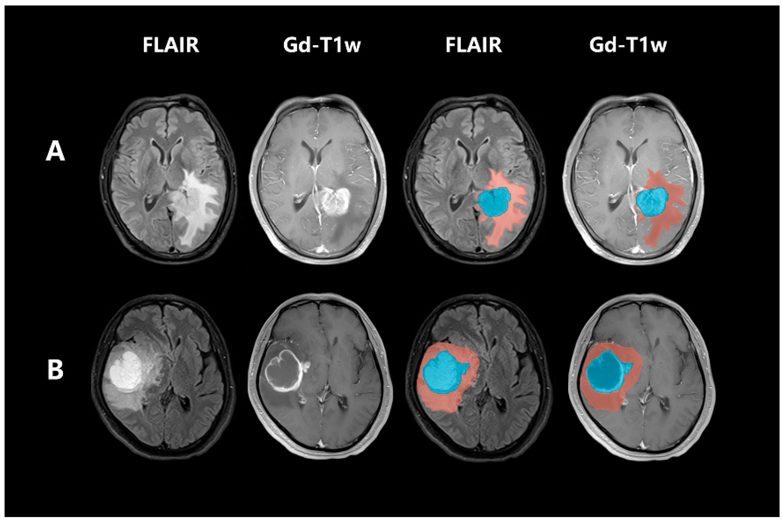
Gd-T1W and FLAIR were registered for each image using a 3D Slicer (https://www.slicer.org/, accessed on 1 July 2021) to ensure that the two sequences had the same spacing and slice thickness with correction, registration, and resampling. FLAIR and Gd-T1W were selected for delineation as Gd-T1W was used to capture tumor necrosis cores and enhanced areas; FLAIR was used to observe the whole tumors and edematous areas. All MR images were normalized to reduce the effects of imaging protocol heterogeneity and apply N4-bias field correction to correct for image bias domains. Each glioma was defined as two sub-compartments of the tumor habitat: (1) the tumor parenchymal area and (2) peritumoral edema. To fully display the lesions, we first delineated the edematous region (including the inner tumor parenchyma) on the FLAIR and then delineated the inner tumor parenchyma on the Gd-T1W. A sketch of this process is shown in (Figure 2). Two radio-neurologists, blinded to the pathological results, outlined all the layers of the regions of interest (ROIs), thus forming the volumes of interest. If the two doctors had differing outlines, they discussed and summarized the final image segmentation results. The original segmentation results from the two doctors were used for consistency analysis, whereas the final results reached after discussion were used for feature extraction and radiomics analysis.

2.5. Radiomic Feature Extraction and Radiomic Signature Construction

The intraclass correlation consistency test examined the segmentation results of the two radiologists. The results showed that the intraclass correlation value of FLAIR was 0.68–1.00, and Gd-T1W was 0.75–1.00. For each sub-compartment (edema, tumor) in every MRI protocol, 1688 radiomic features were extracted using PyRadiomics (version 2.2.0). In total, the radiomic features were divided into four categories. The first category is first-order features, which describe simple statistics based on a gray histogram of the entire ROI, including gray mean, maximum, and minimum. The second category is the shape, which describes the geometric characteristics of the ROIs. Simple shape features include diameter and axis, whereas complex shape features include compactness and sphericity based on surface and volume algorithms. The third category includes texture features, such as the gray level cooccurrence matrix, gray level run length matrix, and gray level size zone matrix. The fourth category is higher-order statistical features, which mainly included first-order statistics and texture features derived from the wavelet transformation of the original images: exponential, square, square root, logarithm, and wavelet (wavelet-LHL, wavelet-LHH, wavelet-HLL, wavelet-LLH, wavelet-HLH, wavelet-HHH, wavelet-HHL, and wavelet-LLL).
For the first objective of building the CDKN2A/B predictive model, three methods of variance threshold, univariate selection, and least absolute shrinkage and selection operator (LASSO) analysis were used to select the most useful features for subsequent analysis to reduce the dimensionality of the extracted features and the redundancy of high-dimensional features (Supplementary Figure S1 and Supplementary Table S1). The LASSO Cox algorithm was used to screen the most significant radiomic features in our second objective of building the survival analysis model (Supplementary Figure S2 and Supplementary Table S2). The radiomic score (rad-score) of each lesion was calculated using a linear combination of selected features, which were weighted by their respective coefficients.

2.6. Clinical and Radiological Factors for Predicting CDKN2A/B

To assess the MRI features of gliomas with CDKN2A/B homozygous deletion, we included all patients with gliomas from the two cohorts. General clinical data (age, sex, WHO grade, and histological diagnosis) were collected. Based on the selected radiomic features, a logistic regression model was used to predict CDKN2A/B homozygous deletion in the training set, and a rad-score was established. A multivariate logistic regression model was established by combining the screened significant imaging features with the general clinical features to predict CDKN2A/B homozygous deletion.

2.7. Prognostic Value Analysis

We screened 104 patients to evaluate the effect of CDKN2A/B homozygous deletion and other clinical characteristics on the prognosis of patients with IDH-mutant LGGs. To investigate the association between clinical characteristics and overall survival (OS), we used a univariate Cox regression to screen out clinical features related to prognosis; we combined these relevant clinical features with radiomic features to construct a prognostic model of IDH-mutant LGGs using a multivariate Cox regression analysis.
For each MRI protocol, each patient in the training and validation set was assigned all risk scores in the high- and low-risk score groups based on the rad-score median point. The prognostic power of clinical and imaging features was assessed using the Kaplan–Meier (K–M) survival analysis.
The K–M survival curve was used to compare the survival period of the high- and low-risk groups. The horizontal and vertical axes on the K–M survival curve represent time and survival probability, respectively. At any given point on the survival curve, the probability that a patient in each group is still alive at that time is shown. In addition, the hazard ratio was used to quantify the effect of individual characteristics on survival. The predictive performance of the model was quantified using the C-index. Adjustments were made based on the agreement between the estimated 1-, 3-, and 5-year survival rates and the actual year survival rates in the training and validation cohorts with corresponding calibration curves. p < 0.05 was considered statistically significant.

2.8. Statistical Analysis

Normalization of features, selection of features, and model construction were performed using Python (version 3.7.0), Scikit-learn version 0.19.2, and PyRadiomics (version 2.2.0). Other statistical analyses were performed using the R software (version 3.3.0). Statistical significance was set at p < 0.05.
For the first objective of predicting CDKN2A/B, univariate analysis was performed to select candidate features with significant differences (p < 0.05). The best subset selection was used for the subsequent model construction. The chi-squared test or Fisher’s exact test and the Mann–Whitney U test were used for categorical and continuous variables, respectively. The receiver operating characteristic (ROC) curve analysis was used to calculate the area under the curve (AUC), precision, sensitivity, and specificity to evaluate the predictive effect of the model.
For the second objective of the survival analysis, the survival probability was analyzed using the K–M method. The log-rank test was used to compare outcomes between patients with high- and low-risk scores. Univariate Cox regression analyses were performed to identify the significant predictors of survival probability.

3. Results

3.1. Clinical Characteristics

Pre-modeling analysis of all clinical features revealed that only age, histological classification, and WHO grade were statistically significant (p < 0.05) between the two groups. These factors were associated with CDKN2A/B homozygous deletion (Table 1).

3.2. CDKN2A/B Predictive Model Construction and Evaluation

The selected radiomic features were used to establish a univariate logistic regression model, and the three statistically significant clinical factors were used to establish a clinical univariate model. A comprehensive model combining radiomic features with clinical factors was then constructed using a multiple logistic regression analysis. The results are shown in (Table 2).
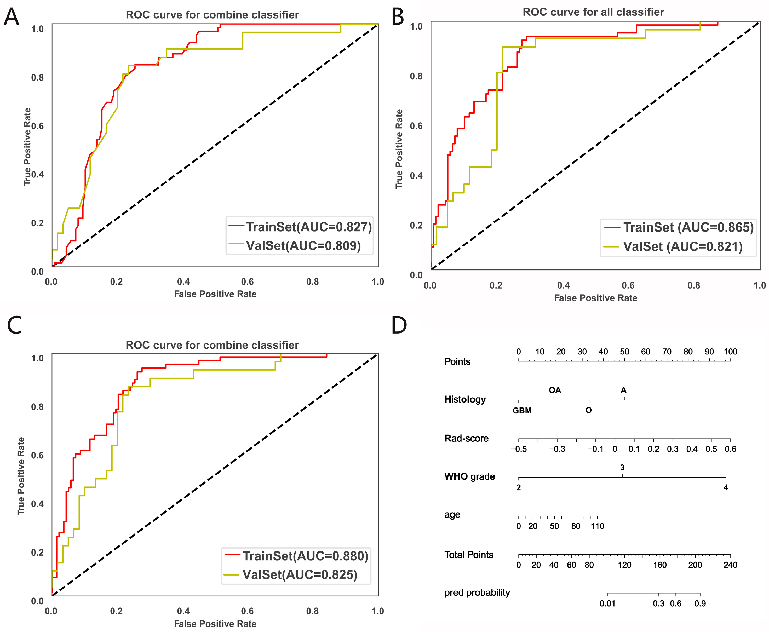
The AUCs of the comprehensive model in the training and validation sets were 0.880 (95% confidence interval [CI], 0.840–0.918) and 0.825 (95% CI, 0.743–0.894), respectively. The results of the multivariate logistic regression model are presented as nomograms (Figure 3). The results showed that the rad-score was strongly associated with CDKN2A/B homozygous deletion. It also showed that the radiomics model based on the conventional MRI sequence had a better predictive effect on CDKN2A/B homozygous deletion.
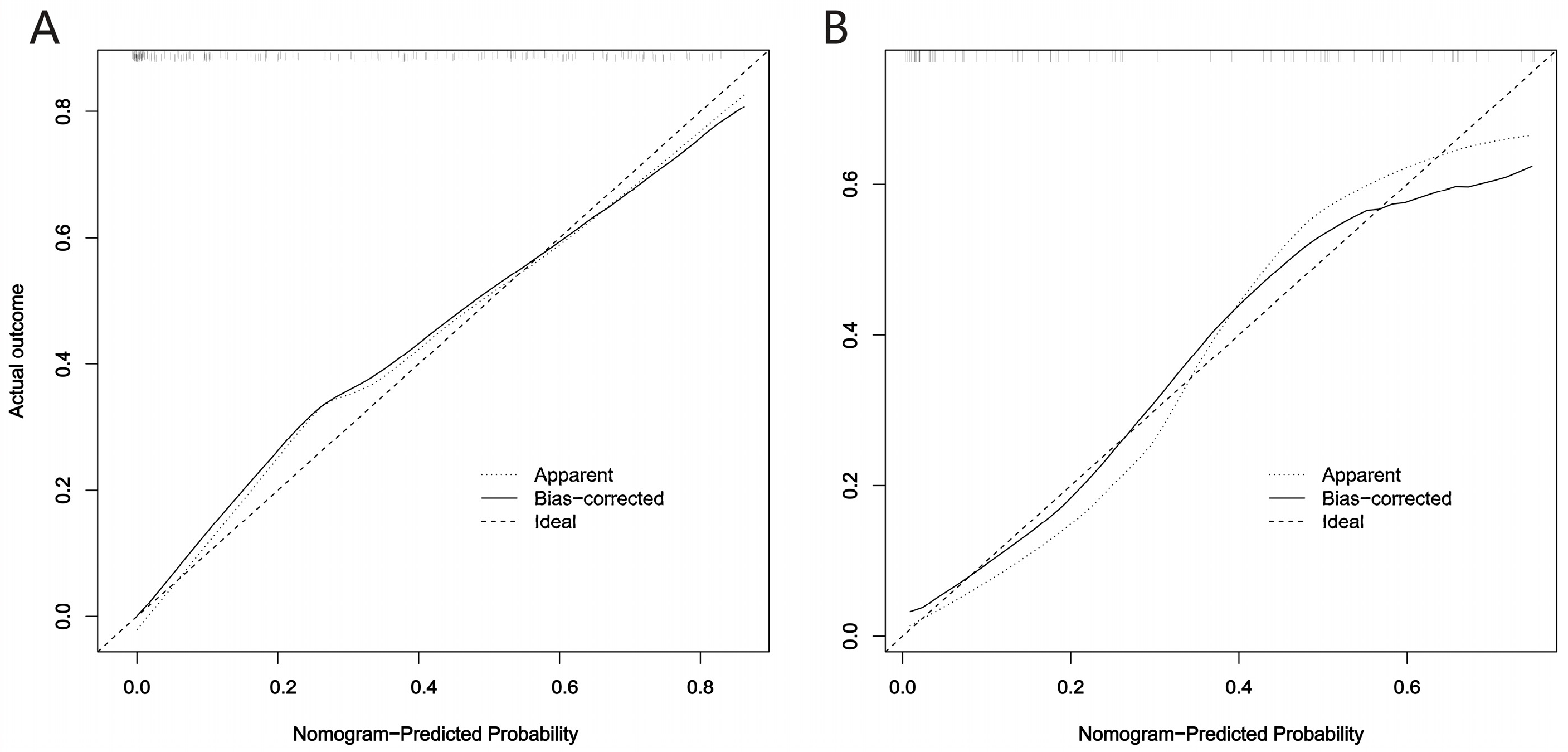
Calibration curve analysis was performed on the model, and the results of the calibration curve reflected the proximity of the predicted results of the model and the actual results. In our study, the calibration curve reflected a high degree of agreement between the predictions of the model in the training and validation sets and the actual status, indicating that the model displayed good performance and reliability in predicting CDKN2A/B homozygous deletion in gliomas (Figure 4).

3.3. Prognostic Value of the Fusion Radiomics Signature and Clinical Characteristics

A total of 1688 features were extracted from every conventional sequence, FLAIR, and Gd-T1W. The 23 most significant image features were extracted.
Univariate Cox analysis of all clinical features showed that only the CDKN2A/B homozygous deletion status was statistically significant (p < 0.05) (Supplementary Table S3). This result was expected as it agrees with the previous studies demonstrating that CDKN2A/B homozygous deletion is an independent prognostic marker in LGGs.
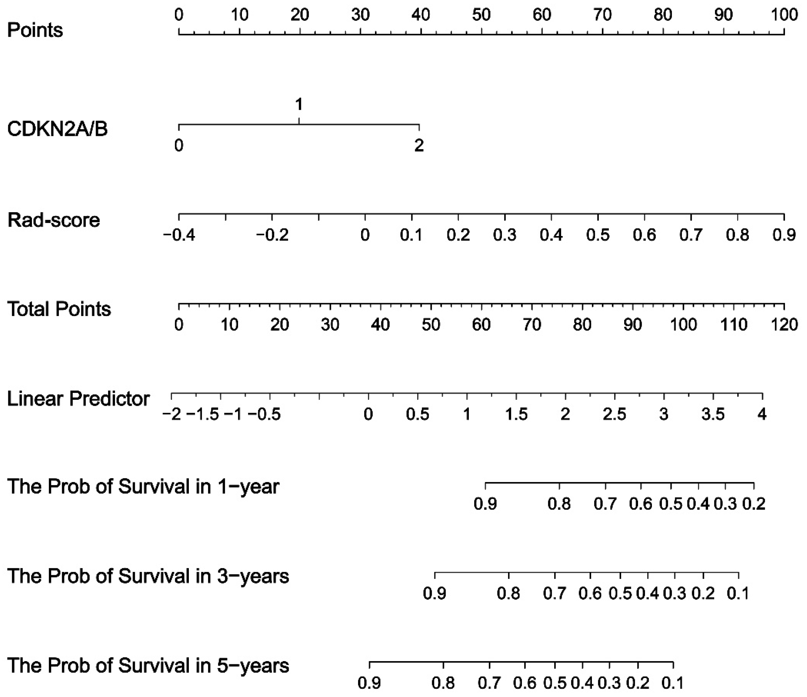
By combining CDKN2A/B homozygosity deletion with radiomic features, a multivariate Cox regression survival analysis model was established. The C-indices in the training and validation sets were 0.876 and 0.818, respectively (Supplementary Table S4). The model was displayed using a nomogram (Figure 5).
Next, all risk scores were divided into high- and low-risk scores according to the median value of patient risk scores. K–M survival analysis was used to perform univariate survival analysis according to the high- and low-risk scores. The results showed that high- and low-risk scores were the key influencing factors for patient survival (p < 0.05). The results of the K–M growth curve showed significant differences between the high- and low-risk groups (Figure 6).
In clinical practice, patient-individualized treatment plans can be guided by the patient’s risk score. The calibration curves indicated that the predicted and actual survival outcomes were good at 2, 4, and 6 years of follow-up in the training and validation sets (Supplementary Figure S3).

4. Discussion

In this study, we investigated a noninvasive imaging method based on conventional MRI to establish radiomics models for predicting CDKN2A/B homozygosity deletion in gliomas, and the results showed that the logistic regression integrated model had the most excellent predictive performance (AUC of 0.880 and 0.825 for the training and validation sets, respectively). This study is the first to propose the use of noninvasive imaging to build radiomic models for CDKN2A/B homozygosity deletion in gliomas. CDKN2A/B homozygosity deletion serves as a prognostic predictor for gliomas, predicting poorer OS, and providing treatment guidance. Therefore, accurately predicting this subtype provides an important clinical adjunct value for accurate diagnosis, clinical decision making, and survival prediction for molecular staging of patients with gliomas.
First, we adopted logistic regression models to analyze the accuracy and reliability of independent imaging and clinical features, and then combined predictions from both. The results showed that combining radiomic features with clinical parameters (age and sex) did not greatly improve the models’ performance, which fully characterized the importance and accuracy of image characteristics in model construction. Previous studies have reported the correlation of MRI features with CDKN2A/B homozygosity deletion. This study expanded on this research and used the newest machine learning algorithms to increase the reliability of gene prediction models.
Since few studies have used MRI features to explore CDKN2A/B homozygous deletion, the Heidelberg University Medical Center analyzed the association between multiparametric, multiregional MRI features and key molecular features of GBM, including CDKN2A, and found that CDKN2A deletion is associated with increased normalized relative cerebral blood volume (nrCBV) and normalized regional cerebral blood flow (nrCBF) within the enhanced tumor volume (ROC range, 63% to 69%; false discovery rate adjusted p < 0.05) [24]. This study found meaningful associations between the multiparametric MRI features and molecular markers. Since it was a multi-genotype study in GBM, it did not investigate the relevant situation in LGGs; however, CDKN2A/B homozygous deletion is implicated in LGGs. Our study comprehensively constructed a CDKN2A/B predictive model based on MRI features for all patients with gliomas and overcame aforementioned shortcomings. Concurrently, Nowak et al. identified significantly different imaging parameters in supratentorial ependymomas according to CDKN2A alterations [25]. Since the stratification of patients by morphological MRI alone is difficult, radiomic technology promises to make such research models more accurate and reliable. Our study innovatively used radiomics to determine the association between MRI features and CDKN2A/B homozygous deletion in gliomas, constructing a CDKN2A/B predictive model. Through this study, we aimed to construct a template for CDKN2A/B homozygous deletion study models for gliomas, contributing to the development of clinical treatment plans.
Second, we investigated the association between OS and all clinical features, including CDKN2A/B homozygous deletion, in IDH-mutant LGGs. We found that only CDKN2A/B, an independent prognostic factor for IDH-mutant LGGs, was associated with prognoses. According to the rad-score median point (−0.07), patients were divided into high- and low-risk groups. Patients in the high-risk group were those with rad-scores greater than −0.07. The K–M curve showed that patients in the high-risk group had different degrees of death over time, and the risk of death was higher than in the low-risk group. However, no patients in the low-risk group died during the follow-up period. This study reveals that IDH-mutant LGGs have an overall optimistic prognosis, but certain patients have a poorer prognosis, such as those with CDKN2A/B homozygous deletion. Radiomics features can help identify which class of patients is at risk. This verifies that in our dataset, patients with CDKN2A/B homozygous deletion and a rad-score of more than −0.07 have a poor prognosis, and radiomic features can be used as noninvasive prognostic markers for patients with LGGs.
The latest CNS tumor classification has indicated that CDKN2A/B homozygous deletion represents a poor prognosis in astrocytoma [14], but there are no reports using CDKN2A/B and MRI features to predict survival in LGGs. This study is novel in that CDKN2A/B and conventional MRI features can be used to predict the survival of patients with preoperative LGGs, providing a reference for clinical prognosis. In addition, this study aimed to predict survival in subgroups of LGGs, including astrocytoma, oligodendroglioma, WHO grade, and whether postoperative adjuvant radiotherapy was required, to determine OS in these LGG subgroups and whether patients would benefit from postoperative adjuvant radiotherapy. However, when we analyzed the clinical characteristics, these indicators were not statistically significant, and only CDKN2A/B homozygous deletion was included in the subsequent analysis. This limited the subgroup analysis, which may have been related to the enrolled patients. We plan to expand the sample size in future studies.
This study has some limitations. First, the data from TCIA and our hospital do not coincide on the MRI scan protocol; therefore, to keep all MR images in a consistent grayscale, we excluded most of the patients from TCIA. Second, although the expert consensus on glioma segmentation has been published, some gliomas—particularly LGGs—have unclear demarcations between tumor parenchyma and edema which makes it difficult to separate the necrotic components in the center from the surrounding strengthened tissue. This is also why we decided to divide the tumor into two parts. Moreover, the segmentation is time-consuming, and the results will vary due to different delineators and times, which makes the reproducibility of radiomics poor. In the future, the development of fully automatic delineation tools and accurate segmentation will be helpful for clinical model reconstruction. However, the validation was retrospective, and the LGG sample size in the survival analysis was small. We plan to include a large-scale multi-site evaluation in further studies. In the future, our study will also focus on incorporating complementary imaging parameters (PET, perfusion, diffusion-weighted imaging), which may further improve survival prediction using radiogenic analysis while also considering subsequent treatments.

5. Conclusions

We were able to successfully develop superior CDKN2A/B homozygous deletion predictive and IDH-mutant LGG survival models. We found that MRI can be used as a noninvasive imaging-based marker for detecting genes and prognostic indicators to guide clinical treatment decisions. Radiomics displays high accuracy and reliability in predicting CDKN2A/B homozygous deletion, which provides a stepping stone for future clinical applications of molecular genetic diagnostic platforms or AI software in diagnosing gliomas.

Supplementary Materials

The following supporting information can be downloaded at: https://www.mdpi.com/article/10.3390/brainsci13040548/s1, Figure S1. Feature selection of the CDKN2A/B homozygous deletions analysis. Figure S2. Feature selection of the survival analysis of the isocitrate dehydrogenase-mutant low-grade glioma. Figure S3. Calibration curves for the survival analysis. (A) Training set, and (B) Validation set. Table S1. Feature Selection Results of CDKN2A/B homozygous deletions. Table S2. Feature Selection Results of Survival Analysis. Table S3. The results of clinical characteristics. Table S4. The results of survival analysis.

Author Contributions

Data collection, data management, statistical analysis, manuscript drafting, and revision: J.Y. Pathological detection, pathological data support: L.L. and T.L. Software technical service support, statistical analysis: J.Y. MRI acquisition: C.N. Lesion segmentation: R.F. and D.L. Clinical evaluation: C.Z., R.Y. and Q.L. Project design, guidance and suggestions, manuscript revision: X.H. Project design and supervision: W.C. All authors have read and agreed to the published version of the manuscript.

Funding

This work was supported by Sichuan Science and Technology Program (2023YFQ0002).

Institutional Review Board Statement

This study was ethically approved by the First Affiliated Hospital of Army Military Medical University (approval number: KY20220305).

Informed Consent Statement

Patient consent was waived due to the retrospective nature of this study.

Data Availability Statement

The datasets used and/or analyzed during the current study are available from the corresponding author on reasonable request.

Acknowledgments

We acknowledge technical support from Yang Jing, Huiying Medical Technology (Beijing) Co., Ltd., Beijing 100192, China.

Conflicts of Interest

The authors declare that they have no competing interest.

References

  1. Lapointe, S.; Perry, A.; Butowski, N.A. Primary Brain Tumours in Adults. Lancet 2018, 392, 432–446. [Google Scholar] [CrossRef]
  2. Weller, M.; Wick, W.; Aldape, K.; Brada, M.; Berger, M.; Pfister, S.M.; Nishikawa, R.; Rosenthal, M.; Wen, P.Y.; Stupp, R.; et al. Glioma. Nat. Rev. Dis. Prim. 2015, 1, 15017. [Google Scholar] [CrossRef] [PubMed]
  3. Yan, H.; Parsons, D.W.; Jin, G.; McLendon, R.; Rasheed, B.A.; Yuan, W.; Kos, I.; Batinic-Haberle, I.; Jones, S.; Riggins, G.J.; et al. Idh1 and Idh2 Mutations in Gliomas. N. Engl. J. Med. 2009, 360, 765–773. [Google Scholar] [CrossRef] [PubMed]
  4. Olar, A.; Wani, K.M.; Alfaro-Munoz, K.D.; Heathcock, L.E.; van Thuijl, H.F.; Gilbert, M.R.; Armstrong, T.S.; Sulman, E.P.; Cahill, D.P.; Vera-Bolanos, E.; et al. Idh Mutation Status and Role of Who Grade and Mitotic Index in Overall Survival in Grade Ii-Iii Diffuse Gliomas. Acta Neuropathol. 2015, 129, 585–596. [Google Scholar] [CrossRef] [Green Version]
  5. Alentorn, A.; Dehais, C.; Ducray, F.; Carpentier, C.; Mokhtari, K.; Figarella-Branger, D.; Chinot, O.; Cohen-Moyal, E.; Ramirez, C.; Loiseau, H.; et al. Allelic Loss of 9p21.3 Is a Prognostic Factor in 1p/19q Codeleted Anaplastic Gliomas. Neurology 2015, 85, 1325–1331. [Google Scholar] [CrossRef] [PubMed] [Green Version]
  6. Gul, A.; Leyland-Jones, B.; Dey, N.; De, P. A Combination of the Pi3k Pathway Inhibitor Plus Cell Cycle Pathway Inhibitor to Combat Endocrine Resistance in Hormone Receptor-Positive Breast Cancer: A Genomic Algorithm-Based Treatment Approach. Am. J. Cancer Res. 2018, 8, 2359–2376. [Google Scholar]
  7. Ichimura, K.; Schmidt, E.E.; Goike, H.M.; Collins, V.P. Human Glioblastomas with No Alterations of the Cdkn2a (P16ink4a, Mts1) and Cdk4 Genes Have Frequent Mutations of the Retinoblastoma Gene. Oncogene 1996, 13, 1065–1072. [Google Scholar]
  8. Liu, L.; Dilworth, D.; Gao, L.; Monzon, J.; Summers, A.; Lassam, N.; Hogg, D. Mutation of the Cdkn2a 5’ Utr Creates an Aberrant Initiation Codon and Predisposes to Melanoma. Nat. Genet. 1999, 21, 128–132. [Google Scholar] [CrossRef]
  9. Cimino, P.J.; Holland, E.C. Targeted Copy Number Analysis Outperforms Histologic Grading in Predicting Patient Survival for Who Grades Ii/Iii Idh-Mutant Astrocytomas. Neuro Oncol. 2019, 21, 819–821. [Google Scholar] [CrossRef]
  10. Aoki, K.; Nakamura, H.; Suzuki, H.; Matsuo, K.; Kataoka, K.; Shimamura, T.; Motomura, K.; Ohka, F.; Shiina, S.; Yamamoto, T.; et al. Prognostic Relevance of Genetic Alterations in Diffuse Lower-Grade Gliomas. Neuro Oncol. 2018, 20, 66–77. [Google Scholar] [CrossRef] [Green Version]
  11. Appin, C.L.; Brat, D.J. Biomarker-Driven Diagnosis of Diffuse Gliomas. Mol. Asp. Med. 2015, 45, 87–96. [Google Scholar] [CrossRef] [PubMed]
  12. Appay, R.; Dehais, C.; Maurage, C.A.; Alentorn, A.; Carpentier, C.; Colin, C.; Ducray, F.; Escande, F.; Idbaih, A.; Kamoun, A.; et al. Cdkn2a Homozygous Deletion Is a Strong Adverse Prognosis Factor in Diffuse Malignant Idh-Mutant Gliomas. Neuro Oncol. 2019, 21, 1519–1528. [Google Scholar] [CrossRef]
  13. Juratli, T.A.; Qin, N.; Cahill, D.P.; Filbin, M.G. Molecular Pathogenesis and Therapeutic Implications in Pediatric High-Grade Gliomas. Pharmacol. Ther. 2018, 182, 70–79. [Google Scholar] [CrossRef] [PubMed]
  14. Louis, D.N.; Perry, A.; Wesseling, P.; Brat, D.J.; Cree, I.A.; Figarella-Branger, D.; Hawkins, C.; Ng, H.K.; Pfister, S.M.; Reifenberger, G.; et al. The 2021 Who Classification of Tumors of the Central Nervous System: A Summary. Neuro Oncol. 2021, 23, 1231–1251. [Google Scholar] [CrossRef]
  15. Zhou, H.; Vallières, M.; Bai, H.X.; Su, C.; Tang, H.; Oldridge, D.; Zhang, Z.; Xiao, B.; Liao, W.; Tao, Y.; et al. Mri Features Predict Survival and Molecular Markers in Diffuse Lower-Grade Gliomas. Neuro Oncol. 2021, 19, 862–870. [Google Scholar] [CrossRef] [PubMed]
  16. Stadlbauer, A.; Zimmermann, M.; Kitzwögerer, M.; Oberndorfer, S.; Rössler, K.; Dörfler, A.; Buchfelder, M.; Heinz, G. Mr Imaging-Derived Oxygen Metabolism and Neovascularization Characterization for Grading and Idh Gene Mutation Detection of Gliomas. Radiology 2017, 283, 799–809. [Google Scholar] [CrossRef] [Green Version]
  17. Lasocki, A.; Anjari, M.; Örs Kokurcan, S.; Thust, S.C. Conventional Mri Features of Adult Diffuse Glioma Molecular Subtypes: A Systematic Review. Neuroradiology 2021, 63, 353–362. [Google Scholar] [CrossRef]
  18. Rogers, W.; Seetha, S.T.; Refaee, T.A.G.; Lieverse, R.I.Y.; Granzier, R.W.Y.; Ibrahim, A.; Keek, S.A.; Sanduleanu, S.; Primakov, S.P.; Beuque, M.P.L.; et al. Radiomics: From Qualitative to Quantitative Imaging. Br. J. Radiol. 2020, 93, 20190948. [Google Scholar] [CrossRef]
  19. Kickingereder, P.; Neuberger, U.; Bonekamp, D.; Piechotta, P.L.; Götz, M.; Wick, A.; Sill, M.; Kratz, A.; Shinohara, R.T.; Jones, D.T.W.; et al. Radiomic Subtyping Improves Disease Stratification Beyond Key Molecular, Clinical, and Standard Imaging Characteristics in Patients with Glioblastoma. Neuro Oncol. 2018, 20, 848–857. [Google Scholar] [CrossRef]
  20. Lambin, P.; Rios-Velazquez, E.; Leijenaar, R.; Carvalho, S.; van Stiphout, R.G.; Granton, P.; Zegers, C.M.; Gillies, R.; Boellard, R.; Dekker, A.; et al. Radiomics: Extracting More Information from Medical Images Using Advanced Feature Analysis. Eur. J. Cancer 2012, 48, 441–446. [Google Scholar] [CrossRef] [Green Version]
  21. Gillies, R.J.; Kinahan, P.E.; Hricak, H. Radiomics: Images Are More Than Pictures, They Are Data. Radiology 2016, 278, 563–577. [Google Scholar] [CrossRef] [PubMed] [Green Version]
  22. Lambin, P.; Leijenaar, R.T.H.; Deist, T.M.; Peerlings, J.; de Jong, E.E.C.; van Timmeren, J.; Sanduleanu, S.; Larue, R.; Even, A.J.G.; Jochems, A.; et al. Radiomics: The Bridge between Medical Imaging and Personalized Medicine. Nat. Rev. Clin. Oncol. 2017, 14, 749–762. [Google Scholar] [CrossRef] [PubMed] [Green Version]
  23. Kumar, V.; Gu, Y.; Basu, S.; Berglund, A.; Eschrich, S.A.; Schabath, M.B.; Forster, K.; Aerts, H.J.; Dekker, A.; Fenstermacher, D.; et al. Radiomics: The Process and the Challenges. Magn. Reson. Imaging 2012, 30, 1234–1248. [Google Scholar] [CrossRef] [PubMed] [Green Version]
  24. Kickingereder, P.; Bonekamp, D.; Nowosielski, M.; Kratz, A.; Sill, M.; Burth, S.; Wick, A.; Eidel, O.; Schlemmer, H.P.; Radbruch, A.; et al. Radiogenomics of Glioblastoma: Machine Learning-Based Classification of Molecular Characteristics by Using Multiparametric and Multiregional Mr Imaging Features. Radiology 2016, 281, 907–918. [Google Scholar] [CrossRef] [Green Version]
  25. Nowak, J.; Jünger, S.T.; Huflage, H.; Seidel, C.; Hohm, A.; Vandergrift, L.A.; von Hoff, K.; Rutkowski, S.; Pietsch, T.; Warmuth-Metz, M. Mri Phenotype of Rela-Fused Pediatric Supratentorial Ependymoma. Clin. Neuroradiol. 2019, 29, 595–604. [Google Scholar] [CrossRef]
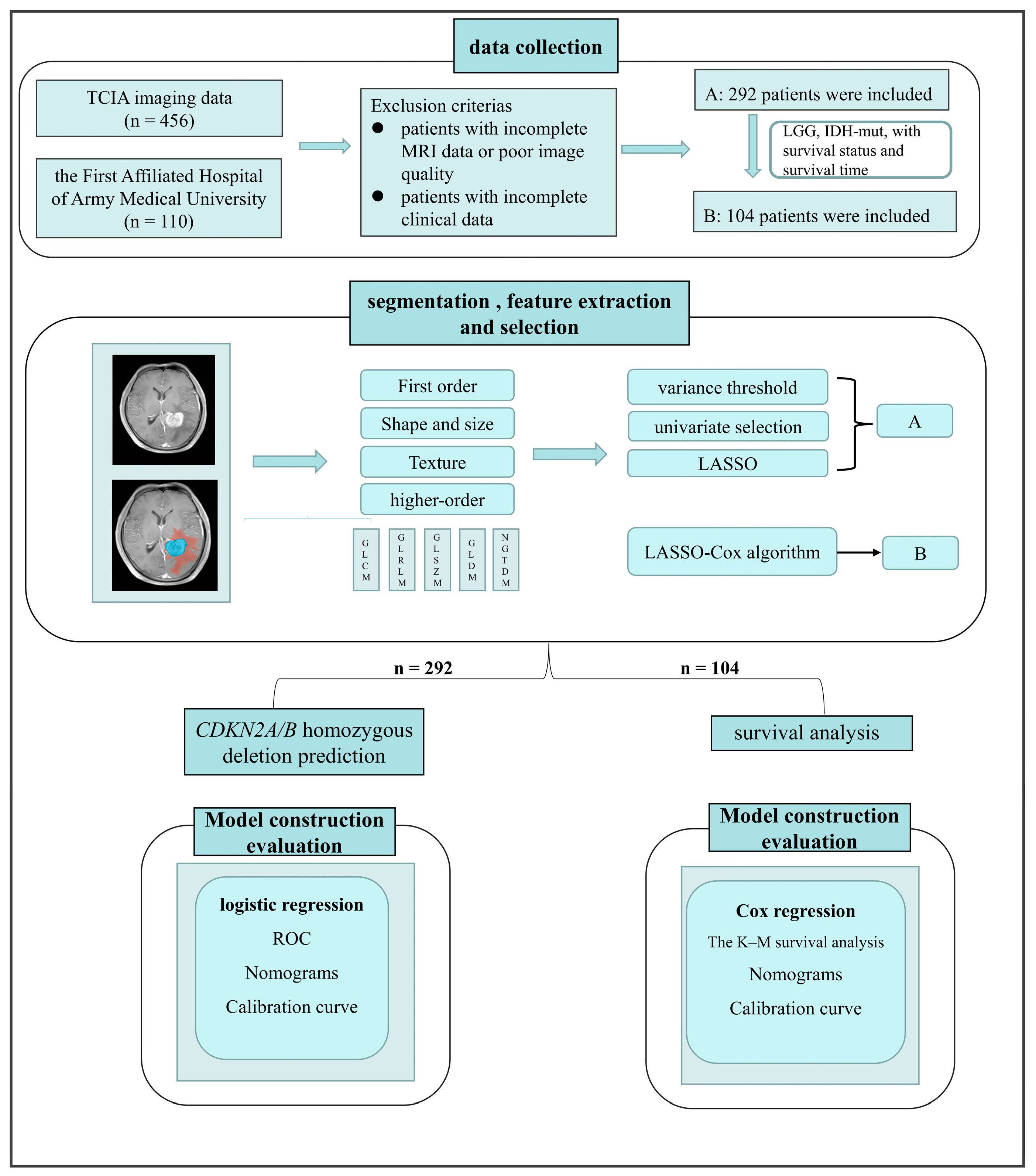
Figure 1. Workflow of this study: A means the project of CDKN2A/B homozygous deletion prediction. B means the project of survival analysis.
Brainsci 13 00548 g001
Figure 2. The delineated and displayed regions of interest. Blue indicates the tumor parenchyma and orange indicates the edematous region. (A) Patients without CDKN2A/B homozygous deletion and (B) Patients with CDKN2A/B homozygous deletion.
Brainsci 13 00548 g002
Figure 3. Establishment and validation of the predictive model of CDKN2A/B homozygous deletion in glioma. (AC): The receiver operating characteristic results of the models. (A) clinical model, (B) radiomics model, (C) the combined clinical and radiomics comprehensive prediction model. (D) Nomogram of the combined prediction model of CDKN2A/B homozygous deletion: the relevant clinical characteristics and rad-score were included in the nomogram, and the results showed that rad-score correlated most strongly with the model.
Brainsci 13 00548 g003
Figure 4. Calibration curves for the prediction model of CDKN2A/B homozygous deletions. (A) Training set and (B) Validation set.
Brainsci 13 00548 g004
Figure 5. Nomogram of the training set of survival analysis: survival rate at 1-, 3-, and 5-years can be calculated from CDKN2A/B homozygous deletion status and rad-score.
Brainsci 13 00548 g005
Figure 6. Kaplan–Meier curves with isocitrate dehydrogenase-mutant low-grade glioma. (A) Training set and (B) Validation set. Mortality was higher in the high-risk group, whereas almost no patients died in the low-risk group (p-value was 0.026 in the training group and 0.0049 in the validation group). The number at risk means the number of people in the high and low groups who were not censored from the endpoint event at different times.
Brainsci 13 00548 g006
Table 1. The baseline characteristics of the patients.
CDKN2A/B Homozygous Deletion
NOYESp-ValueSMD
N 19894
Age (median [IQR]) 41.50 [24.00, 57.75]60.00 [52.25, 66.00]<0.0010.856
Gender (%)Female111 (56.1) 61 (64.9) 0.1920.181
Male87 (43.9) 33 (35.1)
HistologyAstrocytoma43 (21.7) 8 (8.5) <0.0011.249
Oligoastrocytoma27 (13.6) 2 (2.1)
Oligodendroglioma51 (25.8) 1 (1.1) .
GBM77 (38.9) 83 (88.3)
WHO grade (%)65 (32.8) 1 (1.1) <0.0011.262
56 (28.3) 10 (10.6)
77 (38.9) 83 (88.3)
The bold means these clinical characteristics were statistically significant (p < 0.05).
Table 2. Model results for prediction of CDKN2A/B homozygous deletion.
DatasetModelAUC95%CIACCSensitivitySpecificity
Training setradomics0.8650.820~0.9090.7930.6310.87
clinic0.8240.776~0.8710.7830.7230.812
radiomic + clinic0.880.840~0.9180.7930.6620.855
Validation setradiomic0.8210.741~0.8900.7080.4830.817
clinic0.8020.722~0.8750.7530.6550.8
radiomic + clinic0.8250.743~0.8940.730.5170.833
Radomics: Model based on radiomic features only. Clinic: Model based on clinical characteristics only. Radiomic + Clinic: Model based on radiomic features and clinical characteristics.
Disclaimer/Publisher’s Note: The statements, opinions and data contained in all publications are solely those of the individual author(s) and contributor(s) and not of MDPI and/or the editor(s). MDPI and/or the editor(s) disclaim responsibility for any injury to people or property resulting from any ideas, methods, instructions or products referred to in the content.